R E S E A R C H
Open Access
Dietary
ω
-6 polyunsaturated fatty acid
arachidonic acid increases inflammation,
but inhibits ECM protein expression in
COPD
Sandra Rutting
1,2, Michael Papanicolaou
1,3, Dia Xenaki
1, Lisa G. Wood
2, Alexander M. Mullin
1,
Philip M. Hansbro
2and Brian G. Oliver
1,3*Abstract
Background:The obesity paradox in COPD describes protective effects of obesity on lung pathology and inflammation. However, the underlying relationships between obesity, diet and disease outcomes in COPD are not fully understood. In this study we measured the response to dietary fatty acids upon markers of inflammation and remodelling in human lung cells from people with and without COPD.
Methods: Pulmonary fibroblasts were challenged with ω-3 polyunsaturated fatty acids (PUFAs), ω-6 PUFAs, saturated fatty acids (SFAs) or the obesity-associated cytokine TNFα. After 48–72 h release of the pro-inflammatory cytokines interleukin (IL)-6 and CXCL8 was measured using ELISA and mRNA expression and deposition of the extracellular matrix (ECM) proteins fibronectin, type I collagen, tenascin and perlecan were measured using qPCR or ECM ELISA, respectively.
Results: Challenge with theω-6 PUFA arachidonic acid (AA), but notω-3 PUFAs or SFAs, resulted in increased IL-6 and CXCL8 release from fibroblasts, however IL-6 and CXCL8 release was reduced in COPD (n = 19) compared to non-COPD (n = 36). AA-induced cytokine release was partially mediated by downstream mediators of cyclooxygenase (COX)-2 in both COPD and non-COPD. In comparison, TNFα-induced IL-6 and CXCL8 release was similar in COPD and non-COPD, indicating a specific interaction of AA in COPD. In patients with or without COPD, regression analysis revealed no relationship between BMI and cytokine release. In addition, AA, but not SFAs or ω-3 PUFAs reduced the basal deposition of fibronectin, type I collagen, tenascin and perlecan into the ECM in COPD fibroblasts. In non-COPD fibroblasts, AA-challenge decreased basal deposition of type I collagen and perlecan, but not fibronectin and tenascin.
Conclusions:This study shows that AA has disease-specific effects on inflammation and ECM protein deposition. The impaired response to AA in COPD might in part explain why obesity appears to have less detrimental effects in COPD, compared to other lung diseases.
Keywords:COPD,ω-6 PUFAs, Airway inflammation, Remodelling, Human pulmonary fibroblasts
* Correspondence:brian.oliver@uts.edu.au
1Respiratory Cellular and Molecular Biology, Woolcock Institute of Medical
Research, The University of Sydney, Sydney, Australia
3School of Life Sciences, University of Technology Sydney, Sydney, Australia
Full list of author information is available at the end of the article
Introduction
More than two billion people around the world are over-weight or obese, classified by a body mass index (BMI) greater than, or equal to, 25 or 30 kg/m2, respectively [1]. This global obesity epidemic is associated with many chronic diseases and recently, its role in lung disease, in-cluding chronic obstructive pulmonary disease (COPD), has received new interest. Obesity is becoming more common in mild-to moderate COPD with a prevalence that is generally higher than the general population [2– 4]. In the general population, obesity has a major nega-tive impact on health outcomes. However paradoxically, mild-to moderate obesity in moderate to severe COPD is reported to have protective effects on survival, lung function decline and exacerbations [5–8]. The under-lying mechanisms of the protective effects of obesity in COPD are currently poorly understood.
In COPD, structural changes to the airways, also known as airway remodelling, occur. A characteristic feature of airway remodelling is airway wall thickening, which is due to an increase in connective tissue. In-creased airway wall thickness is associated with frequent exacerbations and symptoms of chronic bronchitis [9,
10]. Connective tissue is composed of scaffolding pro-teins termed the extracellular matrix (ECM). Perlecan, fibronectin, collagens and tenascin are ECM proteins and their presence is associated with remodelling and/or inflammation in the airways. Tenascin and fibronectin have been shown to be increased in COPD [11]. Inter-estingly, obesity has also been shown to affect airway re-modelling, with BMI being negatively associated with emphysema and positively associated with airway wall thickness [12]. This finding corresponds with the ‘blue bloater’ phenotype of obese COPD patients, who are more likely to have more bronchitis and less emphysema [9, 13]. Although the exact mechanisms that drive re-modelling are still undefined, ongoing chronic inflamma-tory processes are likely to contribute.
In COPD, airway inflammation is characterized by in-creased numbers of neutrophils, macrophages, and CD8 +−T lymphocytes, as well as increased levels of interleu-kin (IL)-6 and CXCL8 in the airways [14, 15]. Neutro-phils and CXCL8 levels, in particular, are associated with COPD exacerbations [15–17]. Neutrophils are also strongly implicated in causing chronic bronchitis and the destruction of lung tissue in emphysema, through the production of reactive oxygen metabolites and tissue damaging enzymes [16]. Obesity itself is associated with chronic systemic low-grade inflammation, with increased levels of serum IL-6 and TNFα, produced by adipose tis-sue [18,19]. Epidemiological evidence suggests a role for diet in the prevention and management of COPD. In-creased intake of certain nutrients, such as vitamin E, D and C and ω-3 polyunsaturated fatty acids (PUFAs) are
positively associated with lung function in the general population [20, 21]. In addition, epidemiologic studies have demonstrated that increased intake of these nutri-ents is associated with a decreased risk of COPD devel-opment [20]. These effects are thought to be the result of anti-oxidant and anti-inflammatory properties of these nutrients. Little is known about effects of the Western diet in COPD. The Western diet contributes to obesity, being high in energy from macronutrients, in-cluding saturated fatty acids (SFAs) and ω-6 PUFAs. These fatty acids are shown to affect inflammatory pro-cesses and have predominantly been associated with pro-inflammatory effects and negatively associated with outcomes in other lung diseases such as asthma [22,23]. However, the effects of these fatty acids in COPD have not been investigated. ω-3 PUFAs and SFAs affect in-flammation by modifying toll-like receptor 4 (TLR4) sig-nalling, whereasω-6 PUFAs affect inflammation through TLR4-indepenent (independent) mechanisms [24].
A clear causal relation between obesity, diet and disease outcomes in COPD is yet to be proven, but the available data suggest a link between these factors and it is import-ant to understand their effects on airway inflammation and remodelling in COPD. Pulmonary fibroblasts are the major structural cell of the airway and play a crucial role in tissue homeostasis, the production of pro-inflammatory cytokines and ECM proteins and, therefore, are likely to contribute to airway inflammation and remodelling [25,
26]. This study investigated whether pulmonary fibroblasts derived from COPD versus non-COPD patients differ in their inflammatory response to dietary fatty acids (ω-6 PUFAs,ω-3 PUFAs and SFAs) and the obesity-associated cytokine TNFαin vitro.Also, the effect of BMI on this re-sponse was assessed. Secondly, this study investigated whether dietary fatty acids affect the expression and de-position of ECM proteins in fibroblasts.
Methods and materials Subjects
Primary fibroblasts were isolated from the parenchyma of lungs from patients undergoing lung transplantation or lung resection for thoracic malignancies from a total of n = 32 donors with COPD, and a total ofn = 50 do-nors with lung disease other than COPD. The diagnosis of disease was made by thoracic physicians according to current guidelines. Approval for all experiments with human lung was provided by the Human Ethics Com-mittees of the University of Sydney and the Sydney South West Area Health Service. Table 1shows a sum-mary of the patient demographics.
Cell culture
were seeded in 12-well plates at a density of 6.2 × 10⁴ cells/mL in DMEM containing 5% fetal bovine serum (FBS) and 1% antibiotic-antimycotic (Gibco, Grand Island, New York, US). When the cells reached 80% confluency, they were serum starved by incubation in DMEM (Gibco, Grand Island, New York, US) supplemented with 0.1% bo-vine serum albumin (BSA) (Sigma Aldrich, Castle Hill, NSW, Australia) and 1% antibiotic-antimycotic for 24 h prior to stimulation. All experiments were carried out using fibroblasts between passage 2 and 6.
Preparation of BSA-conjugated fatty acids
Stock solutions of 0.5 M ω-3 PUFA (docosahexaenoic acid (DHA)) and SFA (palmitic acid (PA)) and 0.3 M ω-6 PUFA (arachidonic acid (AA)) (Sigma Aldrich) were prepared in 100% EtOH and stored at-20 °C. Working water-soluble solutions of 10 mM were gen-erated by incubating the fatty acids in 10% endotoxin and fatty acid-free BSA (Sigma Aldrich), as previously described by Gupta et al. (2012) and Pillon et al. (2012) [28, 29]. These solutions were further diluted in cell culture medium to obtain final concentrations of 10 and 100 μM. These concentrations are based on physiological concentrations and other in vitro studies [30–33].
Treatment of cells with dietary fatty acids and TNFα Pulmonary fibroblasts from COPD and non-COPD pa-tients were stimulated with 10 and/or 100 μM AA, DHA, PA, or TNFα(1 ng/ml) or vehicle (EtOH/BSA/cell culture medium) and compared to untreated controls. All cells were incubated at 37 °C with 5% CO2for 6, 9,
24, 48 or 72 h. Total RNA or cell-free supernatants were collected and stored at−20 °C until further analysis.
Treatment of cells with indomethacin, celecoxib and dexamethasone
Pulmonary fibroblasts from COPD and non-COPD pa-tients were treated with the non-selective cyclooxygen-ase (COX)-inhibitor, indomethacin (1 μM), the COX-2 selective inhibitor, celecoxib (0.01-1 μM) or corticoster-oid, dexamethasone (0.1-1 μM) (all Sigma-Aldrich) for 60 min prior to challenge with dietary fatty acids.
Determination of IL-6, CXCL8 and PGE2 levels
Levels of IL-6 and CXCL8 in cell culture supernatants were measured using sandwich ELISA. The amount of IL-6 release was assessed with optimized IL-6 anti-body pairs from BD pharmingen, BD, Franklin Lakes, NJ. A specific kit for CXCL8 was purchased from R&D Systems (Minneapolis, Minnesota, USA) and used ac-cording to the manufacturer’s instructions. PGE2 levels were measured by enzyme immunoassay according to the manufacturer’s instructions (R&D systems).
Cytotoxicity assay
Cell toxicity was estimated using a lactate dehydrogenase (LDH) assay according to the manufacturer’s instruc-tions (Sigma-Aldrich).
Determination of COX-2, fibronectin, type I collagen and tenascin mRNA expression
COX-2, fibronectin, type I collagen or tenascin mRNA expression in treated and untreated cell cultures was measured by quantitative PCR (qPCR). Total RNA was purified using the ISOLATE II RNA Mini Kit and tran-scribed into cDNA using the SensiFAST™cDNA Synthe-sis Kit (Bioline, Alexandria, Australia). Both kits were used as per the manufacturer’s instructions. qPCR was performed using the StepOne Plus detection system and data were collected and analysed by StepOne software
Table 1Summary of patient demographics
All patientsn= 82
Non-COPD (n= 50) COPD (n= 32)
Characteristics Resection for thoracic malignancy (n= 17)
End stage lung disease other than COPD (n= 33)
Pathology – 1. IPF (n= 17) (51.5%)
2. Pulmonary hypertension (n= 4) (12.1%) 3. Eisenmenger’s syndrome (n= 2) (6.1%) 4. BOS (n= 2) (6.1%)
5. Bronchiectasis(n= 2) (6.1%) 6. Other (n= 6) (18.2%)
1. Emphysema or COPD (n= 28) (87.5%) 2.α1-antitrypsin deficiency (n= 4) (12.5%)
Sex (n) Female/Male 13/4 11/22 13/19
Mean age (years) (SD) 61.0 (9.7) 55.5 (13.4) 59.1 (8.7)
Mean BMI (kg/m2)(SD) 25.0 (5.2) 25.4 (5.3) 23.8 (4.0)
Smokers/non-smokers/unk (% smokers) 12/2/3(70.5%)
15/9/9 (45%)
28/2/2 (87.5%)
COPDChronic obstructive pulmonary disease,IPFIdiopathic pulmonary fibrosis,BOSBronchiolitis obliterans syndrome,unkdata Unknown,SDStandard deviation,
(Applied Biosystems, Melbourne, Australia). Assays were carried out in triplicate using a reaction mixture con-taining the Bioline SensiFAST Probe Hi-ROX Master Mix, primer for COX-2, fibronectin, type I collagen or tenascin and the ubiquitously expressed ribosomal RNA (18S rRNA) was used as a housekeeping gene. Relative expression and quantification was performed using the 2ΔΔCT method.
Measurement of ECM protein deposition
Fibronectin, type I collagen, tenascin and perlecan de-position into the ECM was measured by ECM ELISA using optimised monoclonal mouse-anti human perlecan (2 μg/ml) (ThermoFisher Scientific), type I collagen (2 μg/ml)(Sigma-Aldrich), fibronectin (0.5 μg/ml) (Merck, Bayswater, Australia) and tenascin (0.5 μg/ml) (Sigma-Aldrich) antibodies as previously described by Kuo et al. (2011) [34].
Statistical analysis
Statistical analysis was conducted using GraphPad Prism version 7 software (GraphPad Software, San Diego, CA). After testing for normal distribution and equal variance, comparisons of the data were carried out by a paired t-test or Analysis of variance (ANOVA) with repeated measures followed by a Bonferroni post-test, where ap-propriate. All data on bar graphs are presented as mean ± standard error of the mean (SEM), unless otherwise specified. A probability (p) value of less than or equal to 0.05 was considered significant.
Results
Reduced arachidonic acid-induced cytokine release in COPD versus non-COPD
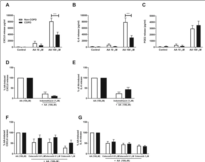
To assess the inflammatory response to dietary fatty acids in pulmonary fibroblasts from COPD versus non-COPD patients, cells were treated with 10 and 100 μM of AA, DHA or PA for 48 h and CXCL8 and IL-6 release was measured. Challenge with DHA or PA did not induce cytokine release from fibroblasts from both COPD and non-COPD patients (data not shown). However, challenge with AA resulted in increased CXCL8 and IL-6 release in both groups. Interestingly, CXCL8 and IL-6 release was greater in the non-COPD group (n= 36) compared to the COPD group (n= 19) (p < 0.001) (Fig. 1a and b). The non-COPD group consists of patients with non-smoking related end-stage lung dis-eases such as pulmonary hypertension and idiopathic pulmonary fibrosis, as well as cells from macroscopically normal lung tissue obtained from resection surgery. As it was possible that pulmonary fibroblasts from patients with disease such as pulmonary hypertension and idio-pathic pulmonary fibrosis had a differential response to fibroblasts from macroscopically normal lung tissue we
investigated if they respond differently to AA. Fibro-blasts derived from patients with non-smoking re-lated end-stage lung disease (n = 24) and patients who were going lung resection for thoracic malig-nancies (n = 12) had similar AA-induced cytokine re-lease (Additional file 1: Fig. S1). These results justify combining these two groups in the non-COPD group and indicate a disease-specific effect of COPD, rather than an effect of smoking.
The response to arachidonic is partially mediated through cyclooxygenase with no differences between COPD and non-COPD
AA can act as a bio-active molecule, and is the precur-sor that is metabolized by COX to produce prostaglan-dins, including PGE2. Prostaglandins are known to play a key role in the generation of inflammatory responses. Since the response to AA is different in COPD versus non-COPD, we next assessed whether AA-induced cytokine release is prostaglandin-dependent and whether there are differences in the effect of inhibition of COX on AA-induced cytokine release. We measured the release of PGE2 and found that PGE2 levels are in-creased upon challenge with AA 100μM in both COPD (n= 11) and non-COPD (n= 12) fibroblasts, with no differences between the two groups (Fig. 1c). The non-selective COX-inhibitor indomethacin and the COX-2 selective inhibitor, celecoxib at a concentration of 1μM both partially suppressed AA-induced IL-6 and CXCL8 release. There were no differences in the per-centage of inhibition of AA-induced cytokine release in the COPD versus the non-COPD group at all concen-trations of indomethacin or celecoxib used (Fig.1d, e, f
and g). These results show that the response to AA in fibroblasts is partially mediated through downstream mediators of COX-2, and partially mediated through COX-2 independent mechanisms.
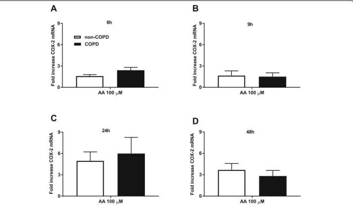
Simular AA-induced COX-2 mRNA expression in COPD and non-COPD
Arachidonic acid does not cause cytotoxicity in COPD and non-COPD fibroblasts
To investigate whether reduced viability is the cause of the impaired response of COPD fibroblasts to AA, we performed a lactate dehydrogenase (LDH) assay and found that AA does not cause cytotoxicity in both COPD (n= 7) and non-COPD (n= 9) fibroblasts (Fig.3).
No differential response to TNFαin COPD versus non-COPD
We also assessed the inflammatory response to TNFα, which is a pro-inflammatory cytokine and known to be elevated in obese individuals. TNFα induced IL-6 and CXCL8 release from fibroblasts from COPD (n = 15) and
non-COPD patients (n = 27), but there was no difference between the two groups (Fig.4).
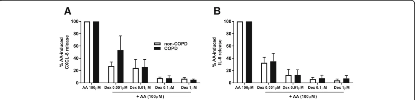
Dexamethasone suppresses AA-induced cytokine release in COPD and non-COPD
occurred in the COPD cells, suggesting a reduced ster-oid sensitivity in COPD versus non-COPD cells.
Body mass index does not affect the production of inflammatory cytokines
Since BMI is associated with clinical outcomes in COPD, we investigated if BMI affects the production of inflam-matory cytokines upon challenge with AA (100μM). We found no associations between CXCL8 or IL-6 release and BMI in either the non-COPD (n = 32) or the COPD group (n = 18) (Fig.6).
Arachidonic acid reduces basal ECM protein mRNA expression and deposition
Since lung pathology is different in obese COPD com-pared to non-obese COPD, we assessed whether chal-lenge with dietary fatty acids leads to changes in the expression and deposition of ECM proteins. Challenge with AA (100 μM) resulted in significantly reduced fi-bronectin and type I collagen mRNA expression com-pared to constitutive levels (n= 5, p< 0.01) (Fig. 7aand
b) in COPD fibroblasts. There was no effect on TNC mRNA expression (Fig. 7c). The effect of AA on fibro-nectin mRNA expression was specific for COPD cells, whereas AA also decreased basal expression of type I Fig. 2Similar arachidonic acid-induced COX-2 mRNA expression in COPD and non-COPD. Pulmonary fibroblasts from COPD (n = 5)and non-COPD patients (n = 5) were unstimulated (control) or challenged withω-6 PUFA arachidonic acid (AA) in 0.1% BSA-DMEM (100μM) for 6 (a), 9 (b), 24 (c) or 48 (d) hours. Total RNA was extracted and cyclooxygenase (COX)-2 mRNA expression was measured using qPCR. The results are normalized to the endogenous control (18S rRNA), and presented as fold change from control (t= 0 h) ± standard error of the mean. Unpaired t-test was used to determined statistical significance. There were no statistical differences
collagen in non-COPD cells (n= 5, p< 0.05). Challenge with DHA or PA did not affect ECM protein expression (data not shown) in COPD and non-COPD cells.
As mRNA levels do not always reflect corresponding protein levels, the deposition of ECM proteins upon challenge with AA 100 μM were also measured using ECM ELISA. Challenge with AA (100μM) resulted in a reduced basal deposition of all ECM proteins measured (fibronectin, tenascin, type I collagen and perlecan) in COPD fibroblasts (n= 5–7) (p< 0.05). In non-COPD fi-broblasts (n= 4–6) challenge with AA reduced the basal deposition of perlecan and type I collagen (p< 0.05), but not fibronectin or tenascin (Fig.8).
Discussion
This study explored the relationship between dietary fatty acids and airway inflammation and remodelling in COPD using human pulmonary fibroblasts. We found that the ω-6 PUFA AA causes substantial CXCL8 and IL-6 release, and interestingly, this was impaired in fi-broblasts from COPD patients. ω-3 PUFAs or SFAs did
not induce inflammatory responses in pulmonary fibro-blasts from either group.
Pulmonary fibroblasts are the major structural cell of the airway and are found at the interface of the lumen and the blood supply and, therefore, they are directly ex-posed to components of blood, including nutrients. These cells play a crucial role in tissue homeostasis and the production of pro-inflammatory cytokines and ECM proteins, and, therefore are likely to contribute to airway inflammation and remodelling in COPD [25,26].
Typically, Western diets are deficient in ω-3 PUFAs and contain excessive amounts ofω-6 PUFAs and SFAs. ω-6 PUFAs and SFAs have predominantly been associ-ated with pro-inflammatory effects, whereas the ω-3 PUFAs are predominantly anti-inflammatory [35]. West-ern dietary pattWest-erns have been associated with an in-creased risk of newly diagnosed COPD [36,37] and with greater lung function decline in COPD [38]. We found a pro-inflammatory effect of AA, with impaired cytokine release occurring in COPD cells.
Potential beneficial effects of ω-3 PUFAs have been observed in many diseases, and there is some evidence
A
B
Fig. 4Similar cytokine release upon TNFαchallenge in COPD versus non-COPD. Pulmonary fibroblasts from COPD (n = 15) and non-COPD patients (n = 27) were unstimulated (control) or challenged with TNFαin 0.1% BSA-DMEM (1 ng/ml) for 48 h. Cell free supernatants were collected and CXCL8 (a) and IL-6 (b) release was measured using ELISA. All data are represented as mean ± standard error of the mean. Two-way ANOVA with Bonferroni post-hoc testing was used to determine statistical significance. There were no statistical differences
A
B
of a potential role in COPD [39,40]. Several studies have shown that ω-3 PUFAs reduce inflammation by inhibit-ing TLR4 signallinhibit-ing [24]. In addition, observational stud-ies have shown that diets rich in fruit, vegetables and ω-3 PUFAs are positively associated with FEV1 and FVC in the general population [41], and reduce the risk of developing of COPD [36, 37, 42, 43]. We found no pro-inflammatory effects of ω-3 PUFAs which is
consistent with most literature, however this study did not investigate potential anti-inflammatory effects. Fur-thermore, SFAs did not induce cytokine release from pulmonary fibroblasts. SFAs, including PA, initiate in-nate immune responses via TLR 2 and 4 signalling [44– 48]. The non-responsiveness to SFAs in this study may be explained by the lack of functional TLR4/CD14 sig-nalling in pulmonary fibroblasts [49,50].
The majority of the patients in the COPD and non-COPD group were smokers, however the percent-age of smokers was higher in the COPD group. The non-COPD group consists of patients with non-smoking related end-stage lung diseases, as well as cells from macroscopically normal lung tissue obtained from resec-tion surgery. We confirmed that there was no difference in the response to AA between these two groups. Fur-thermore, TNFα induced similar cytokine release in the COPD and non-COPD group. Taken together this shows that COPD fibroblasts have a specific impairment in their response to AA, independent of smoking history.
The current study measured IL-6 and CXCL8 as these mediators are important in the pathogenesis of COPD and are increased in serum and BAL fluid of COPD pa-tients [15, 17]. IL-6 is a marker of systemic inflamma-tion, a predictor of mortality and is negatively correlated with lung function [15, 51]. CXCL8 is a potent neutro-phil chemoattractant and activates neutroneutro-phils, leading to the secretion of reactive oxygen metabolites, inflam-matory cytokines and tissue damaging enzymes. In this study AA induced lower levels of IL-6 and CXCL-8 in COPD versus non-COPD pulmonary fibroblasts, sug-gesting that in COPD meals rich in ω-6 PUFAs are not as potent in the induction of inflammatory responses compared to other chronic lung diseases.
AA affects inflammation through TLR4 independent mechanisms. It acts as a bio-active molecule and is converted into eicosanoids, including prostaglandins, through metabolism by COX. Prostaglandins are known
a
b
Fig. 6No correlation between BMI and arachidonic acid induced-CXCL8 and IL-6 release. Body mass index (BMI) was correlated with CXCL8 (a) and IL-6 (b) release upon challenge with arachidonic acid (AA) (100μM), in fibroblasts from both COPD (n = 18)and non-COPD patients (n = 32). The correlation coefficient (r) was determined using linear regression (Pearson analysis). There were no correlations between BMI and CXCL8 or IL-6 release
A
B
C
to play a key role in the generation of inflammatory re-sponses [52] and have been shown to affect cytokine production in immune and lung cells [53, 54]. AA in-duced prostaglandin E2 (PGE2) release, but there was no difference between COPD and non-COPD fibroblasts. The non-selective COX-inhibitor, indomethacin, and the COX-2 selective inhibitor, celecoxib both partially sup-pressed AA-induced IL-6 and CXCL8 release showing that cytokine induction also occurs via COX-2 inde-pendent mechanisms. There were no differences in the percentage of inhibition between COPD and non-COPD fibroblasts. In addition, there were no differences in AA-induced COX-2 mRNA expression between COPD and non-COPD cells.
Since BMI is associated with clinical outcomes in COPD, we investigated whether obesity itself affects the response to AA in pulmonary fibroblasts. We found no effect of BMI on AA-induced CXCL8 or IL-6 release from pulmonary fibroblasts, in both groups. Epidemio-logical studies have reported that moderate obesity in moderate to severe COPD has protective effects on sur-vival, lung function decline and exacerbations [5–8]. Cai et al. (2003) showed beneficial effects of high fat diet versus low fat diet on lung function in COPD. These ef-fects of obesity are contradictory to the known deleteri-ous effects of obesity in the general population and
other diseases [55]. McDonald et al. (2016) investigated the effect of exercise and a low-energy diet in obese COPD patients. They found some clinically significant improvements on COPD outcomes including health sta-tus, but no effects on inflammatory markers and lung function [56]. Clearly, more studies are needed to under-stand the effects of obesity and diet in COPD.
We investigated the ability of dietary fatty acids to affect ECM protein mRNA expression and deposition. AA, but not DHA or PA reduced basal fibronectin and type I collagen mRNA expression and fibronectin, type I collagen, tenascin and perlecan deposition in COPD fibroblasts. The inhibitory effect of AA on ECM protein expression and deposition was less substantial in non-COPD cells.
Fibronectin and tenascin are increased in COPD air-ways and their presence is correlated with airway remod-elling and/or inflammation and is negatively correlated with FEV1 [11, 57, 58]. The effect of obesity on airway remodelling is not well established. However, BMI has been negatively associated with the severity of emphy-sema, independent of gender, age and smoking history and positively associated with airway wall thickness [12]. Our results show that AA suppresses the basal depos-ition of fibronectin, type I collagen, tenascin and perlecan suggesting that AA and possibly other dietary
A
C
D
B
factors could play a role in the regulation of ECM de-position in COPD. ECM proteins play an essential role in maintaining tissue homeostasis affecting many cellular processes including proliferation, migration and repair [59]. Decreased levels of ECM proteins could lead to in-adequate repair mechanisms in COPD.
One limitation of our study is the lack of pulmonary fibroblasts from obese and severely obese COPD and non-COPD patients. In addition, the BMI was lower in the COPD group compared to the non-COPD group. This occurred by chance, as we did not select patients on the basis of BMI; rather we used samples as they were available. It may be that the protective effects of obesity account for the lack of samples from the obese COPD population, as if patients have reduced severity of COPD they would not need lung transplantation. Inter-estingly, a recent meta-analysis has shown that BMI is also inversely associated with lung cancer [60], which could explain the limited resection samples from obese patients with a thoracic malignancy.
Conclusion
Our study demonstrates thatω-6 PUFA AA, but notω-3 PUFA DHA or SFA PA, affects inflammatory processes and ECM deposition in COPD. We found that whilst AA increases inflammation, pulmonary fibroblasts from patients with COPD had a reduced response to AA in comparison to cells from people without COPD. Obesity itself was not associated with the inflammatory response. Moreover, we found that AA had a more substantial in-hibitory effect on basal ECM-protein expression and de-position in COPD cells compared to non-COPD cells. This study suggest that the dietary fatty acid AA and possibly other dietary components have disease-specific effects and could explain differential effects of high fat diets in different lung diseases. The impaired response to AA in COPD might in part explain why obesity ap-pears to have less detrimental effects in COPD, com-pared to other lung diseases.
Additional file
Additional file 1:Figure S1.Similar response to arachidonic acid in patients with non-smoking related end-stage lung disease and patients who underwent lung resection for thoracic malignancies. (DOC 153 kb)
Abbreviations
18sRNA:18S ribosomal RNA; AA: Arachidonic acid; ANOVA: Analysis of variance; BMI: Body mass index; BSA: Bovine serum albumin; COPD: Chronic obstructive pulmonary disease; COX: Cyclooxygenase; CXCL8: Chemokine (C-X-C motif) ligand 8; DHA: Docosahexaenoic acid; ECM: Extracellular matrix; FBS: Fetal bovine serum; IL-6: Interleukin 6; PA: Palmitic acid;
PUFA: Polyunsaturated fatty acid; qPCR: Quantitative polymerase chain reaction; SEM: Standard error of the mean; SFA: Saturated fatty acid
Acknowledgements
We would like to acknowledge the collaborative effort of the cardiopulmonary transplant team and the pathologists at St Vincent’s Hospital (Sydney, Australia), and the thoracic physicians and pathologists at the Royal Prince Alfred Hospital (Sydney) and Strathfield Private Hospital (Strathfield, Australia).
Funding
PH is supported by a fellowship from the National Health and Medical Research Council (NHMRC) of Australia, a Brawn Fellowship from the Faculty of Health and Medicine and funding from and the Rainbow Foundation. The authors thank F. Thomson and M. Thomson for their continued support.BO is supported by a fellowship from the National Health and Medical Research Council (NHMRC) of Australia (APP1110368). LW is supported by a fellowship from the National Health and Medical Research Council (NHMRC) of Australia.
Availability of data and materials
The datasets used and analysed during the current study are available from the corresponding author on request.
Authors’contributions
SR, MP, and BO conceived and wrote the manuscript. LW, DX, PH and BO contributed to the analysis or interpretation of the data. AM, DX, PH and LW revised the manuscript. SR, MP and AM carried out the experiments. All authors read and approved the final manuscript.
Ethics approval and consent to participate
Approval for all experiments with human lung was provided by the Human Ethics Committees (IRB) of the Sydney South West Area Health Service approval number X14–0045 & HREC/09/RPAH/630 and St Vincent’s Hospital IRB approval number SSA/16/SVH/39.
Consent for publication Not applicable.
Competing interests
The authors declare that they have no competing interests.
Publisher’s Note
Springer Nature remains neutral with regard to jurisdictional claims in published maps and institutional affiliations.
Author details
1Respiratory Cellular and Molecular Biology, Woolcock Institute of Medical
Research, The University of Sydney, Sydney, Australia.2Priority Research Centre for Healthy Lungs, Hunter Medical Research Institute and The University of Newcastle, Newcastle, NSW, Australia.3School of Life Sciences,
University of Technology Sydney, Sydney, Australia.
Received: 18 July 2018 Accepted: 21 October 2018
References
1. Ng M, Fleming T, Robinson M, Thomson B, Graetz N, Margono C, Mullany EC, Biryukov S, Abbafati C, Abera SF, et al. Global, regional, and national prevalence of overweight and obesity in children and adults during 1980-2013: a systematic analysis for the global burden of disease study 2013. Lancet. 2014;384:766–81.
2. Vozoris NT, O'Donnell DE. Prevalence, risk factors, activity limitation and health care utilization of an obese, population-based sample with chronic obstructive pulmonary disease. Can Respir J. 2012;19:e18–24.
3. Steuten LM, Creutzberg EC, Vrijhoef HJ, Wouters EF. COPD as a
multicomponent disease: inventory of dyspnoea, underweight, obesity and fat free mass depletion in primary care. Prim Care Respir J. 2006;15:84–91. 4. Eisner MD, Blanc PD, Sidney S, Yelin EH, Lathon PV, Katz PP, Tolstykh I,
Ackerson L, Iribarren C. Body composition and functional limitation in COPD. Respir Res. 2007;8:7.
5. Zapatero A, Barba R, Ruiz J, Losa JE, Plaza S, Canora J, Marco J. Malnutrition and obesity: influence in mortality and readmissions in chronic obstructive pulmonary disease patients. J Hum Nutr Diet. 2013;26 Suppl 1:16–22. 6. Hallin R, Gudmundsson G, Suppli Ulrik C, Nieminen MM, Gislason T,
long-term mortality in hospitalised patients with chronic obstructive pulmonary disease (COPD). Respir Med. 2007;101:1954–60.
7. Lainscak M, von Haehling S, Doehner W, Sarc I, Jeric T, Ziherl K, Kosnik M, Anker SD, Suskovic S. Body mass index and prognosis in patients hospitalized with acute exacerbation of chronic obstructive pulmonary disease. J Cachexia Sarcopenia Muscle. 2011;2:81–6.
8. Celli BR, Thomas NE, Anderson JA, Ferguson GT, Jenkins CR, Jones PW, Vestbo J, Knobil K, Yates JC, Calverley PM. Effect of pharmacotherapy on rate of decline of lung function in chronic obstructive pulmonary disease: results from the TORCH study. Am J Respir Crit Care Med. 2008;178:332–8. 9. Mair G, Maclay J, Miller JJ, McAllister D, Connell M, Murchison JT, MacNee W.
Airway dimensions in COPD: relationships with clinical variables. Respir Med. 2010;104:1683–90.
10. Han MK, Kazerooni EA, Lynch DA, Liu LX, Murray S, Curtis JL, Criner GJ, Kim V, Bowler RP, Hanania NA, et al. Chronic obstructive pulmonary disease exacerbations in the COPDGene study: associated radiologic phenotypes. Radiology. 2011;261:274–82.
11. Annoni R, Lancas T, Yukimatsu Tanigawa R, de Medeiros Matsushita M, de Morais Fernezlian S, Bruno A, Fernando Ferraz da Silva L, Roughley PJ, Battaglia S, Dolhnikoff M, et al. Extracellular matrix composition in COPD. Eur Respir J. 2012;40:1362–73.
12. Gu S, Li R, Leader JK, Zheng B, Bon J, Gur D, Sciurba F, Jin C, Pu J. Obesity and extent of emphysema depicted at CT. Clin Radiol. 2015;70:e14–9. 13. Guerra S, Sherrill DL, Bobadilla A, Martinez FD, Barbee RA. The relation of
body mass index to asthma, chronic bronchitis, and emphysema. Chest. 2002;122:1256–63.
14. Di Stefano A, Caramori G, Ricciardolo FL, Capelli A, Adcock IM, Donner CF. Cellular and molecular mechanisms in chronic obstructive pulmonary disease: an overview. Clin Exp Allergy. 2004;34:1156–67.
15. Donaldson GC, Seemungal TA, Patel IS, Bhowmik A, Wilkinson TM, Hurst JR, Maccallum PK, Wedzicha JA. Airway and systemic inflammation and decline in lung function in patients with COPD. Chest. 2005;128:1995–2004. 16. O'Donnell R, Breen D, Wilson S, Djukanovic R. Inflammatory cells in the
airways in COPD. Thorax. 2006;61:448–54.
17. Chung KF. Cytokines in chronic obstructive pulmonary disease. Eur Respir J Suppl. 2001;34:50s–9s.
18. Bastard JP, Maachi M, Lagathu C, Kim MJ, Caron M, Vidal H, Capeau J, Feve B. Recent advances in the relationship between obesity, inflammation, and insulin resistance. Eur Cytokine Netw. 2006;17:4–12.
19. Maachi M, Pieroni L, Bruckert E, Jardel C, Fellahi S, Hainque B, Capeau J, Bastard JP. Systemic low-grade inflammation is related to both circulating and adipose tissue TNFalpha, leptin and IL-6 levels in obese women. Int J Obes Relat Metab Disord. 2004;28:993–7.
20. Hanson C, Rutten EP, Wouters EF, Rennard S. Diet and vitamin D as risk factors for lung impairment and COPD. Transl Res. 2013;162:219–36. 21. Grievink L, Smit HA, Ocke MC, van‘t Veer P, Kromhout D. Dietary intake of
antioxidant (pro)-vitamins, respiratory symptoms and pulmonary function: the MORGEN study. Thorax. 1998;53:166–71.
22. Woods RK, Raven JM, Walters EH, Abramson MJ, Thien FC. Fatty acid levels and risk of asthma in young adults. Thorax. 2004;59:105–10.
23. Wood LG, Garg ML, Gibson PG. A high-fat challenge increases airway inflammation and impairs bronchodilator recovery in asthma. J Allergy Clin Immunol. 2011;127:1133–40.
24. Rogero MM, Calder PC. Obesity, inflammation, toll-like receptor 4 and fatty acids. Nutrients. 2018;5:e432.
25. Kendall RT, Feghali-Bostwick CA. Fibroblasts in fibrosis: novel roles and mediators. Front Pharmacol. 2014;5:123.
26. White ES. Lung extracellular matrix and fibroblast function. Ann Am Thorac Soc. 2015;12(Suppl 1):S30–3.
27. Krimmer D, Ichimaru Y, Burgess J, Black J, Oliver B. Exposure to biomass smoke extract enhances fibronectin release from fibroblasts. PLoS One. 2013;8:e83938.
28. Gupta S, Knight AG, Gupta S, Keller JN, Bruce-Keller AJ. Saturated long-chain fatty acids activate inflammatory signaling in astrocytes. J Neurochem. 2012; 120:1060–71.
29. Pillon NJ, Arane K, Bilan PJ, Chiu TT, Klip A. Muscle cells challenged with saturated fatty acids mount an autonomous inflammatory response that activates macrophages. Cell Commun Signal. 2012;10:30.
30. Abdelmagid SA, Clarke SE, Nielsen DE, Badawi A, El-Sohemy A, Mutch DM, Ma DW. Comprehensive profiling of plasma fatty acid concentrations in young healthy Canadian adults. PLoS One. 2015;10:e0116195.
31. Chen X, Iqbal N, Boden G. The effects of free fatty acids on
gluconeogenesis and glycogenolysis in normal subjects. J Clin Invest. 1999; 103:365–72.
32. Raatz SK, Bibus D, Thomas W, Kris-Etherton P. Total fat intake modifies plasma fatty acid composition in humans. J Nutr. 2001;131:231–4. 33. Cranmer-Byng MM, Liddle DM, De Boer AA, Monk JM, Robinson LE.
Proinflammatory effects of arachidonic acid in a lipopolysaccharide-induced inflammatory microenvironment in 3T3-L1 adipocytes in vitro. Appl Physiol Nutr Metab. 2015;40:142–54.
34. Kuo C, Lim S, King NJ, Johnston SL, Burgess JK, Black JL, Oliver BG. Rhinovirus infection induces extracellular matrix protein deposition in asthmatic and nonasthmatic airway smooth muscle cells. Am J Physiol Lung Cell Mol Physiol. 2011;300:L951–7.
35. Simopoulos AP. An increase in the Omega-6/Omega-3 fatty acid ratio increases the risk for obesity. Nutrients. 2016;8:128.
36. Varraso R, Fung TT, Hu FB, Willett W, Camargo CA. Prospective study of dietary patterns and chronic obstructive pulmonary disease among US men. Thorax. 2007;62:786–91.
37. Varraso R, Fung TT, Barr RG, Hu FB, Willett W, Camargo CA Jr. Prospective study of dietary patterns and chronic obstructive pulmonary disease among US women. Am J Clin Nutr. 2007;86:488–95.
38. McKeever TM, Lewis SA, Cassano PA, Ocke M, Burney P, Britton J, Smit HA. Patterns of dietary intake and relation to respiratory disease, forced expiratory volume in 1 s, and decline in 5-y forced expiratory volume. Am J Clin Nutr. 2010;92:408–15.
39. de Batlle J, Barreiro E, Romieu I, Mendez M, Gomez FP, Balcells E, Ferrer J, Orozco-Levi M, Gea J, Anto JM, Garcia-Aymerich J. Dietary modulation of oxidative stress in chronic obstructive pulmonary disease patients. Free Radic Res. 2010;44:1296–303.
40. Sugawara K, Takahashi H, Kasai C, Kiyokawa N, Watanabe T, Fujii S, Kashiwagura T, Honma M, Satake M, Shioya T. Effects of nutritional supplementation combined with low-intensity exercise in malnourished patients with COPD. Respir Med. 2010;104:1883–9.
41. Shaheen SO, Jameson KA, Syddall HE, Aihie Sayer A, Dennison EM, Cooper C, Robinson SM. The relationship of dietary patterns with adult lung function and COPD. Eur Respir J. 2010;36:277–84.
42. Tabak C, Smit HA, Heederik D, Ocke MC, Kromhout D. Diet and chronic obstructive pulmonary disease: independent beneficial effects of fruits, whole grains, and alcohol (the MORGEN study). Clin Exp Allergy. 2001;31: 747–55.
43. Hirayama F, Lee AH, Binns CW, Zhao Y, Hiramatsu T, Tanikawa Y, Nishimura K, Taniguchi H. Do vegetables and fruits reduce the risk of chronic obstructive pulmonary disease? A case-control study in Japan. Prev Med. 2009;49:184–9.
44. Ajuwon KM, Spurlock ME. Palmitate activates the NF-kappaB transcription factor and induces IL-6 and TNFalpha expression in 3T3-L1 adipocytes. J Nutr. 2005;135:1841–6.
45. Huang S, Rutkowsky JM, Snodgrass RG, Ono-Moore KD, Schneider DA, Newman JW, Adams SH, Hwang DH. Saturated fatty acids activate TLR-mediated proinflammatory signaling pathways. J Lipid Res. 2012;53:2002–13. 46. Lee JY, Zhao L, Youn HS, Weatherill AR, Tapping R, Feng L, Lee WH,
Fitzgerald KA, Hwang DH. Saturated fatty acid activates but polyunsaturated fatty acid inhibits toll-like receptor 2 dimerized with toll-like receptor 6 or 1. J Biol Chem. 2004;279:16971–9.
47. Nguyen MT, Favelyukis S, Nguyen AK, Reichart D, Scott PA, Jenn A, Liu-Bryan R, Glass CK, Neels JG, Olefsky JM. A subpopulation of macrophages infiltrates hypertrophic adipose tissue and is activated by free fatty acids via toll-like receptors 2 and 4 and JNK-dependent pathways. J Biol Chem. 2007; 282:35279–92.
48. Schaeffler A, Gross P, Buettner R, Bollheimer C, Buechler C, Neumeier M, Kopp A, Schoelmerich J, Falk W. Fatty acid-induced induction of toll-like receptor-4/nuclear factor-kappaB pathway in adipocytes links nutritional signalling with innate immunity. Immunology. 2009;126:233–45. 49. Xing Z, Jordana M, Braciak T, Ohtoshi T, Gauldie J. Lipopolysaccharide
induces expression of granulocyte/macrophage colony-stimulating factor, interleukin-8, and interleukin-6 in human nasal, but not lung, fibroblasts: evidence for heterogeneity within the respiratory tract. Am J Respir Cell Mol Biol. 1993;9:255–63.
51. Celli BR, Locantore N, Yates J, Tal-Singer R, Miller BE, Bakke P, Calverley P, Coxson H, Crim C, Edwards LD, et al. Inflammatory biomarkers improve clinical prediction of mortality in chronic obstructive pulmonary disease. Am J Respir Crit Care Med. 2012;185:1065–72.
52. Ricciotti E, FitzGerald GA. Prostaglandins and inflammation. Arterioscler Thromb Vasc Biol. 2011;31:986–1000.
53. Kalinski P. Regulation of immune responses by prostaglandin E2. J Immunol. 2012;188:21–8.
54. Tavakoli S, Cowan MJ, Benfield T, Logun C, Shelhamer JH. Prostaglandin E(2)-induced interleukin-6 release by a human airway epithelial cell line. Am J Physiol Lung Cell Mol Physiol. 2001;280:L127–33.
55. Cai B, Zhu Y, Ma Y, Xu Z, Zao Y, Wang J, Lin Y, Comer GM. Effect of supplementing a high-fat, low-carbohydrate enteral formula in COPD patients. Nutrition. 2003;19:229–32.
56. McDonald VM, Gibson PG, Scott HA, Baines PJ, Hensley MJ, Pretto JJ, Wood LG. Should we treat obesity in COPD? The effects of diet and resistance exercise training. Respirology. 2016;21:875–82.
57. Laitinen A, Altraja A, Kampe M, Linden M, Virtanen I, Laitinen LA. Tenascin is increased in airway basement membrane of asthmatics and decreased by an inhaled steroid. Am J Respir Crit Care Med. 1997;156:951–8.
58. Kranenburg AR, Willems-Widyastuti A, Moori WJ, Sterk PJ, Alagappan VK, de Boer WI, Sharma HS. Enhanced bronchial expression of extracellular matrix proteins in chronic obstructive pulmonary disease. Am J Clin Pathol. 2006; 126:725–35.
59. Bonnans C, Chou J, Werb Z. Remodelling the extracellular matrix in development and disease. Nat Rev Mol Cell Biol. 2014;15:786–801. 60. Yang Y, Dong J, Sun K, Zhao L, Zhao F, Wang L, Jiao Y. Obesity and