Intraventricular Neurocysticercosis: A Case Report
Ruzbih Bahtiar1), Ferry Wijanarko2)
1)General Surgery Resident, Faculty of Medicine, Universitas Sebelas Maret 2)Neurosurgery Consultant, Dr. Moewardi Hospital, Surakarta
ABSTRACT
Background: Neurocysticercosis is the most common parasitic disease of nervous system in humans and the single most common cause of acquired epileptic seizures in the developing country. But in Indonesia, it was rarely reported. It has wide variety of clinical manifestation due to its pleomorphic nature.
Subjects and Method:We reported a case of a 12 years old male with seizure and hydrocephalus due to intraventricular neurocysticercosis. A ventriculoperitoneal shunt (V-P shunt) performed to reduce the intracranial pressure due to hydrocephalus and followed by endoscopic surgery to taking sample of the neurocysticercosis and reducing the mass.
Results: Pathological anatomy examination of excised mass found worm eggs. The patient received medications of albendazole and metil prednisolon.
Conclusion: Intraventricular neurocysticercosis causing hydrocephalus is the commonest surgical indication, with V-P shunt placement followed by endoscopic surgery as the procedure of choice as it minimally invasive.
Keyword: Intraventricular neurocysticercosis, hydrocephalus, headache, V-P shunt, endoscopic surgery.
Correspondence:
Ruzbih Bahtiar. General Surgery Resident, Faculty of Medicine, Universitas Sebelas Maret. Jl. Ir. Sutami 36A, Surakarta 57126, Central Java.
BACKGROUND
Cysticercosis is a disease caused by a parasitic cyst/larvae infection of the Taenia solium tapeworm (T. solium) at the organs of the body, such as muscles, eyes, skin, brain and other structures. When attacking the nervous system, referred to as neuro-cysticercosis (Gripper and Welburn, 2017).
Taxonomically, T. solium worms are included in Platyhelminthes phylum, Cestoda class, cyclophellidea order, Taenii-dae family, genus taenia. Habitat of this worm is on the intestinal wall and its length can reach 2-7 meters. The body consists of head, neck and strobila consisting of proglotid segments. In a mature proglotid there are reproductive organs. The terminal end of the proglotide will be released from the strobila and excreted out with the stool.
Eggs will be removed at the time before and after the proglotid release (Wiria, 2008).
little immune response, so it can survive for several years.
T. solium could extend to 1 cm in 2-3 months. Adult tapeworms could reach a length of 2-7 meters and produce more than 1000 proglottids and each proglottids contains about 50,000 eggs and lived in the small intestine for years (Wiria, 2008).
Outside the nervous system, cysticer-cosis does not cause significant symptoms, whereas in the nervous system it will cause various clinical manifestations due to its pleomorphic origin, from asymptomatic to endanger patient’s live, such as headache, dizziness, seizures, stroke, dementia/ neurobehavioral disorder, diplopia, hydro-cephalus, epilepsy, migraine, arachnoiditis, and increased intracranial pressure. The symptoms could be subacute or chronic and depend on location, number of parasites, stage of cyst at site, and patient’s immune response. Manifestations of seizures that may arise can be general tonic clonic seizu-res or simple or complex partial seizuseizu-res (Gripper and Welburn, 2017).
Neurocysticercosis is the leading cause of acquired seizures and epilepsy in developing countries, especially in Latin America, India, Africa, and China. In Indo-nesia this disease is endemic especially in North Sumatra, Bali, Papua, Timor, Flores, North Sulawesi and West Kalimantan. Raka (1998) reported that 68% of patients with neurocysticercosis are having epilepsy attack. In the United States there is an increase of the neurocysticercosis incidence since the 1980s due to immigration from Latin America to the US. The spreading in non-endemic areas associated with immi-grants, as neurocysticercosis is a disease that transmission can occur between humans (Wandra et al., 2007).
Case Report
A 12 years old boy with complaint of headache that being felt since 6 months ago
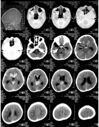
and become worsening, unconscious (-), vomiting (-), seizures (-), history of trauma (-). The patient lives in a poor sanitation environment (neighboring chicken and pig breeders). Two months ago, he was brought by his family to RS PKU Muhammadiyah Solo, handled by Neurologist, head CT-scan performed, hydrocephalus non comunicans and intraventricular mass have been found.
The patient was then consulted to a neurosurgeon and V-P shunt surgery was being performed. Afterwards head MRI was being performed, multiple solid dominant masses at the right and left lateral intraventricles have been found, especially at the anterior cornu, ventricle III, accom-panied by left lateral ventricular dilatation (hydrocephalus non comunicans), resulting slight midline shifting, concluded as choroid tumor plexus.
Figure 2. Head MRI and MRA ahead of neuroendoscopic tumor excision
A month ago, a neuroendoscopic surgery was performed with partial excision of tumor mass. From the results of the PA, worm eggs have being found and suspected as an intraventricular parasitic worm infection. Patient treated with medications of Albendazole and Metil Prednisolone for 1 month.
Figure 3. Pathological anatomy results post neuroendoscopic
excision of tumor
DISCUSSION
Neurocysticercosis has no prevalence of age or gender. Children patients are usually asymptomatic, and there are some differences in child and adult cases: (Zammarchi et al., 2016).
• Children are rarely affected by neuro-cysticercosis due to the difference of transmission mechanism and immune system response to the parasite.
• Asymptomatic neurocysticcosis is more commonly detected by MRI in patients aged > 15 years than < 15 years.
• Adults have higher incidence of headache, extra parenchymal neuro-cysticercosis, increased intra cranial pressure, changes in cerebrospinal fluid examination results, focal neurodeficitis, depression syndrome, and seizure recurrence after discontinuation of anti convulsant administration.
• Children are more likely to experience convulsions and single colloidal parenchymal cysts
When cysticercosis reaches the brain, there are 6 clinical syndromes that could manifestate based on the location of the cyst, namely:
1. Asymptomatic neurocysticercosis
Some patients do not show significant symptoms and the finding of neuro-cysticercosis is usually incidental.
2. Parenchymal neurocysticercosis
number of cysts present in the brain (Rajshekhar, 2016; Zafar, 2016; Gripper and Welburn, 2017).
3. Subarachnoid neurocysticsercosis (racemose)
This form occurs because the cyst located in the subarachnoid space and grows abnor-mally into a large mass and/or racemose cyst and supressing surrounding structures and endanger the patient due to mass effect. Clinical symptoms, approaches and treatments are different from those in parenchyma. Clinical symptoms are often found to be a sign of meningeal excitability, due to the inflammatory process of the meninges and, if continued, may lead to lacunar and hydrocephalus infarction. In general, prognosis is worse than parenchy-mal neurocysticercosis (Zafar, 2016).
The inflammation process because of this cyst could be located in suprasella, quadrigeminal sisterna and aquaductus Sylvii and usually accompanied by signs of meningitis and increased intracranial pressure. The inflammation could lead to lacunar infarction and suppression of the cranial nerves located adjacent to this inflammatory process. If the nerve of the eyeball is pressed, the muscles of the eye-ball become paralyzed, resulting in diplo-pia. If optic nerve and optic chiasma affect-ed, visual field disruption and eyesight decreased may occur.
Neurocysticercosis in the bacillary system is often fatal, especially if the lesions are extensive and located in the serebro-pontine angle and in the fissurea Silvii. If the ongoing inflammatory process conti-nues, it is necessary to provide adequate anti-inflammatory medications (Rajshe-khar, 2016; Gripper and Welburn, 2017). 4. Intraventricular neurosistisercosis
The cyst located in the ventricle and interference the CSF flow resulting to
hydrocephalus and increased intracranial pressure (Jensen and Post, 2016).
5. Spinal neurocysticercosis
This is a rare form, only 1-5% of all cases. However, it could leads to severe neuro-cysticercosis when it occurs. It could be located intramedullary or extramedullary. The most common symptoms are back pain and radiculopathy. Extramedullary is more frequent with symptoms of radicular pain, limb weakness and paresthesia, whereas intramedullary will cause paraparese, sen-sory and sphincter disorders (Rajshekhar, 2016; Gripper and Welburn, 2017).
6. Ocular neurocysticercosis
It occurs when cysticercosis reaches the eyeball, subconjunctiva, extraocular muscle and subretinal space with different symp-toms on each location. Subretinal space is the most common place. The symptoms could be visual impairment, visual field dis-ruption, retinal edema, hemorrhage, even severe blindness. Cysts located in the vitre-ous could causing eyesight decreased and often the patient feels there is something moving in the eye. Cysts in the front chamber may induce severe iridocyclitis, while intraorbital retro-ocular cysticercosis may lead to eyesight decreased due to supression of the optic nerve (Rajshekhar, 2016; Gripper and Welburn, 2017).
Neurocysticercosis is the leading cause of acquaired epileptic seizures (70-90%) in adulthood, especially in endemic areas such as in Latin America, Africa and Asia (Zammarchi et al. 2016). Seizures may
manifestate as generalized clonic tonic seizures or simple or complex partial seizures, although generally the simple partial seizure is the most common. The seizures occured due to:
• Secondary to vasculitis and infarction due to subarachnoid disturbance
• Calcification due to the presence of dead cysts (Gripper and Welburn, 2017). Epileptogenesis in patients with neuro-cysticercosis may caused by various factors, such as: inflammation, gliosis, genetic, and predilection of cyst in the frontal lobe and temporal lobes. Patient’s immune response to degeneration cysts is an important factor in epileptogenesis. In women and children, there is a clear response to parasitic infection in the brain, whereas in adults the response varies. Response in a patient's body also varies, i.e. excessive inflamma-tory responses can occur at one cyst, while at other cysts around it there are no signs of inflammation, so the focus of the seizure usually lies in cysts that degenerate or experience inflammation. There are reports showing the damage to the blood-brain barrier from the release of the serum cysticercosis component to the brain that inducing seizures.
Administration of antihelmintic also causing initiation of inflammatory response from the host to cysts that resemble radiological and histologic features such as degenerated cysts, increasing the risk of seizures. Chronic seizures are often asso-ciated with calcified granulomas, where patients have a high risk of recurrent seizures. The most epileptogenic cysts are those that are active and in the degeneration process. The cyst degenerate about 6 to 12 months after the start of treatment (Rajshekhar, 2016).
Headache could occur as a chronic headache accompanied by nausea and vomiting, and if symptoms of increasing intracranial pressure occured, hydrocepha-lus could be developing, due to cysticercosis located in the ventricular system blocking the CSF flow. Hydrocephalus could be either comunicating hydrocephalus or non
communicating hydrocephalus. The comu-nicating hydrocephalus occured due to inflammatory process, fibrosis of the arachnoid villi or inflammatory reaction in the menigen and blockage in the Luschka and Magendie foramen. Non comunicating hydrocephalus occured due to the presence of cysts in the intraventricles (Zafar, 2016).
Stroke that occurs in patients with neurocysticercosis could be infarction or bleeding. Infarction stroke occured due to blockage or blood vessels damage, while bleeding occured due to rupture of the aneurysm, both as a result of further cyst suppression (Zafar, 2016).
Neurobehavior disorders including depression and psychosis occured due to an increase in intracranial pressure. Diplopia occured due to increased intracranial pressure, arachnoiditis, or suppression of the III, IV, and VI cranial nerves.
Radiological examinations including X-ray examination, CT-Scan and MRI will resulting various image depending on the location and stage of the cyst, namely: (Zafar 2016; Gasparetto et al., 2016; Gripper and Welburn 2017).
• In the brain parenchyme :
CT-Scan will show a skolex inside nodule as a hole with dot as a pathognomonis sign of neurosistcercosis. In subsequent stages, perilesional edema and contrast absorption in degenerated cysts will occured. This stage is called colloidal stage and is often referred to as the "acute phase of ence-phalitis" of neurocysticercosis caused by an immune reaction of the patient.
a dead cysticercosis, CT-Scan and MRI will show calcification.
• On basillary system:
Head CT-Scan will not show any results, while in MRI a Leptomeningeal enhance-ment at the cranial base will be seen clearly.
• In the ventricle:
Initial CT-Scan will show isodense lesion with CSF, making it difficult to see, but in the later phase it will appear as a cystic lesion.
On MRI examination, ventricular cyst will be seen clearly, especially when using FLAIR technique. The ventricular cysts may migrate locally if the patient's head is moved (ventricular migration sign).
• In the spinal:
CT-Scan will show symmetrical enlarge-ment of the spinal cord (intramedullary cyst) or pseudoreticular spinal canal forma-tion (leptomeningeal cyst).
MRI of intramedullary cysticercosis will show a ring-enchancing lesion, whereas skolex will give a thick, oval nodule.
Laboratory tests are not always supporting the diagnosis. Eosinophilia in peripheral blood examination is only positive in 0-37% of cases. Serological tests that aim to detect antibodies, antigens and measurements of oligoclonal immunoglo-bulins or IgE, the sensitivity and specificity of antibody and cysticercosis antigens highly dependent on the examination sample (cerebrospinal or serum fluid), cyst activity (active or inactive) and examination methods. However, most of the tests that use the unfractionated antigen give rise to false-positive due to the ability of the cyst to interact with the immunoglobulin and false-negative due to the high threshold values set by the laboratory to avoid false-positive results (Rajshekhar, 2016; Gripper and Welburn, 2017)
An enzyme-linked immune-transfer blot (EITB) has a high specificity for T.
solium infection and no false positive result found, with a virtually 100% sensitivity in patients with active multiple parenchymal cysts or extraparenchymal neurocystcerco-sis. In patients with single parenchymal lesion, negative result will occur in more than 30% of patients and only positive in calcified cysticercosis. However, in patients with two or more cystic or contrast-absorbing lesions, the specificity is nearly 100% and the sensitivity is 94-98%. The serum gives a more sensitive result than CSF. Levels of parasitic antigen decreased after 3 months of albendazole adminis-tration. DNA testing is another diagnostic technique for ensuring T. solium (Rajshe-khar, 2016; Gripper and Welburn 2017).
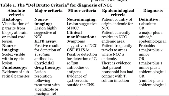
Del Brutto et al. (2001) provided the diagnostic criteria for neurocysticercosis to facilitate diagnosis (See Table 1) (Gripper and Welburn, 2017).
Treatment of neurocysticercosis patients influenced by various factors, such as cyst location, cyst stage, cyst number, inflammation process, cyst size and severity of symptoms. Treatments include: (Zafar, 2016; Rajshekhar, 2016; Gripper and Welburn, 2017).
a. Symptomatic treatment
Symptomatic treatment is given to over-come the symptoms caused by the parasite as well as the result of anti-parasitic treatment. Preparations tahat being used include:
• Anti-convulsant such as phenytoin or carbamazepine generally could control the occuring seizures.
• Manitol may be administered to reduce occuring edema.
Methotrexate may be administered to control the prolonged inflammatory process in meninges.
Table 1. The “Del Brutto Criteria” for diagnosis of NCC Absolute
criteria Major criteria Minor criteria Epidemiological criteria Diagnosis Histology:
Visualization of parasite from biopsy at brain or spinal cord lesion. Neuro-imaging: Scolex visible within cystic lesion. Fundoscopy:
Evidence of sub-retinal parasites Neuro-imaging: Lesion highly suggestive of NCC EITB assay: Positive results for detection of
T. solium antibodies. Cysticidal drug therapy: Lesion resolution following treatment with albendizole or praziquantel Neuroimaging: Lesion suggestive of NCC Clinical manifestation: Symptoms
suggestive of NCC
CSF ELISA:
Positive detection for detection of T. solium
antibodies or antigens Evidence of cysticercosis outside the CNS.
Patient country of origin endemic for NCC.
Patient currently resides in NCC endemic area. Patient frequently travels to areas where NCC is endemic.
There is evidence that patient
household has had contact with T. solium infection
Definitive:
1 absolute OR
2 major plus 1 minor/1
epidemiological. Probable: 1 major plus 2 minor
OR
1 major plus 1 minor plus 1 epidemiological OR
3 minor plus 1 epidemiological
Source: Del Brutto et al. (2001).
b. Anti parasitic treatment
The choice for neurocysticerckosis medi-cations are albendazole 15 mg/kg/day for one month and praziquantel 50 mg/kg/ day for two weeks, which has 60-85% range of eradicating parasites at the brain paren-chym. Vasques and Sotelo in their study stated that administration of albendazole can decrease the occurrence of seizures in patients with cysts less than or equal to 20, and patients will experience seizure free after 3 years. Albendazole has better pene-tration into the CSF and its concenpene-tration is not affected by corticosteroids, while prazi-quantel interacts with corticosteroids and also lowering the serum levels of phenytoin and carbamazepine.
Some authors suggest that adminis-tration of antiparasitic drugs may be unfavorable, as they may worsen the pati-ent condition, such as the gastrointestinal effects, seizures, increased intracranial
pressure, and sometimes death. Neurologic symptoms usually occur on the second day until the fifth day of antiparasit treatment, allegedly caused by the inflammatory response due to cysticercosis death, so corticosteroids should be given.
c. Surgery
Indications for surgical intervention and recommended procedures are as follows:
• Hydrocephalus due to an intraventri-cular cyst: placement of a V-P shunt, followed by surgical extirpation of the cysts and subsequent medical treatment.
• Multiple cysts in subarachnoid space (i.e. the racemose form): urgent surgical extirpation
Other several specific indications for surgery:
• Large parenchymal colloid cysts or racemose cysts that causing mass effects.
• Atypical neurocysticercosis which requi-res a definite diagnosis.
• Epilepsy that does not improve with administration of anti-convulsant drugs.
• Ocular or spinal lesions (either intrame-dullary/ extramedullary) (Rajshekhar 2016).
Intraventricular cysts are best treated with endoscopic excision as procedure of choice compared to open craniotomy and microsurgery excision since the last 2 decades. Neuroendoscopic surgery is a minimally invasive surgery performed through a single burr hole, and often act as a curative therapy in a single intra-ventricular cyst and has minimal surgical side effects when performed by experienced surgeons. Although there are concerns about the possibility of anaphylactic reactions due to rupture of the cyst during surgery, there are no reports about it (Rajshekhar 2016).
Avoid consuming foods contaminated by tapeworm eggs brought by taeniasis carriers and prevent autoinfection is the main effort to prevent cysticercosis. Furthermore, eradicating source of infect-ion transmissinfect-ion in patients with taeniasis by removing the worms and skolex is also important. Other means to prevent tape-worms infection are by sanitation improve-ment, meat health inspection, as well as meat cooking and processing (Zafar, 2016; Rajshekhar, 2016; Gripper and Welburn, 2017).
Based on the results, it can conclude that intraventricular neurocysticercosis causing hydrocephalus is the commonest surgical indication, with V-P shunt place-ment followed by endoscopic surgery as the
procedure of choice as it minimally invasive.
REFERENCE
Gasparetto EL, Alves-Leon S, Domingues FS, Frossard JT, Lopes SP, Souza JM (2016). Neurocysticercosis, familial cerebral cavernomas and intracranial calcifications: differential diagnosis for adequate management. Arquivos de Neuro-Psiquiatria, 74(6): 495– 500.
Gripper LB, Welburn SC (2017). Neuro-cysticercosis infection and disease–A review. Acta Tropica, 166: 218–224. Jensen TO, Post JJ (2016). Intraventricular
neurocysticercosis: Presentation, diagnosis and management. Asian Pacific Journal of Tropical Medicine, 9(8): 815–818.
Rajshekhar V (2016). Neurocysticercosis: Diagnostic problems & current therapeutic strategies. Indian Journal of Medical Research, 144(3): 319. Wandra T, Margono SS, Gafar MS, Saragih
JM, Sutisna P, Dharmawan NS, Sudew AAR, et al. (2007). Taeniasis/ cysticercosis in indonesia, 1996-2006. Southeast Asian Journal of Tropical Medicine and Public Health, 38(1): 140–143.
Wiria AE (2008). Buku Ajar Parasitologi Kedokteran 4th ed., Jakarta: FKUI. Zafar MJ (2016). Neurocysticercosis
treat-ment and managetreat-ment. Available at: emedicine.medscape.com/article/118 656-overview