Pre-eclampsia is one of the leading causes of maternal and perinatal mortality and morbidity worldwide, and places a significant burden on the South African (SA) healthcare system. The soluble fms-like tyrosince kinase (sFlt-1)/placental growth factor (PlGF) ratio can serve as a diagnostic aid for PE, and should be used in combination with clinical judgement and other ancillary tests. The Preeclampsia Advisory Board was convened on 31 March 2017, with experts in the field of PE from various hospitals and universities around the country in attendance. An international expert gave insight into best practices from countries that have implemented the Elecsys immunoassay sFlt-1/PlGF ratio. Others recommend that the sFlt-1/PlGF ratio be implemented in clinical practice when clinical diagnosis is in doubt in patients with suspected PE, in the interests of avoiding unnecessary hospitalisation and interventions. The strength of the test lies in its negative predictive value in ruling out PE. Ruling out PE could drive cost savings, as fewer women would be needlessly admitted to hospital, and there could, in addition, be fewer iatrogenic preterm deliveries, which are associated with considerable morbidity and cost. As most data are derived from high-income countries, multicentre studies are required to assess the clinical performance of this test within the context of SA.
S Afr Obstet Gynaecol 2018;24(2):61-65. DOI:10.7196/SAJOG.2018.v24i2.1411
Consensus statement on the potential implementation of the
sFlt-1/PlGF ratio in women with suspected pre-eclampsia
M Matjila,
1,2BSc, MB ChB, Dip Obs (SA), FCOG (SA), PhD; J Anthony,
1MB ChB, FCOG (SA), MPhil; M Vatish,
3MA, DPhil, FRCOG;
J Moodley,
4MB ChB, FCOG, FRCOG, MD; I Bhorat,
5MB ChB, BSc, DA (SA), Dip Mid COG (SA), FCOG (SA), Cert Maternal
Fetal Med (SA), PhD; E Nicolaou,
6,7MB ChB, FCOG (SA), MD, Dip Fet Med (UK); P Soma-Pillay,
8MB ChB, Dip Obs (SA),
FCOG (SA), MMed, Cert Maternal Fetal Med(SA), PhD); S Monokoane,
9MB ChB, FCOG (SA); H Lombaard,
10MB ChB, MMed
(Obstetr Gynaec, FCOG (SA); L Chauke,
11MB ChB, MMed, FCOG (SA), MSc, Cert Maternal Fetal Med (SA);
T Pillay,
12MB ChB, FCPath (SA), CHEM; E Mokaba,
13BCur, B Hons (Cur), MA (Cur), PGDip (Midwifery), PDPM
;
on behalf of the Preeclampsia Advisory Board
1Department of Obstetrics and Gynaecology, Groote Schuur Hospital and University of Cape Town, South Africa
2 Receptor Biology Unit, Division of Medical Biochemistry, Institute of Infectious Disease and Molecular Medicine, University of Cape Town, South Africa
3Nuffield Department of Women’s & Reproductive Health, University of Oxford, UK
4 Department of Obstetrics and Gynaecology, Women’s Health and HIV Research Group, Nelson Mandela School of Medicine, University of
KwaZulu-Natal, Durban, South Africa
5 Department of Obstetrics and Gynaecology, Nelson Mandela School of Medicine, University of KwaZulu-Natal, Durban, South Africa
6 Department of Obstetrics and Gynaecology, Chris Hani Baragwanath Academic Hospital and University of the Witwatersrand, Johannesburg, South Africa
7Morningside MediClinic, Johannesburg, South Africa
8Department of Obstetrics and Gynaecology, Steve Biko Academic Hospital and University of Pretoria, South Africa
9Department of Obstetrics and Gynaecology, Dr George Mukhari Hospital and Sefako Makgatho Health Sciences University, Pretoria, South Africa
10 Department of Obstetrics and Gynaecology, Rahima Moosa Mother and Child Hospital and University of the Witwatersrand, Johannesburg, South Africa
11 Department of Obstetrics and Gynaecology, Charlotte Maxeke Johannesburg Academic Hospital and University of the Witwatersrand,
Johannesburg, South Africa, National Health Laboratory Services
12 Department of Chemical Pathology, Charlotte Maxeke Johannesburg Academic Hospital and University of the Witwatersrand, Johannesburg,
South Africa, and National Health Laboratory Services
13Woman, Maternal and Reproductive Health, National Department of Health, South Africa
Corresponding author: M Matjila ([email protected])
Pre-eclampsia (PE) is one of the leading causes of maternal and perinatal mortality and morbidity worldwide.[1-3] It complicates
between 2 and 8% of pregnancies globally; however, there is wide variation across different regions of the world.[2,3] South Africa
(SA), as a low- and middle-income country (LMIC), experiences the brunt of most complications associated with PE, in comparison with high-income countries.[4] Hypertensive disorders of pregnancy
are responsible for approximately 25 000 maternal deaths in Africa annually.[5] A Canadian study estimated that for every woman who
dies, another 20 suffer severe morbidity.[6] Ninety-nine percent of
pre-eclampsia-associated maternal deaths occur in LMICs.[2] PE
is associated with one-quarter of stillbirths and neonatal deaths in LMIC countries, and is a common cause of preterm births.[2]
PE is characterised by the presence of hypertension and proteinuria after 20 weeks’ gestation.[7,8] Most recent guidelines
also support the diagnosis of PE on the basis of hypertension and signs of maternal organ dysfunction, other than proteinuria (Table 1).[9-11] This is as a result of the variable clinical presentation
pressure and proteinuria have low sensitivity and specificity in terms of predicting the course of the disease, and/or adverse maternal and perinatal outcomes.[12,13] Their diagnostic value is
also limited when women have pre-existing hypertension and/or proteinuria (e.g. in chronic renal disease).[14]
The pathogenesis of PE is not fully understood. New research has demonstrated that there is altered angiogenesis and an increase in circulating antiangiogenic factors in PE.[13,15] Soluble
fms-like tyrosine kinase-1 (sFlt-1) and placental growth factor (PlGF) are proteins released by the placenta into the circulation of pregnant women and have been demonstrated to be deranged in PE.[14-17] PlGF is a member of the vascular endothelial growth
factor family, and plays a role in angiogenesis, trophoblast invasion and subsequent transformation of the maternal spiral arteries.[18,19] Soluble fms-like tyrosine kinase-1, a soluble form
of VEGF receptor -1, binds and scavenges circulating vascular endothelial growth factor (VEGF) and PlGF, thus antagonising the action of these pro-angiogenic proteins.[16,17,20,21]
Several studies have demonstrated that circulating levels of sFlt-1 and PlGF are altered in women with PE.[13-16] Maternal serum
concentrations of sFlt-1 and PlGF are altered before the onset of clinical signs and symptoms of PE, and correlate with disease severity.[15,22-24] The sFlt-1 levels increase approximately 5 weeks
before the onset of PE, and remain elevated compared with those in unaffected women,[15] while PlGF levels are significantly lower
in women who later develop PE.[15,23,25,26] Elevated sFlt-1 and
diminished PlGF levels are more significantly altered in women with an early- rather than late-onset PE and in women in whom PE is associated with small-for-gestational-age babies.[15,25,27] The
sFlt-1/PlGF ratio is an index of pro- and anti-angiogenic activity that reflects alterations in both biomarkers. This ratio seems to be a better predictor of PE than either measure alone.[28-30]
The sFlt-1/PlGF ratio allows the identification of women at high risk for imminent delivery, whereas measurements of blood pressure and proteinuria are poor indicators of the severity of the disease, clinical course and the impact on maternal and fetal morbidity and mortality.[12,31] Extremely elevated sFlt-1/PlGF values have been
shown to be closely related to the need for immediate delivery.[31]
Use of the sFlt-1/PlGF ratio in women with
signs and symptoms of PE
The sFlt-1/PlGF ratio has been recommended as a diagnostic aid for PE, and should be used in combination with clinical judgement and other diagnostic tests.[30,32,33]
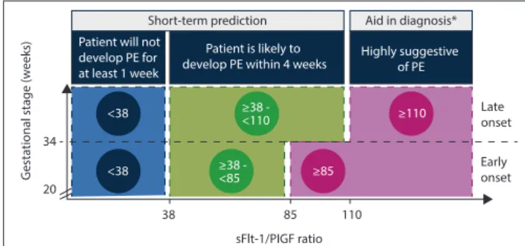
Three subgroups of women can be defined based on the sFlt-1/ PlGF ratio (Fig. 1):[10]
• sFlt-1/PlGF ratio <38: these women will most likely not develop PE for at least 1 week.[10,30]
• sFlt-1/PlGF ratio >85 (early-onset PE) or >110 (late-onset PE): these women are very likely to have PE or another form of placental-based disorder.[32]
• sFlt-1/PlGF ratio 38 - 85 (early-onset PE) or 38 - 110 (late-onset PE): these women do not have a definite diagnosis of PE, but are highly likely to develop PE within 4 weeks.[34]
sFlt-1/PlGF ratio <38
Women with an sFlt-1/PlGF ratio <38 do not have PE at the time of the test, and in all likelihood will not develop PE for at least a week.[10,30,33] The great majority of patients will fall into this category
(negative predictive value of 99.3% (95% confidence interval (CI) 97.9 - 99.9, for PE developing in the next week). This allows clinicians to exclude the majority of patients and to focus on the patients who need appropriate attention and care.[11] The sFlt-1/PlGF
ratio will therefore likely improve clinical decisions with respect to hospitalisation v. outpatient monitoring, and the intensity of outpatient monitoring.[30]
sFlt-1/PlGF ratio >85 (early-onset PE) or >110
(late-onset PE)
Women with an elevated sFlt-1/PlGF ratio >85 (early-onset PE) or >110 (late-onset PE) are highly likely to have PE or some form of placenta-related disorder, and should be managed accordingly.[10,32] Moreover,
a severely elevated sFlt-1/PlGF ratio (>655 in early-onset PE; >201 in late-onset PE) is closely associated with the need to deliver within 48 hours. These patients should be managed in an Table 1. The revised ISSHP definition of PE 2014 [11]
Hypertension developing after 20 weeks’ gestation and the coexistence of one or more of the following new onset conditions:
1. Proteinuria
2. Other maternal organ dysfunction: • renal insufficiency (creatinine >90 μmol/L)
• liver involvement (elevated transaminases and/or severe right upper quadrant or epigastric pain)
• neurological complications (examples include eclampsia, altered mental status, blindness, stroke, or more commonly hyperreflexia when accompanied by clonus, severe headaches when accompanied by hyperreflexia, persistent visual scotomata)
• haematological complications (thrombocytopenia, DIC, haemolysis) 3. Uteroplacental dysfunction:
• fetal growth restriction
ISSHP = International Society for the Study of Hypertension in Pregnancy; PE = pre-eclampsia.
34
20
Late onset
Early onset
38 85 110 sFlt-1/PIGF ratio
G
esta
tional stage (w
eeks)
<38
<38
≥38 - <110
≥38 - <85
≥110
≥85 Patient will not
develop PE for at least 1 week
Patient is likely to
develop PE within 4 weeks Highly suggestive of PE
Short-term prediction Aid in diagnosis*
Fig. 1. Using gestational age-specific cut-offs, the sFlt-1/PIGF ratio can aid in the diagnosis and short-term prediction of PE. (PE = pre-eclampsia; PlGF = placental growth factor; sFlt-1 = soluble fms-like tyrosine kinase-1.)
appropriate clinical setting, and the administration of antenatal corticosteroids to accelerate fetal lung maturation should be seriously considered in early-onset PE.[10]
sFlt-1/PlGF ratio 38 - 85 (early-onset PE) or 38 -
110 (late-onset PE)
Women with a sFlt-1/PlGF ratio of 38 - 85 (early-onset PE) or 38 - 110 (late-onset PE) are unlikely to have PE at the time of the test. Although the majority of these patients will not develop PE, these women may be at risk for developing PE within 4 weeks, and should be more closely monitored.[10]
sFLT-1/PlGF ratio has the potential for
cost-saving in clinical practice
An economic analysis (for the UK) on the use of the sFlt-1/PlGF ratio test suggested that introduction of the test could reduce the number of women hospitalised by more than half (56%). Reduction of hospitalisation was the driver of cost-savings, and it was found that the additional cost of the test was more than offset by a saving in inpatient resource use.[35] The NICE [National Institute for Health
and Care Excellence] economic model also demonstrated significant cost-savings compared with standard clinical assessment, particularly for women presenting with suspected PE before 35 weeks’ gestation (approximately GBP2 488 saving).[33,36]
The Preeclampsia Advisory Board meeting took place at the Roche offices in Midrand on 31 March 2017. In attendance were experts in the field of PE from various hospitals and universities around the country. The National Health Laboratory Service (NHLS) and the Department of Health (DoH) were also represented. Dr M Vatish, an international expert, gave insight into best practices from countries that have successfully implemented the Elecsys immunoassay sFlt-1/ PlGF ratio. The purpose of the meeting was:
• to discuss the burden of disease, economic and clinical impact of PE on the SA healthcare system
• to determine the clinical positioning and value of the Elecsys immunoassay sFlt-1/PlGF ratio in the SA context in relation to current management interventions
• to learn best practices from countries that had clinical utility for the test.
Consensus statements
Statement 1: Pre-eclampsia places a significant burden on the SA
healthcare system.
Pre-eclampsia places a ignificant economic burden on healthcare systems.[35,37] The mortality and morbidity for women and children
affected by PE and its complications are a major burden, particularly in LMICs.[2,38] SA has a high incidence of PE compared with most European
and North American countries.[3] Furthermore, pre-eclampsia and in
particular eclampsia contribute significantly to serious maternal and perinatal mortality in SA.[39-41] Two studies, one in Limpopo Province
and another at Groote Schuur Hospital, Cape Town, found that the most common reasons for obstetric intensive care unit (ICU) admissions were PE and eclampsia.[42,43] Possible reasons for PE and eclampsia’s significant
contribution to maternal mortality and the perinatal morbidity rate are a lack of proper antenatal care, late referrals, poor transport facilities, limited specialist obstetrician and critical-care specialist support, long distances to the referral hospital and inadequate emergency obstetric care at referral centres close to patient residences.[42] Neonatal resources
in SA are limited and oversubscribed.[43,44] As a result and in most
instances, neonates with birth weights of 800 - 1000 g are not ventilated. The severity of PE, late presentation for medical care as well as birthweight and gestational age restriction for ventilation all contribute to high neonatal mortality rates.[43,44]
Statement 2: The sFlt-1/PlGF ratio test is recommended in clinical
practice when the clinical diagnosis is in doubt in patients with suspected PE, in the interests of avoiding unnecessary hospitalisation and unnecessary intervention. The emphasis would be on utilising the negative predictive value of sFlt-1/PlGF ratios to rule out PE.
In the PROGNOSIS study, a single sFlt-1/PlGF ratio cut-off value of 38 was identified as having important negative predictive value.[30] An
sFlt-1/PlGF ratio <38 was validated to reliably rule out PE within 1 week, and had a negative predictive value of 99.3% (95% CI; 97.9 - 99.), with 80.0% sensitivity (95% CI; 51.9 - 95.7) and 78.3% specificity (95% CI; 74.6 - 81.7).[10,30,33]
In routine clinical practice, PE may be over-diagnosed, and suspected PE may be over-investigated and treated.[45] The PreOS
study demonstrated that use of sFlt-1/PlGF ratio test influences clinical decision-making in routine clinical practice towards appropriate hospitalisation in a considerable proportion of women with suspected PE.[45] Reducing inappropriate hospital admissions is an important
goal, in order to avoid unnecessary stress and anxiety for the patient, and to reduce the financial burden for the healthcare provider. The wider adoption of the sFlt-1/PlGF ratio test in maternity care could assist with decision-making in clinical care. Patients deemed low risk can be reassured, and avoid unnecessary hospitalisation.[45] Instances of
immediate delivery of the fetus could also be reduced, with resultant fewer premature babies requiring neonatal ICU admission.[33]
Statement 3: Identifying the clinically suspicious PE group needs
to be contextualised to SA circumstances. Criteria relevant and appropriate to the country’s settings should be specified, regarding when the sFlt-1/PlGF ratio should be implemented, so as to provide clear guidelines as to when the assay could provide the most benefit.
It was suggested that sFlt-1/PlGF ratio test be used in patients who do not present with classic symptoms of PE, and when the diagnosis is uncertain – criteria similar to those utilised in the PreOS and PROGNOSIS studies (Table 2). [46,47]
Statement 4: It would be important for the test results to be
promptly available, in order to derive maximum savings from hospitalisation costs, and to minimise patient anxiety. In order for the sFlt-1/PlGF ratio to be a valuable aide in conjunction with clinical assessment and other tests, laboratories would need to ensure that test results are available within 24 hours.
Under optimal conditions, the established turnaround time of the Elecsys immunoassay sFlt-1/PlGF ratio test is about 18 minutes.[33] In order
to be of additional clinical value, laboratories need to prioritise the sFlt-1/ PlGF ratio test results.
Conclusion
Acknowledgements. The authors acknowledge the contribution of Roche in
creating a platform for enabling a forum of experts from across the country to engage on the potential relevance of these biomarkers in South Africa. How-ever, none of the authors or their relatives are employed by, have any affiliation with, or have financially benefitted from Roche.
Author contributions. All the authors, apart from TP and EM, were involved in
the drafting of the consensus statement, as a result of their independent exper-tise in the field of PE.
Funding. None.
Conflicts of interest. None.
1. World Health Organization. WHO Recommendations for Prevention and Treatment of Pre-eclampsia and Eclampsia. Geneva: WHO, 2011.
2. Duley L. The global impact of pre-eclampsia and eclampsia. Semin Perinatol 2009;33(3):130-137.
https://doi.org/10.1053/j.semperi.2009.02.010
3. Abalos E, Cuesta C, Grosso AL, Chou D, Say L. Global and regional estimates of pre-eclampsia
and eclampsia: A systematic review. Eur J Obstet Gynecol Reprod Biol 2013;170(1):1-7. https://doi.
org/10.1016/j.ejogrb.2013.05.005
4. Abalos E, Cuesta C, Carroli G, et al. Pre-eclampsia, eclampsia and adverse maternal and perinatal outcomes: A secondary analysis of the World Health Organization Multicountry Survey on Maternal
and Newborn Health. BJOG 2014;121(Suppl 1):S14-S24. https://doi.org/10.1111/1471-0528.12629
5. Hutcheon JA, Lisonkova S, Joseph K. Epidemiology of pre-eclampsia and the other hypertensive
disorders of pregnancy. Best Pract Res Clin Obstet Gyanecol 2011;25(4):391-403. https://doi.
org/10.1016/j.bpobgyn.2011.01.006
6. Rusen I, Liston R, Wen S, Bartholomew S. Special Report on Maternal Mortality and Severe Morbidity in Canada. Enhanced Surveillance. The Path to Prevention. Ottawa: Public Health Agency of Canada, 2004.
7. American College of Obstetricians and Gynecologists, Committee on Practice Bulletins – Obstetrics. ACOG Practice Bulletin No. 33. Diagnosis and management of pre-eclampsia and eclampsia. Obstet Gynecol 2002;99(1):159-167.
8. National Collaborating Centre for Women’s and Children’s Health. Hypertension in pregnancy: The management of hypertensive disorders during pregnancy. London: RCOG Press, 2010. 9. American College of Obstetricians and Gynecologists, Task Force on Hypertension in Pregnancy.
Hypertension in pregnancy. Report of the American College of Obstetricians and Gynecologists’
task force on hypertension in pregnancy. Obstet Gynecol 2013;122(5):1122-1131. https://doi.
org/10.1097/01.AOG.0000437382.03963.88
10. Stepan H, Herraiz I, Schlembach D, et al. Implementation of the sFlt‐1/PlGF ratio for prediction and diagnosis of pre‐eclampsia in singleton pregnancy: Implications for clinical practice. Ultrasound
Obstet Gynecol 2015;45(3):241-246. https://doi.org/10.1002/uog.14799
11. Tranquilli A, Dekker G, Magee L, et al. The classification, diagnosis and management of the hypertensive disorders of pregnancy: A revised statement from the ISSHP. Pregnancy Hypertens
2014;4(2):97-104. https://doi.org/10.1016/j.preghy.2014.02.001
12. Zhang J, Klebanoff MA, Roberts JM. Prediction of adverse outcomes by common definitions of hypertension in pregnancy. Obstet Gynecol 2001;97(2):261-267.
13. Verlohren S, Stepan H, Dechend R. Angiogenic growth factors in the diagnosis and prediction of
pre-eclampsia. Clin Sci 2012;122(2):43-52. https://doi.org/10.1042/CS20110097
14. Hagmann H, Thadhani R, Benzing T, Karumanchi SA, Stepan H. The promise of angiogenic markers for the early diagnosis and prediction of pre-eclampsia. Clin Chem 2012;58(5):837-485.
https://doi.org/10.1373/clinchem.2011.169094
15. Levine RJ, Maynard SE, Qian C, et al. Circulating angiogenic factors and the risk of pre-eclampsia.
N Engl J Med 2004;350(7):672-683. https://doi.org/10.1056/NEJMoa031884
16. Powe CE, Levine RJ, Karumanchi SA. Pre-eclampsia, a disease of the maternal endothelium.
Circulation 2011;123(24):2856-2869. https://doi.org/10.1161/CIRCULATIONAHA.109.853127
17. Rana S, Powe CE, Salahuddin S, et al. Angiogenic factors and the risk of adverse outcomes in women
with suspected pre-eclampsia: Clinical perspective. Circulation 2012;125(7):911-919. https://doi.
org/10.1161/CIRCULATIONAHA.111.054361
18. Park JE, Chen HH, Winer J, Houck KA, Ferrara N. Placenta growth factor. Potentiation of vascular endothelial growth factor bioactivity, in vitro and in vivo, and high affinity binding to Flt-1 but not to Flk-1/KDR. J Biol Chem 1994;269(41):25646-25654.
19. Athanassiades A, Lala PK. Role of placenta growth factor (PlGF) in human extravillous trophoblast proliferation, migration and invasiveness. Placenta 1998;19(7):465-473.
20. Ferrara N, Gerber H-P, LeCouter J. The biology of VEGF and its receptors. Nat Med
2003;9(6):669-676. https://doi.org/10.1038/nm0603-669
21. Park HJ, Shim SS, Cha DH. Combined screening for early detection of pre-eclampsia. Int J Mol Sci
2015;16(8):17952-17974. https://doi.org/10.3390/ijms160817952
22. Polliotti BM, Fry AG, Saller DN, Mooney RA, Cox C, Miller RK. Second-trimester maternal serum placental growth factor and vascular endothelial growth factor for predicting severe, early-onset pre-eclampsia. Obstet Gynecol 2003;101(6):1266-1274.
23. Taylor RN, Grimwood J, Taylor RS, McMaster MT, Fisher SJ, North RA. Longitudinal serum concentrations of placental growth factor: Evidence for abnormal placental angiogenesis in pathologic pregnancies. Am J Obstet Gynecol 2003;188(1):177-182.
24. Tsatsaris V, Goffin F, Munaut C, et al. Overexpression of the soluble vascular endothelial growth factor receptor in preeclamptic patients: Pathophysiological consequences. J Clin Endocrinol Metab
2003;88(11):5555-5563. https://doi.org/10.1210/jc.2003-030528
25. Ohkuchi A, Hirashima C, Matsubara S, et al. Alterations in placental growth factor levels before and after the onset of pre-eclampsia are more pronounced in women with
early-onset severe pre-eclampsia. Hypertens Res 2007;30(2):151-159. https://doi.org/10.1291/
hypres.30.151
26. Torry DS, Wang H-S, Wang T-H, Caudle MR, Torry RJ. Pre-eclampsia is associated with reduced serum levels of placenta growth factor. Am J Obstet Gynecol 1998;179(6):1539-1544.
27. Romero R, Nien JK, Espinoza J, et al. A longitudinal study of angiogenic (placental growth factor) and anti-angiogenic (soluble endoglin and soluble vascular endothelial growth factor receptor-1) factors in normal pregnancy and patients destined to develop pre-eclampsia and deliver a
small-for-gestational-age neonate. J Matern Fetal Neonatal Med 2008;21(1):9-23. https://doi.
org/10.1080/14767050701830480
28. Levine RJ, Lam C, Qian C, et al. Soluble endoglin and other circulating antiangiogenic factors in
pre-eclampsia. N Eng J Med 2006;355(10):992-1005. https://doi.org/10.1056/NEJMoa055352
29. Noori M, Donald AE, Angelakopoulou A, Hingorani AD, Williams DJ. Prospective study of placental angiogenic factors and maternal vascular function before and after pre-eclampsia
and gestational hypertension. Circulation 2010;122(5):478-487. https://doi.org/10.1161/
CIRCULATIONAHA.109.895458
30. Zeisler H, Llurba E, Chantraine F, et al. Predictive value of the sFlt-1: PlGF ratio in women with
suspected pre-eclampsia. N Eng J Med 2016;374(1):13-22. https://doi.org/10.1056/NEJMoa1414838
31. Verlohren S, Herraiz I, Lapaire O, et al. The sFlt-1/PlGF ratio in different types of hypertensive pregnancy disorders and its prognostic potential in pre-eclamptic patients. Am J Obstet Gynecol
2012;206(1):58.e1-e8. https://doi.org/10.1016/j.ajog.2011.07.037
32. Verlohren S, Herraiz I, Lapaire O, et al. New gestational phase-specific cutoff values for the use of the soluble fms-like tyrosine kinase-1/placental growth factor ratio as a diagnostic test for
pre-eclampsia novelty and significance. Hypertension 2014;63(2):346-352. https://doi.org/10.1161/
HYPERTENSIONAHA.113.01787
33. National Institute for Health and Care Excellence. PlGF-based testing to help diagnose suspected pre-eclampsia (Triage PlGF test, Elecsys immunoassay sFlt-1/PlGF ratio, DELFIA Xpress PlGF 1-2-3 test, and BRAHMS sFlt-1 Kryptor/BRAHMS PlGF plus Kryptor PE ratio). London: NICE, 2016.
34. Stepan H, Unversucht A, Wessel N, Faber R. Predictive value of maternal angiogenic factors in second trimester pregnancies with abnormal uterine perfusion. Hypertension 2007;49(4):818-824.
https://doi.org/10.1161/01.HYP.0000258404.21552.a3
35. Vatish M, Strunz‐McKendry T, Hund M, Allegranza D, Wolf C, Smare C. sFlt‐1/PlGF ratio test for pre‐eclampsia: An economic assessment for the UK. Ultrasound Obstet Gyencol
2016;48(6):765-771. https://doi.org/10.1002/uog.15997
36. National Institute for Health and Care Excellence. Hypertension in pregnancy: Diagnosis and management. London: NICE, 2010.
Table 2. Criteria contributing to suspicion of clinical diagnosis of PE[46,47]
Clinical signs and symptoms
a. New onset of elevated blood pressure (does not need to be defined hypertension (≥140 mm Hg systolic and/or ≥90 mm Hg diastolic). b. New onset of hypertension (does not need to be defined proteinuria – any protein in the urine is sufficient)
c. Aggravation of pre-existing hypertension d. New onset of protein in urine
e. New onset of proteinuria
f. Aggravation of pre-existing proteinuria
g. One or more other reason(s) for clinical suspicion of PE (see i. and ii. below) i. Pre-eclampsia-related symptoms:
1. Epigastric pain
2. Excessive oedema/severe swelling, (face, hands, feet) 3. Severe or atypical headaches
4. Visual disturbances
5. Sudden weight gain (>1 kg/week in the third trimester) ii. PE-related findings:
1. Low platelets
2. Elevated liver transaminases
3. (Suspected) intrauterine growth restriction
4. Abnormal uterine perfusion detected by Doppler sonography with mean pulsatility index >95th percentile in the second trimester and/or bilateral uterine artery notching
37. Delahaije DH, Smits LJ, van Kuijk SM, et al. Care-as-usual provided to formerly pre-eclamptic women in the Netherlands in the next pregnancy: Healthcare consumption, costs and maternal and child outcome. Eur J Obstet Gynecol Reprod Biol 2014;179:240-245.
https://doi.org/10.1016/j.ejogrb.2014.04.033
38. Firoz T, Sanghvi H, Merialdi M, von Dadelszen P. Pre-eclampsia in low and middle income
countries. Best Pract Res Clin Obstet Gynaecol 2011;25(4):537-548. https://doi.org/10.1016/j.
bpobgyn.2011.04.002
39. Moodley J, National Committee on Confidential Enquiries into Maternal Deaths, National Department of Health, South Africa. Maternal deaths associated with hypertension in South Africa: Lessons to learn from the Saving Mothers report, 2005 - 2007: Cardiovascular topics.
Cardiovasc J Afr 2011;22(1):31-35. https://doi.org/CVJ-21.026
40. Moodley J, Pattinson RC, Fawcus S, et al. The confidential enquiry into maternal deaths in
South Africa: A case study. BJOG 2014;121(Suppl 4):S53-S60.
https://doi.org/10.1111/1471-0528.12869
41. Pattinson R, Rhoda N. Saving Babies 2012 - 2013: Ninth Report on Perinatal Care in South Africa. Pretoria: National Department of Health, 2014.
42. Ntuli TS, Ogunbanjo G, Nesengani S, Maboya E, Gibango M. Obstetric intensive care admissions at a tertiary hospital in Limpopo Province, South Africa. S Afr J Crit Care
2015;31(1):8-10. https:doi.org/10.7196/SAJCC.164
43. Drakeley AJ, Le Roux PA, Anthony J, Penny J. Acute renal failure complicating severe pre-eclampsia requiring admission to an obstetric intensive care unit. Am J Obstet Gynecol 2002;186(2):253-256. 44. Varughese S, Gilbert C, Pieper C, Cook C. Retinopathy of prematurity in South Africa: An
assessment of needs, resources and requirements for screening programmes. Br J Opthalmol
2008;92(7):879-882. https://doi.org/10.1136/bjo.2008.137588
45. Klein E, Schlembach D, Ramoni A, et al. Influence of the sFlt-1/PlGF ratio on clinical decision-making in women with suspected pre-eclampsia. PloS ONE 2016;11(5):e0156013.
https://doi.org/10.1371/journal.pone.0156013
46. Hund M, Allegranza D, Schoedl M, Dilba P, Verhagen-Kamerbeek W, Stepan H. Multicenter prospective clinical study to evaluate the prediction of short-term outcome in pregnant women with suspected pre-eclampsia (PROGNOSIS): Study protocol. BMC Pregnancy
Childbirth 2014;14(1):324. https://doi.org/10.1186/1471-2393-14-324
47. Hund M, Verhagen-Kamerbeek W, Reim M, Messinger D, van der Does R, Stepan H. Influence of the sFlt-1/PlGF ratio on clinical decision-making in women with suspected
pre-eclampsia – the PreOS study protocol. Hypertens Pregnancy 2015;34(1):102-115. https://doi.
org/10.3109/10641955.2014.982331
![Table 2. Criteria contributing to suspicion of clinical diagnosis of PE [46,47] Clinical signs and symptoms](https://thumb-us.123doks.com/thumbv2/123dok_us/8094514.2144953/4.892.451.827.499.1163/table-criteria-contributing-suspicion-clinical-diagnosis-clinical-symptoms.webp)
