Journal of Global Pharma Technology
Available Online at:
www.jgpt.co.in
RESEARCH ARTICLE
Comorbidity of the Metabolic Syndrome: Hyperuricemia, Gallstone
Disease, Hormonal Disorders
Raisa A. Aringazina
*,Yerlan Sh. Bazargaliyev
,Kuanysh G. Suleimenov
,Anes
G. Bekkuzhin
,Manas M. Mukushev
West Kazakhstan Marat Ospanov State Medical University, Aktobe, Kazakhstan.
*Corresponding Author: Raisa A. Aringazina
Abstract
This article presents relationships between metabolic syndrome and gallstone disease, hyperuricemia, hormonal disorders and point out the most relevant picture for today. In the review the general pathogenetic mechanism of endothelial dysfunction formation in metabolic syndrome and hyperuricemia is revealed and described. Shown, that hyperuricemia is a marker of the metabolic syndrome. The symptoms of metabolic syndrome increase with increasing levels of hyperuricemia. The review found that it is obesity and insulin resistance are common leading risk factors for progression metabolic syndrome and gallstone disease. No association between gallstone disease and dyslipidemia unlike metabolic syndrome. Obesity is a common factor in the relationship between metabolic syndrome, gallstone disease and hormonal disorders. Insulin resistance is a common factor metabolic syndrome and polycystic ovary syndrome, the leading factor in the course of metabolic syndrome in menopause, hypopituitarism, and gallstone disease. Also, insulin resistance is a common factor in the comorbidity of gallstone disease and postmenopausal conditions. Obesity and insulin resistance are common factors of metabolic syndrome and benign prostate hyperplasia.
Keywords: Metabolic syndrome, Uric acid, Gallstone disease, Hyperuricemia, Hormonal disorders.
Introduction
One of the most urgent problems of the modern medicine is the search for the risk factors to prevent cardiovascular disease (CVD) [1, 2]. Investigation at the early phase of the development of a clinical and laboratory symptoms set of great social importance in terms of preventing the development of hypertension, atherosclerosis, and acute myocardial infarction. Endothelial dysfunction, as the leading mechanism of the pathogenesis of cardiovascular problems, is formed gradually. It can be associated with hormonal disorders in the body at its early phase [3, 4].
Metabolic syndrome (MetS) means the presence of multiple risk factors for CVD [5, 10]. The worldwide prevalence of MS in the adult population is estimated between 20% and 48% [11, 12]. MetS is characterized by biochemical, physiological and metabolic abnormalities, that contribute to cardiovascular disease, type 2 diabetes mellitus and development of
atherothrombotic complications [5]. It is accompanied by the state of low chronic inflammation and such symptoms as insulin resistance, visceral adiposity, elevated fasting plasma glucose, oxidative stress, atherogenic dyslipidemia (which comprises lower high-density lipid, and higher serum triglyceride levels), endothelial dysfunction, elevated blood pressure and increased coagulation [5, 13, 14].
According to the International Diabetes Federation definition, established in April 2005, Mets is a “cluster of the most dangerous heart attack risk factors: diabetes and prediabetes, abdominal obesity, high cholesterol and high blood pressure” [15]. There are about 775 million obese people in the World including adult, children, and adolescents [16].
overweight population. In 2017, the obesity prevalence in the United States amounted to 31.3 percent among adults aged 20 and over. Around 28 percent of elderly adults were obese in 2018, compared to 25.3 percent in 2013. It is also documented that at least 2.6 million people die annually due to obesity worldwide [17]. Worldwide, over half a billion people are currently classified as obese (body mass index [BMI] ≥ 30 kg/m2), and it is estimated that over 40% of the U.S population will be obese by the year 2030 [18].
Criteria for MetS differ particularly regarding the definition of central obesity and consequently, there could be differences in the assessment of cardiovascular risk [6]. Current evidence supports the role of soluble uric acid as a true mediator of injury, exerting its effects through the induction of growth factors, cytokines, hormones and autacoids [19]. Soluble uric acid has important biologic roles. While it acts as an antioxidant, there is also evidence that uric acid has pro-inflammatory and proliferative effects on smooth muscle cells, and causes dysfunction of endothelial cells [20, 19].
Uric acid may contribute to endothelial dysfunction by inducing antiproliferative effects on endothelium and impairing nitric oxide production [20]. In 1994 it was shown thatthere is no difference in the sex hormone profiles between asymptomatic hyperuricaemic and normouricaemic men [21]. Nevertheless, violation of uric acid metabolism is an indicator of hormone-metabolic shifts [19].
The violation of lipid metabolism, which is observed in patients with metabolic syndrome, increases the release of cholesterol into bile [22]. Pathogenetic mechanisms of formation of cholelithiasis, associated with a violation of lipid metabolism, allow us to conclude that there is an association of this disease with MetS [23].
The role of hormonal disbalance, i.e. hyperleptinemia, hyperinsulinemia, hypercortisolemia, hypercatecholaminemia, hyperestrogenemia and hyperandrogenemia in MetS is proved [24, 25]. A special attention in the development of MetS is paid to the activity of the hypothalamo-pituitary-adrenal axis after stress. There are reliable scientific arguments that the metabotrophic deficit due to reduced neurotrophins could be implicated
in the pathogenesis of MetS, type 2 diabetes mellitus, and atherosclerosis as well [14]. Higher rates of cardiovascular events reported in primary aldosteronism could be due in part to the increased prevalence of the metabolic syndrome in this disorder [7]. The aim of this article was to consider and establish relationships between MetS, GSD, hyperuricemia and hormonal disorders in patients with MetS and point out the most relevant picture for today.
Method
A literature search conducted using PubMed was conducted up to August 2018 using multiple search terms ‘Metabolic syndrome’, ‘Prevalence of metabolic syndrome’. A search for each risk factor and its relation to MetS was conducted. A search for the association between MetS and hyperuricemia, MetS and gallstone disease and MetS and hormonal disorders was conducted. Were used multiple search terms «Metabolic syndrome and hyperuricemia», «Metabolic syndrome and gallstone disease », «Metabolic syndrome and hormonal disorders ».
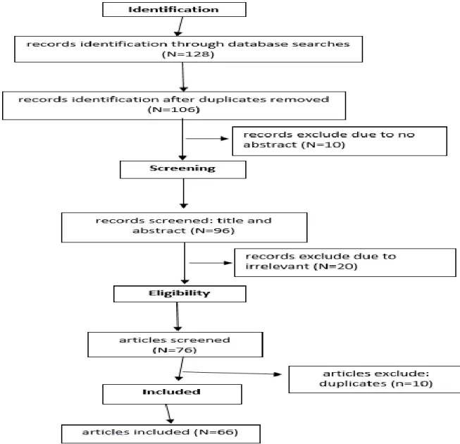
A total of 128 studies were screened, however only 76 were assessed for eligibility and 66 were included in this review, 2 to 1994, 21 from 2002 to 2010 and 43 from 2011 to 2018. All articles identified were English-language. We also searched the reference lists of identified articles for further relevant papers (Figure 1).
Results and Discussions
Comorbidity of MetS and hyperuricemia
The prevalence of the MetS increases substantially with increasing levels of hyperuricemia [26]. The symptoms of MetS increase with increasing levels of hyperuricemia, including abdominal obesity, hypertriglyceridemia, low high-density lipoprotein (HDL) cholesterol, high blood pressure, and high fasting glucose.
components of MetS is often found in medical practice [28, 29]. For example, it was shown that prevalence of the MetS, as well as prevalence of its distinct components like body mass index, hypertension, and diabetes
is in direct dependence to serum uric acid level. In individuals with a higher level of uric acid, the prevalence of MetS is higher Table 1 [30].
Figure 1: Diagram of Studies Selected for Review
Information published on the web sites WHO, IDF and others were also considered.
Table 1: The prevalence of metabolic syndrome depending on serum uric acid level Participants characteristics serum uric acid level prevalence of MetS 8669 men and nonpregnant women aged at
least 20 years who attended the medical examination, had fasted at least 8 hours
before the blood collection, and had complete information to allow definition of the metabolic syndrome and measurement
of serum uric acid levels
<6 mg/dL 18.9%
from 6 to 6.9 mg/dL 36.0%
from 7 to 7.9 mg/dL 40,8%
from 8 to 8.9 mg/dL 59,7%
from 9 to 9.9 mg/dL 62%
>10 mg/dL 70.7%
Serum uric acid level 5.5mg/dL independently predicted the development of hyperinsulinemia at both 6 and 12 months in nondiabetic patients with first-time myocardial infarction [31]. Renal clearance of urate is inversely related to the degree of insulin resistance [30, 32]. Thus, the reduced renal excretion of urate among patients with
reduces levels of endothelial nitric oxide (NO), a key mediator of insulin action. NO increases blood flow to skeletal muscle and enhances glucose uptake, thereby its deficiency causes insulin resistance and other features of the metabolic syndrome. This hypothesis may be proved by the investigation, showing that Feeding fructose to rats causes rapid development of the metabolic syndrome, including obesity, hypertension, insulin resistance, hypertriglyceridemia and hyperinsulinemia.
Case control analysis revealed that mean plasma fasting insulin level was higher in hyperuricemia patients, regardless of sex, age and triglyceride levels [34]. As it was mentioned above, MetS is characterized by endothelial dysfunction. Endothelial functional status as reflected by decreased NO and increased serum endothelin levels along with insulin resistance is seen in MetS [36].At the same time, elevated uric acid level is also able to cause this impairment and, thus, decrease bioavailability of endothelial NO.
Vasorelaxation of arterial rings in response to acetylcholine, a process that is mediated by NO, is blocked by uric acid [33].Given the frequent association between hyperuricemia and the MetS, it is imperative to develop appropriate dietary and other lifestyle guidelines taking into account improving hyperuricemia and overall long-term health effects
Comorbidity of MetS and Gallstone Disease
According to worldwide statistic, the prevalence of gallstone disease (GSD) reaches 10% - 15% among white adults of the European countries [36, 38], while in East Asians and African Americans it is not so high [39]. At the same time in China, the incidence of GSD is rising, thus it is considered a health problem of prime importance.
The prevalence of GSD in Taiwan is a little lower than those in western countries [40, 41, 8]. Fewer than 20% of subjects with gallstones develop clinical events. Larger, multiple, and older gallstones are associated with events [42]. MetS affects almost one-third of the adult population, including over one-half of adults over 60 years, with abdominal obesity being the most prevalent
component. And some studies concluded that GSD might be a component of MetS [26, 6, 43] although it needs to be validated by more evidences. According to other authors MetS is strongly connected with GSD and the more the components of MetS, the higher the prevalence of GSD [44, 45]. In an age- and sex-adjusted logistic regression model, metabolic syndrome was associated with gallstone disease (OR = 1.61; P < 0.0001). Females had a higher odds ratio than males in waist circumference for GSD, whereas males had a lower odds ratio than females in HDL-C for GSD.
Waist circumference and high-density lipoprotein cholesterol are all associated with GSD. Men and women may possibly have different priorities and strategies to reduce the burden of GSD [8]. Age, female sex, BMI, non- HDL cholesterol, and polyps are
independent determinants
for gallstone formation. Incident gallstones and the metabolic syndrome share common risk factors [42, 46, 38]. Risk factors, which may lead to GSD, comprise hyperlipidemia, diabetes and obesity, therefore MetS is also associated with GSD.
Study on Chinese population showed that if 5 components of the MetS are present, risk of GSD will rise by 3.4 times (P < 0.0001) in men and by 5 times in women [45]. More studies are needed for further exploration. Though increased prevalence of GSD is connected with insulin resistance, age and postmenopausal state, obesity remains the most valid risk factor for GSD appearance, because hepatic secretion of cholesterol rises, while gallbladder motility decreases, contributing to gallstone formation [22, 38].
Interestingly, no association between GSD and dyslipidemia was found, regardless the latter is a common symptom of patients with MetS [37, 47]. Despite this GSD is associated with a higher risk of total mortality and disease-specific mortality, including CVD [37, 48].Finding association between MetS and GSD is so important because of possibility to decrease the incidence of GSD by the means of lifestyle factors, which reduce MetS.
Comorbidity of MetS and Hormonal Disorders
It is one of the most common causes of functional infertility and a clinical problem that can be faced by doctors of many specialties. PCOS is characterized by hyperandrogenism, oligoovulations and metabolic disorders. The fundamental parts of the therapy are lifestyle modifications and weight loss. Losing as little as 5% of body mass increases frequency of ovulations, chances of pregnancy and improves hormonal profile [49, 50].Although the pathophysiology of the syndrome is complex and there is no single defect from which it is known to result, it is hypothesized that insulin resistance is a key factor [51].
Insulin resistance is almost always present in women with PCOS, regardless of weight, and they often develop diabetes and MetS [49]. PCOS is a common heterogeneous endocrine disorder associated with MetS. The PCOS and MetS are common disorders that share many characteristics, particularly abdominal obesity and insulin resistance [52]. Results of the meta-analysis demonstrated that the patients with PCOS regardless of age, recruitment sources of samples had higher odds of MetS compared to healthy controls. The prevalence of MetS was higher in women with PCOS than in
controls regardless of the
applied MetS definition [51, 52, 53].
In reproductive-aged women with abdominal obesity without concomitant PCOS in the presence of insulin resistance, the ovarian reserve decreases and the levels of follicle-stimulating hormone and luteinizing hormone increase, which leads to accelerated aging processes in the reproductive system [54]. MetS is often associated with male hypogonadism. The meta-analysis of the available cross-sectional data suggests that MetS can be considered an independent association of male hypogonadism [55]. Abdominal obesity is a major determinant of low testosterone levels irrespective of diabetes status.
The causative relationship between the often low testosterone and type 2 diabetes mellitus might be bidirectional or even multidirectional and interrelated with obesity and MetS [56]. The prevalence of MetS in postmenopause is due to loss of the protective role of estrogens and increased circulating androgens resulting in changes to body fat distribution and development of
abdominal obesity. Pathophysiologic changes during the menopause transition may contribute to the risk of MetS. Body fat composition, particularly visceral fat, is highly associated with increased insulin resistance, abnormal carbohydrate metabolism and high blood pressure [57, 58]. Estrogen deficiency promotes hepatic steatosis via a glucocorticoid receptor-dependent mechanism in mice. These studies prove uncover a regulatory axis between estradiol, follicle-stimulating hormone and hepatic glucocorticoid receptor signaling that, when disrupted, as in menopause, promotes hepatic steatosis [59].
The prevalence of MetS was 49.12% in subclinical hypothyroid women and 46.89% in euthyroid women. MetS in both euthyroid and subclinical hypothyroid women is connected with obesity, visceral fat accumulation, and higher TSH and IL-6 concentrations [58]. In euthyroid subjects free T4 is associated with risk of MetS and its components. The prevalence of MetS decreased from 30.1% in the lowest free T4 tertile to 22.4% in the highest FT4 tertile (p<0.001) [60]. Prevalence of MetS and mortality rates from cardiovascular causes are increased in patients with hypopituitarism.
Features of obesity, visceral adiposity, dyslipidemia, insulin resistance, and hypertension are common in these patients. Unreplaced growth hormone deficiency and inadequate replacement of other hormone insufficiencies may be responsible for the adverse body composition and metabolic profile associated with hypopituitarism. Recently, fatty liver disease was added to this unfavorable metabolic phenotype [61]. 48.59% of benign prostate hyperplasia (BPH) is combined with MetS [62]. The mainstream view supports the correlation between MetS and BPH, but the pathogenesis of MetS-BPH is not fully understood.
glucose have been associated with increased prostate size and increased risk of prostate enlargement, clinical BPH and BPH surgery [66].
Conclusion
There are limitations of this analysis that have to be considered. Not all diagnostic criteria for the metabolic syndrome were investigated as general factors of comorbidity metabolic syndrome with hyperuricemia, gallstone disease, and hormonal disorders. In particular, hypertension is not described at all; there is a little data on dyslipidemia. The frequency of cardiovascular complications in the context of comorbidity of the metabolic syndrome is not described.
The review does not include issues of preventing the various complications development during metabolic syndrome, hyperuricemia, gallstone disease, post menopause, polycystic ovary syndrome and other described conditions, related to each other. In the review the general endothelial dysfunction formation pathogenetic mechanism in metabolic syndrome and hyperuricemia is revealed and described. Shown, that hyperuricemia is a marker of the metabolic syndrome. The symptoms of MetS increase with increasing levels of
hyperuricemia. The review found that it is obesity and insulin resistance are common leading risk factors for metabolic syndrome progression and gallstone disease. There is no association between gallstone disease and dyslipidemia unlike metabolic syndrome. Obesity is a common factor in the relationship between metabolic syndrome, gallstone disease and hormonal disorders. Insulin resistance is a common and the most important factor metabolic syndrome and polycystic ovary syndrome, the leading factor in the course of metabolic syndrome in menopause, hypopituitarism, and gallstone disease. Also, insulin resistance is a common factor in the comorbidity of gallstone disease and postmenopausal conditions.
Obesity and insulin resistance are common factors of metabolic syndrome and benign prostate hyperplasia. Thus, a review of 63 articles from 1984 to 2018 years and information published on the web sites concerning the relationships of the metabolic syndrome has shown that the simultaneous course of the metabolic syndrome with hyperuricemia, gallstone disease and various hormonal disorders has a complex pathogenetic relationships, which can open new horizons to prevention the development of the syndrome complications [67, 69].
References
1. Van Camp G (2014) Cardiovascular disease prevention. Acta Clinica Belgica, 69(6): 407-411.
2. Ruan Y, Guo Y, Zheng Y, Huang Z, Sun S, Kowal P, Wu F (2018) Cardiovascular disease (CVD) and associated risk factors among older adults in six low-and middle-income countries: results from SAGE Wave 1. BMC public health, 18(1): 778.
3. Gkaliagkousi E, Gavriilaki E, Triantafyllou A, Douma S (2015) Clinical Significance of Endothelial Dysfunction in Essential Hypertension. Curr. Hypertens Rep., 17(11): 85.
4. Guajardo I, Ayer A, Johnson AD, Ganz P, Mills C, Donovan C, Scherzer R, Shah SJ, Peralta CA, Dubin RF (2018) Sex differences in vascular dysfunction and cardiovascular outcomes: The cardiac, endothelial function, and arterial stiffness in ESRD (CERES) study. Hemodial. Int., 22(1): 93-102.
5. Grundy SM, Brewer HB Jr, Cleeman JI, Smith SC Jr, Lenfant C (2004) Definition of metabolic syndrome: report of the National Heart, Lung, and Blood Institute. American Heart
Association conference on scientific issues related to definition. Circulation, 109: 433-438.
6. Rosenbaum P, Gimeno SGA, Sanudo A, Franco LJ, Ferreira SRG, Japanese‐Brazilian Diabetes Study Group (2005) Analysis of criteria for metabolic syndrome in a population‐based study of Japanese‐Brazilians. Diabetes, obesity and metabolism, 7(4): 352-359.
7. Fallo F, Pilon C, Urbanet R (2012) Primary aldosteronism and metabolic syndrome. Horm Metab Res., 44(3): 208-214.
8. Lin IC, Yang YW, Wu MF, Yeh YH, Liou JC, Lin YL, Chiang CH (2014) The association of metabolic syndrome and its factors with gallstone disease. BMC Fam. Pract., 29(15): 138.
9. Shabanzadeh DM, Sørensen LT, Jørgensen T (2016) A prediction rule for risk stratification of incidentally discovered gallstones: results
from a large cohort
study. Gastroenterology, 150(1): 156-167. 10. Santilli F, DArdes D, Teresa Guagnano M,
Davi G (2017) Metabolic syndrome: sex-related
cardiovascular risk and therapeutic
11. Ajayi EA, Ajayi OA, Adeoti OA (2014) Metabolic syndrome: prevalence and association with electrocardiographic abnormalities in Nigerian hypertensive patients. Metab. Syndr. Relat. Disord., 12(8): 437-442.
12. De Carvalho, Vidigal F, Ribeiro AQ, Babio N, Salas-Salvadó J, Bressan J (2015) Prevalence of metabolic syndrome and pre-metabolic syndrome in health professionals: LATINMETS Brazil study. Diabetology & metabolic syndrome, 7(1): 6.
13. Grundy SM, Cleeman JI, Daniels S, Donato KA, Eckel RH, Franklin B A, Spertus JA (2005) Diagnosis and management of the metabolic syndrome: an American Heart Association/National Heart, Lung, and Blood Institute scientific statement. Circulation, 112(17): 2735-2752.
14. Hristova MG (2013) Metabolic syndrome--from the neurotrophic hypothesis to a theory. Med Hypotheses, 81(4): 627-634.
15. IDF Epidemiology Task Force Consensus Group (2005) International Diabetes Federation: The IDF consensus worldwide definition of the metabolic syndrome. http://www. idf. org/ webdata/ docs/ Metabolic_syndrome_def. pdf.
16. Report: obesity rates by country (2017) https://renewbariatrics.com/obesity-rank-by-countries/
17. Statista (2019) The portal for statistics. https://www.statista.com
18. Smith KB, Smith MS (2016) Obesity
statistics. Primary care: clinics in office practice, 43(1): 121-135.
19. Sánchez-Lozada LG, Nakagawa T, Kang DH, Feig DI, Franco M, Johnson RJ, Herrera-Acosta J (2006) Hormonal and cytokine effects of uric acid. Current opinion in nephrology and hypertension, 15(1): 30-33.
20. Kanellis J, Kang DH (2005) Uric acid as a mediator of endothelial dysfunction, inflammation, and vascular disease. Semin Nephrol., 25(1): 39-42.
21. Rosen R, Tomer Y, Carel R, Weinberger A (1994) Serum 17-beta-estradiol and testosterone levels in asymptomatic hyperuricaemic men. Clinical rheumatology, 13(2): 219-223.
22. Smelt AHM (2010) Triglycerides and gallstone formation. Clinica Chimica Acta, 411(21-22): 1625-1631.
23. Ponziani FR, Pecere S, Gasbarrini A, Ojetti V (2015) Physiology and pathophysiology of liver lipid metabolism. Expert Rev Gastroenterol. Hepatol., 9(8): 1055-1067. 24. Abraham SB, Rubino D, Sinaii N, Ramsey
S, Nieman LK (2013) Cortisol, obesity, and
the metabolic syndrome: a cross-sectional study of obese subjects and review of the literature. Obesity (Silver Spring), 21(1): E105-117.
25. Hristova MG (2018) Neuroendocrine and immune disequilibrium as a probable link between metabolic syndrome and carcinogenesis. Med. Hypotheses, 118: 1-5. 26. Fam AG (2002) Gout, diet, and the insulin
resistance syndrome. J. Rheumatol., 29: 1350-1355.
27. Baldwin W, McRae S, Marek G, Wymer D, Pannu V, Baylis C, Sautin YY (2011) Hyperuricemia as a mediator of the proinflammatory endocrine imbalance in the adipose tissue in a murine model of the metabolic syndrome. Diabetes, 60(4): 1258-1269.
28. Viitasalo A, Lakka TA, Laaksonen DE, Savonen K, Lakka HM, Hassinen M, Rauramaa R (2014) Validation of metabolic syndrome score by confirmatory factor analysis in children and adults and prediction of
cardiometabolic outcomes in
adults. Diabetologia, 57(5): 940-949.
29. Van der Berg JD, Stehouwer CD, Bosma H, van der Velde JH, Willems PJ, Savelberg HH, Dagnelie PC (2016) Associations of total amount and patterns of sedentary behaviour with type 2 diabetes and the metabolic
syndrome: The Maastricht
Study. Diabetologia, 59(4): 709-718.
30. Choi HK, Ford ES (2007) Prevalence of the metabolic syndrome in individuals with hyperuricemia. The American journal of medicine, 120(5): 442-447.
31. Nakagawa T, Tuttle KR, Short RA, Johnson RJ (2005) Hypothesis: fructose-induced hyperuricemia as a causal mechanism for the epidemic of the metabolic syndrome. Nature Reviews Nephrology, 1(2): 80.
32. Perez-Ruiz F, Aniel-Quiroga MA, Herrero-Beites AM, Chinchilla SP, Erauskin GG, Merriman T (2015) Renal clearance of uric acid is linked to insulin resistance and lower excretion of sodium in gout patients. Rheumatol. Int., 35(9): 1519-1524.
33. Nakagawa T, Tuttle KR, Short RA, Johnson RJ (2005) Hypothesis: fructose-induced hyperuricemia as a causal mechanism for the epidemic of the metabolic syndrome. Nat Clin Pract. Nephrol., 1(2): 80-86.
34. Scragg RK, Calvert GD, Oliver JR (1984) Plasma lipids and insulin in gall stone disease: a case-control study. Br Med. J. (Clin Res Ed), 289(6444): 521-525.
metabolic syndrome. Horm. Mol. Biol. Clin Investig., 24(3): 131-136.
36. Shaffer EA (2005) Epidemiology and risk factors for gallstone disease: has the paradigm changed in the 21st century? Current gastroenterology reports, 7(2): 132-140.
37. Grigoreva IN, Maliutina SK, Voevoda MI (2010) Role of hyperlipidemia in cholelithiasis. Eksperimental'naia i klinicheskaia gastroenterologiia = Experimental & clinical gastroenterology, 4: 64-68.
38. Shabanzadeh DM (2018) New Determinants
for Gallstone Disease?. Danish medical
journal, 65: 2.
39. Shaffer EA (2006) Epidemiology of gallbladder stone disease. Best practice & research Clinical gastroenterology, 20(6): 981-996.
40. Liu CM, Tung TH, Liu JH, Lee WL, Chou P (2004) A community-based epidemiologic study on gallstone disease among type 2 diabetics in Kinmen, Taiwan. Dig Dis., 22: 87-91.
41. Huang J, Chang CH, Wang JL, Kuo HK, Lin JW, Shau WY, Lee PH (2009) Nationwide epidemiological study of severe gallstone disease in Taiwan. BMC Gastroenterol., 9: 63.
42. Shabanzadeh DM, Sørensen LT, Jørgensen T (2016) Determinants for gallstone formation–a new data cohort study and a systematic review with meta-analysis. Scandinavian journal of gastroenterology, 51(10): 1239-1248.
43. Nervi F, Miquel JF, Alvarez M, Ferreccio C, García-Zattera MJ, González R, Pérez-Ayuso RM, Rigotti A, Villarroel L (2006) Gallbladder disease is associated with insulin resistance in a high risk Hispanic population. J. Hepatol., 45: 299-305.
44. Méndez-Sánchez N, Chavez-Tapia NC, Motola-Kuba D, Sanchez-Lara K, Ponciano-Rodríguez G, Baptista H, Uribe M (2005) Metabolic syndrome as a risk factor for gallstone disease. World journal of gastroenterology: WJG, 11(11): 1653.
45. Chen L-Y, Qiao Q-H, Zhang S-C, Chen Y-H, Chao G-Q, Fang L-Z (2012) Metabolic syndrome and gallstone disease. World Journal of Gastroenterology: WJG, 18(31): 4215-4220.
46. Shabanzadeh DM, Holmboe SA, Sørensen LT, Linneberg A, Andersson AM, Jørgensen T (2017) Are incident gallstones associated to sex‐dependent changes with age? A cohort study. Andrology, 5(5): 931-938.
47. Kim SS, Lee JG, Kim DW, Kim BH, Jeon YK, Kim MR, Kim IJ (2011) Insulin resistance as a risk factor for gallbladder stone formation in Korean postmenopausal women. The Korean journal of internal medicine, 26(3): 285.
48. Zheng Y, Xu M, Heianza Y, Ma W, Wang T, Sun D, Qi L (2018) Gallstone disease and increased risk of mortality: Two large
prospective studies in US men and women. Journal of gastroenterology and hepatology, 33(11): 1925-1931.
49. Mortada R, Williams T (2015) Metabolic Syndrome: Polycystic Ovary Syndrome. FP Essent., 435: 30-42.
50. Marciniak A, Lejman-Larysz K, Nawrocka-Rutkowska J, Brodowska A, Songin D (2018) Polycystic ovary syndrome-current state of knowledge. Polski merkuriusz lekarski: organ Polskiego Towarzystwa Lekarskiego, 44(264): 296-301.
51. Williams T, Mortada R, Porter S (2016) Diagnosis and Treatment of Polycystic Ovary Syndrome. American family physician, 94(2): 54.
52. Panidis D, Macut D, Tziomalos K, Papadakis E, Mikhailidis K, Kandaraki EA, Tsourdi A, Mavromatidis G, Katsikis I (2013) Prevalence of metabolic syndrome in women with polycystic ovary syndrome. Clin Endocrinol (Oxf).78(4): 586-592.
53. Behboudi-Gandevani S, Tehrani FR, Hosseinpanah F, Khalili D, Cheraghi L, Kazemijaliseh H, Azizi F (2018) Cardiometabolic risks in polycystic ovary syndrome: long-term population-based follow-up study. Fertility and sterility, 110(7): 1377-1386.
54. Durmanova AK, Otarbaev NK (2016) Anti-Müllerian hormone as an indicator of reproductive health in women with obesity and concomitant polycystic ovary syndrome. Terapevticheskii arkhiv, 88(12): 41-44.
55. Corona G, Monami M, Rastrelli G, Aversa A, Tishova Y, Saad F, Lenzi A, Forti G, Mannucci E, Maggi M (2011) Testosterone and metabolic syndrome: a meta-analysis study. J Sex Med., 8(1): 272-283. 56. Mohammed M, Al-Habori M, Abdullateef
A, Saif-Ali R (2018) Impact of Metabolic Syndrome Factors on Testosterone and SHBG in Type 2 Diabetes Mellitus and Metabolic Syndrome. J. Diabetes Res., 2: 4926789. 57. Meirelles RM (2014) Menopause and metabolic
syndrome. Arq Bras Endocrinol Metabol, 58(2): 91-96.
58. Siemińska L, Wojciechowska C, Walczak K, Borowski A, Marek B, Nowak M, Kos-Kudła B
(2015) Associations between metabolic
syndrome, serum thyrotropin, and thyroid antibodies status in postmenopausal women, and the role of interleukin-6. Endokrynologia Polska, 66(5): 394-403.
60. Mehran L, Amouzegar A, Tohidi M, Moayedi M, Azizi F (2014) Serum free thyroxine concentration is associated with metabolic syndrome in euthyroid subjects. Thyroid, 24(11): 1566-1574.
61. Miljić D, Popovic V (2018) Metabolic Syndrome in Hypopituitarism. Front Horm. Res., 49: 1-19.
62. Chen ZP, Yan Y, Chen CJ, Li M, Chen C, Zhao SC, Song T, Liu T, Zou CH, Xu Q, Li X (2018) The single nucleotide polymorphism rs700518 is an independent risk factor for metabolic syndrome and benign prostatic hyperplasia (MetS-BPH). Andrology, 6(4): 568-578.
63. Gorbachinsky I, Akpinar H, Assimos DG (2010) Metabolic syndrome and urologic diseases. Rev Urol., 12: 157-180.
64. Wang JY, Fu YY, Kang DY (2016) The association between metabolic syndrome and characteristics of benign prostatic hyperplasia:
a systematic review and
meta-analysis. Medicine, 95: 19.
65. Parsons JK, Carter HB, Partin AW, Windham BG, Metter EJ, Ferrucci L (2006) Metabolic
factors associated with benign prostatic hyperplasia. J Clin Endocrinol Metab., 91: 2562-2568.
66. Sarma AV, Parsons JK, McVary K, Wei JT
(2009) Diabetes and benign prostatic
hyperplasia/lower urinary tract
symptoms-what do we know?. The Journal of
urology, 182(6S): S32-S37.
67. Chen CH, Huang MH, Yang JC, Nien CK, Yang CC, Yeh YH, Yueh SK (2006) Prevalence and risk factors of nonalcoholic fatty liver disease in an adult population of taiwan: metabolic significance of nonalcoholic fatty liver disease in nonobese adults. J. Clin. Gastroenterol,. 40, 745-752.
68. Park SK, Rosenthal TR, Williams JS, Shelton JM, Takahashi M, Zhang S, Bobulescu IA (2018) Metabolic and cardiovascular effects of chronic mild hyperuricemia in rodents. J. Investig. Med., 24.