EVALUATING HISTOLOGIC GRADING SYSTEMS AND THE EXPRESSION OF HUMAN CYTOMEGALOVIRUS IN SALIVARY GLAND MUCOEPIDERMOID
CARCINOMA
Sasha Jane Betz
A thesis submitted to the faculty at the University of North Carolina at Chapel Hill in partial fulfillment of the requirements for the degree of Master of Science in the School
of Dentistry (Oral and Maxillofacial Pathology)
Chapel Hill 2017
iii
ABSTRACT
Sasha Jane Betz: Evaluating histologic grading systems and the expression of human cytomegalovirus in salivary gland mucoepidermoid carcinoma
(Under the direction of Ricardo J. Padilla)
Mucoepidermoid carcinoma (sMEC) is the most common salivary gland malignancy. Traditionally, these tumors are histologically graded using point-based systems. Accurate grading is needed to guide treatment; however, current systems are criticized as inconsistent and cumbersome. The finding of human cytomegalovirus (hCMV) as causative to the development of sMEC suggests viral activity may influence tumor grade.
Twenty-three sMEC specimens were independently graded by two oral pathologists and one oral pathology resident using both the Armed Forces Institute of Pathology (AFIP) and Brandwein methods. Inter-observer agreement and predictive value to patient outcome were statistically analyzed. sMEC specimens were then immunohistochemically evaluated using antibodies to two different hCMV proteins.
iv
v
TABLE OF CONTENTS
LIST OF TABLES……….……….……vi
LIST OF FIGURES……….…..vii
LIST OF ABBREVIATIONS………..viii
INTRODUCTION………...1
References………...4
CHAPTER 1: GRADING OF SALIVARY GLAND MUCOEPIDERMOID CARCINOMA Introduction…………...……….…………..6
Materials and Methods………...16
Results………17
Discussion………..22
Conclusions………25
References………..26
CHAPTER 2: HUMAN CYTOMEGALOVIRUS EXPRESSON IN SALIVARY GLAND MUCOEPIDERMOID CARCINOMA Introduction………28
Materials and Methods………...29
Results………31
Discussion………..34
Conclusions………35
vi
LIST OF TABLES
Table 1 Grades of mucoepidermoid carcinomas and their histocytologic
characteristics. Batsakis et al 1990………..8
Table 2 Parameters used for grading intraoral mucoepidermoid carcinoma. Auclair et al 1991………9
Table 3 Proposed grading system for mucoepidermoid carcinoma. Brandwein et al 2001………...12
Table 4 Agreement measures for categorical data. Landis et al 1977………..…..17
Table 5 Grading and inter-observer agreement per case……….19
vii
LIST OF FIGURES
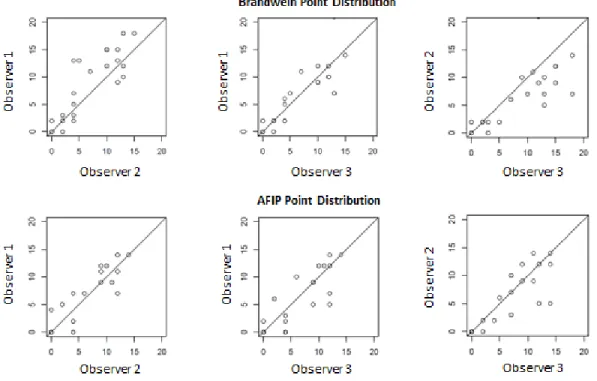
Figure 1 Point distribution between observers using the Brandwein vs AFIP
grading method………..………18 Figure 2 Kaplan-Meier plots of overall survival stratified by prognostic
variables….………...………...………..21 Figure 3 Kaplan-Meier plots of overall survival stratified by dichotomized
grading systems……...………...22 Figure 4 Immunohistochemistry controls, IE1-72 and pp65 antibodies………..32 Figure 5 Immunohistochemistry, sMEC reactivity with IE1-72 and pp65
viii
LIST OF ABBREVIATIONS AFIP Armed Forces Institute of Pathology DOD Died of disease
DFS Disease free survival DSS Disease specific survival
FFPE Formalin fixed and paraffin embedded hCMV Human cytomegalovirus
H&E Hematoxylin and eosin HPF High-power field IHC Immunohistochemistry IRB Institutional Review Board LVI Lymphovascular invasion PNI Perineural invasion
SEER National Cancer Institute Surveillance, Epidemiology, End Results Program
1
INTRODUCTION
Mucoepidermoid carcinoma is the most common malignancy of salivary glands.1 It comprises 10% of all salivary gland tumors and 35% of salivary gland malignancies.5 Clinical outcomes range from no evidence of disease after surgical resection to distant metastasis and death.6 Several grading systems were developed in attempts to predict these outcomes based on histopathological features; however, no single system has been universally adopted.
Tumor grade is based on the microscopic features of a neoplasm. A low-grade
designation indicates well-differentiated neoplastic cells. This infers that the cellular features closely resemble those of the tissue of origin, and these neoplasms tend to have less aggressive behavior.7 Conversely, high-grade neoplasms demonstrate loss of features of a mature cell population and are associated with aggressive behavior.7 Tumor grading is coupled with staging, or the extent of disease, to assess prognosis and guide treatment decisions.7
2
Both perinerual invasion (PNI) and lymphovascular invasion (LVI) allow for tumor spread to distant sites and are associated with metastasis.7,8 Cellular pleomorphism refers to variability in cellular and nuclear size and shape, and anaplasia indicates loss of differentiation.7 These features are present when the malignant cells no longer function to their specialized purpose.
Using these histologic criteria, in addition to others specific to features of
mucoepidermoid carcinoma, multiple scoring systems were developed in an attempt to predict the behavior of this malignancy.3, 4, 9 Due to the lack of a consensus grading system and subjectivity in some of their criteria, better methods of grading sMEC are needed.
Recent studies suggest a causal relationship between human cytomegalovirus (hCMV, human herpesvirus-5, HHV-5) and sMEC.2 hCMV infection is endemic, with positivity shown in every population examined through seroepidemiologic surveys.10 Its tropism for salivary gland epithelium was identified as early as 1932, when hCMV nuclear inclusions were observed in salivary ductal epithelial cells.11 hCMV remains in the latent cycle in the majority of individuals
infected, but reactivation is seen in immunocompromised patients.11 The molecular mechanisms of viral transition from latency to the lytic cycle are not fully established; however, viral gene products can be used to detect transcriptionally active hCMV.12 These include gene products IE1-72, proven to be important in viral replication, and pp65, a protein thought to play a role in immune subversion and incorporation of additional proteins into the virion.13, 14
3
4
REFERENCES
1. Seethala RR. An update on grading of salivary gland carcinomas. Head Neck Pathol. 2009 Mar;3(1):69-77.
2. Melnick M, Sedghizadeh PP, Allen CM, Jaskoll T. Human cytomegalovirus and
mucoepidermoid carcinoma of salivary glands: cell-specific localization of active viral and oncogenic signaling proteins is confirmatory of a causal relationship. Exp Mol Pathol. 2012 Feb;92(1):118-25.
3. Auclair PL, Goode RK, Ellis GL. Mucoepidermoid carcinoma of intraoral salivary glands. Evaluation and application of grading criteria in 143 cases. Cancer. 1992 Apr 15;69(8):2021-30. 4. Brandwein MS, Ivanov K, Wallace DI, Hille JJ, Wang B, Fahmy A, et al. Mucoepidermoid carcinoma: a clinicopathologic study of 80 patients with special reference to histological grading. Am J Surg Pathol. 2001 Jul;25(7):835-45.
5. Spiro RH, Huvos AG, Berk R, Strong EW. Mucoepidermoid carcinoma of salivary gland origin. A clinicopathologic study of 367 cases. Am J Surg. 1978 Oct;136(4):461-8.
6. Seethala RR. Histologic grading and prognostic biomarkers in salivary gland carcinomas. Adv Anat Pathol. 2011 Jan;18(1):29-45.
7. Abbas AK, Aster JC, editors. Chapter 7: Neoplasia. In: Robbins and Cotran Pathologic Basis of Disease. 9th ed. Philadelphia, PA: Elsevier/Saunders; 2015. p. 265-340.
8. Liebig C, Ayala G, Wilks JA, Berger DH, Albo D. Perineural invasion in cancer: a review of the literature. Cancer. 2009 Aug 1;115(15):3379-91.
9. Batsakis JG, Luna MA. Histopathologic grading of salivary gland neoplasms: I. Mucoepidermoid carcinomas. Ann Otol Rhinol Laryngol. 1990 Oct;99(10 Pt 1):835-8.
10. Bate SL, Dollard SC, Cannon MJ. Cytomegalovirus Seroprevalence in the United States: The National Health and Nutrition Examination Surveys, 1988–2004. Clinical Infectious Diseases. 2010 June 01;50(11):1439-47.
11. Riley HD,Jr. History of the cytomegalovirus. South Med J. 1997 Feb;90(2):184-90. 12. Yuan J, Liu X, Wu AW, McGonagill PW, Keller MJ, Galle CS, et al. Breaking human cytomegalovirus major immediate-early gene silence by vasoactive intestinal peptide stimulation of the protein kinase A-CREB-TORC2 signaling cascade in human pluripotent embryonal NTera2 cells. J Virol. 2009 Jul;83(13):6391-403.
5
Biology, Therapy, and Immunoprophylaxis. Cambridge: Cambridge University Press; 2007. Chapter 17.
6
CHAPTER 1: GRADING OF SALIVARY GLAND MUCOEPIDERMOID CARCINOMA
Introduction
Mucoepidermoid “tumor” was first named and characterized by Stewart et al (1945). Their group evaluated approximately 700 salivary gland neoplasms and found 45 that contained mucous, intermediate, and epidermoid cells. These cases were separated into categories of “benign” and “malignant” by correlating histologic appearance to clinical outcome. Features associated with a favorable outcome were the presence of multiple of the aforementioned cell types in large quantities, delineated margins, cystic spaces with mucous pools, and sheets or “plugs” of squamous epithelial cells. Conversely, features associated with malignancy included a predominance of epidermoid cells, anaplastic cells, infiltrative margins, and lack of large cystic spaces with mucous pools. It is emphasized that mucous cells, as demonstrated by a positive mucicarimine histochemical stain, must be present to render a diagnosis of mucoepidermoid tumor.1
Foote et al (1953) further detailed these neoplasms in a review of tumors of the major salivary glands. Due to observed metastases in cases designated as benign, the authors
7
In 1970, Healey et. alstudied 60 cases of sMEC to elaborate criteria for grading and surgical management. Like Foote, the authors adopted a three-tiered system. Grade I was described as well differentiated or low-grade, Grade II as moderately differentiated or medium-grade, and Grade III as poorly differentiated, high-grade neoplasms. Grade I lesions were characterized by cystic spaces lined by mucous-producing cells and epidermoid cells, having a tumor front that infiltrated surrounding tissues with broad borders, and in which mitotic figures were rare. Grade II sMECs comprised solid nests of intermediate or epidermoid cells. Cystic spaces contained increased intraluminal proliferations of intermediate and epidermoid cells as compared to Grade I tumors. The tumor front was less distinct and occasional mitotic figures were present. Grade III was defined by greater proportions of solid nests and glandular
structures, but less cystic space. Pleomorphism, prominent nucleoli, brisk mitotic activity, and aggressive infiltration into adjacent tissue were appreciable. Like Stewart’s publication, mucicarmine positivity was emphasized as critical to the diagnosis.3 Batsakis et al (1990) summarized and made minor modifications to Healey’s grading criteria of sMEC (Table 1).4
Spiro et al (1978) studied 367 cases of sMEC from the major and minor salivary glands and also adopted a 3-tiered system. The histologic criteria used for grading overlapped Healey’s, but focused on the predominance of different cell types within each grade. The low-grade tumors contained well-developed cystic structures lined by mucous cells. Increased solid areas of
epidermoid, squamous, or basaloid cells were observed in intermediate grade lesions; but cystic structures were also mentioned. The high-grade tumors had increased basaloid and epidermoid cells in solid nests or cords as well as prominent nucleoli and conspicuous mitoses. The 5-year DFS rate was 92%, 63%, and 27% for low, intermediate, and high grades, respectively.
8
initial surgery, which was usually simple excision rather than resection with wide margins, or a higher proportion of intermediate and high grade tumors. The authors concluded that the histologic grade correlated to the stage of the tumor, but due to instances of metastasis and tumor-related deaths in low grade lesions, all sMECs should be considered malignant.5
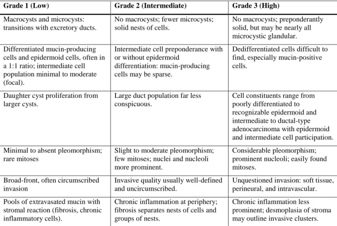
Table 1: Grades of mucoepidermoid carcinomas and their histocytologic characteristics. Batsakis et al 1990.
Grade 1 (Low) Grade 2 (Intermediate) Grade 3 (High)
Macrocysts and microcysts: transitions with excretory ducts.
No macrocysts; fewer microcysts; solid nests of cells.
No macrocysts; preponderantly solid, but may be nearly all microcystic glandular.
Differentiated mucin-producing cells and epidermoid cells, often in a 1:1 ratio; intermediate cell population minimal to moderate (focal).
Intermediate cell preponderance with or without epidermoid
differentiation: mucin-producing cells may be sparse.
Dedifferentiated cells difficult to find, especially mucin-positive cells.
Daughter cyst proliferation from larger cysts.
Large duct population far less conspicuous.
Cell constituents range from poorly differentiated to recognizable epidermoid and intermediate to ductal-type adenocarcinoma with epidermoid and intermediate cell participation.
Minimal to absent pleomorphism; rare mitoses
Slight to moderate pleomorphism; few mitoses; nuclei and nucleoli more prominent.
Considerable pleomorphism; prominent nucleoli; easily found mitoses.
Broad-front, often circumscribed invasion
Invasive quality usually well-defined and uncircumscribed.
Unquestioned invasion: soft tissue, perineural, and intravascular.
Pools of extravasated mucin with stromal reaction (fibrosis, chronic inflammatory cells).
Chronic inflammation at periphery; fibrosis separates nests of cells and groups of nests.
Chronic inflammation less prominent; desmoplasia of stroma may outline invasive clusters.
9
invasion, and proportion of cystic spaces showed statistically significant association to outcome. Too few tumors showed necrosis to achieve statistical power; however, the authors found this feature to be an important indicator when present. These five histologic criteria were weighted and incorporated into a proposed grading system (Table 2). The points for each group were compared with patient outcomes to define high, intermediate, and low tumor grades. This method became known as the Armed Forces Institute of Pathology (AFIP) grading system of mucoepidermoid carcinoma.6
Table 2: Parameters used for grading intraoral mucoepidermoid carcinoma. Auclair et al 1991.
Parameter Point value
Intracystic component <20% +2
Neural invasion present +2
Necrosis present +3
Mitoses (4+ per 10 HPF) +3
Anaplasia +4
Low-grade: 0-4, Intermediate-grade: 5-6, High-grade: 7+ *HPF = high powered field In a 1998 publication, Goode, Auclair, and Ellis applied the AFIP grading system to mucoepidermoid carcinoma of the major salivary glands. Two hundred thirty four cases were divided into four groups according to clinical outcome as elaborated previously. The tumors were then scored with the grading criteria outlined in Table 2.7
When comparing Group 1 and Group 4 patients, independent statistical significance of each grading criteria was confirmed. Groups 1 and 2 had mean scores of 2.0 and 2.3,
10
explain that clinical factors such as large tumor size or conservative treatment may incite metastasis in this group of low-grade appearing neoplasms.7
The average score of Group 4 tumors was 7.56. This score correlated a high-grade to poor prognosis; however, only 52% of these tumors were assigned high grades. Forty percent received a low-grade score. The tumor site accounted for some of this discrepancy, with 75% of Groups 3 and 4 tumors of the submandibular gland given a low histological grade. Conversely, 33% of Group 4 parotid gland tumors were given low grades. The authors concluded that their grading system is useful for parotid tumors, but sMECs of the submandibular gland require aggressive treatment regardless of histological tumor grade.7
Brandwein et al evaluated the AFIP grading system on reproducibility and prediction of outcome in a 2001 publication. Five pathologists independently graded 20 hematoxylin and eosin (H&E) slides of different sMECs using their personal grading method as well as the AFIP
grading system. Out of 100 pairs of results, there were 46 disagreements between the
pathologist’s own grade and the AFIP grade. Forty five of these were “downgrades,” where a pathologist graded a tumor higher using their own criteria than the AFIP grade. In 8 of the 45, the AFIP grading system downgraded the tumor by 2 grades. In one case, the AFIP grade was higher than the pathologist’s. Weighted kappa values were then averaged across observers to determine inter-observer agreement. The agreement between pairs of observers using their own grading criteria was ranged from poor to good (κ = 0.27 – 0.79, average κ = 0.49). Better
11
3mm (p = 0.048). Positive lymph nodes were identified in 33% (14). This correlated to increased tumor grade (p < 0.001), with 85% (12/14) corresponding to grade 3 tumors and the remaining 15% (2/14) corresponding to grade 2 tumors. Three patients developed distant metastases, all with grade 3 tumors. Ultimately, 0% (0/12) of grade 1, 5% (1/20) of grade 2 and 65% (10/16) of grade 3 patients in their study population died of disease. Taken together, increased tumor grade correlated to increased morbidity and mortality.8
The Brandwein group asserted that, based on their findings, the AFIP method downgraded tumors and they proposed a new grading system (Table 3). They re-graded 31 tumors using their method and compared the outcomes to those predicted by the AFIP system. Statistical significance was not achieved in correlating tumors grades to DFS using either grading method; however, the p value showed greater correlation to DFS with the proposed Brandwein grading than the AFIP (p = 0.099 vs 0.249).8
The authors concluded that a standardized grading system improves reproducibility between observers. In contrast to the findings of Goode et al, no decreased prognosis of tumors of the submandibular gland was identified.7, 8 The discrepancy on the behavior of submandibular tumors was theorized to be due to surgical failure to obtain adequate margins for risk of
12
Table 3: Proposed grading system for mucoepidermoid carcinoma. Brandwein et al 2001.
Feature Points
Intracystic component <25% +2
Tumor front invades in small nests and islands +2
Pronounced nuclear atypia +2
Lymphatic and/or vascular invasion +3
Bony invasion +3
> 4 mitoses per 10 HPF +3
Perineural spread +3
Necrosis +3
Low-grade: 0, Intermediate-grade: 2-3, High-grade: 4+ *HPF = high powered field Since the proposal of the three aforementioned grading systems, numerous publications debated their prognostic value and reproducibility. In 2006, Luna et algraded 43 sMECs of the parotid with the modified Healey, AFIP, and Brandwein system. The modified Healey and Brandwein graded similarly, with only one instance of disagreement between the two systems. Their study echoed the tendency of the AFIP system to downgrade tumors. The authors stated that the point-based AFIP and Brandwein systems were easier to reproduce than the modified Healey system; however, this finding was not statistically quantified.9
13
Later that year, a publication by Nance et al assessed 50 additional cases of sMEC from all sites in the head and neck, including larynx, trachea, and nasal cavity using the Brandwein grading method. No statistically significant differences were observed between low and intermediate-grades in either overall survival or DFS, and no patients in these groups DOD. There was statistical difference between high and low-grades and high and intermediate-grades by both measures (p<.001), and 52% of patients with high-grade tumors DOD. Loco-regional recurrence occurred in 30% (7/23) of high, 23% (3/13) intermediate, and 0% (0/14) low-grade cases. Based on a multivariate analysis, histologic grade was the only factor studied that affected both overall survival and DFS. The authors concluded that their study supports the predictive utility of the Brandwein method.11
In a 2009 review, Seethala criticized the modified Healey, AFIP, and Brandwein grading systems as cumbersome with ill-defined criteria. He restated the concern of the AFIP system to downgrade tumors, but also the tendency of the Brandwein system to upgrade. It was
emphasized that the results of these inaccuracies place patients at risk for under or over treatment, both of which can be associated with increased morbidity and/or mortality. The tumors of intermediate-grade were discussed as particularly concerning, with some studies clustering this group with low-grade behavior and others clustering them with high-grade. With the insipid nature of this category, the proper management of intermediate-grade tumors is unclear. However imperfect, Seethala advocated using a grading system for increased
reproducibility. He recommended the Brandwein or Healey system as it is more acceptable for a high-grade tumor to run an indolent course than for a low-grade neoplasm to behave
14
Brandwein re-analyzed her grading system as compared to that of the AFIP in a 2013 multi-institutional review of 76 patients. Forty one percent (31/76) of tumors were upgraded with the Brandwein method as compared to the AFIP. Most of the upgrades increased an AFIP Grade 1 tumor to Brandwein Grade 2 (20/25), but a significant number increased to Brandwein Grade 3 (5/25). Half of AFIP Grade 2 tumors were reclassified as Brandwein Grade 3 (5/10). It was noted that 6 patients with AFIP Grade 1 tumors experienced advanced disease beyond expected for a low grade. Three had positive cervical lymph node metastases, 2 experienced local recurrence, and 1 developed distant metastasis. Statistical power to determine predictive performance of each grading method was not achieved, however; and reliability between observers was not assessed.13
With the prevalent issue of limited sample sizes in previous studies, Chen et al(2014) analyzed Surveillance, Epidemiology, End Result (SEER) data on sMEC of the parotid gland. Patient demographics, tumor characteristics, and survival were correlated to the tumor grade, but the method of grading was unknown. A total of 2,400 adult patients were identified between the years 1988 and 2009. Low-grade sMEC comprised 21.8%, intermediate-grade 47.4%, and high-grade 30.9%. The demographic differences between low and intermediate-high-grades were not statistically significant. Both had increased prevalence in Caucasian females and mean ages of diagnosis of 52-52.8, respectively. In contrast, high-grade tumors were most common in Caucasian men and appeared at a later age (mean age of 66). No statistically significant
15
p < 0.001 and 51.7%, p < 0.001). The intermediate and high-grade tumors were also more likely to metastasize to regional lymph nodes (10.6% vs 15.9%, p = 0.03; and 56.8%, p < 0.001); however, only high-grade tumors were more likely to present with distant metastases (LG 0.2%, IG 0.3%, p > 0.99; HG 3.2%, p < 0.001).14
When evaluating the 5-year disease-specific survival (DSS), there was no statistically significant difference between low and intermediate-grade tumors (98.8% vs 97.4%, p = 0.09). High-grade tumors were associated with decreased 5 year DSS when compared to the grouped low and intermediate-grades (67.0% vs 97.8%, p < 0.001). By Cox multivariate regression, statistically significant indicators of decreased prognosis included histologic high-grade, increasing age, increasing tumor size, extra-parenchymal extension, positive lymph nodes, and distant metastasis.14
The authors concluded that the intermediate and low-grade sMECs were similar in patient demographics and survival. In contrast, high-grade sMECs were associated with male gender, older age at diagnosis, and significantly reduced survival. They attributed the published disparities of intermediate-grade behavior to the use of different grading systems. Because of proven reproducibility and use in WHO classification of head and neck tumors, the authors recommended use of the AFIP grading system.14
16
was no statistical difference in DSS. Although the authors endorsed the AFIP grading system, the method used by pathologists to grade the tumors included in the study was unknown.14 While multiple publications advocate use of a grading system, the question as to which method is best remains unanswered.
Materials and Methods
This project was reviewed and approved by the University of North Carolina Institutional Review Board (IRB 14-2941). Twenty eight formalin fixed and paraffin embedded (FFPE) tissue blocks and their corresponding H&E glass slides of mucoepidermoid carcinoma specimens were retrieved from UNC Hospitals Department of Pathology archives. The accession dates ranged from January 1, 2001 to December 31, 2016. The inclusion criterion was diagnosis of
mucoepidermoid carcinoma of salivary gland origin. Exclusion criteria were insufficient tissue for further study, unreadable H&E slides, and patient age less than 18. From these criteria, 23 cases were selected for study.
Observers were provided with tables of both the Brandwein and AFIP grading criteria. Instructions were given to document the points and grade of each case per grading method. The observers independently graded the same slide from each of the 23 cases. Consensus was
achieved at a round table discussion at a multi-headed microscope for any disagreement in tumor grade. The data generated was collected and tabulated after independent grading and after
consensus grading.
17
original grade given at diagnosis using each grading method. One case was graded at diagnosis as low by the AFIP method and high by Brandwein. This was excluded from the calculations.
Patient outcomes were assessed from the date of diagnosis to end points of last date of follow up or date of death. End dates were determined through review of the medical chart or through the NC State Center for Health Statistics. Log rank tests were used to correlate patient demographics and tumor characteristics to outcome. The staging data was unknown for 3 patients. One case was a recurrence with the initial date of diagnosis unknown. This was excluded from the outcomes assessment. Statistical significance was set at p < 0.05.
Results
Reliability analysis:
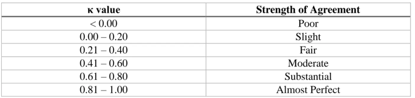
There was higher agreement on tumor grade between observers when using the AFIP grading system over Brandwein (73.9% vs 78.3%; κ = 0.650 vs κ = 0.743) (Figure 1, Table 5). Kappa values from both systems fell within the “substantial agreement” range of the Landis and Koch table of kappa interpretation (Table 4).15 There was also higher agreement between the
grade given at diagnosis and the consensus AFIP grade than the consensus Brandwein grade (81.8% vs 50.0%). Bias toward higher grading was noted in Grader 2 with both grading systems (Figure 1).
Table 4: Agreement measures for categorical data. Landis et al (1977).
κ value Strength of Agreement
< 0.00 Poor
0.00 – 0.20 Slight
0.21 – 0.40 Fair
0.41 – 0.60 Moderate
0.61 – 0.80 Substantial
18
19
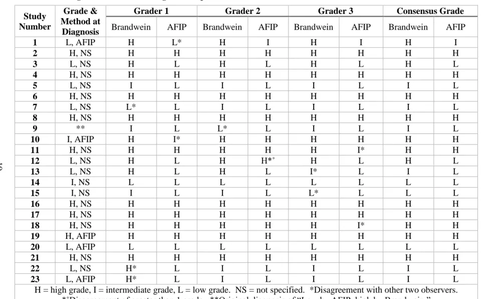
Table 5: Grading and inter-observer agreement per case.
Study Number
Grade & Method at
Diagnosis
Grader 1 Grader 2 Grader 3 Consensus Grade
Brandwein AFIP Brandwein AFIP Brandwein AFIP Brandwein AFIP
1 L, AFIP H L* H I H I H I
2 H, NS H H H H H H H H
3 L, NS H L H L H L H L
4 H, NS H H H H H H H H
5 L, NS I L I L I L I L
6 H, NS H H H H H H H H
7 L, NS L* L I L I L I L
8 H, NS H H H H H H H H
9 ** I L L* L I L I L
10 I, AFIP H I* H H H H H H
11 H, NS H H H H H I* H H
12 L, NS H L H H*˚ H L H L
13 L, NS H L H L I* L I L
14 I, NS L L L L L L L L
15 I, NS I L I L L* L L L
16 H, NS H H H H H H H H
17 H, NS H H H H H H H H
18 H, NS H H H H H I* H H
19 H, AFIP H H H H H H H H
20 L, AFIP L L L L L L L L
21 H, NS H H H H H H H H
22 L, NS H* L I L I L I L
23 L, AFIP H* L I L I L I L
20 Outcomes Assessment:
Follow up data ranged from 3 months to over ll years (median 1.82 years). With a limited sample size, statistical significance was not achieved for most characteristics evaluated. Lymph node status was an exception, with positive nodes being correlated to increased mortality (p = 0.046). Although not statistically significant, a 60% (6/10) death rate was observed in the male group versus 25% (3/12) in the female group. Patient demographics and tumor characteristics are summarized in Table 5.
Table 6: Patient demographics and tumor characteristics.
n Death p value
Sex p = 0.583
Male 10 6
Female 12 3
Race p = 0.146
African American 6 2
Hispanic 1 0
Caucasian 10 2
Unknown 5 5
Grade:Brandwein p = 0.107
Low 3 0
Intermediate 6 1
High 13 8
Grade: AFIP p = 0.088
Low 11 2
Intermediate 1 0
High 10 7
Grade: Original Pathologist p = 0.152
Low 9 1
Intermediate 2 0
High 10 7
Reported Tumor Size p = 0.084
T1 6 0
T2 4 1
T3 3 1
T4 6 4
Node Status p = 0.046
No positive nodes 13 2
21
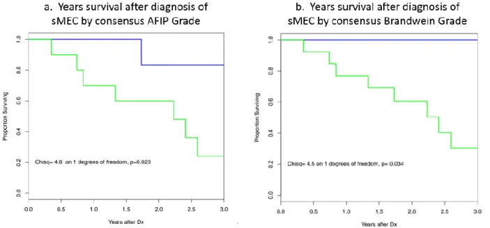
Statistical significance was not achieved to determine the prognostic value of either grading system (Figure 2). Due to the similar behavior between the low and intermediate grade tumors, these groups were combined to dichotomize the data (Figure 3). Statistical significance of the prognostic value of “high grade” versus “not high grade” tumors was achieved for both Brandwein and AFIP grading systems (p = 0.034 and p = 0.028, respectively).
22
Figure 3: Kaplan-Meier plots of overall survival stratified by dichotomized grading systems.
Discussion
Patient demographics:
Among our cases, there was a slightly higher proportion of female patients than males with a ratio of 1.2:1, which is similar to those reported in the literature.5, 6, 7,11,14 The mean female age at diagnosis was 43.7 whereas the mean male age at diagnosis was 85.5. Multiple studies stated an increased incidence of high-grade in males, but, similar to our findings, this has not been statistically significant when controlled for age in multivariate analysis.6,7, 11,14
Tumor characteristics:
23 Grading systems:
Seethala outlined the ideal requirements of a grading system as: Accurately predicts outcome
Can be used to stratify patients into distinct management categories Applicable to all sites where the tumor is seen
Simple criteria
Quick and time efficient
Reproducible with minimal inter- and intra-observer variability
Taking these criteria point by point, both the AFIP and Brandwein grading systems have support for their ability to predict patient outcome of low and high-grade tumors. Although statistical power was not achieved, the AFIP grading system correlated slightly better with patient outcome than the Brandwein system in our study (Figure 2). Several publications failed to prove a
statistically significant difference in survival between the low and intermediate grade
classifications, which was also identified in our study.11,14 This corresponds to the next point, as the similarities in behavior but differences in grade confounds the management of these
intermediate cases.
A lack of statistically significant differences in patient outcome between low and
intermediate grades raises the question of whether the treatment of these two grades should differ or whether the two grades should be combined. While survival was not affected, several studies have shown slightly increased recurrence and regional lymph node metastases of intermediate-grade over low-intermediate-grade tumors.10,11,14 The accepted treatment of low-grade sMECs is local
24
Whether either set of grading criteria can be applied to all sites of sMEC is also
ambiguous. Spiro and Goode noted a site-specific tendency for local metastasis in sMECs of the submandibular gland, with Goode advocating for aggressive treatment of these malignancies regardless of histological stage.5,7 This tendency was not reported in subsequent studies,
however. Brandwein et al (2001) specifically disputed the finding and offered inadequate surgical margins as an explanation of the discrepancy.8 The lack of consensus is likely due to limited cases of the submandibular site.
The simplicity and time efficiency of a task are subjective measures, but could certainly be aided by objective criteria. The criterion of anaplasia (AFIP) or “prominent nuclear atypia” (Brandwein) may be better stated as variation of nuclear sizes, hyperchromasia, or prominent nucleoli.
The reproducibility of the grading system is perhaps one of the most important criteria, so as to ensure these malignancies are studied and measured through the same calibration. By this measure, the AFIP grading method was superior to the Brandwein. This may be due to the reduced number of criteria enhancing the simplicity of the system. The AFIP grading method is recommended in the World Health Organization’s literature and thus is likely the most widely adopted.17 As the largest study in the literature demonstrates the pathology community’s proficiency of determining “high grade” vs “not high grade” tumors, as well as the low grade behavior of the intermediate category, it is of utmost importance that a standard is universally adopted so these tumors may be more adequately studied.14
Conclusions
25
26
REFERENCES
1. Stewart FW, Foote FW, Becker WF. Muco-Epidermoid Tumors of Salivary Glands. Annals of Surgery. 1945;122(5):820-844.
2. Foote FW Jr, Frazell EL. Tumors of the major salivary glands. Cancer. 1953 Nov;6(6):1065-133.
3. Healey WV, Perzin KH, Smith L. Mucoepidermoid carcinoma of salivary gland origin. Classification, clinical-pathologic correlation, and results of treatment. Cancer. 1970 Aug;26(2):368-88.
4. Batsakis JG, Luna MA. Histopathologic grading of salivary gland neoplasms: I. Mucoepidermoid carcinomas. Ann Otol Rhinol Laryngol. 1990 Oct;99(10 Pt 1):835-8. 5. Spiro RH, Huvos AG, Berk R, Strong EW. Mucoepidermoid carcinoma of salivary gland origin. A clinicopathologic study of 367 cases. Am J Surg. 1978 Oct;136(4):461-8.
6. Auclair PL, Goode RK, Ellis GL. Mucoepidermoid carcinoma of intraoral salivary glands. Evaluation and application of grading criteria in 143 cases. Cancer. 1992 Apr 15;69(8):2021-30. 7. Goode RK, Auclair PL, Ellis GL. Mucoepidermoid carcinoma of the major salivary glands: clinical and histopathologic analysis of 234 cases with evaluation of grading criteria. Cancer. 1998 Apr 1;82(7):1217-24.
8. Brandwein MS, Ivanov K, Wallace DI, Hille JJ, Wang B, Fahmy A, et al. Mucoepidermoid carcinoma: a clinicopathologic study of 80 patients with special reference to histological grading. Am J Surg Pathol. 2001 Jul;25(7):835-45.
9. Luna MA. Salivary mucoepidermoid carcinoma: revisited. Adv Anat Pathol. 2006 Nov;13(6):293-307.
10. Aro K. Management and Outcome of Patients With Mucoepidermoid Carcinoma of Major Salivary Gland Origin: A Single Institution's 30‐Year Experience. Laryngoscope. 2008 Feb 1;118(2):258; 258,262; 262.
11. Nance MA, Seethala RR, Wang Y, Chiosea SI, Myers EN, Johnson JT, et al. Treatment and survival outcomes based on histologic grading in patients with head and neck mucoepidermoid carcinoma. Cancer. 2008 Oct 15;113(8):2082-9.
12. Seethala RR. An update on grading of salivary gland carcinomas. Head Neck Pathol. 2009 Mar;3(1):69-77.
27
14. Chen MM, Roman SA, Sosa JA, Judson BL. Histologic grade as prognostic indicator for mucoepidermoid carcinoma: a population-level analysis of 2400 patients. Head Neck. 2014 Feb;36(2):158-63.
15. Landis JR, Koch GG. The measurement of observer agreement for categorical data. Biometrics. 1977 Mar;33(1):159-74.
16. Seethala RR. Histologic grading and prognostic biomarkers in salivary gland carcinomas. Adv Anat Pathol. 2011 Jan;18(1):29-45.
28
CHAPTER 2: HUMAN CYTOMEGALOVIRUS EXPRESSION IN SALIVARY GLAND MUCOEPIDERMOID CARICINOMA
Introduction
Since Agostinos Bassi’s paradigm-shifting discoveries in the mid-1800’s, infectious microorganisms are proven to cause significant human morbidity and mortality.1 In the 1890’s, Robert Koch detailed postulates, or criteria, necessary to establish a causal relationship between an agent and a disease.2 While these postulates explained etiologies of infectious diseases such as tuberculosis and cholera, the impact of microorganisms on human health expanded to include oncogenesis with the discovery of Epstein-Barr virus (EBV, human herpesvirus-4, HHV-4) in Burkitt lymphomas in 1964.3 In 1996, Koch’s postulates were revisited and updated to account for advances made in molecular identification of diseases including viruses.2 Seven
tumor-associated viruses, or oncoviruses, are now appreciated to play causative roles in human cancers.4 Two of these, EBV and Kaposi’s sarcoma herpesvirus 8 (KSV, human herpesvirus-8, HHV-8), are members of the herpesviridae family of DNA viruses.
29
In a second study by their group, two board-certified oral and maxillofacial pathologists graded 39 human sMEC specimens by the modified Healey system. Immunohistochemistry techniques were used to evaluate the tumor tissue with antibodies to hCMV proteins IE1-72 and pp65. IE1-72 reactivity, identified within the nuclei and/or cytoplasm, was identified in 38/39 of their tumors. It was reported that increased IE1-72 reactivity was associated with increased tumor grade; however, this finding was not objectively quantified. Reactivity to pp65 was also seen in the cytoplasm and nucleus of tumor cells, as well as in inflammatory cells within the tumor stroma. No reactivity of either antibody was identified in adjacent, normal salivary gland tissueThe authors concluded that their findings satisfied the causal criteria for hCMV etiology of sMEC by establishing that hCMV is present in most cases of sMEC, only the neoplastic tissue harbors the infectious agent, hCMV-specific gene expression was demonstrated at the cellular level and was positively correlated with sMEC severity, infection was correlated with an upregulation of an oncogenic signaling pathway, and mCMV induced malignant transformation in an in vitro animal model.6
Based on these findings, we hypothesized that a correlation between hCMV IE1-72 and pp65 expression could be quantified and used as an adjunctive prognostic indicator in the pathologic grading of sMEC.
Materials and Methods
30
2001 to December 31, 2016. Inclusion/exclusion criteria and grading methods were previously described in Chapter 1.
All immunohistochemical analyses were performed on 4 µm thick sections at the Translational Pathology Laboratory at the University of North Carolina at Chapel Hill.
Commercial antibodies to IE1-72 (MAB810, clone 8B1.2, Millipore, Temecula, CA) and pp65 (Cytomegalovirus PP65 antibody, Biorbyt, San Francisco, CA) were used for hCMV
identification. Immunohistochemistry (IHC) was performed in the Bond fully-automated slide staining system (Leica Biosystems Inc., Buffalo Grove, IL). Slides were deparaffinized in Bond dewax solution (AR9222) and Bond wash solution (AR9590). Antigen retrieval was performed at 100 C° in Bond-epitope retrieval solution 1, pH 6.0 (AR9961) for 20 minutes. After
pretreatment, pp65 (1:50 dilution) and IE1-72 (1:200 dilution) antibodies were applied for one hour. Bond polymer refine detection system (DS9800), a polymeric horseradish peroxidase-linker conjugate system, was used for antibody detection. Stained slides were dehydrated and cover-slipped.
Positive controls of hCMV infected gastrointestinal tissue were used for each antibody. Two sets of negative controls were used for each antibody. One of these comprised hCMV infected gastrointestinal tissue with no antibody. The other was histologically normal salivary gland lobules included in biopsy specimens of mucoceles from 12 patients approximately age-sex matched to the study population. Fourteen specimens included unaffected, adjacent salivary gland tissue that was evaluated as an internal control.
31
Results
IE1-72 Antibody
The hCMV-infected cells of the positive control were characterized by a dark brown nuclear signal and faint brown, granular cytoplasmic staining (Figure 4c). No staining was identified within the negative controls or the sMEC tumor samples (0/23) (Figures 4 and 5, left column).
pp65 Antibody
32
33
34
Discussion
Immunohistochemistry (IHC) is a helpful diagnostic aid to traditional hematoxylin and eosin (H&E) pathology. This method allows a suspecting pathologist to test cellular expression of specific antigens through the binding of a known antibody coupled with a detection method.7
While these adjunctive procedures are invaluable and revolutionary to the field of pathology, pitfalls confound a correct interpretation or diagnosis. These include biomarkers and/or antibodies lacking in specificity or sensitivity, improper tissue fixation or storage, laboratory techniques, and errors in interpretation.7
Some of these errors may explain the failure of our study to reproduce the findings of hCMV in sMEC. Melnick et al identified positive staining for hCMV protein IE1-72 in 38/39 of tumors studied, while none of our sMEC specimens (0/23) showed positivity using the same clone and manufacturer of that antibody.6 IE1-72 is a viral protein that is synthesized in the cytoplasm and then localizes to the nucleus in the early stages of infection.8 While some reactivity may be seen in the cytoplasm, nuclear staining is expected for positive interpretation per the manufacturer. When evaluating the figures of Melnick et al, cytoplasmic staining is observed but only 1 of 3 images shows evidence of nuclear staining.6 Our contrasting outcomes may be due to differences in interpretation.
Another cause for discrepancy may involve the laboratory techniques utilized. While the underlying chemistry was similar, our study used an automated system and Melnick et al
35
signal. The differing reagents between the two studies may also be contributory to opposing results.6
In our study, the antibody to hCMV protein pp65 showed unspecific binding, possibly due to the polyclonal nature of the antibody cross-reacting with epitopes of the native tissue. This manifested in a positive signal in the myoepithelial cells of the negative control as well as
stromal inflammatory cells and adjacent, unaffected glandular tissue in the tumor specimens. We suspect our results were due to error in choosing a sensitive and specific antibody. This antibody differed from the one used by Melnick et al due to discontinued production by their manufacturer (NCL-CMVpp65 clones 2 and 6 Lecia, Microsystems, Newcastle, UK). While our results were inconclusive, another group published negative findings after evaluating four sMEC cases using the same Lecia antibody and clones.10
Immunohistochemistry is accepted as the gold standard for identification of hCMV-infected tissue, but alternative methods can be used to validate results. Studies have found
polymerase chain reaction (PCR) techniques to be sensitive and specific for identifying hCMV in FFPE tissue.11 Another study of three sMEC cases failed to demonstrate PCR products of major immediate early genes of hCMV.12 Larger, more robust studies using PCR, antibodies to
alternative targets, and/or in-situ hybridization techniques may be indicated to further elucidate the role of hCMV in sMEC.
Conclusions
36
REFERENCES
1. Porter JR. Agostino Bassi bicentennial (1773-1973). Bacteriological Reviews. 1973;37(3):284-288.
2. Fredericks DN, Relman DA. Sequence-based identification of microbial pathogens: a reconsideration of Koch’s postulates. Clinical Microbiology Reviews. 1996;9(1):18-33.
3. Epstein MA, Achong BG, Barr YM. Virus Particles in Cultured Lymphoblasts from Burkitt's Lymphoma. Lancet. 1964 Mar 28;1(7335):702-3.
4. Pagano JS, Blaser M, Buendia MA, Damania B, Khalili K, Raab-Traub N, et al. Infectious agents and cancer: criteria for a causal relation. Semin Cancer Biol. 2004 Dec;14(6):453-71.
5. Melnick M, Abichaker G, Htet K, Sedghizadeh P, Jaskoll T. Small Molecule Inhibitors of the Host Cell COX/AREG/EGFR/ERK Pathway Attenuate Cytomegalovirus-induced
Pathogenesis. Experimental and molecular pathology. 2011;91(1):400-410.
6. Melnick M, Sedghizadeh PP, Allen CM, Jaskoll T. Human cytomegalovirus and
mucoepidermoid carcinoma of salivary glands: cell-specific localization of active viral and oncogenic signaling proteins is confirmatory of a causal relationship. Exp Mol Pathol. 2012 Feb;92(1):118-25.
7. O’Hurley G, Sjöstedt E, Rahman A, Li B, Kampf C, Pontén F, et al. Garbage in, garbage out: A critical evaluation of strategies used for validation of immunohistochemical biomarkers. Molecular Oncology. 2014 6;8(4):783-98.
8. Stinski MF MJ. Immediate-early viral gene regulation and function. In: Arvin A, Campadelli-Fiume G, Mocarski E, Moore PS, Roizman B, editor. Human Herpesviruses: Biology, Therapy, and Immunoprophylaxis. Cambridge: Cambridge University Press; 2007. Chapter 17.
9. Jordan RC, Daniels TE, Greenspan JS, Regezi JA. Advanced diagnostic methods in oral and maxillofacial pathology. Part II: immunohistochemical and immunofluorescent methods. Oral Surg Oral Med Oral Pathol Oral Radiol Endod. 2002 Jan;93(1):56-74.
10. Jayaraj G, Sherlin HJ, Ramani P, Premkumar P, Anuja N. Cytomegalovirus and
Mucoepidermoid carcinoma: A possible causal relationship? A pilot study. J Oral Maxillofac Pathol. 2015 Sep-Dec;19(3):319-24.
37