CBCT USES IN CLINICAL ENDODONTICS
PART 1: EFFECT OF CBCT ON THE ABILITY TO LOCATE MB2 IN MAXILLARY MOLARS; PART 2: OBSERVER EFFECTS IN DETECTING PERIAPICAL
RADIOLUCENCIES
Jeffrey Michael Parker
A thesis submitted to the faculty of the University of North Carolina at Chapel Hill in partial fulfillment of the requirements for the degree of Master of Science in the Endodontics
Department in the School of Dentistry.
Chapel Hill 2016
ABSTRACT
Jeffrey Michael Parker: CBCT uses in Clinical Endodontics
Part 1: Effect of CBCT on the Ability to Locate MB2 in Maxillary Molars; Part 2: Observer Effects in Detecting Periapical Radiolucencies
(Under the direction of Peter Tawil)
CBCT should be considered the imaging modality of choice for initial endodontic treatment of teeth with the potential for extra canals and suspected complex morphology. RCT of maxillary molars often falls into this category due to their complex anatomy and likely MB2 canal that is present. It has been shown that CBCT can be used to locate missed canals, but there are no in vivo studies demonstrating this. The other issue with CBCT is the accuracy of
ACKNOWLEDGEMENTS
I wish to acknowledge the research grant from the American Association of Endodontists Foundation that made this project possible.
Many thanks to the following people:
Dr. Peter Tawil for your mentorship, friendship, and dedication.
Dr. Eric Rivera for your leadership, passion, and giving me the opportunity to excel. Dr. Andre Mol for your guidance and knowledge in the field of oral radiology. Dr. Ceib Phillips for helping with the statistical analysis.
Dr. Asma Khan for always keeping your door open so we could talk about my research progress. Dr. Elena Kan, Dr. Melita Islambasic, Dr. Bryan Mitchell, Dr. TJ Taggar, Dr. Alison St. Paul, Dr. Mark Shallal-Ayzin, Dr. Bill Yeung, and Dr. Tam Trinh for participating in my research study.
Dr. Bruce and Rochelle Parker for their love and support.
TABLE OF CONTENTS
LIST OF FIGURES ... vii
LIST OF TABLES ... viii
LIST OF ABBREVIATIONS ... ix
Thesis Introduction ... 1
References ... 5
Manuscript 1: The effect of CBCT on the ability to locate MB2 in maxillary molars ... 7
Introduction ... 7
Materials and Methods ... 7
Statistical Analysis ... 11
Results ... 11
Discussion ... 12
Conclusion ... 15
References ... 16
Manuscript 2: CBCT and observer effects in detecting periapical radiolucencies ... 18
Introduction ... 18
Results ... 21
Discussion ... 22
Conclusion ... 25
References ... 26
Thesis Summary... 28
References ... 31
Appendix A: Patient Consent Form Part 1 ... 32
Appendix B: Clinician Consent Form Part 2 ... 37
Appendix C: IRB Approval Letter ... 41
Appendix D: Tables ... 43
LIST OF FIGURES
Figure 1: CBCT volume showing an untreated MB2 canal (red arrows),
which was subsequently instrumented at the second appointment. ... 10 Figure 2: Location and root canal fill of MB2 canal located slightly mesial
and palatal to the MB orifice. (A) Pre-op photo (B) Location of MB2
LIST OF TABLES
Table 1: McNemar’s test ... 11
Table 2: Frequency of MB2 canals located ... 12
Table 3: Experience level compared to “gold standard” for each group ... 22
LIST OF ABBREVIATIONS AAE American Association of Endodontists
ALADA As low as diagnostically acceptable ALARA As low as reasonably achievable CBCT Cone beam computed tomography
CT Computed tomography
DDS Doctor of dental surgery FOV Field of view
MB2 Second mesio-buccal canal of a maxillary molar NiTi Nickel titanium
THESIS INTRODUCTION
In 1998 a new volumetric computed tomography (CT) machine was introduced using cone beam technology (1). While this technology was originally used for implant treatment planning, its applications in endodontics quickly became apparent. Cone beam computed tomography (CBCT) has been more frequently used in endodontic practices as awareness of the technology increased and the cost of the equipment decreased. The American Association of Endodontists (AAE) released a position statement in 2015 on CBCT stating that limited field of view CBCT should be considered the imaging modality of choice for initial endodontic treatment of teeth with the potential for extra canals and suspected complex morphology (2). CBCT has been advocated to be used for the location of missed canals (3), (4), (5) and in aiding in the determination of MB2 in vitro. Locating and treating all canals during root canal treatment has been shown to be an important aspect of overall success. A common failure in endodontic treatment of the permanent maxillary first molars is likely to be caused by an inability to locate, clean, and fill the MB2 canal (6). It is also important to understand the anatomy of maxillary molars to successfully treat these teeth (7).
(FOV) are acquired in a complete, or sometimes partial, arc of at least 180° (8). This procedure varies from a traditional medical CT, which uses a fan-shaped x-ray beam in a helical
progression to acquire individual image slices of the FOV and then stacks the slices to obtain a three-dimensional representation. Each slice requires a separate scan and separate
two-dimensional reconstruction. Several advantages of CBCT are a shorter examination time, reduced image distortion due to internal patient movements, and increased x-ray tube efficiency (9).
Recent studies have shown success rates of root canal therapy (RCT) evaluated by CBCT to be significantly lower than those teeth evaluated with two-dimensional radiography. There was a fourteen times increase in failure rate when teeth with no pre-operative periapical
radiolucencies were assessed with CBCT compared to periapical radiographs at one year (10). Symptomatic teeth scanned with CBCT showed periapical lesions 14% of the time compared to periapical radiographs which showed periapical lesions 3% of the time (11). In addition, CBCT revealed a higher prevalence of periapical radiolucencies in teeth diagnosed with non-vital pulps compared to two dimensional imaging (12). Although this technology has been shown to
provide more clinically relevant information than periapical radiographs (13), care should be taken to avoid misinterpretation as artifact formation may lead to inaccurate misinterpretation of CBCT images (14).
In this thesis, a small field of view CBCT was used to maximize resolution and to keep radiation as low as reasonably achievable (ALARA) (15). ALARA is a radiation
safety principle for minimizing radiation doses by employing all reasonable methods as radiation
that are common in dentistry. A CBCT volume taken with the Carestream 9000 of the right posterior maxilla is equivalent to one day of background radiation (9.8 µSv) and a periapical radiograph is equivalent to .6 days of background radiation (16).
Overall, fifteen limited field of view CBCT volumes were made for part 1 of this thesis and an additional eight CBCT volumes were made for part 2, for a total of 23 CBCT volumes. This two-part thesis overlapped as the CBCT volumes from part 1 were used in part 2. The information from part 1 determined the effectiveness of these CBCT volumes and provided some knowledge as to whether three-dimensional imaging may lead to locating and treating more MB2 canals compared to the traditional method of access with a microscope. The information
obtained from CBCT is only valuable if the clinician is able to accurately and reliably interpret the imaging. In part 2, clinicians of varying levels were compared to the “gold standard” of experienced dental radiologists to see how accurately they interpreted the CBCT volumes for the presence or absence of a periapical radiolucency. By having two separate viewing times, with a washout period in between, the intra-rater reliability was assessed to see how consistent
clinicians were in determining if a periapical radiolucency was present at the root apices. The accuracy and consistency of interpreting periapical radiolucencies may lead clinicians to question if they are interpreting CBCT volumes appropriately, and if they are providing the correct diagnosis and etiology to the patient, such as a missed MB2 canal in a maxillary molar.
In this prospective clinical research study, the purpose is two-fold: to determine if a CBCT volume aided in the location of MB2 canals in maxillary molars and to determine the effect that the experience level had on the ability to accurately detect periapical radiolucencies in CBCT volumes. Many clinicians are using CBCT for location of additional canals and to
upon the knowledge of the clinician to interpret CBCT volumes to have the best outcome for the patient. In addition, clinicians may not be on the same level as a dental radiologist in meeting the “standard of care” in interpreting these volumes. Inaccurate interpretations and inconsistency among CBCT readers may ultimately harm the patient by having faulty conclusions of a disease that is present or absent.
REFERENCES
1. Mozzo P, Procacci C, Tacconi A, Martini PT, Andreis IA. A new volumetric CT machine for dental imaging based on the cone-beam technique: preliminary results. European radiology. 1998;8(9):1558-64.
2. Special Committee to Revise the Joint AAEAPSouoCiE. AAE and AAOMR Joint Position Statement: Use of Cone Beam Computed Tomography in Endodontics 2015 Update. Oral surgery, oral medicine, oral pathology and oral radiology. 2015;120(4):508-12.
3. Blattner TC, George N, Lee CC, Kumar V, Yelton CD. Efficacy of cone-beam computed tomography as a modality to accurately identify the presence of second mesiobuccal canals in maxillary first and second molars: a pilot study. Journal of endodontics. 2010;36(5):867-70.
4. Bauman R, Scarfe W, Clark S, Morelli J, Scheetz J, Farman A. Ex vivo detection of mesiobuccal canals in maxillary molars using CBCT at four different isotropic voxel dimensions. International endodontic journal. 2011;44(8):752-8.
5. Vizzotto MB, Silveira PF, Arus NA, Montagner F, Gomes BP, da Silveira HE. CBCT for the assessment of second mesiobuccal (MB2) canals in maxillary molar teeth: effect of voxel size and presence of root filling. International endodontic journal. 2013;46(9):870-6. 6. Chang SW, Lee JK, Lee Y, Kum KY. In-depth morphological study of mesiobuccal root
canal systems in maxillary first molars: review. Restorative dentistry & endodontics. 2013;38(1):2-10.
7. Degerness RA, Bowles WR. Dimension, anatomy and morphology of the mesiobuccal root canal system in maxillary molars. Journal of endodontics. 2010;36(6):985-9.
8. Scarfe WC, Farman AG, Levin MD, Gane D. Essentials of maxillofacial cone beam computed tomography. The Alpha omegan. 2010;103(2):62-7.
9. Scarfe WC, Farman AG. What is cone-beam CT and how does it work? Dental clinics of North America. 2008;52(4):707-30.
10. Patel S, Wilson R, Dawood A, Mannocci F. The detection of periapical pathosis using periapical radiography and cone beam computed tomography - part 1: pre-operative status. International endodontic journal. 2012;45(8):702-10.
12. Abella F, Patel S, Duran-Sindreu F, Mercade M, Bueno R, Roig M. An evaluation of the periapical status of teeth with necrotic pulps using periapical radiography and cone-beam computed tomography. International endodontic journal. 2014;47(4):387-96.
13. Lofthag-Hansen S, Huumonen S, Grondahl K, Grondahl HG. Limited cone-beam CT and intraoral radiography for the diagnosis of periapical pathology. Oral surgery, oral medicine, oral pathology, oral radiology, and endodontics. 2007;103(1):114-9.
14. Estrela C, Bueno MR, Azevedo BC, Azevedo JR, Pecora JD. A new periapical index based on cone beam computed tomography. Journal of endodontics. 2008;34(11):1325-31. 15. Hedesiu M, Baciut M, Baciut G, Nackaerts O, Jacobs R. Comparison of cone beam CT
device and field of view for the detection of simulated periapical bone lesions. Dento maxillo facial radiology. 2012;41(7):548-52.
MANUSCRIPT 1: THE EFFECT OF CBCT ON THE ABILITY TO LOCATE MB2 IN MAXILLARY MOLARS
Introduction
The significance of MB2 was first recognized in 1969 when Weine found a large number of second canals in the mesial buccal root of sectioned maxillary first molars that was done in vitro (17). Weine also stated that the MB and MB2 canals either generally join 1-4 mm from the apex and have a common foramina or remain separate and exit through two foramen. Vertucci showed maxillary first molars have an incidence of MB2 canals 55% of the time and maxillary second molars have an incidence of MB2 canals 29% of the time in vitro (18). With the use of the dental operating microscope, locating MB2 in maxillary molars in a clinical simulation was 82% in vitro (19), 71% in vivo (20), and 93% with experienced clinicians (21). It has been shown in vitro by root sectioning that a separate orifice on the pulpal floor was present 96% of the time, however the canal may only be negotiated 80% of the time (22). Factors affecting negotiation could be debris, pulp stones, calcifications, and anatomical variations (23). The use of CBCT in detecting the existence or absence of MB2 has been shown to be 79% and CBCT has been suggested to be as reliable as the sectioning of teeth (3). The purpose of this study was to determine if a CBCT volume aided in the location of MB2 canals in maxillary molars done in a graduate university based endodontic clinic.
IRB approval was obtained at the University of North Carolina at Chapel Hill (14-0608). The Global G6 dental operating microscope (Global Surgical Corporation, St. Louis, MO, USA) was used by endodontic residents to complete the root canal treatment in this study. The
importance and advantages of using magnification during root canal treatment has been described in the literature (19). The same Carestream 9000 CBCT (Carestream Dental,
Rochester, NY, USA) small volume was used for all of the scans. All endodontic residents had previous training on CBCT through a required dental radiology course for their master’s studies. The inclusion criteria for this study were a patient in need of root canal treatment on a maxillary first or second molar over the age of 18, with an ASA classification of I or II. The exclusion criteria were a patient under the age of 18, an ASA classification of III or higher, or pregnant.
Fifty maxillary first and second molars needing RCT were included in this study and randomly assigned to residents by the clinic front desk. There were no controls in this study. In order to make a pre-operative diagnosis to determine the need for root canal treatment, all appropriate clinical testing was performed and the diagnosis was made according to the AAE guidelines for diagnostic definitions. Written consent was obtained from all patients to
participate in this study. All teeth were accessed using a #2 and #4 carbide round bur (Braessler USA, Savannah, GA, USA), a #169L bur (Miltex Inc., York, PA, USA), and a pulp shaper bur (Dentsply, York, PA, USA). After the access was completed, the clinician determined if MB2 was located or not. Clinical location of MB2 was recorded if the clinician was able to visualize the orifice of the canal and obtain a stick with a six, eight, or ten size C-file (Dentsply
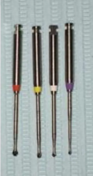
paste of calcium hydroxide powder (Sultan Healthcare Inc., Hackensack, NJ, USA) mixed with saline as an intracanal medicament between appointments. This paste was placed in the canals with a lentulo spiral (Dentsply International Inc., Johnson City, TN, USA). The tooth was temporized with gas sterilized yellow teflon tape (LA-CO Industries Inc., Elk Grove Village, IL, USA) as a space preserver and IRM (Dentsply Caulk, Milford, DE USA). The CBCT volume was interpreted by the same clinician that accessed the tooth who then deemed radiographic CBCT MB2 as present or not present. Figure 1 shows an example of the CBCT volume revealing a missed MB2 canal. At the second appointment Munce discovery burs (CJM Engineering Inc., Santa Barbara, CA, USA) were used for selective troughing along the pulpal floor in the location seen on the CBCT volume in search of MB2. After troughing below the pulpal floor MB2 was deemed as located or not located within 0-2 mm or > 2 mm of depth (troughing). Figure 2 shows the mesial/palatal location of MB2 that is typically present and the root canal fill of the MB2 canal. All subjects had the RCT completed in two visits. Upon completion of the root canal treatment, a core build-up was placed to restore the access and the patient was instructed to have a full coverage restoration placed within sixty days.
Figure 1: CBCT volume showing an untreated MB2 canal (red arrows), which was subsequently instrumented at the second appointment.
Figure 2: Location and root canal fill of MB2 canal located slightly mesial and palatal to the MB orifice. (A) Pre-op photo (B) Location of MB2 (C) Instrumentation of MB2 (D)
Root canal fill of MB2.
A B
Statistical Analysis
McNemar’s test was used to determine the effectiveness of using CBCT to locate MB2 canals in maxillary molars. The level of significance was established at p < .05.
Results
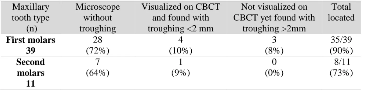
Table 1 shows the McNemar’s test comparing locating MB2 canals with CBCT and troughing. This yielded a p value of .26, which is not statistically significant. There was not a statistically significant discordance between CBCT and access in the identification of MB2. Table 2 provides the distribution of the fifty teeth in this study in terms of maxillary first or second molars. It also shows the MB2 canals located in each group. It is likely MB2 was not present in some of the fifty maxillary molars, and the clinicians may have located all of the possible MB2 canals that were present. Overall, 90% of maxillary first molar and 73% of maxillary second molar MB2 canals were located in these fifty teeth.
TABLE 1: MCNEMAR’S TEST
MB2 seen on CBCT MB2 Located After Access Total
Yes No
Yes 5 0 5
No 3 7 10
Total 8 7 15
TABLE 2: FREQUENCY OF MB2 CANALS LOCATED Maxillary tooth type (n) Microscope without troughing
Visualized on CBCT and found with troughing <2 mm
Not visualized on CBCT yet found with
troughing >2mm Total located First molars 39 28 (72%) 4 (10%) 3 (8%) 35/39 (90%) Second molars 11 7 (64%) 1 (9%) 0 (0%) 8/11 (73%) Discussion
where MB2 was not visualized on the CBCT volume, but MB2 was located clinically after troughing greater than 2 mm. By viewing the CBCT volume in the sagittal, coronal, and axial dimensions, the clinician was able to understand the root anatomy and location of where to trough to locate MB2. Canals are located symmetrically in roots, meaning that if a clinician sees the MB canal is towards the buccal aspect of the root, a MB2 canal is likely present on the lingual aspect of the MB root even if a MB2 is not definitively seen on the CBCT volume. This finding suggests knowledge of how and where to trough for MB2 based on the CBCT image may be important, and could lead to conserving tooth structure while troughing for MB2.
an increase from 60.1% at 0.4 mm voxel size to 93.3% at 0.125 mm voxel size (4). The voxel size of the Carestream 9000 is .076 mm. The Carestream 9000 has the smallest voxel size in the market currently. Another popular CBCT unit in endodontics is the J. Morita Accuitomo 170 (J. Morita USA, Irvine, CA, USA), which has a voxel size of .08 mm.
In terms of the overall success in locating MB2 canals in maxillary molars, the
endodontic residents in our study were consistent with a Stropko study where MB2 was located in 93% of maxillary first molars and 60% of maxillary second molars (21). The residents in our study located MB2 in 90% of maxillary first molars and 73% of maxillary second molars in the fifty teeth. It is important to note that there was faculty assistance in some of these cases, which could explain why the number of MB2 canals found in this in vivo study is on the higher side compared to previous studies (20, 28-30). Another interesting finding showed that MB2 canals were located in 70% of teeth, without clinical troughing. In other words, 70% of MB2 canals were located by using the dental operating microscope. The anatomical location of MB2 is located slightly mesial to the imaginary line between the palatal and MB canal (31). This routine step in accessing maxillary molars could significantly increase locating MB2 canals even
amongst inexperienced practitioners.
Conclusion
REFERENCES
17. Weine FS, Healey HJ, Gerstein H, Evanson L. Canal configuration in the mesiobuccal root of the maxillary first molar and its endodontic significance. Oral surgery, oral medicine, and oral pathology. 1969;28(3):419-25.
18. Vertucci FJ. Root canal anatomy of the human permanent teeth. Oral surgery, oral medicine, and oral pathology. 1984;58(5):589-99.
19. Baldassari-Cruz LA, Lilly JP, Rivera EM. The influence of dental operating microscope in locating the mesiolingual canal orifice. Oral surgery, oral medicine, oral pathology, oral radiology, and endodontics. 2002;93(2):190-4.
20. Buhrley LJ, Barrows MJ, BeGole EA, Wenckus CS. Effect of magnification on locating the MB2 canal in maxillary molars. Journal of endodontics. 2002;28(4):324-7.
21. Stropko JJ. Canal morphology of maxillary molars: clinical observations of canal configurations. Journal of endodontics. 1999;25(6):446-50.
22. Gorduysus MO, Gorduysus M, Friedman S. Operating microscope improves negotiation of second mesiobuccal canals in maxillary molars. Journal of endodontics.
2001;27(11):683-6.
23. Ibarrola JL, Knowles KI, Ludlow MO, McKinley IB, Jr. Factors affecting the
negotiability of second mesiobuccal canals in maxillary molars. Journal of endodontics. 1997;23(4):236-8.
24. Cotton TP, Geisler TM, Holden DT, Schwartz SA, Schindler WG. Endodontic applications of cone-beam volumetric tomography. Journal of endodontics. 2007;33(9):1121-32.
25. Yoshioka T, Kikuchi I, Fukumoto Y, Kobayashi C, Suda H. Detection of the second mesiobuccal canal in mesiobuccal roots of maxillary molar teeth ex vivo. International endodontic journal. 2005;38(2):124-8.
26. Corbella S, Del Fabbro M, Tsesis I, Taschieri S. Computerized Tomography Technique for the Investigation of the Maxillary First Molar Mesiobuccal Root. International journal of dentistry. 2013;2013:614898.
27. Gilles J, Reader A. An SEM investigation of the mesiolingual canal in human maxillary first and second molars. Oral surgery, oral medicine, and oral pathology. 1990;70(5):638-43.
29. Sempira HN, Hartwell GR. Frequency of second mesiobuccal canals in maxillary molars as determined by use of an operating microscope: a clinical study. Journal of
endodontics. 2000;26(11):673-4.
30. Wolcott J, Ishley D, Kennedy W, Johnson S, Minnich S. Clinical investigation of second mesiobuccal canals in endodontically treated and retreated maxillary molars. Journal of endodontics. 2002;28(6):477-9.
31. Hartwell G, Bellizzi R. Clinical investigation of in vivo endodontically treated mandibular and maxillary molars. Journal of endodontics. 1982;8(12):555-7.
MANUSCRIPT 2: CBCT AND OBSERVER EFFECTS IN DETECTING PERIAPICAL RADIOLUCENCIES
Introduction
CBCT is becoming an integral part of endodontic treatment. The accurate interpretation of the three-dimensional volume is critical in identifying pathology correctly and consistently, which leads to proper treatment of patients. While there are services available that have a dental radiologist interpret the volume, many clinicians interpret their patients CBCT volumes to avoid an extra financial cost. It is imperative that the clinicians are up to the same standard of care as a dental radiologist if they are going to take on this responsibility.
CBCT detected periapical lesions in cases where traditional periapical radiography did not reveal a radiolucency (33), (34). However, it was shown that the proportion of apical periodontitis that has been correctly diagnosed is higher using CBCT, while the specificity or proportion of healthy apical tissues correctly diagnosed by CBCT is relatively low (35). In that study, 27% of teeth were diagnosed with apical periodontitis when there was no disease present via the use of CBCT volumes (35).
needs to have more time and focus in the curriculum. New practitioners may need additional training after dental school, and they are encouraged to take continuing education courses to stay current with this technology (39). Without the proper training, practitioners may be providing an incorrect diagnosis by using CBCT imaging.
The role of experience was evaluated in this study by comparing the results between the three groups that had varying levels of experience to a “gold standard” that was established by two experienced dental radiologists. This information determined the effect that the experience level had on the ability to accurately detect periapical radiolucencies in CBCT volumes.
Materials and Methods
IRB approval was obtained at the University of North Carolina at Chapel Hill (14-0608). The same Kodak 9000 CBCT (Carestream Dental, Rochester, NY, USA) was used for all CBCT scans in this study. The inclusion criteria for this study were patients in need of root canal treatment on maxillary first or second molars over the age of eighteen with an ASA classification of I or II. The exclusion criteria were patients under the age of eighteen, an ASA classification of III or higher, or pregnant.
The CBCT volume of each maxillary molar was interpreted by three endodontic faculty, three endodontic residents, and three dental students. The readers were randomly selected by picking the first 3 people in each group that were willing to participate. The readers were then calibrated, blinded, and instructed to look for the presence or absence of periapical
monitors was used for all viewings. Readers rotated through the room viewing each monitor with the CBCT volume pre-loaded using the Carestream software. The lights were kept off for all viewings and there was no time limit. The readers’ responses were compared to the ground truth as determined by the “gold standard.” The expert panel viewed the volumes under the
same conditions as the endodontic faculty, endodontic residents, and dental students. A periapical lesion was defined as a loss of lamina dura with a tear drop shaped
radiolucency extending past the periodontal ligament space. The observers were presented with one question with five answer options for each of the scans, each based on a five point Likert scale. The answer options were as follows: 1= A lesion was definitely not present, 2= A lesion was probably not present, 3= Unsure, 4= A lesion was probably present, 5= A lesion was definitely present.
If disagreement occurred between the dental radiologists, then the volumes were re-evaluated and they came to a consensus. Volumes that were of poor diagnostic quality, as deemed by the experts, were eliminated from the study. This study evaluated the effect that the experience level had on the ability to accurately detect periapical radiolucencies in CBCT volumes.
Statistical Methods
interpreting if a periapical radiolucency was present at the apices of the tooth in the CBCT volumes.
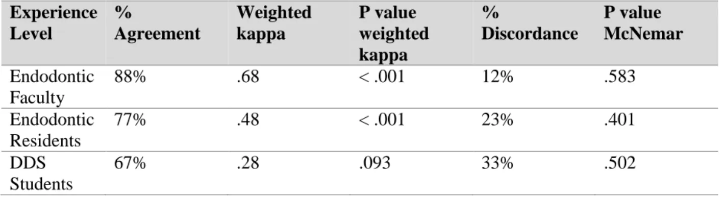
The intra-rater reliability comparing viewing 1 and viewing 2 was determined for each group by calculating the % agreement, weighted kappa, p value of weighted kappa, %
discordances, and p value of McNemar statistics. SPSS version 17 (SPSS Inc, Chicago, IL) was used to calculate the statistics.
Results
Table 3 shows the statistical values for % agreement, weighted kappa, p value of
weighted kappa, % discordances, and p value of McNemar’s test for each group compared to the “gold standard.” The average weighted kappa for endodontic faculty was .49, while endodontic
residents and dental students had an average weighted kappa of .35 and .32 respectively. Landis and Koch’s interpretation for kappa was used for analysis.
TABLE 3: EXPERIENCE LEVEL COMPARED TO “GOLD STANDARD” FOR EACH
GROUP
TABLE 4: INTRA-RATER RELIABILITY COMPARING VIEWING 1 VS VIEWING 2 FOR EACH GROUP Experience Level % Agreement Weighted kappa P value weighted kappa % Discordance P value McNemar Endodontic Faculty
88% .68 < .001 12% .583
Endodontic Residents
77% .48 < .001 23% .401
DDS Students
67% .28 .093 33% .502
Discussion
In this study the two dental radiologists were consistent as the “gold standard.” They decided to eliminate one CBCT volume due to poor image quality. Overall, endodontic faculty had the highest agreement and weighted kappa values with the “gold standard.” The endodontic resident group and the DDS student group had similar weighted kappa values with the “gold standard.” The agreement beyond chance for endodontic faculty and endodontic residents was
Experience Level % Agreement Weighted kappa P value weighted kappa % Discordance P value McNemar Endodontic Faculty
80% .49 .003 20% .521
Endodontic Residents
74% .35 .029 27% .444
DDS Students
moderate as indicated by statistically significant weighted kappa scores (Table 3). All of the readers had a low percent of discordances.
The intra-rater reliability varied widely in this study. Endodontic faculty were the most consistent, followed by the residents and then the DDS students. The endodontic faculty and endodontic residents had p values of weighted kappa that were statistically significant, whereas the DDS students had a non-significant p value of weighted kappa.
In terms of agreement with the “gold standard,” all three groups with varying levels of
experience fall into the moderate range with weighted kappa values ranging on average from .49
to .32. The statistics show that none of the groups regardless of their experience level were in
the excellent range for weighted kappa. This indicates that clinicians may not be as accurate as
dental radiologists in their interpretation of periapical radiolucencies using CBCT. Further
training may be needed to avoid misreading CBCT volumes and incorrectly diagnosing apical
periodontitis, which could lead to incorrect treatment for patients.
Endodontic faculty fell into the strong category for the weighted kappa in terms of
intra-rater reliability with a weighted kappa of .68. This indicates that they were consistent with their
answers between viewing 1 and viewing 2. It is possible that their experience has enabled them
to consistently evaluate CBCT volumes and identify possible pathology. While they were not
always in agreement with the “gold standard,” they were not changing their responses amongst
viewing 1 and viewing 2. The resident group fell into the category of moderate with a weighted
kappa of .48. They were relatively consistent with their answers in viewing 1 and viewing 2, but
not as consistent as endodontic faculty. DDS students fell into the category of fair, with a
viewing 1 and viewing 2. This may be due to lack of experience and confidence in using CBCT
software and interpreting periapical radiolucencies.
Recent studies have shown success rates of RCT evaluated by CBCT to be significantly lower. There was a 14 times increase in failure rate when teeth with no pre-operative periapical radiolucencies were assessed with CBCT compared to periapical radiographs at one year. Diagnosis using CBCT revealed a lower healed and healing rate for primary root canal treatment than periapical radiographs, particularly in roots of molars (40). The follow-up time in the Patel study was only one year, which may mean healing had not taken place yet. However, that study is relevant as both groups were being compared in this same time frame of one year. Strindberg showed healing can take up to ten years and that there were more successes than failures evident at later follow-up times (41). In another study, Kvist and Reit showed healing for nonsurgical retreatment endodontic cases that were followed-up for four years may have slower healing dynamics than surgical cases (42). A different study evaluating healing of periapical radiolucencies after periapical microsurgery using two-dimensional imaging showed that
incomplete healing (scar tissue) was present 2-6 years after surgery (43). It is important to allow time after root canal treatment, retreatment, and surgery cases for periapical radiolucencies to heal, especially when using CBCT imaging to evaluate the healing. As this study has shown, readers may not be in agreement with the “gold standard” in identifying periapical
radiolucencies, and readers are often inconsistent with themselves in interpreting periapical radiolucencies.
periapical radiolucencies using CBCT allows practitioners to be consistent with each other and with themselves. Estrela et al. classified periapical radiolucencies using a CBCT based on a six point system that measured the largest portion of the radiolucency (14). In that study, periapical radiolucencies were identified in 40% of two-dimensional imaging and 61% of
three-dimensional CBCT volumes (14). It is important that further treatment decisions are not based on a short-term analysis of a CBCT volume after root canal treatment as clinicians may be observing incomplete healing following root canal therapy.
Conclusion
Endodontic faculty had the highest agreement with the “gold standard,” while endodontic residents and DDS students had similar agreement with the “gold standard.” None of the three groups were considered to be in the excellent category in terms of the weighted kappa value in comparison to the “gold standard,” which indicates there is room for improvement in identifying
periapical radiolucencies amongst clinicians of varying experience levels. Only the endodontic faculty were in the strong category for weighted kappa when assessing intra-rater reliability. The residents were in the moderate category, while the DDS students were fair. DDS students were inconsistent in their responses between viewing 1 and viewing 2, and would likely benefit from additional CBCT software/interpretation training. Until practitioners have a more elaborate training on this technology, an experienced reader should interpret their patient’s CBCT volumes to avoid potential misdiagnosis. The experience level of the clinician plays a role in accurately and consistently identifying periapical radiolucencies in CBCT volumes.
REFERENCES
33. de Paula-Silva FW, Wu MK, Leonardo MR, da Silva LA, Wesselink PR. Accuracy of periapical radiography and cone-beam computed tomography scans in diagnosing apical periodontitis using histopathological findings as a gold standard. Journal of endodontics. 2009;35(7):1009-12.
34. Ma L, Zhan FL, Qiu LH, Xue M. [The application of cone-beam computed tomography in diagnosing the lesions of apical periodontitis of posterior teeth]. Shanghai kou qiang yi xue = Shanghai journal of stomatology. 2012;21(4):442-6.
35. Pope O, Sathorn C, Parashos P. A Comparative Investigation of Cone-beam Computed Tomography and Periapical Radiography in the Diagnosis of a Healthy Periapex. Journal of endodontics. 2014;40(3):360-5.
36. Allareddy V, Vincent SD, Hellstein JW, Qian F, Smoker WR, Ruprecht A. Incidental findings on cone beam computed tomography images. International journal of dentistry. 2012;2012:871532.
37. Scarfe WC, Li Z, Aboelmaaty W, Scott SA, Farman AG. Maxillofacial cone beam computed tomography: essence, elements and steps to interpretation. Australian dental journal. 2012;57 Suppl 1:46-60.
38. Parashar V, Whaites E, Monsour P, Chaudhry J, Geist JR. Cone beam computed tomography in dental education: a survey of US, UK, and Australian dental schools. Journal of dental education. 2012;76(11):1443-7.
39. Noffke CE, Farman AG, Van der Linde A, Nel S. Responsible use of cone beam computed tomography: minimising medico-legal risks. SADJ : journal of the South African Dental Association = tydskrif van die Suid-Afrikaanse Tandheelkundige Vereniging. 2013;68(6):256, 8-9.
40. Patel S, Wilson R, Dawood A, Foschi F, Mannocci F. The detection of periapical
pathosis using digital periapical radiography and cone beam computed tomography - part 2: a 1-year post-treatment follow-up. International endodontic journal. 2012;45(8):711-23.
41. Strindberg L. The dependence of the results of pulp therapy on certain factors. An analytic study based on radiographic and clinical follow-up examination. Acta odontologica Scandinavica. 1956;14: Suppl. 21.
43. Molven O, Halse A, Grung B. Incomplete healing (scar tissue) after periapical surgery--radiographic findings 8 to 12 years after treatment. Journal of endodontics.
THESIS SUMMARY
This thesis related to the AAE research priority to “address factors affecting success of endodontic treatment.” Part 1 was designed to help clinicians determine if making a CBCT volume on a maxillary molar is warranted to aid in the location of MB2. This study had not been performed before using CBCT scans in vivo. The thesis was also relevant to the AAE research priority of “assessment of new technologies such as devices and materials.” With the increasing popularity and use of CBCT in endodontics, clinicians need to ensure that they are using this technology only when it is necessary to benefit the patient. There is a potential gap in knowledge of when to use CBCT in endodontics while respecting ALARA principles. This study showed that experience and knowledge of the anatomy of maxillary molars using a dental operating microscope may be the over-riding factor in the attempt to locate MB2 canals. While CBCT certainly has many beneficial uses, clinician experience, along with the use of a dental operating microscope are invaluable in locating MB2 canals in maxillary molars. Clinicians need to be judicial in the use of CBCT imaging to aid them in locating MB2 canals.
treatment plans are often based on these CBCT findings and patients may be undergoing unnecessary treatment due to false positive readings that inexperienced clinicians may see as a “periapical radiolucency” on a CBCT volume. There is a need for more continuing education for practitioners who do choose to incorporate this technology in their practice of endodontics to know the appropriate uses and limitations of CBCT, as well as to improve their accuracy and consistency of detecting periapical radiolucencies. Until such training is complete, it would be prudent for practitioners to have an experienced reader interpret their volumes to establish a definite diagnosis through this new technology. Another interesting finding that needs further exploration is the “gold standard” assigning four out seven vital teeth as having periapical radiolucencies on their respective CBCT volumes. It was previously thought using two
dimensional imaging that a tooth had to be necrotic to have a “lesion.” Now with the resolution and spatial relationships of CBCT, perhaps we can detect these smaller periapical radiolucencies in vital teeth at an early stage, which could possibly lead to a better outcome for the patient.
Stashenko described that the breakdown of periapical bone may be possible in vital pulps as a result of bacterial infection of the dental pulps, due to caries, trauma, or iatrogenic insult. Immediate type responses leading to the bony breakdown include increased vascular
permeability due to vasodilation, as well as leukocyte extravasation, which are mediated by prostanoids, kinins, and neuropeptides. Non-specific immune response includes
polymorphonuclear and monocyte migration and activation and cytokine production.
This thesis was a prospective in vivo clinical study, which has a higher level of evidence than past in vitro studies of similar subject matter. This study was randomized, had clinically relevant goals, and had minimal bias (46). One of the weaknesses of in vitro studies is that the results do not necessarily correspond to the circumstances that occur around a living organism. In vivo studies are better suited to have a clinical relevance as they take into account the effects of an experiment on living subjects. The results of this in vivo study are able to make a stronger connection to treating patients in a clinical setting compared to past in vitro studies around the same subject matter.
REFERENCES
44. Stashenko P, Teles R, D'Souza R. Periapical inflammatory responses and their
modulation. Critical reviews in oral biology and medicine : an official publication of the American Association of Oral Biologists. 1998;9(4):498-521.
45. Stashenko P. Role of immune cytokines in the pathogenesis of periapical lesions. Endodontics & dental traumatology. 1990;6(3):89-96.
APPENDIX A: PATIENT CONSENT FORM PART 1
University of North Carolina at Chapel Hill Consent to Participate in a Research Study Adult Participants Patient
Consent Form Version Date: June 1, 2014 IRB Study # 14-0608
Title of Study: CBCT Uses in Clinical Endodontics-Part 1: Effect of CBCT on the ability to locate MB2 in maxillary molars; Part 2: Specificity and Sensitivity of detecting periapical lesions using CBCT
Principal Investigator: Jeffrey Parker, DMD Principal Investigator Department: Endodontics Principal Investigator Phone number: 412-606-6027 Principal Investigator Email Address: [email protected]
Faculty Advisor: Peter Tawil
Faculty Advisor Contact Information: [email protected] Co-Investigators: Eric Rivera, DDS, MS
Andre Mol, DDS, MS
_________________________________________________________________ What are some general things you should know about research studies?
You are being asked to take part in a research study. To join the study is voluntary.
You may refuse to join, or you may withdraw your consent to be in the study, for any reason, without penalty.
Research studies are designed to obtain new knowledge. This new information may help people in the future. You may not receive any direct benefit from being in the research study. There also may be risks to being in research studies.
You will be given a copy of this consent form. You should ask the researchers named above, or staff members who may assist them, any questions you have about this study at any time. What is the purpose of this study?
The purpose of this research study is to determine if the use of a three-dimensional Cone Beam Computed Tomography (CBCT) scan is able to help the clinician locate a fourth canal in the maxillary molar tooth having root canal treatment. Typically maxillary molars have 4 canals between 55%-93% of the time. The fourth canal that is being referred to is often called the second mesial buccal canal or MB2 as it is located in the mesial root of the tooth if it is present. To achieve a disinfected canal system, it is important to locate, clean, and fill each canal in the tooth. If we are not able to locate a fourth canal during the initial search, the CBCT scan may be helpful in determining if it is present or absent in the tooth, as well as the location that we may be able to locate the fourth canal.
If a CBCT scan is made, the second part of the study will have interpreters of different
experience levels read the volume to determine if a periapical lesion is present at the end of the roots on the tooth requiring root canal treatment. These volumes will not have any patient information and will be viewed by dental radiologists, endodontic faculty, endodontic residents, and dental students. We will be looking to see if clinicians with more experience are more accurate in their assessment of the three-dimensional volume.
You are being asked to be in the study because you are scheduled to have root canal treatment on a maxillary tooth.
Are there any reasons you should not be in this study?
You should not be in this study if you have allergic reactions to dental anesthetics or sulfites. If you are please inform the Research Dentist.
You should not be in this study if you are pregnant. If you are please inform the Research Dentist.
You should not be in this study if you have an uncontrolled systemic condition: such as diabetes mellitus, hypertension, tuberculosis, Cushing syndrome. If you are please inform the Research Dentist.
You should not be in this study if you are participating in another study that involves radiation within a one year period.
How many people will take part in this study?
There will be approximately 50 people in this research study. How long will your part in this study last?
412-606-6027.
What will happen if you take part in the study?
If it is determined by a clinician that you have a maxillary first molar that needs root canal treatment, and you qualify based on medical status, then you have the option to participate in this study.
1st visit: You will undergo the standard procedures involved in having a root canal. These procedures are not part of the research study and you would have these procedures even if you did not participate in the research study. Initial exam will be performed and radiographs will be taken of the tooth to be treated. Clinical testing will confirm if root canal treatment is needed. If so, informed consent will be given along with details of the study. Root canal treatment will occur with the same standard of care as any other root canal procedure.
If the fourth canal (MB2) is not located after drilling to a pre-determined depth, a dimensional CBCT volume will be made and you will be dismissed at this point. This three-dimensional CBCT is the only research procedure to be performed. Appointment time is 2-3 hours.
2nd visit: Routine root canal treatment will continue. If the fourth canal (MB2) was seen on the three-dimensional volume, we will attempt to locate it by drilling deeper into the tooth. We will record if we are able to locate the fourth canal or not, and also if we are able to clean the entire canal to the end of the root or not. Root canal treatment will be finished and a core restoration will be placed. You will need to see your dentist for a crown if necessary. Appointment time is 2-3 hours.
If there are problems or concerns, you may contact Dr. Jeff Parker (Principle Investigator) to schedule and appointment to be evaluated at 412-606-6027 or by email: [email protected].
What are the possible benefits from being in this study?
Research is designed to benefit society by gaining new knowledge. The benefits to you from being in this study may be locating the fourth canal (MB2) to potentially increase the prognosis of the treatment.
What are the possible risks or discomforts involved from being in this study? The only potential risk involved in this study is the radiation exposure from the
three-dimensional CBCT volume that will be performed if your dentist cannot locate a fourth canal in the maxillary molar tooth having root canal treatment. The total radiation dose from this study is less than 1 mrem, which is equivalent to radiation everyone receives in one day of background radiation.
There may be uncommon or previously unknown risks. You should report any problems to the researcher.
What if we learn about new findings or information during the study?
You will be given any new information gained during the course of the study that might affect your willingness to continue your participation.
How will information about you be protected?
Every effort will be made to protect your privacy. All information collected in this study will remain confidential and only those directly involved in the study will have access to this information. All participants will be assigned numbers and all data collected will be stored electronically and will have a strong password for access.
Participants will not be identified in any report or publication about this study. Although every effort will be made to keep research records private, there may be times when federal or state law requires the disclosure of such records, including personal information. This is very unlikely, but if disclosure is ever required, UNC-Chapel Hill will take steps allowable by law to protect the privacy of personal information. In some cases, your information in this research study could be reviewed by representatives of the University, research sponsors, or government agencies (for example, the FDA) for purposes such as quality control or safety.
What will happen if you are injured by this research?
All research involves a chance that something bad might happen to you. This may include the risk of personal injury. In spite of all safety measures, you might develop a reaction or injury from being in this study. If such problems occur, the researchers will help you get medical care, but any costs for the medical care will be billed to you and/or your insurance company. The University of North Carolina at Chapel Hill has not set aside funds to pay you for any such reactions or injuries, or for the related medical care. You do not give up any of your legal rights by signing this form.
What if you want to stop before your part in the study is complete?
You can withdraw from this study at any time, without penalty. The investigators also have the right to stop your participation at any time. This could be because you have had an unexpected reaction, or have failed to follow instructions, or because the entire study has been stopped. Will you receive anything for being in this study?
No. Subsidy to you as an incentive to complete the proposed study will not be provided, regardless if you are insured or not.
Will it cost you anything to be in this study?
You will be responsible for paying for the root canal treatment fee in full before starting the procedure, as well as the fee for CBCT if it is made. You are also responsible for parking and transportation and any other costs to come to pay for your treatment following root canal treatment.
What if you have questions about this study?
related injury occurs, you should contact the researchers listed on the first page of this form. What if you have questions about your rights as a research participant?
All research on human volunteers is reviewed by a committee that works to protect your rights and welfare. If you have questions or concerns about your rights as a research subject, or if you would like to obtain information or offer input, you may contact the Institutional Review Board at 919-966-3113 or by email to [email protected].
Participant’s Agreement:
I have read the information provided above. I have asked all the questions I have at this time. I voluntarily agree to participate in this research study.
______________________________________________________ Signature of Research Participant
____________________ Date
______________________________________________________ Printed Name of Research Participant
______________________________________________________ Signature of Research Team Member Obtaining Consent
____________________ Date
______________________________________________________ Printed Name of Research Team Member Obtaining Consent
APPENDIX B: CLINICIAN CONSENT FORM PART 2 University of North Carolina at Chapel Hill
Consent to Participate in a Research Study Adult Participants Clinician
Consent Form Version Date: 6-24-14 IRB Study # 14-0608
Title of Study: CBCT Uses in Clinical Endodontics Part 1: Effect of CBCT on the ability to locate MB2 in maxillary molars. Part 2: Specificity and Sensitivity of detecting periapical lesions using CBCT.
Principal Investigator: Jeffrey Parker
Principal Investigator Department: Endodontics Principal Investigator Phone number: 412-606-6027 Principal Investigator Email Address: [email protected] Faculty Advisor: Peter Tawil
Faculty Advisor Contact Information: 919-667-6051
_________________________________________________________________ What are some general things you should know about research studies?
You are being asked to take part in a research study. To join the study is voluntary.
You may refuse to join, or you may withdraw your consent to be in the study, for any reason, without penalty.
Research studies are designed to obtain new knowledge. This new information may help people in the future. You may not receive any direct benefit from being in the research study. There also may be risks to being in research studies. Deciding not to be in the study or leaving the study before it is done will not affect your relationship with the researcher, your health care provider, or the University of North Carolina-Chapel Hill. If you are a patient with an illness, you do not have to be in the research study in order to receive health care.
Details about this study are discussed below. It is important that you understand this information so that you can make an informed choice about being in this research study.
You will be given a copy of this consent form. You should ask the researchers named above, or staff members who may assist them, any questions you have about this study at any time. What is the purpose of this study?
an endodontic faculty member, an endodontic resident, or a dental student at UNC-CH School of Dentistry.
Are there any reasons you should not be in this study?
You should not be in this study if you are not in good standing with UNC-CH School of Dentistry.
How many people will take part in this study?
There will be approximately 11 people in this research study. How long will your part in this study last?
The study will take one session and approximately one hour to complete. What will happen if you take part in the study?
The CBCT volume of each maxillary first and second molar from Part 1 of the study will be interpreted by two dental radiologists, three endodontic faculty, three endodontic residents, and three dental students. The readers will be able to view the entire CBCT volume. In this study the dental radiologists will represent the “gold standard.” The “gold standard” is composed of a panel of experts in the particular field being studied. The experts determine the ground truth for the study and the data obtained will be compared with what the experts believe to be true. The readers will answer the following question: Is there a periapical lesion present at the apices of tooth # X?
1= A lesion is definitely not present 2= A lesion is probably not present 3= Unsure
4= A lesion is probably present 5= A lesion is definitely present
What are the possible benefits from being in this study?
Research is designed to benefit society by gaining new knowledge. There is little chance you will benefit from being in this research study.
What are the possible risks or discomforts involved from being in this study? There are no known risks involved with this study.
There may be uncommon or previously unknown risks. You should report any problems to the researcher.
What if we learn about new findings or information during the study?
You will be given any new information gained during the course of the study that might affect your willingness to continue your participation.
How will information about you be protected?
Participants will not be identified in any report or publication about this study. Although every effort will be made to keep research records private, there may be times when federal or state law requires the disclosure of such records, including personal information. This is very unlikely, but if disclosure is ever required, UNC-Chapel Hill will take steps allowable by law to protect the privacy of personal information. In some cases, your information in this research study could be reviewed by representatives of the University, research sponsors, or government agencies (for example, the FDA) for purposes such as quality control or safety.
What will happen if you are injured by this research?
All research involves a chance that something bad might happen to you. This may include the risk of personal injury. In spite of all safety measures, you might develop a reaction or injury from being in this study. If such problems occur, the researchers will help you get medical care, but any costs for the medical care will be billed to you and/or your insurance company. The University of North Carolina at Chapel Hill has not set aside funds to pay you for any such reactions or injuries, or for the related medical care. You do not give up any of your legal rights by signing this form.
What if you want to stop before your part in the study is complete?
You can withdraw from this study at any time, without penalty. The investigators also have the right to stop your participation at any time. This could be because you have had an unexpected reaction, or have failed to follow instructions, or because the entire study has been stopped. Will you receive anything for being in this study?
No
Will it cost you anything to be in this study? It will not cost you anything to be in this study. What if you are a UNC student?
You may choose not to be in the study or to stop being in the study before it is over at any time. This will not affect your class standing or grades at UNC-Chapel Hill. You will not be offered or receive any special consideration if you take part in this research.
What if you are a UNC employee?
Taking part in this research is not a part of your University duties, and refusing will not affect your job. You will not be offered or receive any special job-related consideration if you take part in this research.
What if you have questions about this study?
You have the right to ask, and have answered, any questions you may have about this research. If you have questions about the study (including payments), complaints, concerns, or if a research-related injury occurs, you should contact the researchers listed on the first page of this form. What if you have questions about your rights as a research participant?
would like to obtain information or offer input, you may contact the Institutional Review Board at 919-966-3113 or by email to [email protected].
Participant’s Agreement:
I have read the information provided above. I have asked all the questions I have at this time. I voluntarily agree to participate in this research study.
______________________________________________________ Signature of Research Participant
____________________ Date
______________________________________________________ Printed Name of Research Participant
______________________________________________________ Signature of Research Team Member Obtaining Consent
____________________ Date
______________________________________________________ Printed Name of Research Team Member Obtaining Consent
APPENDIX C: IRB APPROVAL LETTER
To: Jeffrey Parker Endodontics
From: Biomedical IRB Approval Date: 8/28/2014
Expiration Date of Approval: 7/27/2015
RE: Notice of IRB Approval by Full Board Review Submission Type: Initial
Study #: 14-0608
Study Title: CBCT Uses in Clinical Endodontics Part 1: Effect of CBCT on the ability to locate MB2 in maxillary molars. Part 2: Specificity and Sensitivity of detecting periapical lesions using CBCT.
This submission has been approved by the IRB for the period indicated. Study Description:
Purpose: The first objective is to determine if the use of a Cone Beam Computed Tomography (CBCT) volume aids the clinician in locating more second mesial buccal canals (MB2) in
maxillary molars during root canal treatment. The second objective is to determine the sensitivity and specificity of detecting periapical lesions using CBCT by comparing CBCT interpretations amongst different experienced CBCT readers.
Participants: Patients needing root canal treatment on maxillary first molars. Endodontic faculty, endodontic residents, dental radiology faculty, dental students.
Procedures (methods): For part 1, patients will have a CBCT volume made if initial searching for MB2 in maxillary molars using a microscope is unsuccessful. This will potentially aid in locating for MB2 canals. These same volumes will be used in part 2 of the study, which will involve a dental radiologist interpreting the volumes as the "gold standard." The experience levels we will be comparing in regards to identifying periapical lesions are dental students, endodontic faculty, and endodontic residents that are at UNC at Chapel Hill School of Dentistry.
Regulatory and other findings:
This approval includes a limited waiver of HIPAA authorization to identify potential subjects for recruitment into this research study, as allowed under 45 CFR 164.512. This temporary waiver provides access to protected health information (PHI) to confirm eligibility and facilitate initial contact, after which consent and HIPAA authorization will be sought when applicable. Access and use is limited to the minimum amount of PHI necessary to review eligibility criteria and to contact potential subjects.
Investigator’s Responsibilities:
You are required to obtain IRB approval for any changes to any aspect of this study before they can be implemented. Any unanticipated problem involving risks to subjects or others (including adverse events reportable under UNC-Chapel Hill policy) should be reported to the IRB using the web portal at http://irbis.unc.edu.
Please be aware that approval may still be required from other relevant authorities or "gatekeepers" (e.g., school principals, facility directors, custodians of records).
This study was reviewed in accordance with federal regulations governing human subjects research, including those found at 45 CFR 46 (Common Rule), 45 CFR 164 (HIPAA), 21 CFR 50 & 56 (FDA), and 40 CFR 26 (EPA), where applicable.
APPENDIX D: TABLES Table 1: Descriptive statistics part 1
Clinical findings Results Percentage
MB2 canals located overall 43/50 86%
MB2 canals located without CBCT 35/50 70%
MB2 canals located overall maxillary first molars 36/40 90% MB2 canals located overall maxillary second molars 7/10 70%
MB2 seen on CBCT volume 5/15 33%
MB2 canals seen on volume AND located 5/5 100%
MB2 canals NOT seen on volume 11/15 73%
MB2 canals not seen on volume AND located 3/15 20%
MB2 canals located after CBCT 8/15 53%
MB2 canals joined MB1 canals 25/43 58%
MB2 canals negotiated 42/43 98%
Table 2: MB2 canals located according to coronal restoration present at time of endodontic access preparation and age (< or > 50 years old)
MB2 canals located Full coverage
restoration
<50 years old: 3/3
(100%) 11/13 (85%)
43/50 (86%) >50 years old: 8/10
(80%) Without full
coverage restoration
<50 years old: 20/23
(87%) 32/37 (86%)
Table 3: Pulp vitality as determined upon endodontic access. 7/22 teeth were deemed to have vital pulps upon access, and 4/7 of these vital teeth were determined by the “gold standard” to have periapical radiolucencies.
Subject Vital (yes/no) Lesion according to “gold
standard” (yes/no)
1 yes no
2 no no
3 no yes
4 no yes
5 no no
6 no no
7 no yes
8 no yes
9 yes yes
10 yes no
11 no yes
12 yes no
13 no yes
14 no yes
15 yes yes
16 no yes
17 no yes
18 no yes
19 yes yes
20 no yes
21 yes yes
Appendix E: FIGURES
Figure 8: CBCT viewing room for part 2