Received 19 Oct 2015
|
Accepted 13 Jan 2016
|
Published 12 Feb 2016
Loss of UBE3A from TH-expressing neurons
suppresses GABA co-release and enhances
VTA-NAc optical self-stimulation
Janet Berrios
1,2,3
, Alice M. Stamatakis
1,3,4,5
, Pranish A. Kantak
3,4
, Zoe A. McElligott
4,6
, Matthew C. Judson
2,3
,
Megumi Aita
2,3
, Marie Rougie
2,3
, Garret D. Stuber
1,3,4
& Benjamin D. Philpot
1,2,3,7
Motivated reward-seeking behaviours are governed by dopaminergic ventral tegmental area
projections to the nucleus accumbens. In addition to dopamine, these mesoaccumbal
terminals co-release other neurotransmitters including glutamate and GABA, whose roles in
regulating motivated behaviours are currently being investigated. Here we demonstrate that
loss of the E3-ubiquitin ligase, UBE3A, from tyrosine hydroxylase-expressing neurons impairs
mesoaccumbal, non-canonical GABA co-release and enhances reward-seeking behaviour
measured by optical self-stimulation.
DOI: 10.1038/ncomms10702
OPEN
1Curriculum in Neurobiology, University of North Carolina, Chapel Hill, 27599 North Carolina, USA.2Department of Cell Biology and Physiology, University of
North Carolina, Chapel Hill, 27599 North Carolina, USA.3Neuroscience Center, University of North Carolina, Chapel Hill, 27599 North Carolina, USA.
4Department of Psychiatry, University of North Carolina, Chapel Hill, 27599 North Carolina, USA.5Inscopix Inc, Palo Alto, 94303 California, USA.6Bowles
Center for Alcohol Studies, University of North Carolina, Chapel Hill, 27599 North Carolina, USA.7Carolina Institute for Developmental Disabilities,
D
opamine projections from the ventral tegmental area
(VTA) target the nucleus accumbens (NAc) and release
dopamine in response to reward-predictive cues, which in
turn initiates reward-seeking and promotes reward learning
1–3.
While phasic dopamine dynamics have been well-characterized
within the NAc
1, dopaminergic terminals are now known to be
capable of co-releasing several neurotransmitters including
gamma-aminobutyric acid (GABA) and glutamate
4–7. However,
the role of mesoaccumbal non-canonical GABA and glutamate
co-release remains to be elucidated.
The mechanisms differ for glutamate and GABA co-release,
presenting an opportunity to dissect their functional roles.
Glutamate co-release in the mesoaccumbal pathway occurs in
restricted axonal microdomains and is not necessarily packaged
within the same synaptic vesicles as dopamine
7. Conversely,
GABA co-release requires the vesicular monoamine transporter-2
(VMAT2), which is also required for dopamine vesicular loading,
suggesting that GABA and dopamine can be packaged within
the same synaptic vesicles at mesoaccumabal terminals
5.
Dopaminergic neurons do not express conventional GABA
synthesizing enzymes, but instead actively uptake ambient
GABA in their terminals within the NAc, a process mediated
by GABA transporter-1 (GAT1)
5,8.
Here, we describe our efforts to optogenetically dissect the
underlying causes of disrupted reward-seeking behaviours in
mice that lack the maternal copy of
Ube3a
, a mouse model
for Angelman syndrome (
Ube3a
m/pþ). In doing so, we
serendipitously discovered that the loss of the E3-ubiquitin
ligase,
Ube3a
, did not directly alter VTA-to-NAc dopamine
release but is essential for GABA co-release and the modulation
of positively reinforced behaviour. Our findings reveal a
novel molecular mechanism underlying GABA co-release from
mesoaccumbal terminals and provide new insights into the
relevance of this non-canonical mode of neurotransmission to
motivated behaviour.
Results
Mesoaccumbal optical stimulation in a
Ube3a-
null model
. In
an effort originally designed to elucidate mechanisms underlying
dopaminergic dysfunction in an Angelman syndrome mouse
model
9, we used a reductionist approach to selectively manipulate
the
mesoaccumbal
dopaminergic
pathway.
To
target
catecholaminergic neurons, mice expressing CRE recombinase
within TH
þneurons (
TH
CRE) were crossed into
Ube3a
m/pþmice or their wild-type littermates (
Ube3a
mþ/pþ). To specifically
manipulate the axon terminals of NAc-projecting TH
þneurons,
we
transduced
CRE-dependent
AAV5-channelrhodopsin-2
(H134R)
fused
to
enhanced
yellow
fluorescent
protein
(ChR2-eYFP) into the VTA and implanted an optical fibre
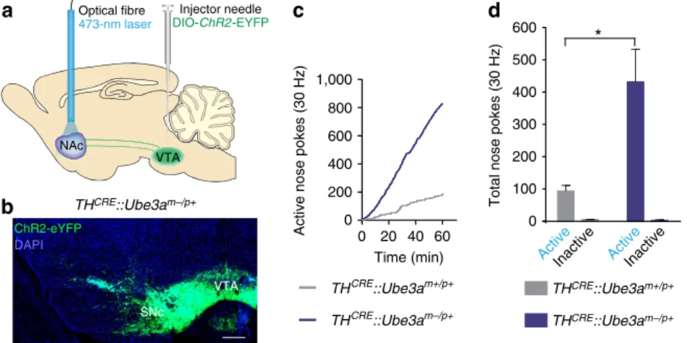
above the NAc (Fig. 1a). We observed qualitatively similar viral
expression within
Ube3a
-deficient lines compared with their
wild-type littermates (Fig. 1b and Supplementary Fig. 1), but
observed
no
expression
following
viral
injection
into
CRE-negative
control mice. To
determine
if there
are
differences in positive-reinforcement behaviour, we trained
TH
CRE::Ube3a
m/pþand
TH
CRE::Ube3a
mþ/pþmice to
nose-poke for optical stimulation with a fixed-ratio one schedule of
reinforcement. We used a 30 Hz optical-stimulation paradigm, as
this stimulation frequency is within a range previously shown to
produce robust DA release at NAc terminals, and has been used
across various behavioural paradigms examining the effects of
neuromodulatory release
in vivo
10–14.
TH
CRE::Ube3a
m/pþand
TH
CRE::Ube3a
mþ/pþmice similarly learned an appetitive
nose-poke operant task (Supplementary Fig. 2) for natural
rewards. However,
TH
CRE::Ube3a
m/pþmice nose-poked to
receive
optical
stimulation
significantly
more
than
TH
CRE::Ube3a
mþ/pþmice (Fig. 1c,d). These data suggest that
the loss of UBE3A enhances motivation driven through TH
þterminals within the NAc, thus increasing optical self-stimulation.
Motivation and dopaminergic physiology in
Ube3a
m/pþmice
.
On the basis of previous findings
9, we hypothesized that UBE3A
loss might enhance optically evoked reward-seeking by increasing
VTA-to-NAc dopamine release. To examine this possibility, we
performed
in vitro
fast-scan cyclic voltammetry in brain slices
from
TH
CRE::Ube3a
m/pþand
TH
CRE::Ube3a
mþ/pþmice
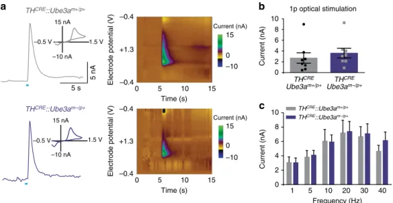
expressing ChR2-eYFP within VTA-to-NAc terminals. To probe
for possible changes in dopamine release, we optically stimulated
either with a single pulse (Fig. 2b) or with stimulation trains across
a range of frequencies (Fig. 2c). Contrary to our initial hypothesis,
the loss of UBE3A had no effect on dopamine availability or
NAc Optical fibre
473-nm laser
VTA
Injector needle
DIO-ChR2-EYFP
a
0 100 200 300 400 500 600
c
b
THCRE::Ube3am–/p+Active Active Inactive Inactive
Total nose pokes (30 Hz)
0 20 40 60
0
Active nose pokes (30 Hz)
Time (min)
*
200 400 600 800 1,000
d
THCRE::Ube3am+/p+
THCRE::Ube3am–/p+ THCRE::Ube3am+/p+
THCRE::Ube3am–/p+ ChR2-eYFP
DAPI
VTA
SNc
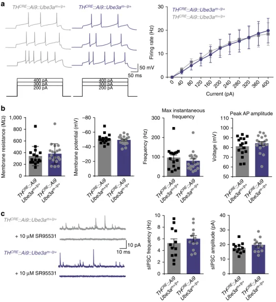
release. Moreover, using
in vitro
whole-cell electrophysiology, we
found that intrinsic excitability and inhibition onto VTA
TH
CREþneurons were unchanged in
TH
CRE::Ube3a
m/pþmice compared
with controls (Fig. 3). Collectively, these findings suggest that
enhanced reward seeking in
Ube3a
m/pþmice is not due to
changes in dopamine release from VTA-to-NAc dopaminergic
terminals or due to altered excitability of dopaminergic VTA
neurons in
Ube3a
m/pþmice.
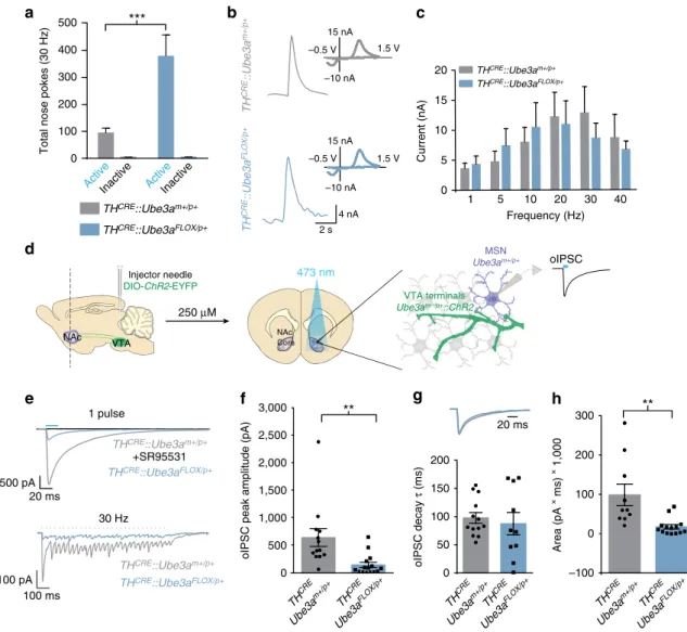
Consequences of selective
Ube3a
deletion in TH
þneurons
.
Because VTA neurons exhibited typical excitability and retain a
normal capacity to release dopamine in
Ube3a
m/pþmice (Figs 2
and 3), we questioned if maternal
Ube3a
deletion selectively in
catecholaminergic neurons would be sufficient to alter
motiva-tional drive. To test this, we used a novel condimotiva-tional
Ube3a
knockout mouse (
Ube3a
FLOX/pþ) to selectively delete maternal
Ube3a
in a
TH
CRE-dependent manner (Supplementary Figs 1 and
3a). We injected
TH
CRE::Ube3a
FLOX/pþand
TH
CRE::Ube3a
mþ/pþmice with CRE-dependent ChR2-eYFP into the VTA to determine
if deletion of
Ube3a
within TH
þneurons would be sufficient to
phenocopy self-stimulation phenotypes observed in AS model
mice. Mice were trained to nose-poke (as described above) for
optical stimulation of CRE
þterminals within the NAc.
TH
CRE::Ube3a
FLOX/pþmice poked significantly more for 30 Hz
stimulation than
TH
CRE::Ube3a
mþ/pþmice (Fig. 4a). This
dif-ference occurred in the absence of observable changes in optically
evoked dopamine release (Fig. 4b,c). These data demonstrate that
the selective loss of UBE3A in TH
þneurons is sufficient to
enhance motivational drive despite the lack of a detectable deficit
in NAc dopamine release.
Dopaminergic terminals in the NAc are also capable of
releasing glutamate and GABA
5–7,15. Thus, we tested whether
Ube3a
loss in TH
þneurons could alter transmitter co-release. To
assess this, we first optogenetically activated VTA-to-NAc
terminals and measured GABAergic currents in ventral striatal
medium spiny neurons while blocking glutamatergic responses
with AMPA and NMDA receptor antagonists (Fig. 4d). We
applied a single light-pulse (20 ms) to measure peak amplitude
and the kinetics of the resulting current by averaging
Z
6
consecutive traces (Fig. 4e). We confirmed
post hoc
that these
currents were GABAR-mediated by bath applying SR95531
(10
mM), a selective GABA-A receptor antagonist (Fig. 4e). To
verify that these GABAergic currents were a consequence of
co-release rather than release by GAD1
þ/TH
neurons
spuriously expressing ChR2 due to non-specific CRE expression
in the
TH
CREline, we treated mice with the VMAT2 inhibitor,
reserpine, thereby selectively inhibiting vesicular loading in
dopaminergic terminals capable of co-release. Similar to
previous electrophysiological studies
15and our own anatomical
evidence showing that the vast majority of ChR2-expressing
neurons in the VTA are TH
þ(Supplementary Fig. 4a,b), we
found that reserpine abolished nearly all optically evoked
GABAergic currents in the NAc (Supplementary Fig. 4c). This
further demonstrates that our optical stimulation almost
exclusively evoked GABA release from dopaminergic terminals.
Despite exhibiting normal glutamatergic co-release with optical
stimulation of VTA-to-NAc terminals (Supplementary Fig. 5a,b),
TH
CRE::Ube3a
FLOX/pþmice showed a
450% reduction in peak
amplitude of GABAergic currents relative to controls, while
current decay kinetics proved normal (Fig. 4e–g). Using an
optical-stimulation paradigm similar to that used in the
behavioural experiments, we also found that
TH
CRE::Ube3a-FLOX/pþ
mice exhibit diminished GABAergic currents at 30 Hz
stimulation of VTA-to-NAc terminals compared with control
mice (Fig. 4h).
Exogenous VGAT reverses UBE3A-deficient phenotypes
. We
tested if GABA co-release could be restored in
TH
CRE::
Ube3a
FLOX/pþmice, and whether this could normalize
motivational drive. The observed decrease in GABA co-release
could arise from changes in GABA uptake, availability, release or
vesicular loading
5,15,16. To enhance vesicular loading of GABA in
TH
þterminals, we used a CRE-dependent virus expressing the
vesicular GABA transporter (VGAT) and introduced this with
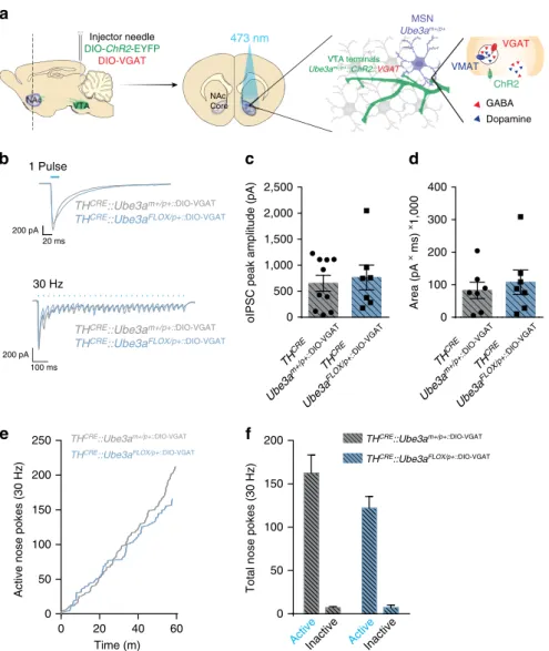
DIO-ChR2-eYFP at a 1:1 ratio within the VTA (Fig. 5a)
15. GABA
co-release evoked by a single light-pulse or a 30 Hz optical
stimulation was similar in
TH
CRE::Ube3a
FLOX/pþ::DIO-VGATmice
compared with
TH
CRE::Ube3a
mþ/pþ::DIO-VGATmice (Fig. 5b–d,
THCRE::Ube3am+/p+
1.5 V
–10 nA 15 nA
5 s
5 nA
–0.5 V
THCRE::Ube3am–/p+
1.5 V
–10 nA 15 nA
–0.5 V
0 5 10 15
0 5 10 15
–0.4 +1.3 –0.4
Electrode potential (V)
Time (s)
Time (s)
0
–10 15 Current (nA)
0 –10 15
Current (nA)
1p optical stimulation 10
8 6 4 2 0
Current (nA)
THCRE Ube3am–/p+ THCRE
Ube3am+/p+
Current (nA)
10
8
6
4
2
0
Frequency (Hz)
a
b
c
Electrode potential (V) –0.4 +1.3 –0.4
1 5 10 20 30 40
THCRE::Ube3am–/p+
THCRE::Ube3am+/p+
Figure 2 | Optically evoked dopamine release is similar in TH-positive VTA-to-NAc terminals inUbe3am/pþ andUbe3amþ/pþ mice.(a) Representative fast-scan voltammetric recordings from ventral striatal slices in bothTHCRE::Ube3am/pþandTHCRE::Ube3amþ/pþmice. Insets represent background-subtracted electrochemical signal characteristic of oxidized dopamine. Right: consecutive background-subtracted voltammogram recorded over an 8-s interval. Applied electrode potential (Eappsversus Ag/AgCl reference electrode) is shown versus time. (b) Light-evoked current is similar in
P
40.05). Exogenous expression of VGAT within TH
þterminals
was also sufficient to normalize optical intracranial
self-stimulation
responding
in
TH
CRE::Ube3a
FLOX/pþ::DIO-VGATmice compared with
TH
CRE::Ube3a
mþ/pþ::DIO-VGATmice
(Fig.
5e,f),
suggesting
a
causal
relationship
between
the observed deficits in GABA co-release and alterations
in motivational drive in the absence of UBE3A (Fig. 5f,
P
40.05).
Discussion
In this study we optogenetically dissected mesoaccumbal circuitry
to investigate the underlying causes of disrupted reward-seeking
behaviours in a
Ube3a-
deficient mouse model. Collectively, our
data indicate that UBE3A regulates circuits involved in motivated
behaviour and, in particular, GABA co-release from putative
dopaminergic mesoaccumbal terminals. Following UBE3A loss,
suppressed GABA co-release and enhanced optical
self-stimula-tion occur in the absence of other measured cellular and synaptic
deficits in the mesoaccumbal pathway. Remarkably, exogenous
expression of VGAT is sufficient to reinstate GABA co-release
and normalize motivational drive in the UBE3A-deficient circuit,
suggesting a causal link between GABA co-release from
VTA-to-NAc terminals and the modulation of motivation.
We previously concluded, based on electrical stimulation of the
medial forebrain bundle in
Ube3a
m/pþmice, that the loss of
UBE3A enhances dopamine release at the mesoaccumbal
pathway, thereby reducing reward threshold
9. Consequently, we
hypothesized here that selective loss of UBE3A in TH
þneurons
would be sufficient to alter dopamine release within the
NAc. Instead, we find that optogenetically evoked release in
VTA-to-NAc terminals is normal (Figs 2b,c and 4b,c). Given that
medial forebrain bundle stimulation is known to activate diverse
circuit pathways
17, including septal nuclei, there are numerous
a
b
sIPSC f
requenc
y (H
z)
0 2 4 6 8
sIPSC amplitude (pA
)
0 10 20 30 40
+ 10 μM SR95531
+ 10 μM SR95531
THCRE::Ai9::Ube3am–/p+
THCRE::Ai9::Ube3am+/p+
0 10 20 30
0 40
80 120 160 200 240 280 320 360 400
F
iring r
a
te
(H
z)
Current (pA)
THCRE::Ai9::Ube3am–/p+
THCRE::Ai9::Ube3am+/p+
10 0
200 800
400 1,000
600
M
e
mbrane resistance (M
Ω
)
–20 –40 –60 –80
0
M
embrane p
o
te
ntial (mV
)
0 100 200 300
Max instantaneous frequency
F
requenc
y
(H
z)
c
Peak AP amplitude
50 100
60 70 80 90
V
oltage (mV
)
110
TH CRE
::Ai9
Ube3a m–/p+ TH
CRE ::Ai9
Ube3a m+/p+
THCRE::Ai9::Ube3am+/p+
50 mV 50 ms
THCRE::Ai9::Ube3am–/p+
200 pA 300 pA 400 pA
200 pA 300 pA 400 pA
10 ms 10 pA
TH CRE
::Ai9
Ube3a m–/p+ TH
CRE ::Ai9
Ube3a m+/p+
TH CRE
::Ai 9
Ube3a m–/p+ TH
CRE ::Ai9
Ube3a m+/p+ TH
CRE ::Ai9
Ube3a m–/p+ TH
CRE ::Ai9
Ube3a m+/p+ TH
CRE ::Ai9
Ube3a m–/p+ TH
CRE ::Ai9
Ube3a m+/p+ TH
CRE ::Ai9
Ube3a m–/p+
TH CRE
::Ai9
Ube3a m+/p+
explanations for our previous observation of enhanced dopamine
release in the NAc of
Ube3a
m/pþmice
9. Regardless, our
new results demonstrate that the loss of UBE3A specifically in
TH-expressing neurons leads to a reduction in GABA
co-release without corresponding reductions in dopamine
release, and that this reduction of GABA co-release is
sufficient to enhance optogenetically mediated, positively
reinforced behaviours. Importantly, GABA co-release may
also regulate natural appetitive behaviours such as copulation,
social interactions and, as shown recently, binge drinking
8.
However, sucrose seeking is unaffected in both
Ube3a
m/pþand
TH
CRE::Ube3a
FLOX/pþ(Supplementary
Figs
2
and
5c),
indicating that GABA co-release is not involved in
food-seeking and suggests at least some degree of specificity
for this release mechanism in the modulation of appetitive
behaviour.
While these data provide the first functional role for GABA
co-release within the ventral striatum in an
in vivo
context, the
utility for GABA co-release regulating motivational drive for
ethologically relevant appetitive stimuli remains to be
deter-mined. Furthermore, we cannot rule out the possibility that our
effect might be driven in part by ectopic CRE expression within
20 ms500 pA
1 pulse
THCRE::Ube3am+/p+
THCRE::Ube3aFLOX/p+
0 500 1,000
oIPSC peak amplitude (pA)
0 50 100 150 200
oIPSC decay
τ
(ms)
**
1,500 2,000 2,500
100 pA 100 ms
30 Hz
TH CRE
Ube3a m+/p+
TH CRE
Ube3a
FLOX/p+ THCRE
Ube3a m+/p+
TH CRE
Ube3a
FLOX/p+ TH
CRE
Ube3a m+/p+
TH CRE
Ube3a FLOX/p+
–100 0 100 200 300
Area (pA
× ms) × 1,000
**
THCRE::Ube3am+/p+
THCRE::Ube3aFLOX/p+
e
f
g
NAc Core
473 nm
Injector needle
DIO-ChR2-EYFP
NAc VTA
250 μM
d
+SR95531
oIPSC MSN
Ube3am+/p+
VTA terminals
Ube3am–/p+::ChR2 0
100 200 300 400 500
Total nose pokes (30 Hz)
a
***
3,000
h
20 ms 0
5 10 15 20
Current (nA)
b
1 5 10 20 30 40
Frequency (Hz)
THCRE::Ube3aFLOX/p+
THCRE::Ube3am+/p+
TH
CRE
::Ube3a
m+/p+
TH
CRE
::Ube3a
FLOX/p+
15 nA
–10 nA 1.5 V –0.5 V
15 nA
–10 nA 1.5 V –0.5 V
Active Active Inactive Inactive
c
4 nA
2 s THCRE::Ube3aFLOX/p+
THCRE::Ube3am+/p+
Figure 4 | DeletingUbe3ain TH-positive neurons decreases GABA co-release and enhances motivational behaviour.(a) Average nose pokes for inactive and active ports triggering 30 Hz optical intracranial self-stimulation in a 60 min behavioural session (n¼5 and 7, one-way ANOVA *Po0.02). Experimental design was similar to that schematized in Fig. 1a, except thatTHCRE::Ube3aFLOX/pþ andTHCRE::Ube3amþ/pþ mice were examined to selectively deleteUbe3aand optically stimulate THþ VTA-to-NAc terminals. (b) Representative fast-scan cyclic voltammograms assessing dopamine release within the ventral striatum ofTHCRE::Ube3aFLOX/pþ andTHCRE::Ube3amþ/pþmice. Dopamine release was evoked by 30 Hz (5-pulses) optical stimulation. Insets represent background-subtracted electrochemical signal characteristic of oxidized dopamine. (c) Averaged optically evoked dopamine release at a range of frequencies demonstrates that there are no statistical differences between genotypes (n¼6 and 7, one-way ANOVAP40.05). (d) Schematic representing protocol for whole-cell optical IPSC (oIPSC) recordings within the NAc ofTHCRE::Ube3aFLOX/pþ mice. IPSCs were
GABAergic neurons
18. However, this possibility seems unlikely
given that we observe a low percentage of ChR2 and GAD1
overlap (
B
3%), and that we also found that the VMAT2 inhibitor
(reserpine) blocked most of the GABAergic currents. Moreover,
GABAergic VTA-to-NAc projections preferentially synapse on
cholinergic interneurons within the NAc
19, further decreasing the
possibility of ectopic CRE expression contributing to our
observed effects.
UBE3A is an E3-ubiquitin ligase that transfers ubiquitin to a
targeted substrate for proteasomal degradation
20, thus UBE3A
loss causes an accumulation of targeted UBE3A substrates. Recent
evidence suggests that GAT1 is a potential UBE3A substrate
16,
proposing that UBE3A loss could directly alter GAT1 transport
function. While the mechanistic details remain to be established,
our findings support the hypothesis that anomalous GABA
uptake on UBE3A loss not only leads to severely attenuated
GABA co-release, but also augments motivational drive via
mesoaccumbal pathway activation.
Methods
Experimental subjects and stereotaxic surgeries
.
Cg-Tg-TH:Cremice (JAX #: 008601),Rosa26-stop-floxed-tdTomato(Ai9, JAX #: 007909) andUbe3a-deficient (JAX #: 016590) mice were obtained through Jackson Laboratories (Bar Harbor, ME).Ube3a-floxedmice (Ube3aFLOX) were engineered in conjunction with theUNC Animal Models Core. Briefly, C57BL/6 mouse embryonic stem cells were electroporated with an AsiSI-linearizedUbe3aKO1sttargeting construct, which was
generated by the trans-NIH Knockout Mouse Project (KOMP) and obtained from the KOMP repository (www.komp.org). To produceUbe3aKO1st-targeted chimeric
mice,Ube3aKO1st-targeted embryonic stem cells were microinjected into C57BL/6-albino blastocysts. Resultant germline chimeric males (determined by the transmission of coat colour in parallel breeding) were then crossed to C57BL/6 female homozygousRosa26-FLPemice (009086, Jackson Laboratories) in order to excise theFRT-flanked lacZ gene trap from theUbe3aKO1stallele and thereby
produce theUbe3aFLOXallele.Ube3aFLOXmice were genotyped using the
following PCR primers:Ube3aFLOXF (50-AAAATTGGGTATGCGAGCTG-30) and
Ube3aFLOXR (50-GGGGTCTAAGGGCCTATGAA-30).
All mice were maintained on a congenic C57BL/6 background, hadad libitum
access to food and water, and were housed on a 12:12 light:dark cycle. Mice for electrophysiological recordings were aged P60–P90 and were compared with wild-type age- and sex-matched controls. The experimenter was blind to genowild-type, and
200
150
100
50
0
Total nose pokes (30 Hz)
250
200
150
100
50
0
Active nose pokes (30 Hz)
Time (m)
0 20 40 60
THCRE::Ube3aFLOX/p+::DIO-VGAT THCRE::Ube3am+/p+::DIO-VGAT
THCRE::Ube3aFLOX/p+::DIO-VGAT THCRE::Ube3am+/p+::DIO-VGAT
THCRE::Ube3aFLOX/p+::DIO-VGAT
THCRE::Ube3am+/p+::DIO-VGAT
THCRE::Ube3aFLOX/p+::DIO-VGAT
THCRE::Ube3am+/p+::DIO-VGAT 100 ms
200 pA
1 Pulse
30 Hz
TH CRE
Ube3a FLOX/p+::
DIO-VGAT TH
CRE
Ube3a m+/p+::
DIO-VGAT
TH CRE
Ube3a FLOX/p+::
DIO-VGAT TH
CRE
Ube3a m+/p+::
DIO-VGAT
oIPSC peak amplitude (pA)
Area (pA
× ms) ×1,000
0 500 1,000 1,500 2,000 2,500
0 100 200 300 400
e
b
200 pA 20 ms
f
ActiveInactive ActiveInactive NAc
Core 473 nm
Injector needle
DIO-ChR2-EYFP
DIO-VGAT
MSN Ube3am+/p+
VTA terminals
Ube3am–/p+::ChR2::VGAT
a
VGAT
VMAT
ChR2
Dopamine GABA
c
d
littermate controls were used when possible. Behavioural mice were group housed until surgery, which was performed when mice weighed 25–30 g (BP60). Mice were anesthetized with ketamine (150 mg kg1) and xylazine (50 mg kg1), and
then placed in a stereotaxic frame (Kopf Instruments) for bilateral injections (0.5ml) of purified adeno-associated virus (B1012viral genomes ml1, packaged and titered by the UNC Viral Vector Core Facility) into the VTA (coordinates from bregma: 3.15 anterior/posterior,±0.75 medial/lateral, 4.75 dorsal/ventral). VTA neurons inTHCRE-positiveUbe3am/pþ,Ube3aFLOX/pþorUbe3aþ/þmice were transduced with virus encoding ChR2-eYFP and/or VGAT under the control
of theEF1apromoter. DIO-VGAT was generously provided by the laboratory of
Bernardo Sabatini15. Mice were individually housed following surgery. For behavioural experiments, mice were implanted with bilateral chronic optical fibres
directed above the NAc (coordinates from bregma: þ1.2 A/P,±1.6 M/L,4.6
D/V at a 10°angle). We performed all experiments 5–8 weeks post-surgery. All procedures were conducted in accordance with the Guide for the Care and Use of Laboratory Animals as adopted by the National Institutes of Health, and with approval of the UNC Institutional Animal Care and Use committees.
Slice preparation for whole-cell electrophysiology and voltammetry
.
Mice were anesthetized with pentobarbital (40 mg kg1) and intracardially perfused with ice-cold dissection buffer (in mM: 87 NaCl, 2.5 KCl, 1.25 NaH2PO4, 26NAHCO3, 75 sucrose, 10 dextrose, 1.3 ascorbic acid, 7 MgCl2and 0.5 CaCl2)
bubbled with 95% O2–5% CO2after disappearance of corneal reflexes. Brains were
then rapidly removed and immersed in ice-cold dissection buffer. VTA sections were dissected and 200-mm thick horizontal slices were prepared using a vibrating microtome (Leica VT1200S). NAc sections were dissected and 250-mm thick cor-onal slices were prepared as described within the VTA. Slices recovered for 20 min in a 35°C submersion chamber filled with oxygenated artificial cerebrospinal fluid (ACSF; in mM: 124 NaCl, 3 KCl, 1.25 NaH2PO4, 26 NAHCO3, 1 MgCl2, 2 CaCl2
and 20 glucose) and then kept at room temperature for440 min until use21.
Voltage-clamp recordings
.
To isolate sIPSCs (spontaneous inhibitory post-synaptic currents), slices were placed in a submersion chamber, maintained at 27°C and perfused at 2 ml min1with oxygenated ACSF (as described above) and held at the AMPAR reversal potential (þ10 mV). AMPAR reversal potential was empirically determined by applying a series of 10 pA current injections (70 toþ60 mV) in the presence of picrotoxin and D,L-APV. sIPSCs were confirmedpost hocby the addition of 10mM SR95531 (Abcam). Cells were visualized using a Zeiss Examiner microscope equipped with infrared differential interference contrast
optics. Putative VTA-to-NAc dopaminergic neurons were identified bytdTomato
fluorescence medial to the medial terminal nucleus of the accessory optic tract in THCRE::Ai9::Ube3am/pþor wild-type mice. Patch pipettes were pulled from
thick-walled borosilicate glass (P2000, Sutter Instruments Novato, CA). Open-tip resistances were between 2.5–5 MOand were backfilled with an internal containing (in mM): 100 CsCH3SO3, 15 CsCl, 2.5 MgCl2, 10 Hepes, 5 QX-314, 5 BAPTA, 4
Mg-ATP, 0.3 Mg-GTP and 0.025 Alexa-488 with pH adjusted to 7.25 with 1 M
CsOH and osmolarity adjusted toB295 mOsM by the addition of sucrose.
Voltage-clamp recordings were performed in the whole-cell configuration using a patch-clamp amplifier (Multiclamp 700B, Molecular Devices), and data were acquired and analysed using pClamp 10 software (Molecular Devices). Pipette seal resistances were41 GO, and pipette capacitive transients were minimized before breakthrough. Changes in series and input resistance were monitored throughout the experiment by giving a test pulse every 30 s and measuring the amplitude of the capacitive current. Cells were discarded if series resistance rose above 30 MO.
Widefield ChR2-mediated photostimulation was provided through a 20/
0.8 NA objective using single-photon excitation through a 470 nml-filter. Light power was provided by a Lambda DG-4 300 W Xenon bulb (Sutter Instruments). This light was coupled to a Mosaic microelectro-mechanical-system digital micromirror device (Andor Technology) and was shuttered via pClamp-delivered TTL pulse to the Lambda DG-4 as previously described22. Co-release from TH-positive terminals originating in the VTA was measured in medium spiny neurons in response to a single 20 ms pulse as well as to a 1 s train of pulses at 30 Hz stimulation. Medium spiny neurons were identified by their shape and passive membrane properties (Cm, Rmand decay constant) measured immediately after
break-in in the voltage-clamp configuration holding at 70 mV.
Activation of ChR2-expressing fibres was performed by using square illumination patterns (as described in ref. 22) in animals previously used in the optical intracranial self-stimulation paradigm. In response to these stimuli, mean E/IPSC amplitude, decay/rise tau and total charge were measured by averagingZ6 consecutive traces. Optical inhibitory/excitatory postsynaptic currents (oIPSCs/ oEPSCs) were measured in ventral striatum medium spiny neurons and were selected by their membrane properties. oIPSCs were isolated by including DNQX (20mM, Abcam) and DL-APV (100mM, Abcam) in the external solution, whereas oEPSCs were isolated by including picrotoxin (50mM, Sigma) in the external solution. Patch pipette open-tip resistances were between 2.5–6 MOand were backfilled with (in mM): 125 CsCl, 10 TEA-Cl, 0.1 EGTA (CsOH), 10 Hepes, 3.3 QX-314, 1.8 MgCl2, 4 ATP, 0.3 GTP, 8 Na2-Phosphocreatine with pH adjusted to
7.25 with 1 M CsOH and osmolarity adjusted toB295 by the addition of sucrose for oIPSC experiments or with (in mM): 100 K-gluconate, 20 KCl, 10 Hepes, 0.2 EGTA, 4 ATP, 0.3 GTP, 10 Na2-Phosphocreatine with pH adjusted to 7.25 with
1 M KOH and osmolarity adjusted toB295 by the addition of sucrose for oEPSC experiments. The high internal-chloride concentration increased the chloride-driving force and allowed for oIPSCs to be more easily resolved at 70 mV. Changes in series and input resistance were monitored throughout the experiment and did not differ between genotypes (P40.05). Recordings were discarded if series resistance rose above 30 MO.
For validation of VMAT2-mediated GABA co-release from THþneurons,
mice were injected intraperitoneally 24 h before killing with reserpine (5 mg kg1, Tocris 2742). Mice were then killed if they displayed ptosis and paralysis the following day. Slices of ventral striatum were prepared as described above. Reserpine was also included within the slice recovery chamber (1mM), and slices were constantly incubated in reserpine before recording. Recordings of GABA co-release were performed as described above.
Current-clamp recordings
.
Intrinsic excitability experiments were performed at50 to60 mV in ACSF containing picrotoxin (50mM, Sigma), DNQX (20mM,
Abcam) and DL-APV (100mM, Abcam) to block excitatory and inhibitory
transmission. Putative VTA-to-NAc dopaminergic cells were selected as described above and pipettes were backfilled with (in mM): 100 K-gluconate, 20 KCl, 10 Hepes, 0.2 EGTA, 4 ATP, 0.3 GTP, 10 Na2-Phosphocreatine and 0.015 Alexa-488
(Life Technologies) with pH adjusted to 7.25 with 1 M KOH and osmolarity adjusted toB295 by the addition of sucrose. For frequency–current plots, current was injected at 40 pA steps and average action potential frequency was calculated. Peak amplitude was calculated by averaging the max amplitude for all events across all collected traces. Maximum instantaneous frequency was calculated by taking the inverse of the shortest inter-event interval across all collected traces. Changes in series and input resistance were monitored throughout the experiment by giving a test pulse every 30 s and measuring the amplitude of the capacitive current. Cells were discarded if series resistance rose above 30 MO.
Fast-scan cyclic voltammetry
.
Ventral striatum sections were prepared as described above. Electrochemical data were acquired using a custom-written software in LabVIEW (Tar Heel CV) and filtered offline at 1 kHz. Briefly, carbon-fibre microelectrodes (50mM in length) were scanned from0.4 V to 1.3 V at a rate of 400 V s1. Samples were acquired at a rate of 10 Hz. Light pulses (5 ms,473 nm, 1 mW) were delivered through a 40 objective via a high-powered LED
(Thorlabs) to evoke dopamine release. A single pulse or 5 light pulses were delivered at 1, 5, 10, 20, 30 or 40 Hz in a randomized order. Immediately after optical stimulation of the slice, background-subtracted cyclic voltammograms were generated, which were characteristic of dopamine (peak oxidation potential of 600–700 mV).
Immunohistochemistry
.
Mice were anesthetized with pentobarbital and then perfused with 4% paraformaldehyde in phosphate-buffered saline (PBS; pHB7.3). Samples were placed in 10, 20 and then 30% sucrose in PBS before being cut at 40mm using a cryostat (Leica). Sections were collected, rinsed (PBS) and blocked with 5% normal goat serum and 0.2% Triton X-100 in PBS. Sections were then tumbled in this blocking solution with primary antibody for 24 h at 4°C. The primary antibodies used in this study were rabbit anti-TH (1:650 Millipore, AB152), chicken anti-EGFP (1:1,000 Aves) and mouse anti-UBE3A (1:750 Sigma clone 3E5, SAB1404508). Transgenic fluorescent proteins expressed viaCRE -mediated recombination (Ai9 mice) were not further antibody enhanced. Sec-ondary detection was performed with Alexa Fluor 488, 568 or 633 conjugated goat anti-rabbit, anti-chicken or anti-mouse antibodies (Invitrogen). Mounted sectionswere imaged on a Zeiss LSM 710 Confocal Microscope using 20/0.8 or40/
1.3 NA objectives. Immunohistochemistry was used to validate viral injection, fibre placement and recombination efficiencypost hocin experimental animals.
Fluorescencein situhybridization
.
Mice were rapidly decapitated after 2 weeks post-operationally and brains were snap frozen in dry ice in O.C.T. Compound (Fisher Scientific). Fresh, frozen brains were sectioned at 20mm on a cryostat (CM1950, Leica) onto charged slides (Leica). Samples were hybridized toGAD1 antisense or sense riboprobes. A 950-bp riboprobe complementary toGAD1-sense cDNA was inserted into the pcrII-TOPO vector (Life Technologies). Plasmid DNA was then digested withEcoRV orAsp718 in order to create sense and antisense template forin vitrotranscription. All probes were created using florescein-labelled nucleotides for detection.EcoRV template was transcribed with Sp6 RNA polymerase for the generation of the sense riboprobe andAsp718 template was transcribed with T7 RNA polymerase for the generation of the antisense riboprobe. Fluorescencein situhybridization was performed at room temperature unless otherwise specified. Tissue was dried at 50°C, fixed in 4% DEPC-PFA for 15 min and washed in DEPC-PBS three times for 5 min. The tissue was then acetylated in 1triethanolamine-HCl with 0.25% acetic anhydride for 10 min and subsequently washed in DEPC-PBS 3 times for 5 min each. Next, the tissue was pre-hybridized for 3 h at 65°C in hybridization buffer containing 5saline sodium citrate (SSC),50% formamide, 1 mg ml1yeast tRNA, 0.1 mg ml1heparin, 0.1% tween-20,
pre-warmed buffers: 115 min in 2SSC and 320 min in 0.2SSC buffer. After the stringency washes, tissue was washed additionally at room temperature 210 min in TS7.5 (0.1 M Tris-HCl, pH 7.5, 0.15 M NaCl). Tissue was then incubated in 3% H2O2in methanol and washed 35 min in TS7.5 to quench
endogenous hydrogen peroxidase activity. Sections were then incubated for 1 h in 1% blocking buffer (Perkin Elmer), followed by incubation for 24 h at 4°C in
anti-Fluorescein-POD (1:350). Then, sections were washed 310 min in TNT wash
buffer (0.1 M Tris-HCl (pH 7.5), 0.15 M NaCl, 0.05% Tween-20), sections under-went a tyramide signal amplification with TSA plus TM POD-DNP (Perkin Elmer, NEL747B) 1:50 in amplification diluent. Following a 4–7 min incubation, sections
were washed with TNT wash buffer 410 min and incubated in a DNP primary
antibody conjugated to Alexa Fluor 488 (1:500, Molecular Probes) at 3 h at room temperature. Sections were then washed 310 min in PBS and coverslipped with a mounting media containing DAPI (Life Technologies).
In vivooptogenetic stimulation
.
For all behavioural experiments, mice wereinjected with AAV5-EF1a-DIO-ChR2-eYFP and/or AAV8-EF1a-DIO-VGAT (at a
1:1 ratio) virus and implanted with bilateral custom-made optical fibre targeted to the NAc core23. Mice were connected to a ‘dummy’ optical patch cable 5 days
before the experiment each day for 60 min to habituate them to the tether procedure. Following the tethering procedure, we ran the mice in several behavioural procedures (detailed below). We used a 10 mW, 473 nm laser with a stimulation frequency of 30 Hz and a 5 ms pulse width duration for all behavioural assays unless otherwise noted.
Positive-reinforcement procedures and optical self-stimulation
.
Behavioural training and testing occurred in mouse operant chambers (Med Associates) interfaced with optogenetic stimulation equipment. Behavioural paradigms were performed during their respective dark cycle. Food-restricted male mice (90% of their free-feeding bodyweight) were trained on a fixed-ratio (1:1) training schedule for one session per day for 60 min, in which each nose-poke resulted in 20ml of a 15% sucrose administration until the number of nose pokes did not vary420% across 3 consecutive days. In addition, active nose-poke ports were coupled with a cue light that remained on. With each successful nose-poke, the cue light turned off and a tone would be presented for 3 s. Once the mice reached a stable number of nose pokes, they were habituated for 5 consecutive days to the patch cable with optical stimulation (3 s of 30 Hz) time-locked to the cue following each active nose-poke. After the 5-day habituation phase, mice were then tested following a 2-day break. Active and inactive nose pokes were recorded in addition to time-stamps.Statistics and data analysis
.
We plotted all data and performed all statistical analyses using GraphPad Prism software. All graphs are represented as the mean±s.e.m. For statistical analyses, we used two-way analysis of variance (Figs 1d, 2c and 4a), one-way analysis of variance (Figs 2c and 3c) or two-tailed Student’st-test (Figs 2b, 3, 4f–h and 5c,d, Supplementary Figs 2b, 3a, 4b,c and 5b,c). Statistical significance is represented as follows: *Po0.05, **Po0.02 and ***Po0.001. Minimum sample sizes were estimated from previously published data sets with similar experimental parameters. The only data points that were discarded were done so before unblinding and only because the data points did not meeta prioricriteria for data inclusion (for example, series resistance in a whole-cell recording was above our established limit for inclusion). No outlier test was used to discount any data point, and all data points are included within the summarized graphs.References
1. Schultz, W. Predictive reward signal of dopamine neurons.J. Neurophysiol.80,
1–27 (1998).
2. Schultz, W. The phasic reward signal of primate dopamine neurons.Adv. Pharmacol.42,686–690 (1998).
3. Yun, I. A., Wakabayashi, K. T., Fields, H. L. & Nicola, S. M. The ventral tegmental area is required for the behavioral and nucleus accumbens neuronal firing responses to incentive cues.J. Neurosci.24,2923–2933 (2004). 4. Tritsch, N. X. & Sabatini, B. L. Dopaminergic modulation of synaptic
transmission in cortex and striatum.Neuron76,33–50 (2012).
5. Tritsch, N. X., Oh, W. J., Gu, C. & Sabatini, B. L. Midbrain dopamine neurons sustain inhibitory transmission using plasma membrane uptake of GABA, not synthesis.Elife3,e01936 (2014).
6. Stuber, G. D., Hnasko, T. S., Britt, J. P., Edwards, R. H. & Bonci, A. Dopaminergic terminals in the nucleus accumbens but not the dorsal striatum corelease glutamate.J. Neurosci.30,8229–8233 (2010).
7. Zhang, S.et al.Dopaminergic and glutamatergic microdomains in a subset of rodent mesoaccumbens axons.Nat. Neurosci.18,386–392 (2015).
8. Kim, J. I.et al.Aldehyde dehydrogenase 1a1 mediates a GABA synthesis pathway in midbrain dopaminergic neurons.Science350,102–106 (2015). 9. Riday, T. T.et al.Pathway-specific dopaminergic deficits in a mouse model of
Angelman syndrome.J. Clin. Invest.122,4544–4554 (2012).
10. Bass, C. E.et al.Optogenetic stimulation of VTA dopamine neurons reveals that tonic but not phasic patterns of dopamine transmission reduce ethanol self-administration.Front Behav. Neurosci.7,173 (2013).
11. Adamantidis, A. R.et al.Optogenetic interrogation of dopaminergic modulation of the multiple phases of reward-seeking behavior.J. Neurosci.31,
10829–10835 (2011).
12. Choe, H. K.et al.Oxytocin mediates entrainment of sensory stimuli to social cues of opposing valence.Neuron87,152–163 (2015).
13. Knobloch, H. S.et al.Evoked axonal oxytocin release in the central amygdala attenuates fear response.Neuron73,553–566 (2012).
14. Gunaydin, L. A.et al.Natural neural projection dynamics underlying social behavior.Cell157,1535–1551 (2014).
15. Tritsch, N. X., Ding, J. B. & Sabatini, B. L. Dopaminergic neurons inhibit striatal output through non-canonical release of GABA.Nature490,262–266 (2012).
16. Egawa, K.et al.Decreased tonic inhibition in cerebellar granule cells causes motor dysfunction in a mouse model of Angelman syndrome.Sci. Transl. Med.
4,163ra157 (2012).
17. Gallistel, C. R. The role of the dopaminergic projections in MFB self-stimulation.Behav. Brain Res.20,313–321 (1986).
18. Lammel, S.et al.Diversity of transgenic mouse models for selective targeting of
midbrain dopamine neurons.Neuron85,429–438 (2015).
19. Brown, M. T.et al.Ventral tegmental area GABA projections pause accumbal cholinergic interneurons to enhance associative learning.Nature492,452–456 (2012).
20. Mabb, A. M., Judson, M. C., Zylka, M. J. & Philpot, B. D. Angelman syndrome: insights into genomic imprinting and neurodevelopmental phenotypes.Trends. Neurosci.34,293–303 (2011).
21. Philpot, B. D., Espinosa, J. S. & Bear, M. F. Evidence for altered NMDA receptor function as a basis for metaplasticity in visual cortex.J. Neurosci.23,
5583–5588 (2003).
22. Larsen, R. S.et al.Synapse-specific control of experience-dependent plasticity
by presynaptic NMDA receptors.Neuron83,879–893 (2014).
23. Sparta, D. R.et al.Construction of implantable optical fibers for long-term optogenetic manipulation of neural circuits.Nat. Protoc.7,12–23 (2012).
Acknowledgements
We thank Rylan Larsen, James Otis and Mike Wallace for critical readings of the manuscript, Dale Cowley and George Altshuller at the UNC Animal Models Core and Ji Eun Han for genotyping help. We thank Karl Deisseroth for the DIO-ChR2-eYFP virus provided via the UNC viral vector core. We also thank Bernardo Sabatini and Nicolas Tritsch for providing the DIO-VGAT virus and technical expertise. Confocal imaging was supported by NINDS Center Grant (P30 NS045892) and NICHD Center Grant (U54 HD079124). This work was supported by grants awarded to B.D.P. from the Simons Foundation (SFARI Award 274426), Angelman Syndrome Foundation, NINDS (R01NS085093) and NIMH (R01MH093372), and NIH grants HD079124 and DA032750 awarded to G.D.S.
Author contributions
J.B. designed experiments with guidance from G.D.S. and B.D.P. J.B., A.M.S., and Z.A.M. performed the experiments and analysed the data. P.A.K. provided technical expertise and training. M.A. performedin situhybridizations and provided histological assistance. M.R. quantifiedin situhybridizations. M.C.J. engineered and characterized the Ube3aFLOXmouse line. J.B., G.D.S. and B.D.P. wrote the manuscript. All authors contributed scientific insights and provided critical readings of the manuscript.
Additional information
Supplementary Informationaccompanies this paper at http://www.nature.com/ naturecommunications
Competing financial interests:The authors declare no competing financial interests.
Reprints and permissioninformation is available online at http://npg.nature.com/ reprintsandpermissions/
How to cite this article:Berrios, J.et al.Loss of UBE3A from TH-expressing neurons suppresses GABA co-release and enhances VTA-NAc optical self-stimulation. Nat. Commun.7:10702 doi: 10.1038/ncomms10702 (2016).