SMILE PREFERENCES & AGE
Peter E. Weber
A thesis submitted to the faculty at the University of North Carolina at Chapel Hill in partial fulfillment of the requirements for the degree of Master of Science in the School of Dentistry
(Orthodontics).
Chapel Hill 2014
ABSTRACT
Peter E. Weber: Smile Preferences & Age (Under the direction of Tung T. Nguyen)
Introduction: Increasing numbers of adults are seeking orthodontic treatment to
improve their smile esthetics. The objective of this study was to determine laypeople’s
ACKNOWLEDGEMENTS
I would like to say thank you to my advisor and committee chair, Dr. Tung Nguyen, for the all of the mentorship and guidance he provided on this project from start to finish. Thank you to Dr. Ceib Phillips for the statistical analysis and insight. Thanks also to Dr. Lorne Koroluk for his valuable input. I would like to recognize and thank Liz Consky for all of her work in creating the images used in this project. Thank you to Debbie Price for her work with data entry and statistics. Thank you to Virginia Mayo, Katya Avanesyan, Marc Weber, and Jenna Yu for their assistance with data collection. Thank you to the Southern Association of Orthodontists for supporting this project with a research grant. Most importantly, I would like to thank my wife Jenny for being a wonderful wife and mom and for all of her love, support, patience, and
TABLE OF CONTENTS
LIST OF TABLES ... vi
LIST OF FIGURES ... vii
LITERATURE REVIEW ...1
Buccal Corridor ...2
Gingival Display ...6
Aging and the Smile...10
References ...14
SMILE PREFERENCES & AGE ...19
Introduction ...19
Material and Methods ...22
Statistical Analysis ...25
Results ...26
Discussion ...28
Conclusions ...35
Tables ...36
Figures...40
LIST OF TABLES
Table 1 - Mean rank ± standard deviation of respondents’ preference
of gingival display for subjects of varying age ...36 Table 2 - Mean rank ± standard deviation of respondents’ preference
of buccal corridor size for subjects of varying age ...36 Table 3 - Mean rank ± standard deviation of respondents’ preference
of gingival display for subjects of varying age and gender ...37 Table 4 - Mean rank ± standard deviation of respondents’ preference
of buccal corridor size for subjects of varying age and gender ...37 Table 5 - Percentage of top ranking for gingival display for subjects
with significant differences based on respondent age ...38 Table 6 - Percentage of top ranking for buccal corridor size for subjects
with significant differences based on respondent age ...38 Table 7 - Percentage of top ranking for gingival display for subjects
with significant differences based on respondent gender ...39 Table 8 - Cropped smile: percentage of top ranking for buccal corridor
LIST OF FIGURES
Figure 1 - Original extra-oral photograph of young male with measurement gauge ...40
Figure 2 - Intraoral photograph of young male with measurement gauge ...41
Figure 3 - Composite image of young male with measurement gauge ...42
Figure 4 - Five gingival display photos for young male (-4mm, -2mm, 0mm, 2mm, 4mm) ...43
Figure 5 - Four buccal corridor display photos for young male (0mm, 2mm, 4mm, 6mm) ...43
Figure 6 - Page 1 of photo booklet: older female gingival display ...44
Figure 7 - Page 2 of photo booklet: older female buccal corridors ...45
Figure 8 - Page 3 of photo booklet: young male gingival display ...46
Figure 9 - Page 4 of photo booklet: young male buccal corridors ...47
Figure 10 - Page 5 of photo booklet: middle female gingival display ...48
Figure 11 - Page 6 of photo booklet: middle female buccal corridors ...49
Figure 12 - Page 7 of photo booklet: young female gingival display ...50
Figure 13 - Page 8 of photo booklet: young female buccal corridors ...51
Figure 14 - Page 9 of photo booklet: older male gingival display ...52
Figure 15 - Page 10 of photo booklet: older male buccal corridors ...53
Figure 16 - Page 11 of photo booklet: middle male gingival display ...54
Figure 17 - Page 12 of photo booklet: middle male buccal corridors ...55
Figure 18 - Page 13 of photo booklet: cropped smile gingival display ...56
Figure 19 - Page 14 of photo booklet: cropped smile buccal corridors ...57
Figure 21 - Mean rank (± standard deviation) of respondents’ preference
of buccal corridor size of subjects of varying age ...59 Figure 22 - Mean rank (± standard deviation) of respondents’ preference
of gingival display for subjects of varying age and gender ...60 Figure 23 - Mean rank (± standard deviation) of respondents’ preference
LITERATURE REVIEW
Adult treatment has quickly grown to become a major part of orthodontic practice. As recently as the 1970’s adult orthodontics was rare, but today approximately 30% of all
orthodontic patients receiving comprehensive treatment are above the age of 18.1 Esthetic concerns are frequently most important to the orthodontic patient, as the desire for an attractive smile is often the main reason people of all ages seek out orthodontic treatment.2, 3 It is
commonly held that soft-tissue and esthetic considerations should also come first in orthodontic treatment planning.2-4 Ackerman et al3 stated that the emerging soft-tissue paradigm in
diagnosis and treatment planning places greater emphasis on clinical examination of soft tissue function and esthetics than has previously been the case and called for new information in these areas. Many authors and researchers have answered this call, as smile esthetics has become a popular topic in the orthodontic literature. It must be stated that a confounding factor influencing any esthetic research is the fact that definitions of beauty and attractiveness are not static over time and can vary a great deal between individuals, and racial, social, and economic groups.5-10 However, there is a significant amount of consensus on attractiveness across different ethnic and social groups.5
cautioned that subjective analysis can detect differences between repeated posed smiles. The posed smile is evaluated on the basis of two major characteristics during orthodontic treatment planning: the amount of maxillary incisor and gingival display and the transverse dimension or width of the smile.13
Buccal Corridor
area, the smaller the buccal corridors appear.20 Some have suggested that orthodontic treatment involving the extraction of premolars may result in a narrow arch and therefore larger buccal corridors, but multiple studies seem to disprove this contention.21-24 Isiksal et al23 found that extraction patients actually showed a slightly wider dental arch relative to the soft tissue when compared to non-extraction patients and untreated controls. Johnson and Smith24 analyzed smiling photographs of 30 extraction and 30 non-extraction patients and found that buccal corridor size was not affected by premolar extractions. Gianelly21 measured anterior and posterior arch widths on post-treatment study models from 25 non-extraction and 25 extraction patients. He found no differences in arch widths, except the mandibular canine width was 1mm larger in the extraction group.
(one original, one modified) were thus created, and presented to laypeople, dentists, and
orthodontists for evaluation. They found that the presence of buccal corridors did not influence smile esthetics. However, this study did not quantify the size of the buccal corridors used in the images, which appear small, and there is a chance that they might not have been large enough to elicit a response.
Other authors have claimed, and many studies have shown that laypeople and clinicians prefer absent or minimal buccal corridors as opposed to larger ones.20, 27-35 Morley and Eubank35 stated that larger buccal corridors can make a smile less esthetic and used the term “deficient vestibular reveal” or DVR to describe this condition. Parekh et al28, 29
published two papers which used a web-based survey to assess attractiveness and acceptability of various sizes of buccal corridors and various shapes of smile arcs simultaneously. Excessive buccal corridors were rated as less attractive and acceptable than ideal or absent buccal corridors. Neither the gender of the subject whose smile was being rated nor the gender of the rater appeared to affect the results. Moore et al32 created full-face color slides of 5 men and 5 women who had just completed orthodontic treatment and were randomly selected to be enrolled into the study. The slides were digitally altered to produce 5 different sizes of buccal corridor. 15 male and 15 female laypeople evaluated a series of pairs of photos of the same patient with different sized buccal corridors. The smaller the buccal corridor, the more attractive the smile was rated. They concluded that minimal buccal corridors are esthetic on men and women and suggested that large buccal corridors should be added to a patient’s orthodontic problem list. Martin et al30
photos. Both laypeople and orthodontists preferred smiles with no buccal corridors or smaller buccal corridors. Orthodontists were more sensitive than laypeople when evaluating buccal corridors and attractiveness. Mild buccal corridor asymmetry was not perceived by laypeople. Ioi et al31 combined a cropped extra-oral photo of one female patient with an intraoral photo of another patient and made the image symmetrical digitally. 6 modified images were created with different sized buccal corridors (from 0 to 25% of intercommisural width). The images were rated by Japanese and Korean orthodontists and patients using a visual analog scale.
Orthodontists and patients rated buccal corridors similarly. Male patients/laypeople were more tolerant of wider buccal corridors than their female counterparts. Overall, respondents preferred small buccal corridors (0-10%). Dunn et al36 found that laypeople prefer a smile which has a greater number of teeth displayed.
Gingival Display
The major vertical characteristic used in the evaluation of the posed smile is the amount of maxillary incisor and gingival display.13 Similar to the previous discussion of buccal
corridors, there are multiple factors which affect gingival display in a smile. Some of these factors are: philtrum and upper lip height, vertical maxillary dimension, maxillary incisal angulation, and muscle recruitment involved in animating the smile.15, 37-40 Sarver37 explained that there are multiple reasons for excessive gingival display in a smile including: vertical maxillary excess, short philtrum height, excessive animation, and maxillary incisor angulation which is too upright. The opposite of these factors can lead to insufficient maxillary incisor display. Rubin’s15
findings regarding smile style and muscular factors involved in animation (discussed previously) are also relevant to gingival display. Dickens et al38 noted that gingival and maxillary incisor display is largely affected by facial soft tissue dimensions. They
recommended consideration of referral to a plastic surgeon in cases where soft tissue issues such as lip proportion or dimension were involved or at fault in dentofacial problems encountered by the orthodontist. Peck et al39 found that the presentation of a gingival smile was related to anterior vertical maxillary excess and ability to raise the upper lip much higher than average. They concluded that people who display significant gingiva when smiling have significantly more efficient lip elevation muscle action than people with more average lip lines.
Sexual dimorphism also seems to affect gingival display, as females tend to have
the ages of 20 and 30 that females were twice as likely to have high smiles and males were twice as likely to have low smiles. Peck et al42 found that high smile lines were significantly more common in females, and low smile lines more common in males in a sample of 88 subjects with a mean age of 15. Peck and Peck43 reported mean gingival display of approximately 1mm for females and -1mm for males aged 15 years on average.
There are differences in the literature when it comes to the ideal relationship of the upper lip to the maxillary incisors and gingiva in a smile. Some studies and authors claim that the ideal elevation for the upper lip in a smile is such that the lower margin of the upper lip is even with the gingival margins of the maxillary central incisors.20, 25, 44 Mackley44 stated that the most attractive smiles have the lower margin of the upper lip even with the margin of the maxillary central incisors and suggested that the orthodontist must consider moving the maxillary incisors vertically in order to achieve maximum improvement to a patient’s smile. The ratings of attractiveness by laypeople who evaluated cropped photos of smiles in Hulsey’s25 study were influenced by the height of the upper lip to the maxillary central incisors. The smiles ranked most attractive had the gingival margin of the maxillary centrals at the height of the upper lip.
Other authors have stated that complete maxillary incisor display with some gingival display is more attractive than lack of gingival display or partial tooth coverage by the upper lip.13, 38, 45, 46 Sarver13 suggested that some amount of gingival display is acceptable and is often esthetic and youthful appearing. Chiche et al46 stated that 1mm of gingival display in a smile is esthetically ideal and 2 to 3mm can be esthetically acceptable. No research was cited to back up this claim. Kokich et al8, in what may have been the first study to use computer-based
gingival display was created by moving the upper lip to create gingival levels from 6mm visible gingival display to 2mm of maxillary central incisor coverage by 2mm increments.
Orthodontists, dentists, and laypeople evaluated the images and rated them according to
attractiveness. No group (orthodontists, dentists, laypeople) discriminated between 2mm of lip coverage or 0mm. All three groups discriminated between those two levels and 2mm of gingival display though. Laypeople rated the smile as unattractive when the gingival level hit 4mm. Orthodontists were more sensitive. The authors suggested that since there was a significant difference in esthetic interpretation of gingival display between laypeople and orthodontists with the laypeople being more tolerant of increased gingival display, orthodontists and surgeons must be prudent in suggesting or planning maxillary impaction to correct a “gummy smile”. In a follow-up study of similar design, Kokich et al45 tested gingival display preferences in smaller increments. Orthodontists and laypeople found 3mm of gingival display or greater to be
unattractive. Based on the results of both studies, the authors concluded that 1 to 2mm of visible gingiva in a smile is generally not regarding as unattractive and suggested that it is probably better for the patient to show some gingiva in a smile rather than none at all. The authors
recommended that clinicians be aware of the fact that not every esthetic problem they perceive as a professional will be noticed by laypeople. In a previous publication, Kokich47 gave treatment tips for orthodontic improvement of the smile with excess gingival display, including indications for surgical and non-surgical options.
gingival display increased. Ker et al27 digitally created a cropped image of a smile and analyzed various characteristics involved in smile esthetics. 243 laypeople took the computer-based survey and were able to adjust the variable being analyzed through a continuous range by using a slider bar. The respondents were asked to select an ideal value and a range of acceptability for each smile esthetic variable in the study. One of the factors analyzed was buccal corridors (results cited previously) and another was gingival display. Gingival display was rated as ideal at -2mm or 2mm of lip coverage over the maxillary central incisors. The range of acceptability ranged from -6mm to 2mm of gingival display. Based on the wide range of values that laypeople tolerated, the authors cautioned clinicians not to sensitize patients and lead them to desire
gingival display for the female model and 6mm of coverage to 2mm of gingival display for the male model. The authors noted that the respondents did not seem to like any gingival display at all at the central incisors. No differences were found between male and female respondents. Kaya and Uyar48 studied gingival display and smile arc simultaneously. A composite cropped smile image was created from a female intraoral photo and extra-oral photo. 5 levels of gingival display were digitally created: -4mm, -2mm, 0mm, 2mm, and 4mm and combined with various shapes of smile arcs. 70 dentists, 70 orthodontists, and 70 laypeople evaluated the resultant images and rated them according to attractiveness. The highest rated smiles included 2mm of maxillary central incisor coverage by the upper lip for dentists and laypeople, and 0mm for the orthodontists. Positive gingival display was a negative influence on smile attractiveness across all rater groups. The authors noted that when gingival display is insufficient, flat smile arcs are preferred and with excessive gingival display vaulted smile arcs are preferred. Age, group (dentist, orthodontist, laity), and sex of the respondents all had no effect on perception of attractiveness.
Aging and the Smile
The soft tissues of the face have a significant influence on the major vertical and
transverse characteristics of the smile.15-17, 37-40 Several authors investigated changes in the lips during growth and aging of younger patients, but did not present data beyond early adulthood. 50-52
Mamandras50 used serial computerized cephalometry to study changes in the lips of 32 untreated subjects. The lips became thicker and longer as the subjects aged from 8 to 18 years-old. Vig and Cohen51 took cephalometric radiographs as patients grew from 4 to 20 years-old and found that lip competence increases with age. Genecov et al52 took cephalometric
position of the lip to a vertical reference line stayed constant during maturation and the anterior growth and projection of the nose continued after the completion of skeletal growth in males and females. The authors also noted that the angular shapes and relative positions of the nose, lips, and chin stayed fairly constant throughout the development period.
The perioral soft tissue sags with advancing age in adulthood as skin stretches, flattens, and loses elasticity.53 Plastic surgery literature commonly discusses this age-related sagging of the facial soft tissues including changes in the lips.54 These changes have a direct effect on smile esthetics. Formby et al55 studied cephalometric radiographs on patients between 18 and 42 years of age and found that the lips become thinner with aging. Bishara et al56 analyzed cephalometric radiographs on patients at age 25 and then 46 and discovered that the lips appear more retrusive with age.
As adults age, the philtrum, upper lip, and commissure lengthen,17, 20, 38, 57 and the ability of the muscle to raise the upper lip in a smile decreases.16, 17, 58 Janzen58 noted that with loss of tonicity of the facial muscles, the lip will move less. Chetan et al17 made video recordings of 241 patients aged 0 to 50 years. They found that upper lip length on smile increase with age,
decreasing maxillary incisor and gingival display in men and women, and these changes were evident earlier in men. They also noted that the intercomissural width increased at rest but decreased on smile with advancing age. The authors concluded that changes in lip elevation contributed less than the sagging of the upper lip in lowering the lip line of the smile with aging. Dickens et al38 used a cross-sectional sample of 1367 patients of various ages from an
at age 11 in females and 12 in males. The continued lengthening of the philtrum and
commissure results in decreased maxillary incisor and gingival display on smile as a person ages. The authors cautioned orthodontists to carefully consider these age-related changes prior to initiating maxillary incisor intrusion on a patient. Singh et al57 made video recordings of 195 subjects between 15 and 55 years of age and took measurements of the smile. They found that upper lip length on smile increased for males and females, but maxillary incisor display
decreased more significantly for males. In conclusion, the authors stated that the smile narrows vertically with age, especially in men.
Multiple other authors and studies have also stated or demonstrated that the soft tissue changes associated with aging result in less maxillary incisor and gingival display in the smile.10, 13, 16, 20, 39, 41
al41 (ages 20 to 30 ) and noted that a gingival smile line was three times more common in the younger sample.
Sarver and Ackerman20 summarized the changes caused by maturation and aging of soft tissues by explaining that with increasing age, there is lengthening of the resting philtrum and commissure heights, decrease in tissue turgor, decrease in maxillary display at rest and decrease in maxillary incisor and gingival display when smiling. They stated that time is the 4th
REFERENCES
1. Proffit WR, Fields HW, Sarver DM. Contemporary Orthodontics. 5th ed. St Louis, MO: Elsevier Mosby; 2013.
2. Proffit WR. The soft tissue paradigm in orthodontic diagnosis and treatment planning: a new view for a new century. J Esthet Dent. 2000;12(1):46-9.
3. Ackerman JL, Proffit WR, Sarver DM. The emerging soft tissue paradigm in orthodontic diagnosis and treatment planning. Clin Orthod Res. 1999 May;2(2):49-52.
4. Sarver DM, Ackerman JL. Orthodontics about face: the re-emergence of the esthetic paradigm. Am J Orthod Dentofacial Orthop. 2000 May;117(5):575-6.
5. Collins M. The Eye of the Beholder: Face Recognition and Perception. Semin Orthod. 2012 September 2012;18(3):229-34.
6. Czarnecki ST, Nanda RS, Currier GF. Perceptions of a balanced facial profile. Am J Orthod Dentofacial Orthop. 1993 Aug;104(2):180-7.
7. Frush J, Fisher R. The dynesthetic interpretation of the dentogenic concept. J Prosthet Dent. 1958;8(4):558-81.
8. Kokich VO,Jr, Kiyak HA, Shapiro PA. Comparing the perception of dentists and lay people to altered dental esthetics. J Esthet Dent. 1999;11(6):311-24.
9. Rigsbee OH,3rd, Sperry TP, BeGole EA. The influence of facial animation on smile characteristics. Int J Adult Orthodon Orthognath Surg. 1988;3(4):233-9.
10. Vig RG, Brundo GC. The kinetics of anterior tooth display. J Prosthet Dent. 1978 May;39(5):502-4.
11. Ackerman JL, Ackerman MB, Brensinger CM, Landis JR. A morphometric analysis of the posed smile. Clin Orthod Res. 1998 Aug;1(1):2-11.
12. Walder JF, Freeman K, Lipp MJ, Nicolay OF, Cisneros GJ. Photographic and videographic assessment of the smile: objective and subjective evaluations of posed and spontaneous smiles. Am J Orthod Dentofacial Orthop. 2013 Dec;144(6):793-801.
13. Sarver DM. The importance of incisor positioning in the esthetic smile: the smile arc. Am J Orthod Dentofacial Orthop. 2001 Aug;120(2):98-111.
15. Rubin LR. The anatomy of a smile: its importance in the treatment of facial paralysis. Plast Reconstr Surg. 1974 Apr;53(4):384-7.
16. Desai S, Upadhyay M, Nanda R. Dynamic smile analysis: changes with age. Am J Orthod Dentofacial Orthop. 2009 Sep;136(3):310.e1,10; discussion 310-1.
17. Chetan P, Tandon P, Singh GK, Nagar A, Prasad V, Chugh VK. Dynamics of a smile in different age groups. Angle Orthod. 2013 Jan;83(1):90-6.
18. Ackerman MB. The effect of maxillary position on anterior tooth display [thesis]. Rochester, NY: University of Rochester; 2000.
19. Sarver DM. The face as determinant of treatment choice. In: Frontiers of dental and facial esthetics. Craniofacial Growth Series. Vol 38. Ann Arbor, MI: Center for Human Growth and Development; University of Michigan; 2001. p.19-54.
20. Sarver DM, Ackerman MB. Dynamic smile visualization and quantification: Part 2. Smile analysis and treatment strategies. Am J Orthod Dentofacial Orthop. 2003 Aug;124(2):116-27. 21. Gianelly AA. Arch width after extraction and nonextraction treatment. Am J Orthod Dentofacial Orthop. 2003 Jan;123(1):25-8.
22. Golwalkar SA, Shetty V. Arch widths after extraction and nonextraction treatment in class I patients. J Contemp Dent Pract. 2013 Mar 1;14(2):312-5.
23. Isiksal E, Hazar S, Akyalcin S. Smile esthetics: perception and comparison of treated and untreated smiles. Am J Orthod Dentofacial Orthop. 2006 Jan;129(1):8-16.
24. Johnson DK, Smith RJ. Smile esthetics after orthodontic treatment with and without extraction of four first premolars. Am J Orthod Dentofacial Orthop. 1995 Aug;108(2):162-7. 25. Hulsey CM. An esthetic evaluation of lip-teeth relationships present in the smile. Am J Orthod. 1970 Feb;57(2):132-44.
26. Roden-Johnson D, Gallerano R, English J. The effects of buccal corridor spaces and arch form on smile esthetics. Am J Orthod Dentofacial Orthop. 2005 Mar;127(3):343-50.
27. Ker AJ, Chan R, Fields HW, Beck M, Rosenstiel S. Esthetics and smile characteristics from the layperson's perspective: a computer-based survey study. J Am Dent Assoc. 2008
Oct;139(10):1318-27.
29. Parekh SM, Fields HW, Beck M, Rosenstiel S. Attractiveness of variations in the smile arc and buccal corridor space as judged by orthodontists and laymen. Angle Orthod. 2006
Jul;76(4):557-63.
30. Martin AJ, Buschang PH, Boley JC, Taylor RW, McKinney TW. The impact of buccal corridors on smile attractiveness. Eur J Orthod. 2007 Oct;29(5):530-7.
31. Ioi H, Kang S, Shimomura T, Kim SS, Park SB, Son WS, et al. Effects of buccal corridors on smile esthetics in Japanese and Korean orthodontists and orthodontic patients. Am J Orthod Dentofacial Orthop. 2012 Oct;142(4):459-65.
32. Moore T, Southard KA, Casko JS, Qian F, Southard TE. Buccal corridors and smile esthetics. Am J Orthod Dentofacial Orthop. 2005 Feb;127(2):208,13; quiz 261.
33. Springer NC, Chang C, Fields HW, Beck FM, Firestone AR, Rosenstiel S, et al. Smile esthetics from the layperson's perspective. Am J Orthod Dentofacial Orthop. 2011
Jan;139(1):e91-e101.
34. Gul-e-Erum, Fida M. Changes in smile parameters as perceived by orthodontists, dentists, artists, and laypeople. World J Orthod. 2008 Summer;9(2):132-40.
35. Morley J, Eubank J. Macroesthetic elements of smile design. J Am Dent Assoc. 2001 Jan;132(1):39-45.
36. Dunn WJ, Murchison DF, Broome JC. Esthetics: patients' perceptions of dental attractiveness. J Prosthodont. 1996 Sep;5(3):166-71.
37. Sarver DM. Esthetic Orthodontics and Orthognathic Surgery. Anonymous St. Louis, MO: Mosby; 1998.
38. Dickens S, Sarver D, Proffit WR. Changes in Frontal Soft Tissue Dimensions of the Lower Face by Age and Gender. World J Orthodontics. 2002;3(4):313-20.
39. Peck S, Peck L, Kataja M. The gingival smile line. Angle Orthod. 1992 Summer;62(2):91,100; discussion 101-2.
40. Epker BN, Fish L. Surgical-orthodontic correction of open-bite deformity. Am J Orthod. 1977 Mar;71(3):278-99.
41. Tjan AH, Miller GD, The JG. Some esthetic factors in a smile. J Prosthet Dent. 1984 Jan;51(1):24-8.
43. Peck S, Peck L. Selected aspects of the art and science of facial esthetics. Semin Orthod. 1995 Jun;1(2):105-26.
44. Mackley RJ. An evaluation of smiles before and after orthodontic treatment. Angle Orthod. 1993 Fall;63(3):183,9; discussion 190.
45. Kokich VO, Kokich VG, Kiyak HA. Perceptions of dental professionals and laypersons to altered dental esthetics: asymmetric and symmetric situations. Am J Orthod Dentofacial Orthop. 2006 Aug;130(2):141-51.
46. Chiche G, Kokich VG, Caudill R. Diagnosis and Treatment Planning of Esthetic Problems. In: Chiche G, Pinault A, editors. Esthetics of Anterior Fixed Prosthodontics. Quintessence; 1994. p.33.
47. Kokich V. Esthetics and anterior tooth position: an orthodontic perspective. Part II: Vertical position. J Esthet Dent. 1993 Jul-Aug;5(4):174-8.
48. Kaya B, Uyar R. Influence on smile attractiveness of the smile arc in conjunction with gingival display. Am J Orthod Dentofacial Orthop. 2013 Oct;144(4):541-7.
49. Ioi H, Kang S, Shimomura T, Kim SS, Park SB, Son WS, et al. Effects of vertical positions of anterior teeth on smile esthetics in Japanese and korean orthodontists and orthodontic patients. J Esthet Restor Dent. 2013 Aug;25(4):274-82.
50. Mamandras AH. Linear changes of the maxillary and mandibular lips. Am J Orthod Dentofacial Orthop. 1988 Nov;94(5):405-10.
51. Vig PS, Cohen AM. Vertical growth of the lips: a serial cephalometric study. Am J Orthod. 1979 Apr;75(4):405-15.
52. Genecov JS, Sinclair PM, Dechow PC. Development of the nose and soft tissue profile. Angle Orthod. 1990 Fall;60(3):191-8.
53. Peck S, Peck H. The aesthetically pleasing face: an orthodontic myth. Trans Eur Orthod Soc. 1971:175-84.
54. Fanous N. Aging lips. Esthetic analysis and correction. Facial Plast Surg. 1987 Spring;4(3):179-83.
55. Formby WA, Nanda RS, Currier GF. Longitudinal changes in the adult facial profile. Am J Orthod Dentofacial Orthop. 1994 May;105(5):464-76.
57. Singh B, Ahluwalia R, Verma D, Grewal SB, Goel R, Kumar PS. Perioral age-related changes in smile dynamics along the vertical plane: a videographic cross-sectional study. Angle Orthod. 2013 May;83(3):468-75.
SMILE PREFERENCES & AGE
Introduction
It is critical for the orthodontist to understand what characteristics make up a desirable smile or ideal esthetic result in the eyes of the patient. In fact, esthetic considerations are
commonly considered to be of primary importance in developing orthodontic treatment plans.1-3 Adult treatment is now a major part of orthodontic practice, with approximately 30% of patients above the age of 18 in the US.4 The pursuit of an attractive smile is often the main motivation for patients of all ages who seek orthodontic treatment,1, 3 so orthodontists need to know if variability in smile preferences exists based on the age of the patient being treated.
The posed smile is evaluated on the basis of two major characteristics during orthodontic treatment planning: the amount of maxillary incisor and gingival display and the transverse dimension or width of the smile.5 The social or posed smile is reproducible reliably,6-8 and the vertical and transverse characteristics of an ideal smile have been studied in depth. However, studies that address these characteristics have often used a cropped smile photo without a face, 9-21
and the few that have included the full face have not been specifically designed to consider the age of the subject/smile in question.22-25 Patients tend to evaluate their smile esthetics in the context of a full face view in a mirror.25 This perspective is a better depiction of what is visually presented during normal conversation.23
defined as the space created between the buccal surfaces of the posterior teeth and the corner of the lips when a patient smiles.26 Buccal corridors have often been considered a function of maxillary width, but are actually influenced by a large variety of factors.5 The appearance of buccal corridors is strongly influenced by lighting: they appear larger in ambient light but much smaller or obliterated altogether if light can penetrate into the smile from the front.27 Smile animation factors, including muscle group recruitment and function, influence buccal corridors as well.28-30 The anterior-posterior positioning of the maxilla with respect to the lips also affects the appearance of buccal corridors. The wider portion of a retrusive maxilla is positioned farther away from the anterior oral commissure which causes buccal corridors to appear larger.31-33
As with buccal corridors, there are multiple factors that affect gingival display in a smile. These include: vertical maxillary dimension, philtrum height, maxillary incisor angulation, and muscle recruitment involved in animation.28, 37-40 Sexual dimorphism also plays a role as females tend to have increased gingival display when compared to males.6, 41-44 Some authors claim that the ideal elevation for the upper lip in a smile is such that the lower margin of the upper lip is even with the gingival margins of the maxillary central incisors.20, 33, 45 It has also been stated that complete maxillary incisor display with some gingival display is more attractive than lack of gingival display or partial tooth coverage by the upper lip.5, 10, 38, 46 On the contrary, other studies have found that laypeople identify 2mm of maxillary central incisor coverage as most
attractive,11, 18, 23 or prefer partial coverage of the maxillary incisors as opposed to any gingival display.17, 18, 21, 23
Considering the significant influence of the soft tissues on the major vertical and transverse characteristics of the smile,28-30, 37-40 it is only logical that aging significantly affects the appearance of the smile. The perioral soft tissue sags with advancing age as skin stretches, flattens, and loses elasticity.47 Plastic surgery literature commonly discusses this age-related sagging of the facial soft tissues including changes in the lips.48 With aging, the lips get thinner and more retrusive.49, 50 The philtrum, upper lip, and commissure lengthen.30, 33, 38, 51 The ability of the muscles to raise the upper lip in a smile decreases.29, 30, 52 These age-related soft tissue changes result in less maxillary incisor and gingival display and more mandibular incisor display in the smile.5, 29, 30, 33, 38, 39, 41, 42, 51 The smile gets relatively narrower vertically and wider
consideration of time, or the 4th dimension, mandatory during smile analysis and orthodontic treatment planning.33
People are more aware than ever of the treatment possibilities that exist with esthetic dentistry. Even patients who are advanced in age may request a dentition that appears youthful.53 Should the orthodontist attempt to give the adult patient as youthful a smile as possible? The patient’s vision of a youthful smile may not be what the orthodontist has in mind. Patients may also be concerned that their smile looks natural at a certain age. The esthetic preferences, ideals, and goals of the clinician may not always agree with those of the patient.9-11, 14, 15, 21
The aim of this study is to determine laypeople’s preferences for gingival and maxillary tooth display and buccal corridor size of the smile as a function of the age of the subject whose smile is being evaluated as well as the age of the evaluator. If smile preferences do vary based on age, this should encourage orthodontists to discuss specific characteristics of smile design with each patient individually rather than applying theoretical standards.
Material and Methods
their involvement and the study was approved by the University of North Carolina institutional review board.
Full face smiling photographs and retracted frontal, center intraoral photographs were taken of each subject. The subjects were instructed to animate significantly on the smiling photographs, in order to create adequate vertical space between the lips to allow manipulation of the final composite image. Multiple center intraoral photos were taken at various vertical
angulations (pitch). Additionally, for one subject (the young female), zoomed extra-oral photos were taken which captured the lips and surrounding tissues in a full smile but not the rest of the face, resulting in a cropped smile photo. The length of one maxillary central incisor was measured clinically and recorded for each of the 6 subjects.
For each subject, one central intraoral photo was selected which matched the pitch of the occlusal plane in the smiling photograph. Each set of two photos was loaded into Adobe
Photoshop Elements 8.0 (Adobe Systems, San Jose, CA) and all manipulations of the photos were performed using this software. A measurement gauge was made using the clinically measured length of a central incisor (Fig 1). This gauge included marks at each mm along the long axis of the incisor and contour lines representing the outline form of the incisor. Using this measurement gauge, the size of the central intraoral photo was proportionally adjusted to match exactly with the size of the smiling photograph (Fig 2). Gingival and tooth shades of the
For each of the 6 subjects, two sets of altered photos were then created. Two sets of altered photos were also created for the cropped smile photo (described previously) which did not include the rest of the subject’s face. The first altered photo set included 5 photos with different vertical orientations, which resulted in 5 different amounts of gingival display: 4mm, 2mm, 0mm, -2mm, and -4mm. Similar to other studies which used digital modification, gingival display was defined as the distance between the inferior margin of the upper lip and the gingival margin of the maxillary central incisors.9-11, 17, 18, 23 The intraoral photo was first positioned vertically such that the gingival margins of the maxillary central incisors were even with the margin of the upper lip. This position was used for the 0mm photo in the vertical set. 1mm buccal corridors were added bilaterally, and these minimal corridors were identical in all 5 vertical photos. Using the measurement gauge, the central intraoral photograph was moved down 2mm, and then 2mm more, to create the 2mm and 4mm gingival display photos. The intraoral photograph was then moved up 2mm from the 0 position, and then up another 2mm to create the -2mm and -4mm photos, which included 2mm and 4mm of maxillary central incisor coverage by the upper lip, respectively (Fig 4).
High-resolution 11” x 13” photo booklets (Fig 6-19) and response forms were generated. Each page of the 14 page booklet included either the 5 gingival display images for a given subject or the 4 buccal corridor images. The order of the 6 subjects and the arrangement of the images on a given page were both determined using a random number generator, and the altered sets using the cropped smile photo (no face) were placed last. Respondents were asked to rank each page of photographs from most attractive to least attractive. After the ranking was
completed, respondents were asked to provide some demographic information including age and gender.
Data was collected in various locations on the UNC – Chapel Hill campus, including but not limited to the School of Dentistry and the hospital. Respondents were required to be 18 years of age or older, and were excluded if associated with the dental profession (dentists, specialists, dental assistants, hygienists, dental students, etc).
Statistical analysis
Attractiveness rankings relating to subject age and gender were summarized by
between respondent’s gender and top choice was analyzed adjusting for respondent age group. Level of significance was set at 0.05.
Results
413 complete responses were collected, and there were a relatively equal number of respondents from each age group (18-26, 27-49, 50+) with equal gender distribution within the groups: 65 young male, 58 young female, 83 middle male, 62 middle female, 83 older male, 62 older female.
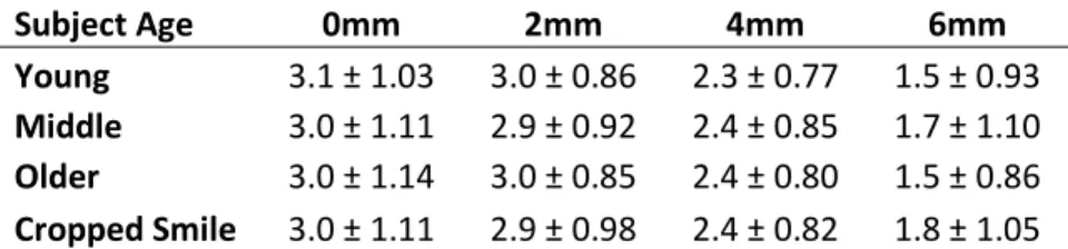
The data was initially analyzed by grouping the 2 subjects of each age group together and calculating mean rankings to test for differences in attractiveness for gingival display (Table 1) (Fig 20) and buccal corridor size (Table 2) (Fig 21) related to subject age. For all 3 age groups and the cropped smile, the highest ranked amount of gingival display was 0mm and the second highest ranked was -2mm. In all age groups and the cropped smile, the lowest ranked amount of gingival display was 4mm. All age groups and the cropped smile had 0mm buccal corridors ranked as most attractive. 2mm buccal corridors ranked as second most attractive for the
cropped smile and both young and middle age groups while tying for most attractive in the older age group. In all age groups and the cropped smile the 6m buccal corridors ranked as least attractive. When evaluating this data using the frequency of a top attractiveness ranking for each image, the results matched with those using mean ranking.
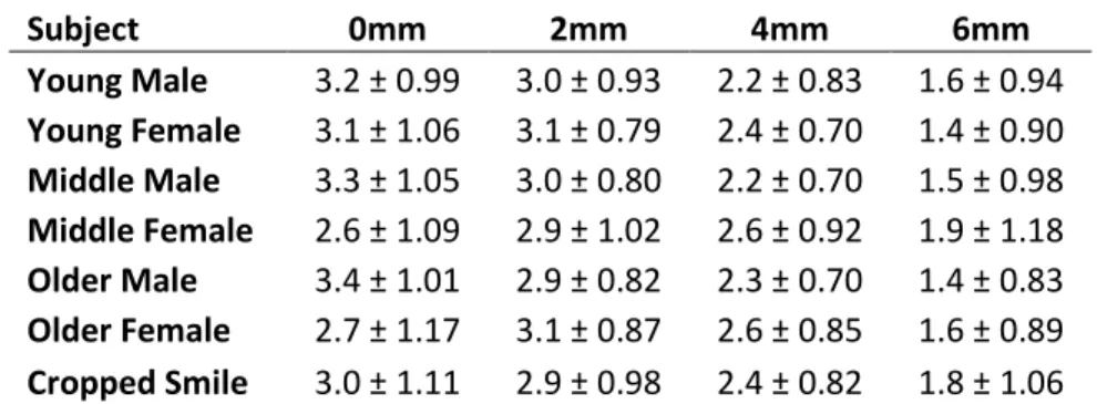
close second. For the rest of the subjects, -2mm gingival display ranked second most attractive, except for the middle female, where 2mm gingival display was second. Least attractive for all subjects was 4mm of gingival display, except for the middle female where -4mm ranked least attractive. All the male subjects had 0mm buccal corridors ranked as most attractive and 2mm as second most attractive. For the female subjects the reverse was true, with 2mm ranked as most attractive and 0mm as second most attractive, except in the young female where the mean values for the two were equal. For all subjects, the largest 6mm buccal corridors were ranked least attractive. When evaluating this data using the frequency of a top attractiveness ranking for each image, the results again matched with those using mean ranking.
To determine any differences based on respondent age, individual subjects were isolated and only the respondents’ top choices were analyzed, adjusting for respondent gender.
Respondents in different age groups made similar choices for the most attractive gingival displays except when evaluating the young male subject (p=0.002) and the older female subject (p<0.0001) (Table 5). For both the young male and older female subject, young respondents were, in general, much more likely to choose 0mm as their preference than middle and older respondents who were less harmonious in their preference. The percentage of older respondents who preferred -2mm of gingival display for the older female patient was nearly twice as high as those who preferred 0mm.
sizes. A similar pattern was seen for the older female subject. For the middle female subject, young respondents had a clear preference for choosing 2mm and rejecting 6mm, while in the middle and older groups there was no distinct preference for any of the buccal corridor options. For the cropped smile, the young respondents were more decisive in selecting the 0mm buccal corridors as top choice, while middle and older respondents had more equal preference for 0mm and 2mm.
To study differences based on respondent gender, subjects were again analyzed
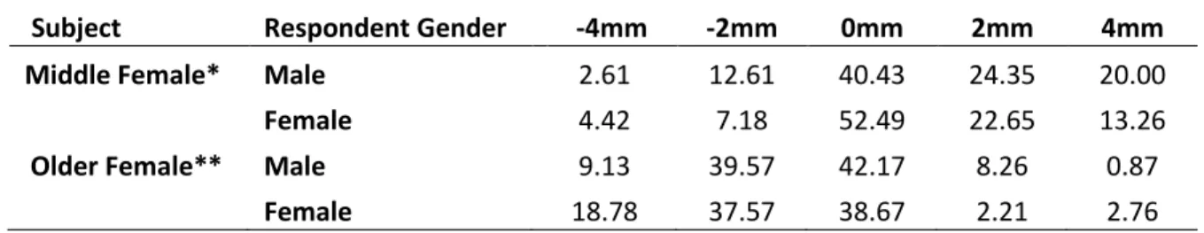
individually and only the respondents’ top choices for attractiveness were considered, adjusting for respondent age. For gingival display, significant differences based on the gender of the respondents were limited to the older (p=0.049) and middle (0.002) female subject and were very minor (Table 7). For the older female subject, both male and female respondents selected 0mm as most attractive with the highest frequency, and -2mm with the second highest frequency, but female respondents were more likely to select -4mm as most attractive. For the middle female, male and female respondents again agreed with most frequently preferred (0mm) and second most frequently preferred (2mm) levels of gingival display, but females were more likely to choose 0mm and males more likely to choose 4mm.
When evaluating buccal corridor size, the male and female respondents only differed significantly for the cropped smile (p=0.022) (Table 8). The male respondents clearly preferred 0mm while the females were split evenly between 0mm and 2mm as the top choice.
Discussion
than has previously been the case and called for new information in these areas. Many authors and researchers have answered this call, as smile esthetics has become a popular topic in the orthodontic literature. Increasing numbers of adults are seeking orthodontic care,4 and it is important for orthodontists to consider smile esthetics as they relate to the adult patient. Perioral soft-tissues change with advancing age and this affects the smile. Therefore, the 4th dimension (time) must be considered during smile assessment in orthodontic treatment planning.33
This is the first study to evaluate laypeople’s preferences for the major vertical and transverse characteristics of the smile on different aged subjects. Many studies have used cropped photos to analyze these factors,9-21 while few have evaluated smile esthetics in the context of the full face.22-25 In this study it was critical to provide full face views of the subjects to the respondents to display the cues which differentiate people according to age. Laypeople were targeted as respondents in this study as they are the group that receives orthodontic treatment and their opinion of the result is critical to clinical success.
incisor length relative to the vertical space between the lips in the smile. Not only was a high percentage of her maxillary central incisors hidden on the negative gingival display photos, a greater amount of mandibular incisor and tissue was displayed than most of the other subjects.
Since there doesn’t seem to be an age-related pattern to these minor differences in overall gingival display preferences, they should not be ascribed to subject age. Any slight differences in preference likely have to do with differences in other facial features and dimensions which may not be determined by age. Flores-Mir et al25, in a study which used full face photos,
reported a similar effect as esthetic perceptions of anterior dentition varied significantly between the subjects whose smiles were evaluated. Springer et al23 also noted significant differences in esthetic preferences between the two different subjects whose full face images were used in their study. The authors attributed these differences to the subjects’ facial appearances.
In this study, no differences in overall preferences for buccal corridor size were noted corresponding to subject age. Though the mean ranks for buccal corridor size are very close for 0mm and 2mm, it is worth noting that 2mm is slightly preferred on the female subjects and 0mm on the males. This may be evidence of a small gender-specific difference in laypeople’s
preference for buccal corridor size. This finding would disagree with the studies of Parekh et al12 and Moore et al22 but may be supported by the study of Gul-E-Erum and Fida24 who found that laypeople preferred small (10%) buccal corridors on a woman and no buccal corridors on a man.
gingival display, respectively. Assessing gingival display, respondents varied by age in their preferences for the young male and older female subjects. For both subjects, as respondent age increased, there was a gradual shift in top preference from the 0mm level to 2mm of upper incisor coverage. This trend of older respondents preferring a slightly “older” amount of gingival display and younger respondents a slightly more youthful gingival display would carry more weight if it was seen with more subjects in the study. For buccal corridor size, in three of the subjects (young male, middle female, older female) and the cropped smile, significant differences in preference were found based on respondent age. Young respondents were more decisive in selecting minimal buccal corridors as most attractive, while middle and older respondents were more varied in their rankings.
The results of this study are in keeping with authors that have stated that the ideal elevation of the lip in a smile places its lower margin even with the gingival margin of the maxillary central incisors20, 33, 45 and disagree with those who have found 2mm of maxillary incisor coverage to be ideal.11, 18, 23 Laypeople in this study did not tend to prefer slight gingival display over slight maxillary incisor coverage, even in the younger subjects. The only exception to this generalization was found with the middle female subject. This finding stands in
opposition to the statements of several authors,5, 10, 38, 46 but agrees with other studies which found that some coverage of the maxillary incisors is preferable to any gingival display.17, 18, 21, 23 However, if the 0mm gingival display level is usually preferred for all ages and the -2mm is second most desirable, orthodontists may need to consider keeping gingival display slightly greater than ideal in a young patient so that lip coverage will stay within the most desirable zone for as much of the patient’s adult life as possible (considering the soft tissue changes that occur with aging).
findings11-13, 15, 16, 22-24, 33, 36 and disagrees with those who have stated that buccal corridors do not influence smile attractiveness.14, 20, 21
Some studies that have digitally manipulated gingival display have done so by changing the lip position or the frame of the smile.9, 10 In this study, the frame was kept constant. The advantage of this was the elimination of any confounding variables which are created by changing lip position and frame size. A possible disadvantage was the need for a significantly animated smile to allow enough vertical space to manipulate the images while avoiding any lower lip coverage of the maxillary incisors, even in the 4mm gingival display photos. There was concern that previous studies which had manipulated images similarly, but allowed the maxillary incisors to be covered by the lower lip in the images with positive gingival display11, 17, 18
had added a confounding variable to bias respondents against those images with increased gingival display. However, a side-effect of creating a larger frame vertically for this study was increased mandibular incisor show on the negative gingival display images.
Many other studies have used a percentage or ratio to quantify buccal corridors,11, 15, 16, 22, 29
but this study was designed using linear measurements. Ker et al11 presented linear
measurements alongside percentages, but the measurements seem far larger than what is depicted in the images. To the authors’ knowledge, this is the first study to use the method described for accurately measuring and representing linear measurements when performing digital image modifications.
considered acceptable.9-11, 15, 17, 23 The results of this study should not lead the orthodontist to overly sensitize patients to desire esthetic ideals that are not clinically achievable for their case, nor should they push the orthodontist to try to attain these ideals when it is not realistic or even beneficial for the patient. For example, as other authors have stated,15, 22 the findings of
preference for minimal buccal corridors should not justify routine expansion in normal maxillae. Some patients have narrower jaws and significant transverse animation of soft tissue on smile that will lead to increased buccal corridors. This isn’t necessarily a problem that must be addressed. Furthermore, one must keep in mind that this study looked at two isolated
characteristics of the smile which in reality may interact with many other esthetic features. For instance, the interaction of the smile arc with gingival display and buccal corridors has been studied by Kaya et al18 and Parekh et al,12, 13 respectively.
It is critical to discuss the esthetic goals of each case with the patient on an individual basis to ensure that the orthodontist and patient are on the same page. The orthodontist must find out specifically what a patient would like to change about his or her smile. Beauty is in the eye of the beholder and what is attractive to one person may not be to another. Definitions of beauty and attractiveness are not static over time and can vary a great deal between individuals, and racial, social, and economic groups.6, 9, 26, 41, 54, 55 In this study alone there were multiple
respondents who ranked the least popular values for gingival display and buccal corridor size as most attractive.
continuous adjustment of variables. Secondly, the cropped smile was intended as a sort of control for comparison, but it is clearly female and not from an elderly person. However, this made it similar to the cropped smiles used in several other studies which used digital image manipulation.11-18 Thirdly, as noted by Martin et al,15 it is extremely difficult to artificially create natural looking buccal corridors. Other studies used very sharply defined buccal corridors,11-16, 22 whereas this study attempted to soften the edges of the artificial buccal corridors slightly. Fourthly, as previously discussed, the large vertical frame needed for manipulation resulted in more mandibular tooth show on the negative gingival display photos. A future study could perhaps use intraoral photos with the teeth just slightly apart in order to avoid excessive lower incisor exposure in some of the modified images. Finally, this study simply used one male and one female subject from three age groups to assess smile preferences and age. Future studies may need to use many more subjects of various ages, or come up with a way to digitally “age” a patient in a realistic fashion, to allow more definitive statements to be made.
Conclusions
1. Preferences for gingival display and buccal corridor size in a given person’s smile do not seem to be determined simply by the person’s age.
2. Preferences for gingival display and buccal corridor size can vary depending on the person whose smile is being assessed and may involve consideration of various facial features and dimensions.
4. There may be a gender specific preference for no buccal corridors on males and minimal (2mm) buccal corridors on females.
Tables
Table 1 - Mean rank* ± standard deviation of respondents’ preference of gingival display for subjects of varying age.
Subject Age -4mm -2mm 0mm 2mm 4mm
Young 2.9 ± 1.17 3.9 ± 0.98 4.2 ± 0.97 2.7 ± 0.97 1.4 ± 0.84
Middle 2.2 ± 1.13 3.4 ± 1.24 4.0 ± 1.07 3.2 ± 1.18 2.1 ± 1.38
Older 2.8 ± 1.04 4.0 ± 0.88 4.2 ± 0.99 2.8 ± 0.98 1.2 ± 0.66
Cropped Smile 2.4 ± 0.99 3.8 ± 1.02 4.4 ± 0.86 3.0 ± 1.04 1.4 ± 0.92
* Higher ranks correspond to increased attractiveness as rated by respondents.
Table 2 - Mean rank* ± standard deviation of respondents’ preference of buccal corridor size for subjects of varying age.
Subject Age 0mm 2mm 4mm 6mm
Young 3.1 ± 1.03 3.0 ± 0.86 2.3 ± 0.77 1.5 ± 0.93
Middle 3.0 ± 1.11 2.9 ± 0.92 2.4 ± 0.85 1.7 ± 1.10
Older 3.0 ± 1.14 3.0 ± 0.85 2.4 ± 0.80 1.5 ± 0.86
Cropped Smile 3.0 ± 1.11 2.9 ± 0.98 2.4 ± 0.82 1.8 ± 1.05
Table 3 - Mean rank* ± standard deviation of respondents’ preference of gingival display for subjects of varying age and gender.
Subject -4mm -2mm 0mm 2mm 4mm
Young Male 3.4 ± 1.17 3.8 ± 1.07 4.0 ± 1.03 2.5 ± 1.01 1.4 ± 0.93
Young Female 2.3 ± 0.92 4.1 ± 0.85 4.4 ± 0.84 2.8 ± 0.90 1.3 ± 0.75
Middle Male 2.6 ± 1.12 3.9 ± 1.05 4.0 ± 1.11 2.8 ± 1.14 1.6 ± 1.13
Middle Female 1.8 ± 1.00 2.9 ± 1.22 4.1 ± 1.02 3.6 ± 1.08 2.5 ± 1.47
Older Male 2.4 ± 0.90 3.9 ± 0.84 4.5 ± 0.82 3.0 ± 1.00 1.3 ± 0.64
Older Female 3.2 ± 1.02 4.1 ± 0.91 4.0 ± 1.06 2.6 ± 0.91 1.2 ± 0.69
Cropped Smile 2.4 ± 0.99 3.8 ± 1.02 4.4 ± 0.86 3.1 ± 1.03 1.4 ± 0.91
* Higher ranks correspond to increased attractiveness as rated by respondents.
Table 4 - Mean rank* ± standard deviation of respondents’ preference of buccal corridor size for subjects of varying age and gender.
Subject 0mm 2mm 4mm 6mm
Young Male 3.2 ± 0.99 3.0 ± 0.93 2.2 ± 0.83 1.6 ± 0.94
Young Female 3.1 ± 1.06 3.1 ± 0.79 2.4 ± 0.70 1.4 ± 0.90
Middle Male 3.3 ± 1.05 3.0 ± 0.80 2.2 ± 0.70 1.5 ± 0.98
Middle Female 2.6 ± 1.09 2.9 ± 1.02 2.6 ± 0.92 1.9 ± 1.18
Older Male 3.4 ± 1.01 2.9 ± 0.82 2.3 ± 0.70 1.4 ± 0.83
Older Female 2.7 ± 1.17 3.1 ± 0.87 2.6 ± 0.85 1.6 ± 0.89
Cropped Smile 3.0 ± 1.11 2.9 ± 0.98 2.4 ± 0.82 1.8 ± 1.06
Table 5- Percentage of top ranking for gingival display for subjects with significant differences based on respondent age.
Subject Respondent Age -4mm -2mm 0mm 2mm 4mm
Young Male* Young 15.45 25.20 52.03 6.50 0.81
Middle 24.83 31.72 37.24 3.45 2.76
Older 18.49 33.56 31.51 10.27 6.16
Older Female** Young 4.88 32.52 56.91 4.88 0.81
Middle 15.86 35.17 38.62 6.90 3.45
Older 17.81 48.63 28.08 4.79 0.68
*MH test detects significant differences with p=0.002 **MH test detects significant differences with p<0.0001
Table 6- Percentage of top ranking for buccal corridor size for subjects with significant differences based on respondent age.
Subject Respondent Age 0mm 2mm 4mm 6mm
Young male* Young 55.28 35.77 3.25 5.69
Middle 48.28 30.34 10.34 11.03
Older 57.82 25.17 10.88 6.12
Middle Female** Young 28.46 42.28 21.14 8.13
Middle 28.08 28.08 23.29 20.55
Older 31.03 24.14 22.76 22.07
Older Female*** Young 39.02 49.59 8.13 3.25
Middle 36.30 31.51 22.60 9.59
Older 37.24 39.31 18.62 4.83
Cropped Smile**** Young 55.28 23.58 6.50 14.63
Middle 40.28 34.72 13.19 11.81
Older 44.52 37.67 9.59 8.22
Table 7- Percentage of top ranking for gingival display for subjects with significant differences based on respondent gender.
Subject Respondent Gender -4mm -2mm 0mm 2mm 4mm
Middle Female* Male 2.61 12.61 40.43 24.35 20.00
Female 4.42 7.18 52.49 22.65 13.26
Older Female** Male 9.13 39.57 42.17 8.26 0.87
Female 18.78 37.57 38.67 2.21 2.76
*MH test detects significant differences with p=0.049 **MH test detects significant differences with p=0.002
Table 8- Cropped smile: percentage of top ranking for buccal corridor size by respondent gender.*
Respondent Gender 0mm 2mm 4mm 6mm
Male 52.63 27.63 10.09 9.65
Female 38.46 37.91 9.89 13.74
Figures
Figure 4 - Five gingival display photos for young male (-4mm, -2mm, 0mm, 2mm, 4mm)
REFERENCES
1. Ackerman JL, Proffit WR, Sarver DM. The emerging soft tissue paradigm in orthodontic diagnosis and treatment planning. Clin Orthod Res. 1999 May;2(2):49-52.
2. Sarver DM, Ackerman JL. Orthodontics about face: the re-emergence of the esthetic paradigm. Am J Orthod Dentofacial Orthop. 2000 May;117(5):575-6.
3. Proffit WR. The soft tissue paradigm in orthodontic diagnosis and treatment planning: a new view for a new century. J Esthet Dent. 2000;12(1):46-9.
4. Proffit WR, Fields HW, Sarver DM. Contemporary Orthodontics. 5th ed. St Louis, MO: Elsevier Mosby; 2013.
5. Sarver DM. The importance of incisor positioning in the esthetic smile: the smile arc. Am J Orthod Dentofacial Orthop. 2001 Aug;120(2):98-111.
6. Rigsbee OH,3rd, Sperry TP, BeGole EA. The influence of facial animation on smile characteristics. Int J Adult Orthodon Orthognath Surg. 1988;3(4):233-9.
7. Ackerman JL, Ackerman MB, Brensinger CM, Landis JR. A morphometric analysis of the posed smile. Clin Orthod Res. 1998 Aug;1(1):2-11.
8. Walder JF, Freeman K, Lipp MJ, Nicolay OF, Cisneros GJ. Photographic and videographic assessment of the smile: objective and subjective evaluations of posed and spontaneous smiles. Am J Orthod Dentofacial Orthop. 2013 Dec;144(6):793-801.
9. Kokich VO,Jr, Kiyak HA, Shapiro PA. Comparing the perception of dentists and lay people to altered dental esthetics. J Esthet Dent. 1999;11(6):311-24.
10. Kokich VO, Kokich VG, Kiyak HA. Perceptions of dental professionals and laypersons to altered dental esthetics: asymmetric and symmetric situations. Am J Orthod Dentofacial Orthop. 2006 Aug;130(2):141-51.
11. Ker AJ, Chan R, Fields HW, Beck M, Rosenstiel S. Esthetics and smile characteristics from the layperson's perspective: a computer-based survey study. J Am Dent Assoc. 2008
Oct;139(10):1318-27.
12. Parekh S, Fields HW, Beck FM, Rosenstiel SF. The acceptability of variations in smile arc and buccal corridor space. Orthod Craniofac Res. 2007 Feb;10(1):15-21.
13. Parekh SM, Fields HW, Beck M, Rosenstiel S. Attractiveness of variations in the smile arc and buccal corridor space as judged by orthodontists and laymen. Angle Orthod. 2006
14. Roden-Johnson D, Gallerano R, English J. The effects of buccal corridor spaces and arch form on smile esthetics. Am J Orthod Dentofacial Orthop. 2005 Mar;127(3):343-50.
15. Martin AJ, Buschang PH, Boley JC, Taylor RW, McKinney TW. The impact of buccal corridors on smile attractiveness. Eur J Orthod. 2007 Oct;29(5):530-7.
16. Ioi H, Kang S, Shimomura T, Kim SS, Park SB, Son WS, et al. Effects of buccal corridors on smile esthetics in Japanese and Korean orthodontists and orthodontic patients. Am J Orthod Dentofacial Orthop. 2012 Oct;142(4):459-65.
17. Ioi H, Kang S, Shimomura T, Kim SS, Park SB, Son WS, et al. Effects of vertical positions of anterior teeth on smile esthetics in Japanese and korean orthodontists and orthodontic patients. J Esthet Restor Dent. 2013 Aug;25(4):274-82.
18. Kaya B, Uyar R. Influence on smile attractiveness of the smile arc in conjunction with gingival display. Am J Orthod Dentofacial Orthop. 2013 Oct;144(4):541-7.
19. Johnson DK, Smith RJ. Smile esthetics after orthodontic treatment with and without extraction of four first premolars. Am J Orthod Dentofacial Orthop. 1995 Aug;108(2):162-7. 20. Hulsey CM. An esthetic evaluation of lip-teeth relationships present in the smile. Am J Orthod. 1970 Feb;57(2):132-44.
21. Isiksal E, Hazar S, Akyalcin S. Smile esthetics: perception and comparison of treated and untreated smiles. Am J Orthod Dentofacial Orthop. 2006 Jan;129(1):8-16.
22. Moore T, Southard KA, Casko JS, Qian F, Southard TE. Buccal corridors and smile esthetics. Am J Orthod Dentofacial Orthop. 2005 Feb;127(2):208,13; quiz 261.
23. Springer NC, Chang C, Fields HW, Beck FM, Firestone AR, Rosenstiel S, et al. Smile esthetics from the layperson's perspective. Am J Orthod Dentofacial Orthop. 2011
Jan;139(1):e91-e101.
24. Gul-e-Erum, Fida M. Changes in smile parameters as perceived by orthodontists, dentists, artists, and laypeople. World J Orthod. 2008 Summer;9(2):132-40.
25. Flores-Mir C, Silva E, Barriga MI, Lagravere MO, Major PW. Lay person's perception of smile aesthetics in dental and facial views. J Orthod. 2004 Sep;31(3):204,9; discussion 201. 26. Frush J, Fisher R. The dynesthetic interpretation of the dentogenic concept. J Prosthet Dent. 1958;8(4):558-81.
28. Rubin LR. The anatomy of a smile: its importance in the treatment of facial paralysis. Plast Reconstr Surg. 1974 Apr;53(4):384-7.
29. Desai S, Upadhyay M, Nanda R. Dynamic smile analysis: changes with age. Am J Orthod Dentofacial Orthop. 2009 Sep;136(3):310.e1,10; discussion 310-1.
30. Chetan P, Tandon P, Singh GK, Nagar A, Prasad V, Chugh VK. Dynamics of a smile in different age groups. Angle Orthod. 2013 Jan;83(1):90-6.
31. Ackerman MB. The effect of maxillary position on anterior tooth display [thesis]. Rochester, NY: University of Rochester; 2000.
32. Sarver DM. The face as determinant of treatment choice. In: Frontiers of dental and facial esthetics. Craniofacial Growth Series. Vol 38. Ann Arbor, MI: Center for Human Growth and Development; University of Michigan; 2001. p.19-54.
33. Sarver DM, Ackerman MB. Dynamic smile visualization and quantification: Part 2. Smile analysis and treatment strategies. Am J Orthod Dentofacial Orthop. 2003 Aug;124(2):116-27. 34. Gianelly AA. Arch width after extraction and nonextraction treatment. Am J Orthod Dentofacial Orthop. 2003 Jan;123(1):25-8.
35. Golwalkar SA, Shetty V. Arch widths after extraction and nonextraction treatment in class I patients. J Contemp Dent Pract. 2013 Mar 1;14(2):312-5.
36. Morley J, Eubank J. Macroesthetic elements of smile design. J Am Dent Assoc. 2001 Jan;132(1):39-45.
37. Sarver DM. Esthetic Orthodontics and Orthognathic Surgery. Anonymous St. Louis, MO: Mosby; 1998.
38. Dickens S, Sarver D, Proffit WR. Changes in Frontal Soft Tissue Dimensions of the Lower Face by Age and Gender. World J Orthodontics. 2002;3(4):313-20.
39. Peck S, Peck L, Kataja M. The gingival smile line. Angle Orthod. 1992 Summer;62(2):91,100; discussion 101-2.
40. Epker BN, Fish L. Surgical-orthodontic correction of open-bite deformity. Am J Orthod. 1977 Mar;71(3):278-99.
41. Vig RG, Brundo GC. The kinetics of anterior tooth display. J Prosthet Dent. 1978 May;39(5):502-4.
43. Peck S, Peck L, Kataja M. Some vertical lineaments of lip position. Am J Orthod Dentofacial Orthop. 1992 Jun;101(6):519-24.
44. Peck S, Peck L. Selected aspects of the art and science of facial esthetics. Semin Orthod. 1995 Jun;1(2):105-26.
45. Mackley RJ. An evaluation of smiles before and after orthodontic treatment. Angle Orthod. 1993 Fall;63(3):183,9; discussion 190.
46. Chiche G, Kokich VG, Caudill R. Diagnosis and Treatment Planning of Esthetic Problems. In: Chiche G, Pinault A, editors. Esthetics of Anterior Fixed Prosthodontics. Quintessence; 1994. p.33.
47. Peck S, Peck H. The aesthetically pleasing face: an orthodontic myth. Trans Eur Orthod Soc. 1971:175-84.
48. Fanous N. Aging lips. Esthetic analysis and correction. Facial Plast Surg. 1987 Spring;4(3):179-83.
49. Bishara SE, Treder JE, Jakobsen JR. Facial and dental changes in adulthood. Am J Orthod Dentofacial Orthop. 1994 Aug;106(2):175-86.
50. Formby WA, Nanda RS, Currier GF. Longitudinal changes in the adult facial profile. Am J Orthod Dentofacial Orthop. 1994 May;105(5):464-76.
51. Singh B, Ahluwalia R, Verma D, Grewal SB, Goel R, Kumar PS. Perioral age-related changes in smile dynamics along the vertical plane: a videographic cross-sectional study. Angle Orthod. 2013 May;83(3):468-75.
52. Janzen EK. A balanced smile--a most important treatment objective. Am J Orthod. 1977 Oct;72(4):359-72.
53. Davis BK. Dental aesthetics and the aging patient. Facial Plast Surg. 2006 May;22(2):154-60.
54. Collins M. The Eye of the Beholder: Face Recognition and Perception. Semin Orthod. 2012 September 2012;18(3):229-34.
55. Czarnecki ST, Nanda RS, Currier GF. Perceptions of a balanced facial profile. Am J Orthod Dentofacial Orthop. 1993 Aug;104(2):180-7.