The handle
http://hdl.handle.net/1887/30243
holds various files of this Leiden University
dissertation.
Author: Donjacour, Claire Elisabeth Henrica Maria
endocrine, metabolic and other aspects
Chipsoft, and Heinen en Löwenstein.
ISBN 978-90-90283-87-6
Layout Renate Siebes, Proefschrift.nu
Cover design Claire Donjacour, Alex van der Lecq
Printed by Ipskamp Drukkers B.V.
© 2014 Claire E.H.M. Donjacour
endocrine, metabolic and other aspects
Proefschrift
ter verkrijging van
de graad van Doctor aan de Universiteit Leiden, op gezag van Rector Magnificus prof. mr. C.J.J.M. Stolker,
volgens besluit van het College voor Promoties te verdedigen op donderdag 18 december 2014
klokke 16.15 uur
door
Claire Elisabeth Henrica Maria Donjacour
Copromotores Dr. G.J. Lammers
Dr. S. Overeem (Radboud umc, Nijmegen)
Overige leden Prof. dr. J.A. Romijn (AMC, Amsterdam)
Dr. N.A. Aziz
Prof. dr. J.G. van Dijk
Chapter 1 General introduction and aim of the thesis 9
Part I Endocrine studies in narcolepsy
Chapter 2 Sodium oxybate increases prolactin secretion in narcolepsy
patients and healthy controls
23
Chapter 3 The effect of sodium oxybate on growth hormone secretion
in narcolepsy patients and healthy controls
39
Chapter 4 Plasma total ghrelin and leptin levels in human narcolepsy and
matched healthy controls: Basal concentrations and response to sodium oxybate
57
Chapter 5 Altered circadian rhythm of melatonin concentrations in
hypocretin-deficient men
71
Part II Metabolic studies in narcolepsy
Chapter 6 Glucose and fat metabolism in narcolepsy and the effect of sodium
oxybate: A hyperinsulinemic-euglycemic clamp study
87
Chapter 7 The effects of sodium oxybate on core body and skin temperature
regulation in narcolepsy
103
Part III Other aspects of narcolepsy
Chapter 8 Month of birth is not a risk factor for narcolepsy with cataplexy
in the Netherlands
123
Chapter 9 Delusional confusion of dreaming and reality in narcolepsy 131
Chapter 10 Summary, conclusions and future perspectives 141
Appendix A Samenvatting, conclusies en toekomstperspectieven 153
Appendix B Dankwoord 167
Appendix C Curriculum Vitae 171
Appendix D List of publications 175
general introduction and
aim of the thesis
Based on ‘Clinical and pathophysiological aspects of narcolepsy’. International Journal of Sleep and Wakefulness 2007; 1(2):55-65.
IntroductIon
Narcolepsy with cataplexy is a chronic sleep disorder caused by hypocretin deficiency, that affects approximately 25–50 per 100,000 people.1 The first symptoms usually appear from childhood to midlife with a large peak around 15 years of age and a smaller one around 36.2 It has a profound effect on the quality of life3 that might exceed the burden of other serious chronic diseases like epilepsy.4 Narcolepsy is associated with reduced vigilance,5 severe fatigue,6 anxiety and mood disorders7 and a higher rate of car accidents.8-10 In addition the unemployment rate among patients is higher than in controls.11
SIgnS and SymptomS of narcolepSy
Excessive daytime sleepiness
The excessive daytime sleepiness (EDS) may come in various forms, with a continuous feeling of sleepiness at one end of the spectrum, and sudden involuntary and irresistible “sleep attacks” at the other. Narcolepsy patients often experience a combination of these. Daytime sleep episodes are brief, often < 20 min, and are typically reported to be refreshing. Although sleep episodes may occur several times a day, the total amount of sleep is not or only slightly increased over a 24-h period due to nocturnal sleep fragmentation.12
Nocturnal sleep disturbances
Fragmented nocturnal sleep is a major and common problem in narcolepsy. While patients fall asleep very quickly numerous awakenings may follow. These awakenings are usually short but can sometimes last for hours, forcing patients to get out of bed. Importantly, fragmented night-time sleep is not the cause of EDS; while improving nocturnal sleep may sometimes alleviate daytime sleepiness, EDS will never disappear.13
Cataplexy
diversity of emotions. The most often reported triggers are laughter, hearing a joke, or feeling excited. Although most cataplectic attacks do have a trigger, 40% of the patients have experienced attacks without an identified trigger. Most cataplectic attacks are partial, resulting for example in sagging of the jaw with blurred speech or buckling of the knees. Partial attacks may evolve into a complete loss of skeletal muscle tone leading to a fall but, because of its gradual onset, patients can usually support themselves preventing injury.14
Hypnagogic hallucinations
Hypnagogic hallucinations are vivid dreamlike experiences that occur upon falling asleep. The auditory, visual, or tactile sensations are usually felt to be real, and are often bizarre and frightening. The perception of intruders, with people or animals standing over or lying under the bed is a common reported phenomenon.15
Sleep paralysis
Sleep paralysis is the inability to move at sleep onset or, more commonly, when awakening.16 While the patient is awake, it is impossible for them to move their arms or legs or sometimes even open their eyes, and it can be extremely distressing, especially when occurring for the first time. It often occurs in combination with hypnagogic hallucinations. About half of the patients experience sleep paralysis. This phenomenon is much less common where it only affects 5% of the population.17
addItIonal SIgnS and SymptomS of narcolepSy
In addition of the above mentioned symptoms, there are various other features, frequently seen in narcolepsy patients. A few of them, most relevant for this thesis will be discussed.Obesity
in narcolepsy is unknown. Some patients complain about a tendency to binge eat, especially during waking periods at night. However, in a study using cross-checked dietary histories, it was shown that over a period of 24 h individuals with narcolepsy tended to consume fewer calories than controls, particularly carbohydrates.21;22 Patients with idiopathic hypersomnia have been shown to have a lower BMI than narcoleptics, indicating that inactivity due to EDS is not likely to account for the obesity.18 It seems possible that mechanisms such as a lowered metabolic rate underlie the increase in body weight seen in subjects with narcolepsy.
Temperature differences
Narcolepsy patients have an altered thermoregulatory profile with warmer hands and feet and lower proximal skin temperature than controls, a state in which sleep propensity is high.23 Temperature manipulation studies offsetting these temperature changes show improvement in narcolepsy symptoms.24;25
Memory complaints
Many narcolepsy patients have memory complaints. Interestingly, when formally tested, they do not always show objective memory deficits.26;27 The reason for this is unclear; patients may, for example, be more vigilant in a testing situation than in everyday life.
Delusional confusion of dreaming and reality
Narcolepsy patients may report difficulty distinguishing between dreams and reality. However, although frequently heard in the consulting room, it is rarely mentioned in the literature. So far just a few case reports have described more severe examples of memory source confusion in patients suffering from narcolepsy, in which false accusations of sexual assault occurred when patients mistook a dreamed assault for the memory of an actual event.28;29
Endocrine alterations
larger group of narcolepsy patients.32;33 For the somatotropic axis, the circadian secretion of GH appears to be altered, so that a relatively large fraction of total production occurs during daytime in narcoleptics.34 In addition, 24-h thyrotropin (TSH) levels were reduced in narcolepsy patients. In contrast, thyroxin (T4) and triiodothyronin (T3) were normal, so the clinical relevance of these findings remains to be elucidated.35 A blunted total and basal adrenocorticotropic hormone (ACTH) production was found in narcolepsy patients whereas the pulsatile secretion was normal35 which seemed to indicate that hypocretins are involved in the basal but not pulsatile secretion of ACTH. The circadian rhythm of cortisol and ACTH was normal, showing that the circadian timekeeper is intact in narcolepsy. Finally, a role for hypocretin-1 in luteinizing hormone (LH) release was observed in rats.36 Accordingly, a decreased 24-h mean LH concentration and pulsatile secretion was found in human hypocretin deficiency.37 A high prevalence of precocious puberty is found in children with narcolepsy.38
Diabetes
In the sixties and eighties some reports have been published on a higher incidence of insulin resistance and type II diabetes mellitus (T2DM) in narcolepsy.39-41 Another study confirmed a higher risk of metabolic syndrome independent of BMI by comparing narcolepsy patients to idiopathic hypersomnia patients.22 However, in a recent study where a large group of patients were compared to healthy matched controls, no differences in insulin resistance were found by calculating pro-insulin to insulin ratio and using homeostatic model assessment method (HOMA).42 Unfortunately in the last study, patients were medicated. This might have influenced the results. Anticataplectic drugs e.g. imipramine may produce a significant increase of fasting blood glucose levels.43
pathophySIology of narcolepSy
Hypocretin deficiency
Two simultaneously published papers from different laboratories then reported a loss of hypocretin cells in brains of human narcolepsy patients.47;48 Peyron et al. also reported a single case of early onset narcolepsy associated with by a hypocretin mutation. Thannickal et al. reported elevated levels of gliosis in the hypothalamic region where hypocretin cells had been lost, consistent with the autoimmune hypothesis.48;49 Further work showed that the number of hypothalamic neurons containing dynorphin and Narp, both substances known to be localized to hypocretin cells, were reduced by 90% in narcoleptics with cataplexy.50;51 This finding suggests that hypocretin cells are lost, rather than simply being unable to produce hypocretin.
Autoimmune hypothesis
treatment of narcolepSy
Every narcolepsy patient should be advised to live a regular life, trying to have similar bedtimes every day. Scheduled naps can alleviate sleepiness for a while, and may be advised.67 Likewise, a short nap before demanding activities may be helpful. However, in the majority of patients, pharmacological treatment is necessary.15;68 In practice, it is recommended to start treating the most disabling symptom first, and to tailor drug schedules and dosages individually. Combination therapy is often necessary.68 It must be emphasized that drug therapy is purely symptomatic. While cataplectic attacks can often be completely abolished, EDS will never completely disappear. Drugs to treat narcolepsy are usually divided into two groups; stimulant drugs to treat EDS and anti-cataplectic drugs that also tackle dream related symptoms.
Stimulants
Examples of wake promoting agents are amphetamines, and amphetamine like drugs e.g. methylphenidate. The mode of action of these drugs is complex, but the central mechanism entails the enhancement of the release of catecholamines (dopamine and noradrenaline) as well as uptake inhibition.69;70 A frequently prescribed non-amphetamine like substance is modafinil. Its mode of action is still a matter of debate. Several studies indicate that modafinil, like other stimulants, inhibits the reuptake of dopamine (DA) via the DA transporter (DAT).71 However in vitro studies show that modafinil binds to the DAT with lower affinity than methylphenidate and other psychostimulants drugs.71 Recent research identified several non-dopaminergic effects of modafinil, such as the increase of electrical neuronal coupling, or the enhancement of histamine and orexin neurotransmission that might be of primary importance to explain its efficacy as a wake-promoting and cognitive-enhancing medication.71
Anticataplectic drugs
publications on its efficacy are scarce. Acute withdrawal from antidepressants may severely aggravate cataplexy, sometimes leading to a status cataplecticus (sequential cataplexy attacks that may last for hours or even longer).
Sodium oxybate
A more recently approved compound to treat narcolepsy is sodium oxybate (SXB,γ-hydroxybutyrate; GHB). SXB is a hypnotic and is taken twice a night (at bedtime, and a second dose 3–4 hours later). Interestingly, despite the short half-life of the drug and its nighttime administration, the effects on daytime cataplexy are already obvious when using low dosages (3–6 gr/night).72 Additional studies show that the use of higher doses of SXB (up to 9 gr/ night) has additional positive effects on the excessive daytime sleepiness, and the quality of nocturnal sleep.73 Moreover SXB may reduce weight, a side effect that could be beneficial to an overweight patient.74 GHB occurs naturally in the brain, but its mechanism of action in narcolepsy is not precisely known. In healthy subjects, it was shown that administration of SXB has a slow wave sleep (SWS) promoting effects associated with an increase in GH secretion.75 Its effects are thought to be mediated through γ-aminobutyric acid B receptor (GABAB) and has an impact on dopamine and serotonin release.76;77 The existence of a specific GHB receptor is still under debate.
aIm of the theSIS
data, it has been hypothesized that SXB may act by stimulating the GHRH secretion in the central nervous system and thereby promoting SWS and the release of GH.75 When administered during nocturnal sleep in narcoleptics, it may again confine GHRH secretion to the night and decrease possible sleep inducing effects of GHRH as a consequence of daytime release.
Part I Endocrine studies in narcolepsy
The objective is to investigate whether the secretion of Prolactin (PRL) (chapter 2), Growth Hormone (GH) (chapter 3), Leptin and Ghrelin (chapter 4), and Melatonin (chapter 5) is altered in narcolepsy patients compared with individually matched healthy controls. In addition, the effect of short term administration of SXB on the aforementioned substances is described.
Earlier reports on PRL secretion in narcolepsy patients have been inconclusive, showing either increased, decreased or normal levels.81-83 Discrepancies seemed to be due to methodological issues. The major physiological regulator of PRL release is dopamine.84 Alterations in the dopaminergic system in the post-mortem brains of narcoleptics have been described making changes in PRL release in narcolepsy conceivable.85 Although the exact mode of action is still unclear, SXB acts on gamma-aminobutyric acid type B (GABAB) and has an impact on dopamine and serotonin release.77 Therefore, SXB treatment may be expected to alter PRL secretion. We hypothesised that changes in hypothalamic hypocretin signaling in narcoleptic patients may give rise to altered PRL secretion. In addition, we aimed to determine the effect of SXB administration on PRL secretion and sleep, both in a healthy and in a hypocretin-deficient state.
Leptin and ghrelin are both strong regulators of energy homeostasis, and their relationship with hypocretin has been shown to be of importance. Receptors for leptin are found on hypocretin cells,86 and leptin can directly inhibit the expression of isolated hypocretin neurons.87 Indirectly, leptin can affect the activity of hypocretin cells via energy-regulating neurons in the arcuate nucleus of the hypothalamus.88 Ghrelin is an important endogenous regulator of energy balance and it excites hypocretin neurons. An interaction between these two systems has been shown to be involved in ingestive behavior.87 Its expression is complex89 and influenced by sympathetic nervous system activity.90 Across the wake period, plasma concentrations wax and wane episodically, providing an appetite-stimulating signal to the brain.91 Hypocretin neurons directly sense ghrelin and leptin but it is unknown if these connections are uni-or-bidirectional.92 Therefore a study of hypocretin deficient narcolepsy patients provides a unique opportunity to explore the nature of these relationships. In addition we hypothesized that SXB administration would alter total ghrelin levels, which might be involved in its GH-promoting effects.
Melatonin is a modulator of sleep and high levels induce sleepiness.93 Animal models indicate that the hypocretinergic innervation of the pineal gland could be important for the regulation of diurnal melatonin synthesis and secretion.94 It is therefore conceivable that melatonin secretion is hampered in narcolepsy. Here we describe a study in which hourly melatonin levels are compared between narcolepsy patients and controls.
Part II Metabolic studies in narcolepsy
Hypocretins have a major role in glucose sensing and homeostasis78 and therefore it is conceivable that hypocretin deficiency may lead to problems in glucose regulation and diabetes. Conflicting reports on the risk of diabetes in narcolepsy have been published. Several studies reported a higher prevalence of type 2 diabetes in narcolepsy.22;39;41;95 More recent studies reported no difference in diabetes prevalence between narcolepsy patients and matched controls.42;96 In chapter 6 we describe the first study to compare insulin sensitivity in nine narcolepsy with cataplexy patients compared with matched healthy controls, measured with the gold standard, the hyperinsulinemic euglycemic clamp technique. In addition, the effect of long term sodium oxybate treatment on insulin sensitivity is evaluated.
a positive effect on the sleep disturbances in narcolepsy.73 The effect of both compounds are the same but the exact mechanisms are still unclear.98 Altered thermoregulation is one of the effects of GHB described in animal studies and human case reports. Rodent studies demonstrate a slight increase in core body temperature after administration of a low dose of GHB and a clear decrease in core body temperature in higher doses.99 In addition several studies describe hypothermia in humans with GHB intoxication.100;101 Given the impact of GHB on temperature regulation, the altered pattern of skin temperature in narcolepsy and the positive effects of SXB on sleep in narcolepsy patients, the treatment effect of SXB may be, at least in part, mediated by restoring the physiological temperature regulation. Chapter 7 aims to determine the effect of short term Sodium Oxybate administration on the 24-hour temperature and sleep-wake profile in narcolepsy patients and controls.
Part III Other aspects of narcolepsy
Hypnagogic hallucinations are frequently described dream related experiences in narcolepsy. Confusions between dreams and reality are an often heard issue in narcolepsy, however so far no systematic studies have been published about these confusions. Here we describe an explorative study on delusional confusion of dreaming and reality in narcolepsy (chapter 8).
sodium oxybate increases
prolactin secretion in narcolepsy
patients and healthy controls
Claire E. H. M. Donjacour N. Ahmad Aziz Marijke Frölich Ferdinand Roelfsema
Sebastiaan Overeem Gert Jan Lammers
Hanno Pijl
European Journal of Endocrinology 2011; 164(3):363-370.
AbstrAct
Objective: Hypocretin deficiency causes narcolepsy and may affect neuroendocrine
systems, including TSH, ACTH and LH secretion. Symptoms can be treated effectively with sodium oxybate (SXB) in many patients. This study was performed to compare prolactin (PRL) secretion in patients and matched controls and establish the effect of SXB administration on PRL and sleep in both the groups.
Design: Open label intervention. Blood was sampled before and after 5 days of SXB
treatment. The study was performed at the Leiden University Medical Centre, Leiden, The Netherlands.
Methods: Subjects were admitted to the clinical research centre on both occasions.
Patients or participants: Eight male hypocretin-deficient narcolepsy with cataplexy
patients and eight controls matched for sex, age, body mass index, waist-to-hip ratio and fat percentage were enrolled.
Interventions: SXB two times 3 g per night for five consecutive nights.
results: Patients and controls underwent 24 h blood sampling at 10 min intervals for
measurement of PRL concentrations. The PRL concentration time series was analysed with a new deconvolution programme, approximate entropy (ApEn) and Cosinor analysis. Sleep was polygraphically recorded. Basal and pulsatile PRL secretion, as well as pulse regularity and frequency, ApEn and diurnal parameters were similar in patients and controls. SXB treatment caused similar nocturnal increase in PRL secretion, advance of the acrophase and decrease in ApEn in patients and controls. Slow wave sleep was increased to a similar extent in patients and controls.
conclusion: This detailed study did not demonstrate altered PRL secretion in
2
IntrODuctIOn
subjects AnD MethODs
subjects
Eight male narcolepsy patients with definite cataplexy were included, who fulfilled the diagnostic criteria of the 2nd edition of the International Classification for Sleep Disorders.118 All narcolepsy patients were hypocretin-1 deficient and free of medication for at least 2 weeks before the study. Only one of the patients received prolonged SXB administration in advance. He did stop taking SXB 20 days prior to the study. One patient took SXB in the past for a short period of time and took stimulants on demand. Another patient was tapered off antidepressants. Five patients were not taking any drugs at the time of enrolment. All consecutive male patients eligible for the study were asked to participate. Eight healthy controls, matched for sex, age, body mass index (BMI), waist-to-hip ratio (WHR) and fat percentage, were included for comparison. Bioelectrical impedance analysis (Bodystat, Douglas, Isle of Man, UK) was used to estimate fat percentage. Subjects were eligible for study after exclusion of hypertension, any known history of pituitary, psychiatric or neurological disease (other than narcolepsy), alcohol or drug abuse, recent weight change (>3 kg weight change within the last 3 months), a sleep disorder history assessed through clinical interview (controls) and endurance sports. Routine laboratory tests were performed to rule out diabetes (fasting plasma glucose >6.9 mmol/l), anaemia, as well as hepatic and renal failure. The study was performed at Leiden University Medical Centre, Leiden, The Netherlands. The study was approved by the ethics committee of the Leiden University Medical Centre. Written informed consent was obtained from all subjects.
Protocol
2
0830, 1300 and 1800 h (Nutridrink, Nutricia, Zoetermeer, The Netherlands; 1.5 kcal/ml, 2100kcal/day; macronutrient composition per 100 ml: protein, 6 g; fat, 5.8 g; carbohydrate, 18.4 g). Subjects remained (semi)supine except for bathroom visits. Daytime naps were allowed. Lights were switched off at 2300 h and turned on at 0730 h the next day. Twenty-four hour sampling was performed at baseline and on the 5th day of SXB administration in two night-time doses of 3 g at 2300 h and 0300 h. This starting dose, higher than the usual of 2.25 g, permitted to elicit some effect in a few days of administration. To monitor side effects, the first night of administration was done clinically.
sleep analysis
Sleep was polygraphically recorded throughout both sampling occasions, using an Embletta X100 recorder (Embla, Broomfield, CO, USA). The recordings were scored visually by an experienced sleep technician at 30 s intervals according to the AASM criteria.119 To allow assessment of the association between changes in serum PRL levels (measured every 10 min) and sleep stages (scored every 30 s), sleep profiles were divided into 10 min segments, separating consecutive PRL measurements as described previously.120 These segments were condensed from the 30 s scoring intervals by calculating the percentage of time spent in stages I and II non-rapid eye movement (REM) sleep, slow wave sleep (SWS) and REM sleep.
Assays
Serum PRL concentrations were measured with a sensitive time-resolved immunofluorometric assay with a detection limit of 0.04 µg/l (Delfia, Wallac Oy, Turku, Finland). The assay was calibrated against the 3rd WHO standard 84/500, 1 ng/mlZ36 mU/l. The intra-assay coefficient of variation varied from 3.0 to 5.2%, while the inter-assay coefficient of variation was between 3.4 and 6.2% (in the 0.1–250 µg/l concentration range). In order to minimise inter-assay variability, samples from each patient and matched control were analysed in the same run.
Calculations and statistics
Deconvolution analysis
series.121 For PRL, the fast half-life was represented as 3.5 min constituting 37% of the decay amplitude and the slow half-life was represented as an unknown variable between 20 and 50 min.122;123 All candidate pulse-time sets were deconvolved. Statistical model selection was then performed to distinguish among the independently framed fits of the multiple candidate pulse-time sets using the Akaike information criterion.124 The parameters (and units) are frequency (number of bursts per total sampling period, l of the Weibull distribution), regularity of inter-pulse intervals (unitless g of Weibull), slow half-life (min), basal and pulsatile secretion rates (concentration units/session), mass secreted per burst (concentration units) and wave form shape (mode or time delay to maximal secretion after objectively estimated burst onset, min).
Approximate entropy
Approximate entropy (ApEn) (1, 20%), was used as a scale- and model-independent regularity statistic to quantify the orderliness (regularity) of PRL release. Higher ApEn denotes greater disorderliness (irregularity) of the secretion process. Mathematical models and clinical experiments have established that greater irregularity signifies decreased feedback control with high sensitivity and specificity (both >90%).125
Diurnal variation
The wave form of individual PRL profiles was quantified by a best-fit curve obtained using a locally weighted regression procedure with a regression window of 4 h and a Gaussian kernel.126 The values (and timings) of the acrophase and the nadir were defined as the levels (the timings) corresponding to the maximum and the minimum of the best-fit curve respectively. The amplitude was defined as 50% of the difference between the acrophase and the nadir values.
Statistical analysis
2
results
Narcoleptic patients and controls did not differ with respect to gender (male), age (38.0 ± 4.7 vs 37.9 ± 4.1, P = 0.984 respectively), BMI (28.1 ± 1.6 vs 27.4 ± 1.4, P = 0.742), fat percentage (23.6 ± 2.1 vs 23.4 ± 1.7, P = 0.946) and WHR (0.92 ± 0.03 vs 0.90 ± 0.02, P = 0.579). As expected, in narcolepsy patients the mean BMI was in the overweight range. Ingestion of SXB was well tolerated and apart from mild drowsiness no other side effects were reported. The mean 24 h PRL concentration in narcolepsy patients was 5.13 ± 0.47 µg/l at baseline and 5.65 ± 0.52 µg/l during the SXB administration (P < 0.001, see Figure 2.1). In controls, it was 6.78 ± 1.68 and 7.40 ± 1.66 µg/l respectively (P < 0.001). The PRL concentration strongly increased shortly after the administration of each dose of SXB. In patients, an increase from 4.56 ± 0.26 to 12.08 ± 1.61 µg/l occurred (P < 0.001), and in controls PRL increased from 6.28 ± 1.72 to 14.07 ± 2.85 µg/l (P < 0.001) during the first SXB administration. After the second
Figure 2.1 Mean 24 h plasma prolactin concentration ± SEM, before and after the SXB administration
table 2.1
Dec
on
volution of serum pr
olactin c
oncen
tr
ation pr
ofiles in nar
colep
sy pa
tien
ts and health
y c on tr ols Nar colep sy Con tr ols Nar colep sy v er sus Con tr ols Baseline SX B Baseline SX B Baseline SX B Tr ea tmen t In ter action Pulse fr
equency (no/24 h)
23.5 ± 0.9
16.6 ± 1.6
21.3 ± 0.8
18.1 ± 1.8
0.08 0.74 0.006* 0.24 Half -lif e (min)
39 ± 4.7
33 ± 2.5
39 ± 4.1
33 ± 2.5
0.93
0.83
0.16
0.96
Basal secr
etion (μg
/L/24 h)
91 ± 16.8
115 ± 18
144 ± 59
163 ± 62
0.40 0.43 0.04* 0.83 Pulsa tile secr etion (μg /L/24h)
93 ± 8.0
105 ± 17.2
105 ± 15.0
147 ± 28
0.48 0.26 0.06 0.29 Tot al secr
etion (μg
/L/24 h)
184 ± 20.0
220 ± 32.0
249 ± 73
310 ± 85
0.40
0.31
0.0008*
0.83
Mean pulse mass (μg
/L)
3.99 ± 0.37
6.69 ± 1.15
4.89 ± 0.55
7.87 ± 1.07
0.20
0.69
0.005*
0.25
Mode (min)
19 ± 2.0
20.6 ± 1.8
18.2 ± 1.7
18.1 ± 3.1
0.77 0.41 0.75 0.71 λ (e ven
ts/ 24 h)
21.4 ± 0.7
15.1 ± 1.5
19.3 ± 0.7
16.3 ± 1.7
0.06
0.69
0.005*
0.25
γ (dimensionless)
2.19 ± 0.17
2.05 ± 0.11
2.18 ± 0.12
2.30± 0.20 0.93 0.54 0.97 0.31 Da ta ar e sho wn
as mean ± SEM.
Each gr oup consis ted of eigh t subjects. Comparisons w er
e made using
Studen t’s t -tes t (c olumn 8) and tw o-w ay ANO VA f or r epea ted measur emen ts. Secr etion da ta ar e e xpr
essed in mass units per litr
e hormone dis
tribution v
olume. *
P
2
dose, serum PRL increased from 5.12 ± 0.36 to 12.26 ± 2.74 µg/l in patients (P < 0.001)and from 7.28 ± 1.94 to 13.82 ± 1.86 µg/l in controls. The effect of SXB was not statistically different between patients and controls.
Deconvolution analysis
At baseline, there were no significant differences in basal, total or pulsatile PRL secretion between narcolepsy patients and controls (Table 2.1). However, there was a clearly stimulatory effect of SXB on PRL secretion. Basal secretion increased slightly after SXB administration, whereas total PRL secretion increased with 20% in patients and nearly 25% in controls, most likely resulting from an increase of more than 60% in mean pulse mass in both the groups (Table 2.1).
Diurnal variation
The acrophase of serum PRL concentrations shifted after SXB administration to 1.5 h earlier in controls and 3 h earlier in narcolepsy patients (Table 2.2). Similarly, the nadir advanced in both the groups after the SXB administration. The amplitude increased significantly in both the groups indicating a greater degree of diurnal variation in PRL secretion after SXB treatment. Intergroup differences as well as group x treatment interaction effects were not significant, indicating a similar effect of SXB on both narcolepsy and control subjects.
Approximate entropy
The ApEn values decreased in both the groups after the SXB administration, reflecting greater regularity of PRL release. There were no differences in baseline ApEn values nor was the group x treatment interaction effect significant (Figure 2.2).
table 2.2 Diurnal variation in prolactin concentrations
Baseline SXB
Treatment effect (P) Controls Patients Controls Patients
Acrophase 5:05 ± 51 5:45 ± 84 3:37 ± 43 2:34 ± 44 0.01* Nadir 15:53 ± 48 14:06 ± 38 13:19 ± 47 13:33 ± 42 0.02*
Amplitude (μg/L) 2.19 ± 0.17 2.08 ± 0.30 3.35 ± 0.47 2.71 ± 0.49 0.015*
sleep recordings
On average, compared with controls, narcolepsy patients spent significantly less time awake both during basal conditions and SXB treatment (Table 2.3). During the day (defined as the lights on period between 0730 and 2300 h), narcolepsy patients spent significantly less time awake, while significantly more time was spent in non-REM sleep, regardless of treatment (Table 2.3). The SXB administration resulted in a significant decrease in stages I and II non-REM and non-REM sleep over 24 h in both the groups (P = 0.011 and 0.009 respectively), while it significantly increased the time spent in SWS (P = 0.001). During the day, SXB treatment reduced the time spent in stages I and II non-REM and REM sleep (P = 0.038 and 0.041 respectively), while it tended to increase wakefulness (P = 0.098). The percentage of SWS during the night more than doubled in both the groups in response to SXB treatment (narcolepsy: 6.5 ± 1.9 vs 16.5 ± 3.0%; controls: 7.1 ± 1.9 vs 18.5 ± 2.4%; P = 0.001 for treatment effect), whereas there were trends for decreases in the percentages of stages I and II non-REM and non-REM sleep. The cross-correlation between SWS and PRL release strongly increased after SXB treatment in both narcolepsy patients (-0.03 ± 0.03 vs 0.47 ± 0.12) and controls (0.09 ± 0.06 vs 0.50 ± 0.07), P ≤ 0.001 for treatment effect (Figure 2.3). However, the SXB administration did not significantly affect the cross-correlation between PRL release and REM sleep in either narcolepsy patients (0.12 ± 0.05 vs 0.04 ± 0.03) or controls (0.22 ± 0.04 vs 0.10 ± 0.07), P = 0.070 for treatment effect. Similarly, the cross-correlation between PRL Figure 2.2 Approximate entropy of serum prolactin concentration series in patients and controls
2
table 2.3 Sleep patterns before and after sodium oxybate administration
Baseline Sodium oxybate Patients Controls P Patients Controls P
Wake total (%) 60.8 ± 2.9 68.7 ± 2.0 0.044* 60.8 ± 2.2 70.1 ± 2.4 0.013* Wake day (%) 79.4 ± 4.2 95.6 ± 2.1 0.004* 82.9 ± 3.2 97.3 ± 1.0 0.001* Wake night (%) 25.8 ± 5.7 18.4 ± 4.0 0.310 19.2 ± 4.3 19.2 ± 5.8 0.999 Stage I/II total (%) 29.1 ± 1.4 25.0 ± 2.4 0.155 26.3 ± 1.4 21.1 ± 2.2 0.063 Stage I/II day (%) 14.6 ± 3.0 2.5 ± 1.6 0.003* 11.1 ± 2.5 1.6 ± 1.0 0.005* Stage I/II night (%) 55.1 ± 2.5 65.6 ± 5.7 0.114 53.5 ± 3.7 56.4 ± 5.3 0.647 SWS total (%) 3.7 ± 0.7 2.5 ± 0.7 0.239 7.6 ± 1.2 6.6 ± 0.9 0.534 SWS day (%) 2.1 ± 0.6 0.03 ± 0.03 0.005* 2.7 ± 1.1 0.05 ± 0.05 0.041* SWS night (%) 6.5 ± 1.9 7.1 ± 1.9 0.843 16.5 ± 3.0 18.5 ± 2.4 0.611 REM total (%) 6.3 ± 1.8 3.7 ± 0.8 0.191 4.7 ± 1.0 2.1 ± 0.8 0.070 REM day (%) 2.9 ± 1.4 0.8 ± 0.5 0.203 1.2 ± 0.5 0.0 ± 0.0 0.032* REM night (%) 12.6 ± 3.0 8.8 ± 1.8 0.305 10.8 ± 2.1 5.8 ± 2.3 0.127 Data are shown as mean ± SEM. The data are presented as percentages of sleep stages during the 24 h of study, before and after the SXB administration. Unpaired t-tests were used to assess the differences between the two groups. *P < 0.05.
Figure 2.3 Cross-correlation coefficients between PRL levels and SWS. Sodium oxybate administration
resulted in a substantial increase in the coupling between PRL release and SWS as evidenced by a significant increase in the cross-correlation (P < 0.001 for treatment effect).
DIscussIOn
This is the first study in which advanced endocrinological modelling has been applied to accurately assess the secretory dynamics of PRL secretion and its response to SXB challenge in narcolepsy patients. As PRL secretion dynamics was similar in hypocretin-deficient narcolepsy patients and healthy controls, our findings indicate that hypocretin is unlikely to be a major physiological regulator of PRL secretion. We showed that the SXB administration markedly increased PRL secretion and that it enhanced the association between PRL release and SWS. As SXB is known to modulate dopamine release, the major regulator of PRL secretion, these findings suggest that changes in the tubero-infundibular dopaminergic output could underlie the effect of SXB on PRL release. These effects of SXB are unlikely to involve the hypocretin system, since SXB treatment-stimulated PRL release did not significantly differ between narcolepsy patients and controls. PRL secretion is under inhibitory control of dopamine released from the tubero-infundibular dopaminergic neurons (TIDA).109 Other inhibitors of PRL release include somatostatin and neuropeptide Y (NPY), while TRH, serotonin, oestrogen, oxytocin, as well as stress and light stimulate PRL secretion.109 GABA has a dual effect on PRL secretion: by inhibiting TIDA neurons it stimulates PRL secretion whereas it inhibits PRL release via a non-dopaminergic pathway.109 The effect of hypocretins on PRL release is still subject to debate. A study in male rats showed that i.c.v. administration of hypocretin-1 (orexin A) reduces plasma PRL through a pathway that appears to be partly independent of the dopaminergic system.108 Fasting upregulates hypocretin-1 and NPY, which in turn stimulates TIDA neuronal activity and inhibits PRL secretion.128 This effect of hypocretin-1 on dopamine, however, seems to be indirect and mediated through NPY.78 As we found no indications for altered PRL secretion in a hypocretin-deficient state, our findings do not support a role of hypocretin in the regulation of PRL secretion. Conversely, our data also do not provide evidence for the possibility that disrupted PRL release contributes to sleep disturbance in narcolepsy.
2
a marked circadian variation, is more variable in women, and is positively associated withbody weight, differences in these factors may have been responsible for the differences in PRL levels between narcoleptics and controls in this study.109 SXB administration resulted in a marked increase in PRL secretion in both narcoleptics and controls. This finding is well in line with previous reports. A nearly threefold increase in PRL within 15 min of a 2.5 g GHB injection and a fivefold increase after 1 h were reported in healthy young men.113 Likewise, van Cauter et al. reported a dose-dependent increase in PRL secretion after the SXB administration in healthy humans.75 The mechanism through which SXB stimulates PRL secretion is still unclear. SXB can influence dopaminergic, serotonergic and GABAB signalling, and activation of these systems could initiate PRL release.77;112 Systemic administration with low amounts of SXB generally induces hyperpolarisation of dopaminergic structures with a reduction in dopamine release, thereby enhancing PRL secretion.112 In addition, SXB increases the turnover of serotonin, a PRL-releasing factor, most likely due to an increase in available tryptophan, a precursor of serotonin.84;112 Additionally, GABA
In conclusion, we found no evidence for altered PRL secretion in hypocretin-deficient narcolepsy patients either during the basal state or after the SXB administration. Therefore, hypocretin signalling is unlikely to be a major regulator of the lactotrophic system. Moreover, our findings suggest that the marked stimulatory effect of SXB on PRL release and SWS is mediated through its influence on the hypothalamic dopaminergic system.
Acknowledgements
the effect of sodium oxybate
on growth hormone secretion
in narcolepsy patients and
healthy controls
Claire E. H. M. Donjacour N. Ahmad Aziz Ferdinand Roelfsema Marijke Frölich Sebastiaan Overeem Gert Jan Lammers Hanno Pijl
American Journal of Physiology Endocrinology & Metabolism 2011; 300(6):1069-1075.
AbstrAct
3
IntroductIon
Classically, narcolepsy is defined as a sleep disorder with excessive daytime sleepiness and cataplexy as the main symptoms.130 However, in recent years, there is increasing attention to other core features of the syndrome. For example, fragmented nighttime sleep is a prominent symptom in many narcoleptic patients, and often warrants treatment.131 In addition, patients are frequently overweight, storing excess fat in abdominal depots.18 The increasing interest for the broad symptomatology of narcolepsy was further fuelled by new insights in the pathophysiology of the disease. In the last decade, it has been shown that deficiencies in hypothalamic hypocretin (orexin) neurotransmission are the primary cause of narcolepsy both in humans and in several animal models of the disease.44;45;132 The hypocretin system is involved in a broad range of functions, including autonomic and hormonal regulation. Recent research therefore focused on consequences of the hypocretin deficiency in narcolepsy beyond disordered sleep regulation, such as metabolic and endocrine changes.22;133
Given the relation between sleep and the somatotropic axis, changes in growth hormone (GH) dynamics have received particular attention in narcolepsy.34;106;134;135 In healthy subjects, there is a clear association between GH secretion and sleep. This is especially clear in young males, in which the majority of 24-hour GH is secreted during the first period of slow wave sleep (SWS) at night.82;120 In a previous study, we showed that GH secretion was less strictly confined to the night in hypocretin-deficient narcolepsy.34 As the relation between SWS and GH secretion was preserved, it was suggested that a shift of SWS episodes to the day was paralleled by a daytime shift of GH secretion.
Sodium oxybate (SXB) has evolved into a first-line treatment for narcolepsy.72;73;136;137 SXB is a short-acting hypnotic which is dosed twice at night, at bedtime and 2.5-4 hours later. It significantly consolidates nighttime sleep, ameliorates cataplexy and in higher doses it may decrease excessive daytime sleepiness. In contrast to other hypnotics, SXB is one of the few compounds that increases rather than decreases SWS. This led to the hypothesis that it may also act as a GH-secretagogue. Indeed, it has been shown that single-dose SXB administration leads to an increase in GH secretion in healthy young men, paralleled by an increase in SWS.75
concomitant sleep registrations at baseline, and after 5 nights of SXB. We hypothesized that SXB administration would lead to a persistent increase in nocturnal GH secretion in both patients and controls, paralleled by an increase in SWS.
MAterIAls And MetHods
subjects
We included 8 male narcolepsy patients who fulfilled the diagnostic criteria for narcolepsy with cataplexy according to the 2nd edition of the International Classification for Sleep Disorders.118 All patients were hypocretin-1 deficient, using a standardized cerebrospinal fluid assay.132 All patients were free of medication for at least 2 weeks before study. Eight male control subjects were individually matched for age, body mass index (BMI), waist-to-hip ratio (WHR) and body fat percentage. Medical exclusion criteria were hypertension, any known (history of) pituitary, psychiatric or neurological disease, and any other chronic conditions except narcolepsy as assessed by clinical examination. Routine laboratory tests were performed to rule out diabetes (fasting plasma glucose > 6.9 mmol/L), anaemia, as well as hepatic and renal failure. Furthermore, we excluded recent weight change (> 3 kg weight gain or loss within the last 3 months), a sleep disorder history assessed through clinical interview (controls), endurance sports and alcohol or drug abuse. The study was approved by the ethics committee of the Leiden University Medical Center. All subjects provided written informed consent to participate.
study design
All subjects underwent two 24-hour blood sampling studies, with a 5-day interval. After the baseline sampling study, subjects received SXB for 5 consecutive nights (see below). The second sampling occasion took place on the 5th day of SXB use.
Medication protocol
3
effects occurred, subjects were allowed to continue the study protocol and take SXB at homeduring the next three nights. The 5th night on SXB took place at the Clinical Research Center during the next sampling occasion.
clinical protocol
Subjects were admitted to the Clinical Research Center for 24-hour blood sampling. A cannula was inserted into an antecubital vein 45 minutes before the start of blood sampling at 12:00 h. Blood samples were collected with S-Monovette (Sarstedt, Etten-Leur, The Netherlands) from a three-way stopcock attached to a 0.9% NaCl and heparin (1 U/ml) infusion (500 ml/24 h) to keep the cannula from clotting. Sampling was performed through a long line to prevent sleep disruption by investigative manipulations. Samples for IGF-1 and IGFBP-3 were both taken just before breakfast at 8:30 on each day of study. For GH measurements, blood was collected at 10-minute intervals. After clotting, the blood was centrifuged within 30 minutes of sampling (20 minutes, 1250 g, 4°C). Serum was then stored at -80°C until hormonal assays. Bioelectrical impedance analysis (Bodystat, Douglas, Isle of Man, UK) was used to assess lean body mass and fat percentage at 8:25 (just before breakfast).
Subjects remained sedentary except for bathroom visits. Lights were switched off at 23:00 and switched on at 07:30 the next morning. Three standardized meals were served at 08:30, 13:00, and 18:00 (Nutridrink, Nutricia, Zoetermeer, The Netherlands; 1.5 kcal/ml, 2100 kcal/d; macronutrient composition per 100 ml protein, 6 g; fat, 5.8 g; carbohydrate, 18.4 g). Subjects were asked to complete each meal provided. Water and caffeine free redbush tea were the only drinks available during the study.
sleep analysis
Assays
Serum GH was measured by a time-resolved fluoroimmunoassay (DELFIA® hGH, PerkinElmer Life and Analytical Sciences, Turku, Finland). The detection limit of the assay was 0.03 mU/L, and the interassay variation ranged from 1.6 to 8.4%. Samples from each patient and matched control were handled in the same run. Total serum insulin-like growth factor IGF-1 and insulin-like growth factor binding protein IGFBP-3 concentrations were measured by radioimmunoassay (Serono, Biomedica, Milan, Italy; and Nichols, San Juan Capristano, CA, respectively). Glycosylated hemoglobin (HbA1c) levels were measured with a high performance liquid chromatography (HPLC) system (Variant, Biomed, Hercules, CA, USA). Urinary epinephrine, norepinephrine and dopamine concentrations were assessed by HPLC with electron capture detection (ESTA-Coulochem, Chelmsford, MA, USA).
Deconvolution analysis
A recently developed, fully automatic, multi-parameter deconvolution procedure, AutoDecon, was used to estimate various specific measures of secretion and serum disappearance rate of GH, considering all serum hormone concentrations and their dose-dependent intra-sample variance simultaneously.138;139 The AutoDecon process is a statistically based algorithm to test the significance of hormone secretion events, obviating the subjective nature of previously used deconvolution methods. Apart from the initial concentration and the basal secretion rate, which both were initialized to zero, the AutoDecon algorithm requires only two approximations of the parameter values that are to be estimated: 1) The standard deviation of the Gaussian-shaped secretion events (SecretionSD) which is generally initialized as half of the data-sampling interval, and 2) a starting value for the elimination parameter, or hormone half-life. Thus, for 10-minute sampled data, the SecretionSD was initialized to 5-minutes together with a starting value for the GH half-life of 16-minutes. To account for intrinsic errors in the estimates of hormone secretion and clearance rates, the AutoDecon
3
Approximate entropy (Apen)
ApEn is a model-independent statistic used to quantify the regularity of a time series, which estimates, within a predefined tolerance r given a pattern of window length m, the likelihood of a similar pattern in the next incremental window.140 Greater regularity yields smaller ApEn values, whereas greater independence among sequential values of a time series yields larger ApEn values. ApEn parameters of m = 1 and r = 20% of the intra-series standard deviation were used, the statistical suitability of which has been established previously.140
Statistical analyses
Results are expressed as mean ± standard error (SE), unless otherwise specified. Unpaired t-tests were used to assess differences in means between the two groups. In order to account for the two repeated measurements within each individual, mixed-effects models were used to assess the effects of SXB treatment and potential interaction effects. Cross-correlation analysis was applied to assess the association between serum GH concentrations and the percentage of time spent in slow wave sleep in the preceding 10 min sampling interval, taking into account all of the sampling intervals during sleep. Because of the individual matching of patients and controls and small number of subjects in each group , paired parametric (paired samples t-test) and non-parametric tests were also performed (Wilcoxon signed rank test). All tests were two-tailed and significance level was set at P < 0.05. Statistical calculations were performed using Systat software (version 11, Systat Software, Inc, San Jose, CA) and SPSS (release 17.0, SPSS, Inc., Chicago, IL).
results
0.62 and P = 0.035 for group and treatment effect, respectively. (Wilcoxon signed rank test:
P for intergroup difference = 0.58, P for treatment effect in patients = 0.48; P for treatment effect in controls = 0.024).
sleep analysis
On average, compared to controls, narcolepsy patients spent significantly less time awake both during basal conditions and after SXB (Table 3.2); paired t-tests and Wilcoxon signed-rank tests yielded similar results (all P ≤ 0.039) During the day (defined as the lights-on
period between 07:30 h-23:00 h), narcolepsy patients also spent significantly less time awake, while the time spent in non-REM sleep was significantly higher regardless of treatment; paired t-tests and Wilcoxon signed-rank tests yielded similar results (all P ≤
0.047). SXB administration resulted in a significant decrease in stages I/II non-REM and REM sleep over 24 hours in both groups (P = 0.011 and P = 0.009, respectively), while the time spent in SWS significantly increased (P = 0.001). During the day, SXB administration also reduced the time spent in stages I/II non-REM and REM sleep (P = 0.038 and P = 0.041, respectively), while there was a trend for a longer period of wakefulness as well (P
= 0.098). The percentage of SWS during the night more than doubled in both groups after SXB administration (narcolepsy: 6.5 ± 1.9 % vs.16.5 ± 3.0 %, controls: 7.1 ± 1.9 % vs. 18.5 ± 2.4 %; P = 0.001 for treatment effect), whereas there were trends for a decline in the percentages of stages I/II non-REM and REM sleep. During the night, SXB administration also significantly reduced the number of awakenings (P = 0.002), while sleep efficiency was not affected (P = 0.082) (Table 3.2).
table 3.1 Deconvolution of serum prolactin concentration profiles in narcolepsy patients and
healthy controls
Patients Controls P-value Age (yrs) 38.0 ± 4.7 37.9 ± 4.1 0.98 BMI (kg/m2) 28.1 ± 1.6 27.4 ± 1.4 0.74
3
table 3.2 Sleep pa tt erns be fore and a
fter S XB adminis tr ation Nar colep sy Con tr ols Nar colep sy vs. c on tr ols (Baseline) Nar colep sy vs. c on tr ols (S XB) Tr ea tmen t ef fect In ter action (Gr oup × Tr ea tmen t) Baseline SX B Baseline SX B W ak e t ot al (%)
60.8 ± 2.9
60.8 ± 2.2
68.7 ± 2.0
70.1 ± 2.4
0.044* 0.013* 0.58 0.57 W ak e da y (%)
79.4 ± 4.2
82.9 ± 3.2
95.6 ± 2.1
97.3 ± 1.0
0.004** 0.001** 0.098 0.60 W ak e nigh t (%)
25.8 ± 5.7
19.2 ± 4.3
18.4 ± 4.0
19.2 ± 5.8
0.31 1.00 0.40 0.087 St ag
e I/II t
ot
al (%)
29.1 ± 1.4
26.3 ± 1.4
25.0 ± 2.4
21.1 ± 2.2
0.16 0.063 0.011* 0.62 St ag
e I/II da
y (%)
14.6 ± 3.0
11.1 ± 2.5
2.5 ± 1.6
1.6 ± 1.0
0.003** 0.005** 0.038* 0.23 St ag
e I/II nigh
t (%)
55.1 ± 2.5
53.5 ± 3.7
65.6 ± 5.7
56.4 ± 5.3
0.11 0.65 0.056 0.13 SW S t ot al (%)
3.7 ± 0.7
7.6 ± 1.2
2.5 ± 0.7
6.6 ± 0.9
0.24 0.53 0.001** 0.90 SW S da y (%)
2.1 ± 0.6
2.7 ± 1.1
0.03 ± 0.03
0.05 ± 0.05
0.005** 0.041* 0.49 0.56 SW S nigh t (%)
6.5 ± 1.9
16.5 ± 3.0
7.1 ± 1.9
18.5 ± 2.4
0.84 0.61 0.001** 0.76 REM t ot al (%)
6.3 ± 1.8
4.7 ± 1.0
3.7 ± 0.8
2.1 ± 0.8
0.19 0.070 0.009** 0.93 REM da y (%)
2.9 ± 1.4
1.2 ± 0.5
0.8 ± 0.5
0.0 ± 0.0
0.20 0.032* 0.041* 0.51 REM nigh t (%)
12.6 ± 3.0
10.8 ± 2.1
8.8 ± 1.8
5.8 ± 2.3
0.31
0.13
0.063
0.53
No. of a
w
ak
enings
50.5 ± 10.5
35.0 ± 4.8
35.5 ± 7.1
15.3 ± 1.7
0.26 0.005** 0.002** 0.85 Sleep e fficiency (%)
66.9 ± 7.0
81.5 ± 4.9
81.2 ± 4.0
81.9 ± 6.0
0.10 0.96 0.082 0.06 Da ta ar e sho
wn as mean ± SEM.
SXB , sodium oxyba te; SW S, slo w -w av
e sleep; REM, r
apid ey e mo vemen t. Per cen tag es of sleep s tag es during
the 24 h
of study , be for e and aft er SXB adminis tr ation. Unpair ed t -tes ts w er e used t
o assess dif
fer
ences
be
tw
een
the 2 gr
oup s. Mix ed-e ffects models w er e applied
to assess the e
ffect of tr ea tmen t and pot en tial in ter action e ffects be tw een gr
oup (i.e. nar
colep
sy or c
on
tr
ol) and tr
ea
tmen
t. *
P
< 0.05 and **
P
Deconvolution analysis of GH time series
The deconvolution-derived GH secretory kinetics in patients and controls at baseline and after SXB are shown in Table 3.3. At baseline and after SXB administration, there were no significant differences between the groups. However, SXB resulted in a significant increase in total 24 hour GH secretion rate in narcolepsy patients (73 ± 21 vs. 112 ± 36 mU/Ldv), but not in controls (120 ± 19 vs. 102 ± 12 mU/Ldv), P = 0.047 for treatment × group interaction.
Regularity of serum GH concentration time series
The ApEn values of the GH time series were not significantly different between narcolepsy patients and controls, either during basal conditions (0.31 ± 0.06 vs. 0.45 ± 0.07, P = 0.17) or following SXB (0.23 ± 0.03 vs. 0.27 ± 0.05, P = 0.47). SXB administration, however, increased the regularity of GH secretion as indicated by lower ApEn values during the second study occasion in both patients and controls (P = 0.002 for treatment effect).
GH release and sleep association
After SXB, the ratio between GH released at night to total GH secretion significantly increased in both narcolepsy patients (0.72 ± 0.06 vs. 0.84 ± 0.03) and controls (0.55 ± 0.06 vs. 0.79 ± 0.05); P < 0.001 and P = 0.456 for treatment and group effect, respectively (Figure 3.1). We also compared the first GH secretory burst right after SXB administration (at 23:00 and 3:00 h). Compared to the baseline condition, the first dose of SXB at 23:00 h led to a significant GH secretory burst in both patients and controls (P = 0.005 for treatment effect; Table 3.3). However, the effect of SXB administration on the first GH secretory burst was not different between patients and controls (P = 0.063) (paired t-test: P = 0.071; Wilcoxon signed-rank test: P = 0.093). After the second dose the increase in GH secretion was less pronounced (Table 3.3).
3
table 3.3
Dec
on
volution analy
sis of 24-hour serum GH c
oncen tr ations Nar colep sy Con tr ols Nar colep sy vs. c on tr ols (Baseline) Nar colep sy vs. c on tr ols (S XB) Tr ea tmen t ef fect In ter action (Gr oup × Tr ea tmen t) Basal SX B Basal SX B Half -lif e (min)
13.9 ± 1.0
15.4 ± 0.8
13.1 ± 0.9
15.5 ± 0.6
0.59 0.92 0.017* 0.56 Pulse half -dur ation (min)
17.6 ± 2.4
17.9 ± 1.0
26.9 ± 3.6
19.8 ± 1.1
0.051
0.22
0.13
0.08
Pulse fr
equency (no./24 h)
20.8 ± 2.3
18.0 ± 1.5
19.0 ± 1.7
16.4 ± 1.5
0.55 0.47 0.15 0.97 Mean secr et
ed mass/pulse (mU/L)
3.5 ± 1.0
6.7 ± 2.4
6.3 ± 1.2
6.1 ± 0.8
0.099 0.81 0.21 0.13 Mean v alue (mU/L)
1.0 ± 0.3
1.8 ± 0.6
1.6 ± 0.2
1.6 ± 0.2
0.19
0.75
0.12
0.091
24-h Basal pr
oduction r
at
e (mU/L
dv
)
2.4 ± 0.43
1.9 ± 0.44
5.5 ± 0.19
2.7 ± 0.66
0.13 0.32 0.09 0.23 24-h Pulsa tile pr oduction r at e (mU/L dv )
69 ± 21
109 ± 36
112 ± 18
98 ± 12
0.14 0.77 0.43 0.048* 24-h T ot al pr oduction r at e (mU/L/L dv )
73 ± 21
112 ± 36
120 ± 19
102 ± 12
0.11 0.79 0.33 0.047* Per cen t pulsa tile (%)
93 ± 1.8
96 ± 1.0
93 ± 1.7
96 ± 0.8
0.95
0.84
0.002**
0.800
Amoun
t of GH secr
et
ed in the 1
st secr
et
or
y
bur
st, mU/lⱡ After 23:00
8.3 ± 7.7
20.2 ± 7.8
12.6 ± 6.3
41.7 ± 7.3
0.673 0.063 0.005** 0.169 Aft er 3:00
1.3 ± 0.6
6.0 ± 3.9
4.9 ± 3.5
12.5 ± 5.8
0.331 0.369 0.139 0.725 Da ta ar e sho
wn as mean
± SEM.
GH,
gr
ow
th hormone; L
dv , lit er dis tribution volume. Unpair ed t -tes ts w er e used t
o assess dif
fer
ences
be
tw
een
the 2 gr
oup s. Mix ed-e ffects models w er e applie d to
assess the e
ffect of tr ea tmen t and pot en tial in ter action ef fects be tw een gr oup (i.e. nar colep
sy or c
on tr ol) and tr ea tmen t. * P
< 0.05 and
**
P
<
0.01. ⱡS
XB w
as adminis
ter
ed a
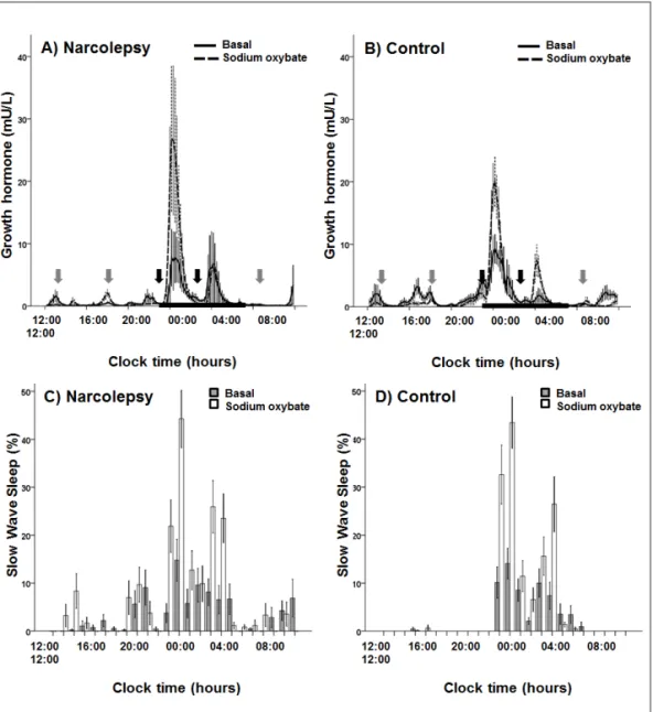
Figure 3.1 Mean serum growth hormone concentrations and slow-wave sleep in narcolepsy and
matched control subjects. Blood sampling started at 1200 and was continued at 10-min intervals for 24 h, whereas sleep electroencephalogram was recorded continuously. Sodium oxybate (SXB) administration induced an immediate rise in growth hormone levels in both narcolepsy patients (A)
and controls (b). Similarly, after 5 consecutive days of SXB treatment (including the 2nd sampling occasion), the percentage of slow-wave sleep had significantly increased in both narcolepsy patients
3
dIscussIon
We have shown that twice-a-night administration of SXB for 5 consecutive days consistently increases nocturnal GH secretion in both healthy controls and hypocretin-deficient narcolepsy patients. This was paralleled by a concomitant increase in SWS. Both the increase in GH and SWS were most prominent after the first dose of SXB at sleep onset. SXB reinforced the relation between GH and SWS, as evidenced by an almost doubled cross-correlation between the two.
GH secretion in narcolepsy has been the topic of a number of studies. Several groups found diminished or even unmeasurable GH concentrations around sleep onset.82;83;134 In contrast, we did not find lowered total 24-hour concentrations of GH in a previous study, but rather a more dispersed pattern with a shift towards daytime secretion.34 However, in both scenarios, SXB may partly restore the nocturnal GH ‘deficit’, by increasing nighttime GH secretion. The potency of SXB as a GH-secretagogue was previously shown in two daytime studies without sleep recordings,113;141 and more recently in a controlled single-dose study using repeated sampling together with sleep registrations in healthy young males.75 Even at the
Figure 3.2 Cross-correlation coefficient between growth hormone (GH) levels and slow-wave sleep
lowest dose (2.5 grams) a twofold increase in sleep-related GH secretion was observed. We confirmed and extended these observations, showing that a second nighttime dose may further enhance GH secretion, albeit to a lesser extent than the first dose. Furthermore, stimulation of GH secretion persists after repeated use, at least after 5 consecutive days. However, in controls we did not find a difference in 24 h GH secretion before and during SXB administration. Van Cauter, Gerra, and Takahara did find an increase in GH secretion after administration of a single dose of SXB in healthy controls as well as in narcoleptic patients.75;113;141 As a putative explanation of our findings, we believe that subchronic administration of SXB may elevate GH levels to induce feedback inhibition in controls, but not in narcoleptic patients, suggesting that narcolepsy does indeed disrupt normal control of GH release. Obviously, a single dose of SXB will not evoke such feedback inhibition, which explains the fact that other authors did not report a reduction of GH release in healthy humans. As SXB was well tolerated by subjects, this indeed suggests a potential for SXB as a strategy to counteract the relative growth hormone deficiency and sleep disturbances in the elderly, as was previously suggested.75;80
The close relation between sleep and the activity of the somatotropic axis has been known for a long time.142;143 There is a wealth of data supporting the hypothesis that this relation is brought about by the simultaneous promotion of sleep and GH release by GHRH.80;144;145 The mechanism through which SXB promotes GH secretion is unknown.75 Some researchers claim that SXB may exert its central nervous system effects through dedicated GHB-receptors in the brain, but the existence of these receptors has been disputed.146;147 There is clear evidence that SXB does modulate GABAergic tone through agonism of GABAB receptors, also in sleep-promoting regions of the hypothalamus.80;146 Our data showed that SXB further strengthened the relation between SWS and GH secretion, so its effect may be mediated by an increase in GHRH activity. SXB increased the regularity of GH secretion as well. This may imply that sodium oxibate simultaneously promotes endogenous somatostatin release, as negative feedback has been shown to increase secretory regularity.148 Although animal studies showed that hypocretin administration induced a dose-dependent reduction of GH concentrations in rats,106 the effects of SXB on GH secretion are unlikely to be mediated by altering hypocretin tone, as results were not different between controls and hypocretin-deficient patients.
3
studies.18;19;149 In fact, there often is a clear increase in body weight around the first onsetof symptoms of narcolepsy, especially excessive daytime sleepiness. Obesity in narcolepsy is not due to decreases in motor activity throughout the day.22;150 Furthermore, the total amount of calories consumed is not increased in narcolepsy.21 Basal metabolic rate has been studied by several groups, but inconsistent results have been reported.151;152 The same holds true for well-known endocrine factors regulating bodyweight, such as leptin.30-33 Obesity in narcolepsy is notoriously difficult to treat. This lends particular interest to a recent case series suggesting that SXB may decrease body weight in patients with narcolepsy.74 In 54 treated patients, the average reduction in body weight amounted to 3.4 kg. In the patients with cataplexy, the mean weight reduction was even larger: 5.1 kg. GH has a potent lipolytic activity, while GH deficiency leads to decreases in lean body mass and an increased fat mass.153;154 It is therefore tempting to speculate that the putative weight reducing effect of SXB is mediated by its stimulatory effect on the somatotropic axis.
We report a relatively low sleep efficiency in controls. It is conceivable that the laboratory setting disrupts sleep more than a natural environment. However the percentages of SWS and awekenings are comparable with earlier studies.75;155
Proper assessment of the secretion pattern of hormones that fluctuate during the day, requires repeated blood sampling over longer periods of time. Obviously, this complicates study design, and limits the number of subjects that can be included. Furthermore, five nights of SXB administration may not correctly reflect the long-term effects of SXB. Our results therefore need confirmation in future long-term studies. Nevertheless, our results suggest that future prospective long-term studies should especially focus on the effects of SXB on body weight, as this would provide a major improvement in the treatment of narcolepsy.
Acknowledgements
Plasma total ghrelin and leptin
levels in human narcolepsy and
matched healthy controls:
basal concentrations and
response to sodium oxybate
Claire E. H. M. Donjacour Daniel Pardi N. Ahmad Aziz Marijke Frölich Ferdinand Roelfsema Sebastiaan Overeem Hanno Pijl Gert Jan Lammers
Journal of Clinical Sleep Medicine 2013; 9(8):797-803.
AbstrAct
study objectives: Narcolepsy is caused by a selective loss of hypocretin neurons and is
associated with obesity. Ghrelin and leptin interact with hypocretin neurons to influence energy homeostasis. Here, we evaluated whether human hypocretin deficiency, or the narcolepsy therapeutic agent sodium oxybate (SXB), alter the levels of these hormones.
Methods: Eight male, medication free, hypocretin deficient, narcolepsy with cataplexy
patients, and 8 healthy controls matched for age, sex, body mass index (BMI), waist-to-hip ratio, and body fat percentage were assessed. Blood samples of total ghrelin and leptin were collected over 24 hours at 60 and 20-min intervals, respectively, during 2 study occasions: baseline, and during the last night of 5 consecutive nights of SXB administration (2 × 3.0 g/night).
results: At baseline, mean 24-h total ghrelin (936 ± 142 vs. 949 ± 175 pg/mL, P = 0.873)
and leptin (115 ± 5.0 vs. 79.0 ± 32 mg/L, P = 0.18) levels were not different between hypocretin deficient narcolepsy patients and controls. Furthermore, SXB did not significantly affect the plasma concentration of either one of these hormones.
conclusions: The increased BMI of narcolepsy patients is unlikely to be mediated by