Metformin Treatment Does Not Inhibit
Growth of Pancreatic Cancer Patient-Derived
Xenografts
Matthew B. Lipner1,2, Raoud Marayati2, Yangmei Deng2, Xianxi Wang2, Laura Raftery3,
Bert H. O’Neil4, Jen Jen Yeh1,2,5*
1Department of Pharmacology, School of Medicine, The University of North Carolina, Chapel Hill, NC, United States of America,2Lineberger Comprehensive Cancer Center, The University of North Carolina, Chapel Hill, NC, United States of America,3ProHealth Care Regional Cancer Center, Waukesha, WI, United States of America,4Department of Medicine, Division of Medical Oncology, University of Indiana, Indianapolis, IN, United States of America,5Department of Surgery, Division of Surgical Oncology and Endocrinology, The University of North Carolina, Chapel Hill, NC, United States of America
Abstract
There is currently tremendous interest in developing anti-cancer therapeutics targeting cell signaling pathways important for both cancer cell metabolism and growth. Several epidemi-ological studies have shown that diabetic patients taking metformin have a decreased inci-dence of pancreatic cancer. This has prompted efforts to evaluate metformin, a drug with negligible toxicity, as a therapeutic modality in pancreatic cancer. Preclinical studies in cell line xenografts and one study in patient-derived xenograft (PDX) models were promising, while recently published clinical trials showed no benefit to adding metformin to combination therapy regimens for locally advanced and metastatic pancreatic cancer. PDX models in which patient tumors are directly engrafted into immunocompromised mice have been shown to be excellent preclinical models for biomarker discovery and therapeutic develop-ment. We evaluated the response of four PDX tumor lines to metformin treatment and found that all four of our PDX lines were resistant to metformin. We found that the mechanisms of resistance may occur through lack of sustained activation of adenosine monophosphate-activated protein kinase (AMPK) or downstream reactivation of the mammalian target of rapamycin (mTOR). Moreover, combined treatment with metformin and mTOR inhibitors failed to improve responses in cell lines, which further indicates that metformin alone or in combination with mTOR inhibitors will be ineffective in patients, and that resistance to met-formin may occur through multiple pathways. Further studies are required to better under-stand these mechanisms of resistance and inform potential combination therapies with metformin and existing or novel therapeutics.
OPEN ACCESS
Citation:Lipner MB, Marayati R, Deng Y, Wang X, Raftery L, O’Neil BH, et al. (2016) Metformin Treatment Does Not Inhibit Growth of Pancreatic Cancer Patient-Derived Xenografts. PLoS ONE 11(1): e0147113. doi:10.1371/journal.pone.0147113
Editor:Marie-Josée Boucher, Université de Sherbrooke, CANADA
Received:July 25, 2014
Accepted:December 29, 2015
Published:January 13, 2016
Copyright:© 2016 Lipner et al. This is an open access article distributed under the terms of the Creative Commons Attribution License, which permits unrestricted use, distribution, and reproduction in any medium, provided the original author and source are credited.
Data Availability Statement:All relevant data are within the paper and its Supporting Information files.
Funding:This work was partially supported by the Lineberger Comprehensive Cancer Center (B.H.O.) and CA140424 and CA193650 (J.J.Y.) from the National Cancer Institute.
Introduction
Pancreatic cancer is one of the most aggressive and lethal malignancies, with 80% of patients presenting with locally advanced or metastatic disease that portends a 6–12 month median sur-vival and a dismal 6% five-year sursur-vival rate [1]. Chemotherapy produces only modest
improvements in survival, and novel therapies are desperately needed to improve treatment options for this large patient population [2]. There is currently tremendous interest in develop-ing anti-cancer therapeutics that target cell signaldevelop-ing pathways important in both cell metabo-lism and cell growth [3]. The 5' adenosine monophosphate-activated protein kinase (AMPK) pathway has gained increasing interest, as AMPK physiologically inhibits the mammalian tar-get of rapamycin (mTOR) to maintain homeostasis in conditions of decreased available cellular energy sources [4,5]. Studies have shown that mTOR signaling plays key roles in survival and proliferation of malignant cells [6,7]. Thus, AMPK activators have generated substantial inter-est as potential antineoplastic agents that function by altering metabolism and inhibiting the mTOR pathway [3].
Metformin is the first-line agent for treatment of type 2 diabetes mellitus. Metformin inhib-its mitochondrial oxidative phosphorylation, thereby increasing the ratio of AMP to ATP [8, 9]. High levels of AMP activate AMPK, which then inhibits energy-consuming pathways such as protein synthesis, in part by downregulating mTOR signaling by direct phosphorylation of the tumor suppressor TSC2 and the mTOR binding partner Raptor [9–13]. The state of energy conservation induced by metformin has been proposed to explain the cytostatic effect of met-formin on cancer [9] and the apparent protective effect observed in diabetic patients treated with metformin who subsequently develop pancreatic cancer [14].
Several epidemiological studies have indicated that patients with diabetes taking metformin have a decreased incidence of pancreatic cancer [14–17]. This has prompted a great deal of excitement to evaluate metformin, a widely used drug with negligible toxicity, as a therapeutic modality in pancreatic cancer. There are currently 3 clinical trials evaluating metformin in combination with various chemotherapies in pancreatic cancer (cancer.gov/clinicaltrials). Pre-clinical studies in cell line xenografts and one recent study in patient-derived xenograft (PDX) models have shown promise [18–22].
PDX models in which patient tumors are directly engrafted into immunocompromised mice have been shown to recapitulate primary tumor architecture and genetic characteristics, even after passaging and expanding the tumors in successive generations of mice [23,24]. Fur-thermore, PDX models are superior to traditional cell line xenografts, which are adapted to in vitro growth and lack the heterogeneity of patient tumors, for evaluating responses to therapies and novel biomarkers [23–27]. Until recently, there have been very limited studies of PDX responses to many proposed oncological agents, and results for metabolic therapies like met-formin are still severely lacking [27]. Thus, the objective of this study was to evaluate the response of pancreatic cancer PDX models to metformin and to investigate metformin’s mech-anism of action and compensatory resistance pathways.
Materials and Methods
Drugs and reagents
AMPKα(Thr172), AMPKα, AMPKα1, AMPKα2, phosphorylated mTOR (Ser2448), mTOR, phosphorylated p70S6K (Thr389), p70S6K, phosphorylated 4E-BP1 (Thr37/46), and 4E-BP1 were from Cell Signaling (Beverly, MA, USA). Anti-glyceraldehyde phosphate dehydrogenase (GAPDH) and horseradish peroxidase-conjugated goat anti-rabbit IgG were from Santa Cruz Biotechnology (Santa Cruz, CA, USA). Pierce1ECL Western Blotting Substrate was from Thermo Scientific (Rockford, IL, USA). Apo-ONE Homogeneous Caspase-3/7 assay kit was from Promega (Madison, WI, USA).
Cell culture and transduction with lentivirus
Pancreatic cancer cell lines Capan-2, CFPAC-1, HPAF-II, and SW1990 were obtained from the American Type Culture Collection (ATCC), authenticated via short–tandem repeat (STR) pro-filing (Genetica, Burlington, NC, USA), and tested negative for mycoplasma by indirect stain-ing. Cell lines were cultured in RPMI 1640 medium supplemented with 10% fetal bovine serum (FBS), 100 U/ml penicillin, and 100μg/ml streptomycin (Invitrogen, Carlsbad, California, USA) at 37°C in a humidified 5% CO2 atmosphere.
The puromycin domain of the AMPKα1–859 pLKO.1 reporter plasmid (generously donated by the laboratory of Channing Der, PhD at The University of North Carolina, Chapel Hill, NC, USA) was replaced with a blasticidin domain by restriction enzyme digestion with BamHI and KpmI. The second generation replication-incompetent lentivirus was generated in 293T cells with a four-plasmid system: the reporter plasmid, pMDL gag/pol RRE, pRSV-Rev, and pCMV VSV-G. For transduction with lentivirus, 1×10⁶CFPAC-1 and HPAF-II cells were seeded in 100 mm plates with lentivirus and a final polybrene concentration of 8μg/mL. After 24 hours, the medium was replaced, and the cells were cultured for another 4 days with 2μg/ml of puromycin or 10 days with 10μg/ml of blasticidin. The cells were trypsinized and analyzed by western blotting to determine gene knockdown.
The expression vector for myc-mTOR transient overexpression was obtained from Addgene (plasmid 1861, Cambridge, MA, USA). Transfection of 5x10⁵CFPAC-1 or HPAF-II cells was carried out with Lipofectamine 2000 (Invitrogen, Carlsbad, California, USA) using manufac-turer guidelines. Following 24 hour incubation, transfected cells were treated with 5 mM met-formin for an additional 24 hours, at which point cells were washed with PBS, harvested by scraping, and stored at−80°C until protein isolation.
MTT assay for cell proliferation
Western blot conditions
After the indicated time of incubation with metformin, cells were washed with PBS, harvested by scraping, and then lysed in 200μL RIPA buffer containing 50 mM Tris-HCl (pH 7.4), 150 mM NaCl, 1 mM EDTA, 1% Triton X, 1 mM NaF, and 0.25% Na deoxycholate and prote-ase inhibitors. Protein extracts (30μg) were electrophoresed on 10% SDS polyacrylamide gels and electrotransferred to polyvinylidene difluoride (PVDF) membranes. For determination of knockdown of AMPKα1 and AMPKα2, membranes were blocked with 5% non-fat dried milk in Tris-buffered saline and then incubated overnight at 4˚C with 1:1000 dilutions of anti-AMPKα1, anti-AMPKα2, and anti-AMPKαantibodies. For cell and tissue lysates isolated following metformin treatments, membranes were incubated at 4°C overnight with 1:1000 dilutions of phospho-mTOR, mTOR, phospho-p70S6K, p70S6K, anti-phospho-4E-BP1, anti-4E-BP1, anti-phospho-AMPKα, and anti-AMPK antibodies. Mem-branes were then washed and incubated with a 1:5000 dilution of horseradish peroxidase-conjugated goat anti-rabbit secondary antibody (Santa Cruz Biotechnology, Santa Cruz, CA, USA). Immunoreactive bands were detected by chemiluminescence using the Pierce1ECL Western Blotting Substrate. Intensity of each immunoreactive band was quantified by densi-tometry using Image J software (NIH, Bethesda, Maryland, USA), and expressed relative to the PBS-treated cells or mice. GAPDH was used to ensure equivalent protein loading. Statistical significance was determined using Student’st-tests for two sample comparisons and one-way ANOVA analysis with Dunnett’s multiple comparisons test for three or more sample comparisons.
PDX cohort expansion
Pancreatic ductal adenocarcinoma tissue from de-identified patients with localized pancreatic cancer who underwent curative surgical resection were obtained from the University of North Carolina Institutional Review Board (IRB) approved Tissue Procurement Facility after IRB approval (08–1153). Tumor tissue was engrafted subcutaneously into the flanks of NSG/NOD mice, expanded, and passaged over time, as described previously [28,29]. 7–8 week old Nu/nu mice with an average weight of 18–20 g were used in all experiments. All animal experiments were carried out in accordance with the U.S. National Institutes of Health (NIH) Guide for the Care and Use of Laboratory Animals under protocols approved by the University of North Carolina Institutional Animal Care and Use Committee (12–314).
Metformin treatment of PDX cohort
Results
Metformin does not inhibit growth of PDX tumors
We evaluated the response of four pancreatic cancer PDX tumor lines to metformin (200 and 400 mg/kg) for 28 days. These doses were chosen as higher doses have been shown to be neces-sary in mice to produce a decrease in blood glucose in diabetic animals [19,30,31]. In addition, higher doses of metformin (0.1% w/w) have been shown to increase the longevity of mice [32]. No tumor growth inhibition or regression was seen in any of the four PDX tumor lines at any time point measured (Fig 1). No change in body weights occurred over the course of the study and tumor architecture remained grossly unchanged following 28 days of treatment as assessed by hematoxylin and eosin staining (S1 Fig).
Activation of AMPK and inhibition of p70S6K phosphorylation in PDX
tumors is not sustained after a 28 day treatment with metformin
To determine whether metformin treatment altered AMPK and mTOR signaling, we evaluated all four PDX tumor lines for phosphorylation of AMPK (Thr172) and p70S6K (Thr389) at the end of the 28-day treatment (Fig 2A and 2BandS2 Fig). In this long-term treatment cohort, no
Fig 1. Metformin does not inhibit growth of PDX tumors.No significant growth inhibition was observed in four different pancreatic cancer PDX tumor lines at any time point during a 28 day treatment course with 200 mg/kg or 400 mg/kg metformin administered by daily oral gavage.
Fig 2. Activation of AMPK and inhibition of p70S6K phosphorylation in PDX tumors is not sustained after 28 days of metformin treatment.
Phosphorylation of AMPKαand p70S6K in (A) P505 and (B) P710 PDX tumors after 28 day treatment with 400 mg/kg metformin. Phosphorylation of AMPKα and p70S6K in (C) P505 and (D) P710 PDX tumors after 3 day treatment with 400 mg/kg metformin (*p<0.05).
change in phosphorylation of AMPK and p70S6K was seen in the metformin compared to the vehicle treated tumors. We then evaluated the effect of metformin on two PDX tumors after only 3 days of treatment. In contrast to the long-term treatment tumors, the short-term treat-ment tumors showed increased phosphorylation of AMPK and decreased phosphorylation of p70S6K (Fig 2C and 2D).
Metformin inhibits growth and alters AMPK and mTOR signaling in
pancreatic cancer cell lines
Since the lack of sustained response in our PDX models was surprising, we next examined the effects of metformin on the proliferation of four pancreatic cancer cell lines (Capan-2, CFPAC-1, HPAF-II, and SW1990). Metformin inhibited cell proliferation in a dose-dependent fashion in all four cell lines (Fig 3A). Metformin treatment activated AMPK as determined by phosphorylation of AMPK at Thr(172) in all cell lines tested, with a peak activation occurring at 4–8 hours after treatment (Fig 3B). Given that AMPK activation is known to inhibit mTOR, we further analyzed the effects of metformin treatment on the phosphorylation status of mTOR and its downstream targets p70S6K and 4E-BP1. Interestingly, we observed a delayed inhibition of mTOR and downstream target phosphorylation, with the nadir of observed phos-phorylation of p70S6K and 4E-BP1 occurring at 48 hours in both CFPAC-1 and HPAF-II cells (Fig 3C and 3D).
AMPK is only partially required for the anti-proliferative effect of
metformin
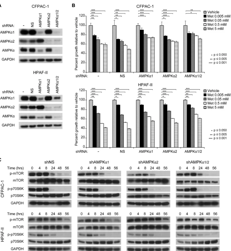
The anti-proliferative effect of metformin has been primarily attributed to its ability to activate the AMPK pathway. We hypothesized that the lack of sustained AMPK activation seen in all four PDX tumor lines may explain the lack of tumor growth response. Thus, we performed shRNA-induced knockdown of one or both of the catalytic subunits of AMPK in pancreatic cancer cell lines to determine whether the anti-proliferative effects of metformin would be affected. We found that knockdown of AMPKα1 and/or AMPKα2 partially but not completely reversed metformin’s ability to inhibit cell proliferation (Fig 4A and 4B).
Metformin appears to act on mTOR in an AMPK-independent manner
We next investigated whether the lack of sustained p70S6K inhibition seen in the P710 PDX tumor line despite evidence of some sustained AMPK activation may have been due to an AMPK-independent mechanism. We evaluated the effect of metformin on phosphorylation of mTOR and p70S6K following knockdown of AMPKα1 and/or AMPKα2. CFPAC-1 and HPAF-II cell lines with stable knockdown of NS, AMPKα1 and/or AMPKα2 (Fig 4A) were treated with 5 mM metformin (Fig 4B and 4C). In both cell lines, knockdown of one or both subunits did not rescue the ability of metformin to inhibit growth or phosphorylation of mTOR and p70S6K, suggesting that the ability of metformin to inhibit mTOR and p70S6K is at least partially independent of AMPK activation in these cell lines.
mTOR overexpression is sufficient to overcome the anti-proliferative
effects of metformin, but combinatorial treatment with metformin and
mTOR inhibitors does not produce synergy
determined whether mTOR activation alone was sufficient to abrogate the effects of metfor-min. We found that overexpression of mTOR using a myc-mTOR construct was sufficient to produce complete resistance growth inhibition and to limit the decrease in p70S6K phosphory-lation following metformin treatment (Fig 5A and 5B). To determine whether combining met-formin with targeted mTOR inhibitors may be a logical therapeutic strategy, we treated cell lines with constant-ratio doses of metformin and either the allosteric mTOR inhibitor
Fig 3. Metformin inhibits growth and alters AMPK and mTOR signaling in pancreatic cancer cell lines.(A) Cells were plated in quadruplicate into 96-well plates at a density of 5x10³per well, incubated overnight, and then treated with media containing either PBS as a vehicle control or different concentrations of metformin (0–5 mM). After 48 hours, proliferation indices were determined using the MTT assay and normalized to those of the vehicle-treated cells. All assays were performed in triplicate. (B) Phosphorylation of AMPKαand total AMPKαat various time points after treatment with 5 mM metformin. Treatment began at 0 hours (hrs). (C) Phosphorylation of mTOR, p70S6K, and 4E-BP1 at various time points after treatment with 5 mM metformin. (D) Densitometry of phosphorylated AMPKα, mTOR, p70S6K, and 4E-BP1 relative to total levels shown in (B) and (C).
Fig 4. AMPK is only partially required for the anti-proliferative effects of metformin.(A) shRNA knockdown of AMPKαsubunits in CFPAC-1 and HPAF-II cell lines. (B) Proliferation of CFPAC-1 and HPAF-II cell lines with stable knockdown of AMPKαsubunits after treatment with different concentrations of metformin (0–5 mM). (C) Phosphorylation of mTOR and p70S6K in CFPAC-1 and HPAF-II cell lines with stable knockdown of AMPKαsubunits.
rapamycin or the catalytic mTOR inhibitor BEZ235, which are known to inhibit pancreatic cancer cell line growth. Growth inhibition was not enhanced at any dose combination relative to single drug treatment, and no synergism between metformin and either mTOR inhibitor was calculated at any dose using the Chou-Talalay median effect equation (Fig 5C and 5D).
Fig 5. mTOR expression is sufficient to promote resistance to metformin in cell lines, but combinatorial treatment with metformin and mTOR inhibitors does not produce synergy.(A) Phosphorylation of p70S6K in and (B) proliferation of HPAF-II cells after treatment with 5 mM metformin following transient expression of a transfected myc-mTOR construct. Using the median effect equation calculation for combination index (CI) following 3 day treatment with constant-ratio doses of metformin and either (C) the allosteric mTOR inhibitor rapamycin or (D) the catalytic mTOR inhibitor BEZ235 failed to produce synergy (CI<1) at any dose combination. CI values at 50% growth inhibition: (C) CFPAC-1 1.54, HPAF-II 1.43; (D) CFPAC-1 1.30, HPAF-II 1.29. (*p<0.050, **p<0.005,***p<0.001).
Discussion
Epidemiological studies in diabetic patients have found that patients treated with metformin have a decreased incidence of multiple cancers, including pancreatic cancer [14–17]. Several preclinical studies of metformin as an anti-cancer therapeutic have been promising, demon-strating impressive tumor growth inhibition [20,22,33] and apoptosis [34] of pancreatic can-cer cell lines. However, cell line xenografts have been generally unreliable predictors of drug responses in humans. Lonardo et al. evaluated the effect of metformin on four pancreatic can-cer PDX tumor lines and, similar to previous cell line xenograft studies, found substantial growth inhibition [21]. In contrast, emerging clinical trials evaluating metformin in pancreatic cancer have tempered the optimism created by this preclinical work. A double-blind, random-ized, placebo-controlled phase II trial evaluating metformin in combination with gemcitabine and erlotinib in patients with advanced pancreatic cancer showed no difference in outcome as a result of metformin treatment [35]. Another phase II trial combining metformin with pacli-taxel in patients with gemcitabine-refractory disease failed to meet its primary endpoint of dis-ease control rate [36].
In this study, we observed a uniform lack of response to metformin in the four PDX tumor lines that we evaluated. Several potential reasons exist for the disparate results between previ-ous preclinical work compared to our study and recent clinical trials. First, PDX tumors are inherently highly heterogeneous because PDX tumors are passaged in bulk and are representa-tive‘biopsies’of similarly heterogeneous source patient tumors. Second, although the dose range in our study overlaps with previous studies, the pharmacokinetics of metformin uptake are still unclear [37]. Tumor microenvironment and stromal content appear to influence met-formin’s access to tumor cells, which may lead to different outcomes between studies [21]. Third, the tumor volume at which treatment was initiated varies between studies, which may affect tumor composition, specifically the cancer stem cell burden. Lonardo et al. and others have shown that only this stem cell subpopulation undergoes apoptosis as a result of metfor-min treatment, while the vast majority of tumor cells experience reversible growth arrest [18, 21]. Interestingly, although Lonardo et al. found that metformin was able to initially slow PDX tumor growth, they noted that all PDX tumors eventually progressed on therapy [21], suggest-ing that metformin monotherapy will not be effective in patients.
silencing of AMPKα1. In addition, metformin treatment was able to attenuate proliferation of both AMPKα1/2 wild-type and AMPKα1/2 deficient mouse embryonic fibroblasts (MEFs), although AMPKα1/2 deficient MEFs were slightly less sensitive to metformin [43]. In breast cancer cell lines, inhibition of HER2 by metformin was found to be completely AMPK-inde-pendent [44]. Recent studies in pancreatic cancer cell lines found that metformin may inhibit growth independently of AMPK through upregulation of miR-26a [45], while metformin’s effects on pancreatic cancer stem cells may be mediated through reexpression of specific miR-NAs [18]. These results suggest that modulation of miRNA expression may be yet another important mechanism underlying the biological effects of metformin.
Taken together with the above studies, our results that knockdown of AMPK subunits did not rescue the inhibitory effects of metformin on mTOR/p70S6K phosphorylation but that mTOR reexpression was able to reverse the anti-proliferative effects of metformin, suggest that the activation of AMPK and inhibition of the mTOR/p70S6K pathway by metformin are inde-pendent events that may both contribute to cancer cell growth inhibition. Moreover, regulation of AMPK by metformin may be cell type dependent, and in pancreatic cancer, the anti-prolifer-ative effects of metformin may be partially or largely AMPK-independent.
Overall, our study shows that although metformin inhibits pancreatic cancer cell line prolif-eration, its effect on patient tumors will likely be transient and much more complex. While resistance mechanisms likely involve mTOR pathway activation, simultaneous treatment with metformin and mTOR inhibitors may do little to enhance the efficacy of either therapy. Fur-ther studies are needed in order to determine wheFur-ther metformin may someday provide benefit to pancreatic cancer patients by leveraging its complex metabolic and signaling effects in com-bination with chemotherapeutics or targeted therapies.
Supporting Information
S1 Fig. Long-term metformin treatment does not affect mouse weight or tumor histology.
(A) Weights of mice over a 28 day treatment course with either 200 mg/kg or 400 mg/kg met-formin shown relative to baseline weights. Hematoxylin and eosin staining of (B) P505 and (C) P710 patient-derived xenograft tumors following 28 day treatment with vehicle (left panels) or 400 mg/kg metformin (right panels) show no difference in tumor architecture, ductal forma-tion, or stromal content. Scale bars are 300μm.
(TIF)
S2 Fig. Activation of AMPK and inhibition of p70S6K phosphorylation in PDX tumors is not sustained after 28 days of metformin treatment.Phosphorylation of AMPKαand p70S6K in (A) P722 and (B) PT4 PDX tumors after 28 day treatment with 400 mg/kg metfor-min.
(TIF)
Acknowledgments
The authors thank Charlene M. Santos at the University of North Carolina (UNC) Lineberger Comprehensive Cancer Center Animal Studies Core, the UNC PDX Program, the UNC Tissue Procurement Facility, and the UNC Translational Pathology Laboratory for technical assistance.
Author Contributions
References
1. Jemal A, Siegel R, Xu J, Ward E (2010) Cancer statistics, 2010. CA: a cancer journal for clinicians 60: 277–300.
2. Chaulagain CP, Ng J, Wazer D, Saif MW (2012) Adjuvant therapy of pancreatic cancer. JOP: Journal of the pancreas 13: 349–353. doi:10.6092/1590-8577/935PMID:22797387
3. Vakana E, Altman JK, Platanias LC (2012) Targeting AMPK in the treatment of malignancies. Journal of cellular biochemistry 113: 404–409. doi:10.1002/jcb.23369PMID:21928327
4. Inoki K, Kim J, Guan KL (2012) AMPK and mTOR in cellular energy homeostasis and drug targets. Annual review of pharmacology and toxicology 52: 381–400. doi: 10.1146/annurev-pharmtox-010611-134537PMID:22017684
5. Carling D, Mayer FV, Sanders MJ, Gamblin SJ (2011) AMP-activated protein kinase: nature's energy sensor. Nature chemical biology 7: 512–518. doi:10.1038/nchembio.610PMID:21769098
6. Bjornsti MA, Houghton PJ (2004) The TOR pathway: a target for cancer therapy. Nature reviews Can-cer 4: 335–348. PMID:15122205
7. Sabatini DM (2006) mTOR and cancer: insights into a complex relationship. Nature reviews Cancer 6: 729–734. PMID:16915295
8. Hardie DG, Ross FA, Hawley SA (2012) AMPK: a nutrient and energy sensor that maintains energy homeostasis. Nature reviews Molecular cell biology 13: 251–262. doi:10.1038/nrm3311PMID: 22436748
9. Pollak MN (2012) Investigating metformin for cancer prevention and treatment: the end of the begin-ning. Cancer discovery 2: 778–790. doi:10.1158/2159-8290.CD-12-0263PMID:22926251
10. Bolster DR, Crozier SJ, Kimball SR, Jefferson LS (2002) AMP-activated protein kinase suppresses pro-tein synthesis in rat skeletal muscle through down-regulated mammalian target of rapamycin (mTOR) signaling. The Journal of biological chemistry 277: 23977–23980. PMID:11997383
11. Hardie DG (2008) AMPK and Raptor: matching cell growth to energy supply. Mol Cell 30:263–265. doi: 10.1016/j.molcel.2008.04.012PMID:18471972
12. Gwinn DM, Shackelford DB, Egan DF, Mihaylova MM, Mery A, Vasquez DS, et al. (2008) AMPK phos-phorylation of raptor mediates a metabolic checkpoint. Mol Cell 30:214–226. doi:10.1016/j.molcel. 2008.03.003PMID:18439900
13. Shaw RJ (2009) LKB1 and AMP-activated protein kinase control of mTOR signaling and growth. Acta Physiol (Oxf) 196:65–80
14. Sadeghi N, Abbruzzese JL, Yeung SC, Hassan M, Li D (2012) Metformin use is associated with better survival of diabetic patients with pancreatic cancer. Clinical cancer research: an official journal of the American Association for Cancer Research 18: 2905–2912.
15. Evans JM, Donnelly LA, Emslie-Smith AM, Alessi DR, Morris AD (2005) Metformin and reduced risk of cancer in diabetic patients. The BMJ 330: 1304–1305. PMID:15849206
16. Libby G, Donnelly LA, Donnan PT, Alessi DR, Morris AD, and Evans JM (2009) New users of metformin are at low risk of incident cancer: a cohort study among people with type 2 diabetes. Diabetes care 32: 1620–1625. doi:10.2337/dc08-2175PMID:19564453
17. Soranna D, Scotti L, Zambon A, Bosetti C, Grassi G, Catapano A, et al. (2012) Cancer risk associated with use of metformin and sulfonylurea in type 2 diabetes: a meta-analysis. The oncologist 17: 813– 822. doi:10.1634/theoncologist.2011-0462PMID:22643536
18. Bao B, Wang Z, Ali S, Ahmad A, Azmi AS, Sarkar SH, et al. (2012) Metformin inhibits cell proliferation, migration and invasion by attenuating CSC function mediated by deregulating miRNAs in pancreatic cancer cells. Cancer prevention research 5: 355–364. doi:10.1158/1940-6207.CAPR-11-0299PMID: 22086681
19. Kim YD, Park KG, Lee YS, Park YY, Kim DK, Nedumaran B, et al. (2008) Metformin inhibits hepatic glu-coneogenesis through AMP-activated protein kinase-dependent regulation of the orphan nuclear receptor SHP. Diabetes 57: 306–314. PMID:17909097
20. Kisfalvi K, Eibl G, Sinnett-Smith J, Rozengurt E (2009) Metformin disrupts crosstalk between G protein-coupled receptor and insulin receptor signaling systems and inhibits pancreatic cancer growth. Cancer research 69: 6539–6545. doi:10.1158/0008-5472.CAN-09-0418PMID:19679549
21. Lonardo E, Cioffi M, Sancho P, Sanchez-Ripoll Y, Trabulo SM, Dorado J, et al. (2013) Metformin targets the metabolic achilles heel of human pancreatic cancer stem cells. PLoS One 8: e76518. doi:10.1371/ journal.pone.0076518PMID:24204632
23. Rubio-Viqueira B, Jimeno A, Cusatis G, Zhang X, Iacobuzio-Donahue C, Karikari C, et al. (2006) An in vivo platform for translational drug development in pancreatic cancer. Clin Cancer Res 12: 4652–4661. PMID:16899615
24. Tentler JJ, Tan AC, Weekes CD, Jimeno A, Leong S, Pitts TM, et al. (2012) Patient-derived tumour xenografts as models for oncology drug development. Nat Rev Clin Oncol 9: 338–350. doi:10.1038/ nrclinonc.2012.61PMID:22508028
25. Rubio-Viqueira B, Hidalgo M (2009) Direct in vivo xenograft tumor model for predicting chemotherapeu-tic drug response in cancer patients. Clin Pharmacol Ther 85: 217–221. doi:10.1038/clpt.2008.200 PMID:19005462
26. Hidalgo M, Amant F, Biankin AV, Budinska E, Byrne AT, Caldas C, et al. (2014) Patient-derived xeno-graft models: an emerging platform for translational cancer research. Cancer Discov. 4: 998–1013. doi: 10.1158/2159-8290.CD-14-0001PMID:25185190
27. Gao H, Korn JM, Ferretti S, Monahan JE, Wang Y, Signh M, et al. (2015) High-throughput screening using patient-derived tumor xenografts to predict clinical trial drug response. Nature medicine 21: 1318–1325. doi:10.1038/nm.3954PMID:26479923
28. Neel NF, Stratford JK, Shinde V, Ecsedy JA, Martin TD, Der CJ, Yeh JJ (2013) Response to MLN8237 in Pancreatic Cancer Is Not Dependent on RalA Phosphorylation. Molecular cancer therapeutics 13: 122–33. doi:10.1158/1535-7163.MCT-12-1232PMID:24222664
29. Torphy RJ, Tignanelli CF, Kamande JW, Moffitt RA, Herrera Loeza SG, Soper SA, Yeh JJ (2014) Circu-lating tumor cells as a biomarker of response to treatment in patient-derived xenograft mouse models of pancreatic adenocarcinoma. PLoS One 9: e89474. doi:10.1371/journal.pone.0089474PMID: 24586805
30. Foretz M, Hebrard S, Leclerc J, Zarrinpashneh E, Soty M, Mithieux G, et al. (2010) Metformin inhibits hepatic gluconeogenesis in mice independently of the LKB1/AMPK pathway via a decrease in hepatic energy state. The Journal of clinical investigation 120: 2355–2369. doi:10.1172/JCI40671PMID: 20577053
31. Wilcock C, Bailey CJ (1990) Sites of metformin-stimulated glucose metabolism. Biochemical pharma-cology 39: 1831–1834. PMID:2111705
32. Martin-Montalvo A, Mercken EM, Mitchell SJ, Palacios HH, Mote PL, Scheibye-Knudsen M, et al. (2013) Metformin improves healthspan and lifespan in mice. Nature communications 4: 2192. doi:10. 1038/ncomms3192PMID:23900241
33. Kawanami T, Takiguchi S, Ikeda N, Funakoshi A (2012) A humanized anti-IGF-1R monoclonal antibody (R1507) and/or metformin enhance gemcitabine-induced apoptosis in pancreatic cancer cells. Oncol-ogy reports 27: 867–872. doi:10.3892/or.2011.1597PMID:22200743
34. Feng YH, Velazquez-Torres G, Gully C, Chen J, Lee MH, Yeung SC (2011) The impact of type 2 diabe-tes and antidiabetic drugs on cancer cell growth. Journal of cellular and molecular medicine 15: 825– 836. doi:10.1111/j.1582-4934.2010.01083.xPMID:20455996
35. Kordes S, Pollak MN, Zwinderman AH, Mathot RA, Weterman MJ, Beeker A, et al. (2015) Metformin in patients with advanced pancreatic cancer: a double-blind, randomised, placebo-controlled phase 2 trial. Lancet Oncology 16: 839–847. doi:10.1016/S1470-2045(15)00027-3PMID:26067687
36. Braghiroli MI, de Celis Ferrari AC, Pfiffer TE, Alex AK, Nebuloni D, Carneiro AS, et al. (2015) Phase II trial of metformin and paclitaxel for patients with gemcitabine-refractory advanced adenocarcinoma of the pancreas. Ecancermedicalscience 11: 563.
37. Pollak M (2012) Metformin and pancreatic cancer: a clue requiring investigation. Clinical cancer research: an official journal of the American Association for Cancer Research 18: 2723–2725.
38. Dowling RJ, Zakikhani M, Fantus IG, Pollak M, Sonenberg N (2007) Metformin inhibits mammalian tar-get of rapamycin-dependent translation initiation in breast cancer cells. Cancer research 67: 10804– 10812. PMID:18006825
39. Zakikhani M, Dowling R, Fantus IG, Sonenberg N, Pollak M (2006) Metformin is an AMP kinase-depen-dent growth inhibitor for breast cancer cells. Cancer research 66: 10269–10273. PMID:17062558
40. Gotlieb WH, Saumet J, Beauchamp MC, Gu J, Lau S, Pollak MN, Bruchim I (2008) In vitro metformin anti-neoplastic activity in epithelial ovarian cancer. Gynecologic oncology 110: 246–250. doi:10.1016/ j.ygyno.2008.04.008PMID:18495226
41. Ben Sahra I, Laurent K, Loubat A, Giorgetti-Peraldi S, Colosetti P, Auberger P, et al. (2008) The antidia-betic drug metformin exerts an antitumoral effect in vitro and in vivo through a decrease of cyclin D1 level. Oncogene 27: 3576–3586. doi:10.1038/sj.onc.1211024PMID:18212742
43. Rattan R, Giri S, Hartmann LC, Shridhar V (2011) Metformin attenuates ovarian cancer cell growth in an AMP-kinase dispensable manner. Journal of cellular and molecular medicine 15: 166–178. doi:10. 1111/j.1582-4934.2009.00954.xPMID:19874425
44. Vazquez-Martin A, Oliveras-Ferraros C, Menendez JA (2009) The antidiabetic drug metformin sup-presses HER2 (erbB-2) oncoprotein overexpression via inhibition of the mTOR effector p70S6K1 in human breast carcinoma cells. Cell cycle 8: 88–96. PMID:19106626