PHYSIOLOGICALLY-BASED PHARMACOKINETICS IN CRITICALLY ILL CHILDREN
Kevin M. Watt
A dissertation submitted to the faculty of the University of North Carolina at Chapel Hill in partial fulfillment of the requirements for the degree of Doctor of Philosophy in the Department of Pharmaceutical Sciences in the UNC Eshelman School of Pharmacy
Chapel Hill 2016
Approved by: Kim L.R. Brouwer Dhiren Thakker Julie Dumond
Michael Cohen-Wolkowiez
© 2016 Kevin M. Watt
iii ABSTRACT
Kevin M. Watt: Physiologically-based Pharmacokinetics in Critically Ill Children (Under the direction of Kim L.R. Brouwer)
Extracorporeal membrane oxygenation (ECMO) is used to support cardiorespiratory failure in critically ill infants, children, and adults. In these
vulnerable populations, the effect of ECMO on drug disposition leaves clinicians with uncertainty about dosing. The goal of this dissertation research was to develop a physiologically-based pharmacokinetic (PBPK) modeling approach that translated results from ex vivo ECMO studies to bedside dosing recommendations. To
dosing recommendations showed good agreement with recommendations based on the Fluconazole ECMO PK Trial. The micafungin ECMO PBPK Model also over-predicted exposure (1.16 fold error), but, again, dosing recommendations were in close agreement with recommendations determined from the trial. The two clinical PK trials of fluconazole and micafungin in children on ECMO were performed in parallel with the PBPK model building. Both the Fluconazole and Micafungin ECMO PK Trials showed that exposure was significantly lower in children on ECMO
iv
ACKNOWLEDGMENTS
I wish to acknowledge and thank all of my committee members (Drs. Kim Brouwer, Danny Benjamin, Dhiren Thakker, Julie Dumond, Jeff Barrett, and Micky Cohen-Wolkowiez) for providing me with guidance and support during my Ph.D. degree. I especially want to thank Dr. Brouwer for welcoming me into her lab, being enthusiastic about pediatric research, and tolerating my clinical schedule.
The PBPK modeling would not have been possible without the mentorship from Jeff Barrett, Andrea Edginton, Ping Zhao, and the folks at Bayer Technology Services, especially Tobias Kanacher, Martin Hobe, and Michael Sevestre. I
especially want to single out Andrea Edginton for her help in developing the ECMO compartment and troubleshooting when the model did not work.
vi
Additionally I need to acknowledge many others that have taught me a great deal: Danny Gonzalez, Christoph Hornik, Dan Crona, Nicole Zane, Mary Paine, Heyward Hull, and Edmund Capparelli.
I wish to thank the Thrasher Research Fund for my Early Career Award and the NICHD at the National Institutes of Health for my Loan Repayment and K23 Career Development Awards.
TABLE OF CONTENTS
List of tables ... x
List of figures ... xii
Abbreviations ... xiv
Chapter 1. Introduction: Altered Pharmacokinetics due to Extracorporeal Life Support .... 1
Chapter 2. Antifungal Extraction by the Extracorporeal Membrane Oxygenation (ECMO) Circuit Ex Vivo ... 23
Introduction ... 23
Methods ... 25
Results ... 29
Discussion ... 32
Chapter 3. Pharmacokinetics of Fluconazole in Children Supported with Extracorporeal Membrane Oxygenation ... 50
Part 1. Pharmacokinetics and Safety of Fluconazole in Young Infants Supported with Extracorporeal Membrane Oxygenation ... 50
Introduction ... 50
Methods ... 51
Results ... 56
Discussion ... 58
Part 2. Fluconazole Population Pharmacokinetics and Dosing for Prevention and Treatment of Invasive Candidiasis in Children Supported with Extracorporeal Membrane Oxygenation ... 74
Introduction ... 74
Methods ... 75
Results ... 81
Discussion ... 87
Chapter 4. Physiologically-Based Pharmacokinetics of Fluconazole in Children on ECMO ... 105
viii
Methods ... 107
Results ... 115
Discussion ... 117
Chapter 5. Pharmacokinetics and Safety of Micafungin in Infants Supported with Extracorporeal Membrane Oxygenation (ECMO) ... 146
Introduction ... 146
Methods ... 147
Results ... 152
Discussion ... 155
Chapter 6. Physiologically-Based Phamacokinetics of Micafungin in Children on ECMO……. ... 172
Introduction ... 172
Methods ... 174
Results ... 183
Discussion ... 186
Chapter 7. Summary and Future Directions ... 220
Appendices ... 243
Appendix 1. Ex vivo data ... 243
Appendix 2. Fluconazole data ... 265
Appendix 3. Micafungin data ... 331
LIST OF TABLES
2.1. Antifungal drug physicochemical properties and clearance pathways ... 38
2.2. ECMO circuit components ... 38
2.3. Number of circuits by configuration and drug ... 39
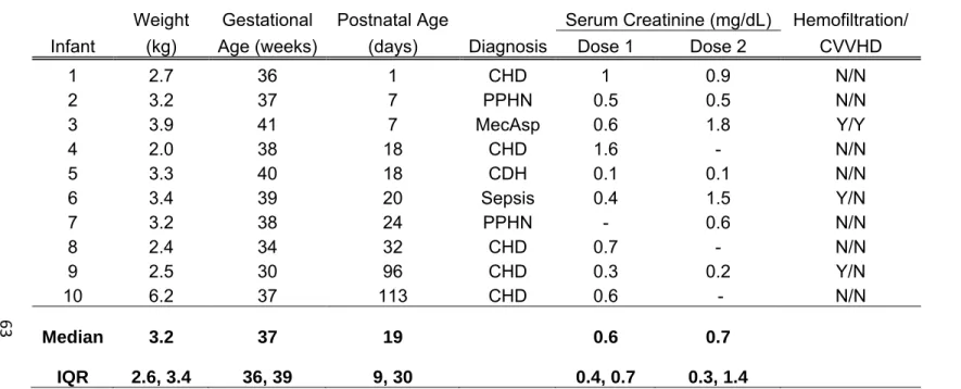
3.1.1. Demographics ... 63
3.1.2. Pharmacokinetic indices ... 64
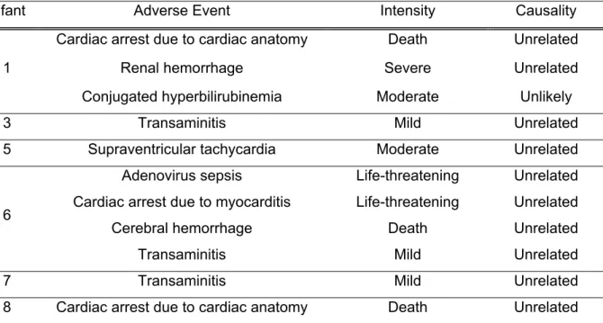
3.1.3. Adverse events ... 65
3.1.4. Pharmacokinetic Indices in Infants on ECMO after the First Dose of Intravenous Fluconazole 25 mg/kg Compared with Historical Controls not on ECMO who Received 1 Dose of Intravenous Fluconazole 25 mg/kg ... 66
3.2.1. Clinical data ... 91
3.2.2. Population PK model development ... 92
3.2.3. Final population PK model parameter estimates ... 94
3.2.4. Bayesian estimates of V and CL overall and by age group based on ECMO ... 95
3.2.5. Exposure in children on ECMO after different simulated dosing regimens ... 96
4.1. Fluconazole physicochemical properties and elimination pathways ... 122
4.2. Assumptions used in the model building process ... 123
4.3. Studies used in model development and evaluation ... 124
4.4. Observed versus PBPK Predicted AUCs for adult studies used in model development and validation. ... 125
5.1. Clinical Characteristics ... 162
5.2. Pharmacokinetic Parameters ... 164
6.1. Micafungin physicochemical properties and elimination pathways ... 193
6.2. Studies used in model development and evaluation ... 194
6.3. Assumptions used in the model building process ... 195
x
6.5. Observed versus predicted area under the concentration time curve for a 24 hour
LIST OF FIGURES
1.1. ECMO Circuit Schematic ... 15
1.2. PBPK Model Structure ... 16
2.1. Molecular Structure of Antifungals ... 40
2.2. ECMO circuit configurations. ... 41
2.3. Recovery by circuit configuration for each drug. ... 42
2.4. Micafungin recovery stratified by albumin concentration. ... 45
3.1.1. Fluconazole concentration time profiles ... 67
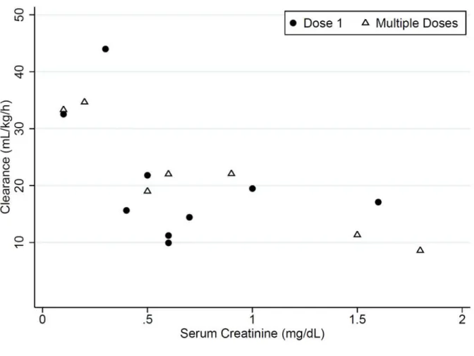
3.1.2. Serum creatinine versus clearance ... 69
3.1.3. Fluconazole exposure in the first 24 hours (AUC0–24) after dose 1 and multiple doses .... 70
3.2.1. Final population PK model diagnostic plots ... 97
3.2.2. Visual predictive check ... 98
3.2.3. Simulated fluconazole plasma concentrations and exposure ... 99
4.1. PBPK model structure ... 126
4.2. PBPK workflow ... 127
4.3. Adult optimized model. PBPK model predictions ... 128
4.4. Adult model validation. ... 129
4.5. Pediatric PBPK Model predictions. ... 131
4.6. Pediatric PBPK Model AUC0-24 predicted versus observed. ... 132
4.7. ECMO PBPK Model development. ... 133
4.8. ECMO PBPK Model AUC0-24 predicted versus observed. ... 136
4.9. ECMO PBPK Model-predicted optimized fluconazole dosing and exposure in children on ECMO across the pediatric age spectrum. ... 137
4.10. ECMO PBPK Model-predicted fluconazole dosing and exposure in children on ECMO across the pediatric age spectrum. ... 138
xii
5.2. Micafungin concentration-time profiles ... 166
5.3. Micafungin exposure ... 167
5.4. Pharmacokinetic parameters vs covariates ... 168
6.1. PBPK model structure ... 198
6.2. PBPK workflow ... 199
6.3. ECMO compartment extraction calculations ... 200
6.4. Adult PBPK Model development ... 201
6.5. Adult model validation ... 206
6.6. Pediatric PBPK Model predictions versus observed data for children (2-<18y) ... 208
6.7. Pediatric PBPK Model predictions versus observed data for infants (0-2y) ... 209
6.8. ECMO PBPK Model development ... 210
6.9. ECMO PBPK Model-predicted versus observed ... 213
6.10. ECMO PBPK Model-predicted optimized micafungin dosing and exposure in children on ECMO across the pediatric age spectrum... 214
LIST OF ABBREVIATIONS AAG Alpha-1-acid glycoprotein
ADME Absorption, distribution, metabolism, elimination
AE Adverse event
ALT Alanine aminotransferase AST Aspartate aminotransferase AUC Area under the curve
Cbc Concentration in blood cells Cin Concentration coming in Clast Last measured concentration Cmax Maximum concentration Cmin Minimum concentration
Cout Concentration going out Cpls Concentration in plasma
CDH Congenital diaphragmatic hernia CHD Congenital heart disease
CI Confidence interval
CL Clearance
CVVH Continuous veno-venous hemofiltration CVVHD Continuous veno-venous hemodialysis
CYP Cytochrome P450
ECMO Extracorporeal membrane oxygenation EDTA Ethylenediaminetetraacetic acid
xiv
IDSA Infectious Disease Society of America IQR Interquartile range
IV Intravenous k Elimination rate constant MecAsp Meconium aspiration
MIC Minimum inhibitory concentration OATP Organic anion-transporting polypeptide
OSF Ontogeny scaling factor
PBPK Physiologically-based pharmacokinetics PD Pharmacodynamics
PK Pharmacokinetics
PNA Postnatal age
PPHN Persistent pulmonary hypertension Q Flow
SCR Serum creatinine
t½ Half-life
Tlast Time at last measurement
tMIC Time above minimum inhibitory concentration UGT UDP-Glucuronosyltransferase
V Volume of distribution VA Venoarterial VPC Visual predictive check
CHAPTER 1. INTRODUCTION: ALTERED PHARMACOKINETICS DUE TO EXTRACORPOREAL LIFE SUPPORT
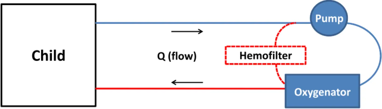
Extracorporeal membrane oxygenation (ECMO) is life-saving in patients with cardiorespiratory failure. ECMO is a cardiopulmonary bypass device that provides complete respiratory and cardiac support (Figure 1.1). Mechanically, blood is drained from the venous system, pumped through an artificial lung membrane, and then
returned to either the venous or arterial circulation. ECMO was first used successfully in 1972 to support an adult with respiratory failure and three years later was used
2 The ECMO Circuit
ECMO support comes in two forms: veno-venous (VV) and veno-arterial (VA). VV ECMO provides pulmonary support and VA ECMO provides both pulmonary and cardiac support. In VV ECMO, deoxygenated blood is drained from the venous system, pumped through the oxygenator where oxygen is added and carbon dioxide removed, and then returned to the venous system. From the venous circulation, this oxygenated blood then drains into the heart where native cardiac function distributes it to the arterial circulation. In VA ECMO, deoxygenated blood is also drained from the venous system and pumped through the oxygenator but returned to the arterial circulation, completely bypassing the heart and lungs. Oxygenated blood is distributed to the arterial circulation via non-pulsatile flow by the ECMO pump.
ECMO oxygenators employ a gas-permeable membrane with a countercurrent gas mixture (sweep gas) flowing on one side of the membrane and the patient’s blood on the other.23 Gas diffuses down a concentration gradient with oxygen crossing the membrane into the blood and carbon dioxide diffusing from blood into the sweep gas. Early oxygenators had a silicone membrane, but high resistance to permeating gases necessitated a large surface area to ensure adequate oxygenation and ventilation. To enhance gas transfer, microporous hollow-fiber membranes were developed. However, these membranes were plagued by plasma leakage across the membrane that resulted in an oxygenator lifespan of 2-3 days. Oxygenators in use today employ a
non-microporous, hollow-fiber, polymethylpentane membrane. These devices are low resistance, require much smaller surface area, do not suffer from plasma leakage, and have a lifespan measured in weeks. The type and amount of material to which the blood is exposed can have a substantial impact on drug interaction with the ECMO circuit.
Pharmacokinetic changes during ECMO support
While ECMO allows for the survival of a very critically ill subset of patients, it also presents new challenges related to their management. One of these challenges is understanding and appropriately compensating for the effect of ECMO on drug
4 Direct extraction by the circuit
Drug extraction by the ECMO circuit is a well-recognized contributor to PK alterations across all patient populations that depends on both the circuit component materials and the drug physicochemical properties. The interaction between the drug and the ECMO circuit has been studied primarily in ex vivo experiments in which drug is administered to isolated ECMO circuits.24-33 Because there is no human attached to the circuit, any change in concentration over time is due to extraction by the circuit or drug degradation. In general, ex vivo studies have demonstrated increased extraction of highly lipophilic and protein bound drugs.28,31,32
However, the degree of extraction, even for the same drug, can be markedly affected by circuit materials. A study of fentanyl (LogP 4, protein binding 80%) in an ECMO circuit using a silicone membrane oxygenator showed >99% fentanyl loss in 180 minutes.28 When the same experiment was repeated in a microporous, hollow-fiber polypropylene oxygenator, fentanyl loss was only 66% at 180 minutes.28 Another study compared the effect of six different coatings on drug extraction by the PVC tubing by administering fentanyl to circuits constructed with only a pump and tubing (no
oxygenator).34,35 In the uncoated circuit, fentanyl loss at 120 minutes was 80%.34 In the coated tubing, fentanyl loss at 120 minutes ranged from 40-75%.35
Drug extraction by the circuit is likely due to non-specific adsorption,33-37
hydrophobic interactions. Interactions with polymers such as those used in ECMO circuit equipment tend to be due to hydrophobic adsorption (i.e., hydrophobic drugs adsorb to hydrophobic alkyl groups on polymers). Electrostatic interactions tend to dominate when surface coatings are applied to ECMO circuit components. As noted above, surface coatings are applied to ECMO circuit components to minimize the inflammatory response triggered when blood comes into contact with a foreign
material,20-22 but coatings also can change the nature of interaction between the drug and material surface. When the drug and surface are oppositely charged, the degree of ionization driven by blood pH and drug pKa can influence the degree of adsorption.
The extent and irreversibility of adsorption depend on the number of binding sites on the material surface and the affinity of the drug for the surface. In the ECMO system data are conflicting as to whether binding is saturable. Two ex vivo studies have
attempted to answer this question by comparing extraction in freshly primed circuits versus circuits that were used clinically and exposed to multiple drugs during their clinical use. Dagan et al. showed that morphine extraction in the freshly primed circuit was 36% after four hours but only 16% in a circuit that had been in clinical use for five days.25 In contrast, Wildschut et al. showed no significant difference in morphine extraction at three hours between fresh and used circuits (76% vs 70%, respectively).28 The higher absolute extraction of morphine in Wildschut et al. was likely due to
6
two studies is less clear, but may be related to one of three factors: 1) the dose of morphine administered to the circuit; 2) other drugs administered with morphine during the experiment, or 3) drugs administered to the used circuit while it was in use clinically. Dagan et al dosed morphine to achieve a concentration of 0.008 mg/L while Wildschut et al achieved an initial concentration over 200x higher (~1.7 mg/L). It is possible that the higher dose in the Wildschut et al study saturated binding sites for the new circuit as well as the old circuit. Secondly, both sets of experiments were designed to measure extraction of multiple drugs in a single circuit. The co-administered drugs were different between studies, and it is possible that co-administered drugs confounded the results by competing for binding sites. Finally, the drugs that were administered to the
patient/circuit prior to the ex vivo experiment were neither controlled nor reported, and differences in clinical drug administration between the two studies could impact
morphine extraction. More work is needed to determine if drug extraction by the ECMO circuit is saturable. Further, it is unknown whether extraction is reversible. While the volume of distribution (V) may be increased as drug is extracted, clearance (CL) may be decreased if drug is slowly released back into the systemic circulation.
Increased volume of distribution
ECMO support can increase V via multiple mechanisms: 1) drug extraction via direct interaction with the circuit as mentioned above; 2) hemodilution; and 3)
transfusions of blood products, and administration of crystalloid to maintain circuit flows. Hemodilution has the largest effect on drugs whose distribution is limited to the plasma compartment (i.e. low V drugs). Drugs with a large V may be less impacted because drug extracted by the circuit may be replaced by drug stored in the tissue. The impact of hemodilution is likely inversely related to age. For a 3kg infant, the circuit prime volume (250-400mL) might exceed the infant’s native blood volume (~250mL), while in a 70kg adolescent, the prime volume is ~8% of the child’s blood volume (~5L). Ongoing
hemolysis and the need to maintain hemostasis results in frequent transfusions of blood products, often totaling 6-8L over the course of an ECMO run.39 Additionally, on
occasion ECMO circuits fail and need to be replaced with a freshly primed circuit, which can further increase the amount of exogenous fluid administered.
The disease state can also impact V. Exposure to the ECMO circuit results in an inflammatory response.18,19,40 Inflammation often results in capillary leak and edema, which can increase V.18,41,42 In addition, patients on ECMO can have altered blood pH, which can affect a drug’s ionization and distribution into tissues. Finally, the renin-angiotensin system in the kidney can be upregulated, possibly related to non-pulsatile blood flow seen in VA EMCO.43 Upregulation of the renin-angiotensin system alters handling of fluids and can change the ratio of fluids in the body fluid compartments.
8
addition to the different ratio of exogenous to native blood volume described above, infants have a higher proportion of body water and lower protein binding, both of which can impact V.44-46 For these reasons, extrapolation of infant ECMO data to older
children and adults must be done with caution.
Altered clearance
ECMO alters the PK of certain drugs by the effect it has on various organ systems. Renal dysfunction is common in patients on ECMO, occurring in >30% of ECMO patients.17 Reasons for the renal dysfunction are not entirely clear but appear to be multifactorial. Hypoxia and poor organ perfusion prior to ECMO support likely
contributes. Non-pulsatile blood flow seen with VA ECMO is associated with decreased glomerular filtration rate (GFR).43 Of note, in VV ECMO where blood flow is pulsatile, the incidence of renal dysfunction (32%) is almost as high as that observed in VA ECMO (47%).17 Altered renal function can substantially increase exposure of renally-cleared drugs and places patients at risk for toxicity. The effect is exacerbated if hemofiltration or dialysis are combined with ECMO support.47
The impact of ECMO on metabolic capacity is not well described. It is postulated that decreased regional flow to the liver could result in decreased metabolism of
glucuronosyltransferase 2B7 (UGT2B7) are down regulated, likely mediated by inflammatory cytokines.50-53 On the other hand, rat models of liver disease show upregulation of arylsulfatase activity. 54,55 There are no data describing the impact of ECMO on drug transporters. Transporters also are impacted by inflammation, so it is reasonable to assume that ECMO could also impact disposition of drugs that rely on transporters.56-58
Because patients on ECMO are exposed to multiple drugs, it is important to understand the impact of ECMO support on drug PK. Such PK alterations are complex and challenging to investigate, arguing for a targeted approach based on the frequency of use of the drug, medical need, delayed clinical effect, and expected PK changes on ECMO. Some drugs have a readily apparent clinical effect and are easily titrated (e.g., epinephrine). Easily titratable drugs can be dosed clinically and dedicated PK studies are less necessary. However, many drugs have effects that are not easily measured. Antimicrobials are a good example, and multiple studies have been conducted to describe the effect of ECMO on the PK of antibiotics.24,59-68 However, an important category of antimicrobial agents that have not been investigated extensively are the antifungal drugs.
Fungal infection on ECMO
10
substantial morbidity and mortality70 and are difficult to eradicate due to the organism’s ability to adhere to indwelling catheters. For this reason, routine management for candidiasis consists not only of antifungal agents but also removal of catheters.71 Catheter removal for patients on ECMO is often impossible, because the ECMO cannulas connect the patient to the ECMO circuit. Therefore, therapy on ECMO relies on either prevention of invasive candidiasis or optimal therapeutic dosing in children with infection. Optimal dosing for prevention or treatment of candidiasis in children on ECMO is unknown due to the PK changes induced by the ECMO circuit.
Fluconazole and micafungin are first-line agents for prevention and treatment of candidiasis in children. 72,73 Amphotericin B deoxycholate is used less frequently
because of its renal toxicity and safer alternatives, but remains a mainstay of treatment for serious invasive fungal infections. Each of these antifungal drugs has potential strengths and limitations in the ECMO system.72-77 Fluconazole penetrates tissue well, and its hydrophilicity, neutral charge, and low binding to plasma proteins may protect it from adsorption by the ECMO circuit.32,74 However, fluconazole is eliminated primarily by the kidneys, which may prolong its half-life and alter dosing in this patient population that frequently develops renal insufficiency.74,78 Amphotericin B deoxycholate is a fungicidal against many clinically important species of fungi and undergoes renal clearance similar to fluconazole. Amphotericin is a slightly lipophilic (LogP 0.8) zwitterion that is 90% protein bound; it is unknown how these physicochemical
These biofilms can form in the different components of the ECMO circuit, which makes micafungin a promising antifungal drug in this setting. However, micafungin is more than 99% protein-bound and negatively charged at physiologic pH, which may result in high adsorption by the ECMO circuit and significantly decrease exposure.
Approaches to determining drug dosing on ECMO
12
the dosing recommendations based on the old ECMO equipment would no longer be valid and a new trial would be necessary.
Physiologically-based pharmacokinetics (PBPK) offers an alternative modeling platform with greater flexibility, efficiency, and the ability to account for the complex physiology of critically ill children. PBPK models expand on traditional compartmental models by incorporating the key physiological, biochemical, and physicochemical determinants of drug disposition. The models are parameterized using a physiologic structure (Figure 1.2) with mathematical equations that describe the volume of the compartments, flows into and out of the compartments, and drug disposition within the compartments. These models can account for the effect that physiologic changes have on organ function (e.g., decreased renal perfusion) and provide a mechanistic
understanding of drug disposition.Understanding the mechanism(s) of drug disposition enables more accurate dose predictions and optimization of trial design for different disease states.
In children on ECMO, an ECMO compartment can be added to the PBPK model to account for the effect of ECMO on drug disposition. The ECMO compartment is assigned a volume based on the volume of blood required to prime the ECMO circuit, and assigned an equal blood flow into and out of the compartment. Drug interaction (e.g. adsorption) within the ECMO compartment is informed by administering a drug of interest to an isolated ex vivo ECMO circuit. Drug extraction by the ECMO circuit can be described mathematically with a disposition function that is added to the ECMO
the trial design more efficient. Further, if ECMO technology changes, the ex vivo experiment can be repeated with the new circuit to understand whether the drug
interaction is different. Based on the interaction with the new circuit, parameterization of the ECMO compartment can be updated in the PBPK model and new predictions of drug disposition can be generated. While the critically ill child, especially with
extracorporeal support (e.g., ECMO, dialysis), represents an ideal population for the use of PBPK modeling, this approach has never been applied in this population.
The hypothesis of this proposal is that PBPK modeling can be combined with ex vivo ECMO experiments to mechanistically understand drug disposition and provide evidence-based dosing recommendations in children on ECMO.
Specific Aim 1: Evaluate the ECMO circuit extraction of three antifungal drugs ex
vivo.
Hypothesis 1: Fluconazole and amphotericin B deoxycholate will undergo limited extraction due to their physicochemical properties (low lipophilicity, neutral or
14
Specific Aim 2: Develop PBPK models of fluconazole and micafungin in critically
ill children.
Hypothesis 2: PBPK models will predict drug exposure of fluconazole and micafungin as measured by area under the concentration time curve (AUC) within 0.7-1.3 fold of
observed exposure in critically ill children and can be adapted to different physiologic states including ECMO.
Specific Aim 3: Prospectively evaluate through clinical trials PBPK models of
fluconazole and micafungin in children on ECMO.
Hypothesis 3: Addition of the ECMO compartment into PBPK models will predict drug exposure of fluconazole and micafungin as measured by AUC within 0.7-1.3 fold of observed exposure in children on ECMO.
Figure 1.1. ECMO Circuit Schematic. An ECMO circuit has at least three components: tubing,
pump, and oxygenator. Some circuits will also employ a hemofilter that can remove fluid and perform dialysis. Ex vivo ECMO experiments would replace the child in this schematic with a reservoir.
Child
HemofilterPump
Q (flow)
Pump
16
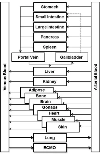
Figure 1.2. PBPK Model Structure. Each organ compartment is assigned a volume and flow
based on the age, weight, and height of the individual or population. Drug-specific information such as physicochemical properties and clearance pathways determine drug disposition within each compartment. In children on ECMO, an ECMO compartment can be added.
REFERENCES
1. Bartlett RH, Gazzaniga AB, Jefferies MR, Huxtable RF, Haiduc NJ, Fong SW. Extracorporeal membrane oxygenation (ECMO) cardiopulmonary support in infancy. Transactions - American Society for Artificial Internal Organs 1976;22:80-93.
2. Hill JD, O'Brien TG, Murray JJ, et al. Prolonged extracorporeal oxygenation for acute post-traumatic respiratory failure (shock-lung syndrome). Use of the Bramson membrane lung. N Engl J Med 1972;286:629-34.
3. Bartlett RH, Roloff DW, Cornell RG, Andrews AF, Dillon PW, Zwischenberger JB.
Extracorporeal circulation in neonatal respiratory failure: a prospective randomized study. Pediatrics 1985;76:479-87.
4. O'Rourke PP, Crone RK, Vacanti JP, et al. Extracorporeal membrane oxygenation and conventional medical therapy in neonates with persistent pulmonary hypertension of the newborn: a prospective randomized study. Pediatrics 1989;84:957-63.
5. UK collaborative randomised trial of neonatal extracorporeal membrane oxygenation. UK Collaborative ECMO Trail Group. Lancet 1996;348:75-82.
6. Morris AH, Wallace CJ, Menlove RL, et al. Randomized clinical trial of pressure-controlled inverse ratio ventilation and extracorporeal CO2 removal for adult respiratory distress syndrome. American journal of respiratory and critical care medicine 1994;149:295-305. 7. Zapol WM, Snider MT, Hill JD, et al. Extracorporeal membrane oxygenation in severe
acute respiratory failure. A randomized prospective study. JAMA 1979;242:2193-6. 8. Bessereau J, Chenaitia H, Michelet P, Roch A, Gariboldi V. Acute Respiratory Distress
Syndrome following 2009 H1N1 virus pandemic: When ECMO come to the patient bedside. Annales francaises d'anesthesie et de reanimation.
9. Mitchell MD, Mikkelsen ME, Umscheid CA, Lee I, Fuchs BD, Halpern SD. A systematic review to inform institutional decisions about the use of extracorporeal membrane oxygenation during the H1N1 influenza pandemic. Crit Care Med 2010;38:1398-404. 10. Buckley E, Sidebotham D, McGeorge A, Roberts S, Allen SJ, Beca J. Extracorporeal
membrane oxygenation for cardiorespiratory failure in four patients with pandemic H1N1 2009 influenza virus and secondary bacterial infection. Br J Anaesth;104:326-9.
11. Davies A, Jones D, Bailey M, et al. Extracorporeal Membrane Oxygenation for 2009 Influenza A(H1N1) Acute Respiratory Distress Syndrome. JAMA 2009;302:1888-95. 12. Firstenberg MS, Blais D, Louis LB, Stevenson KB, Sun B, Mangino JE. Extracorporeal
membrane oxygenation for pandemic (H1N1) 2009. Emerg Infect Dis 2009;15:2059-60. 13. Grasselli G, Foti G, Patroniti N, et al. A case of ARDS associated with influenza A - H1N1
infection treated with extracorporeal respiratory support. Minerva Anestesiol 2009;75:741-5.
18
15. Kumar A, Zarychanski R, Pinto R, et al. Critically ill patients with 2009 influenza A(H1N1) infection in Canada. JAMA 2009;302:1872-9.
16. Peek GJ, Elbourne D, Mugford M, et al. Randomised controlled trial and parallel economic evaluation of conventional ventilatory support versus extracorporeal membrane
oxygenation for severe adult respiratory failure (CESAR). Health Technol Assess 2010;14:1-46.
17. ELSO. ECLS Registry Report: International Summary; 2014 January 2015.
18. Butler J, Pathi VL, Paton RD, et al. Acute-phase responses to cardiopulmonary bypass in children weighing less than 10 kilograms. Ann Thorac Surg 1996;62:538-42.
19. Kozik DJ, Tweddell JS. Characterizing the inflammatory response to cardiopulmonary bypass in children. Ann Thorac Surg 2006;81:S2347-54.
20. Palatianos GM, Foroulis CN, Vassili MI, et al. A prospective, double-blind study on the efficacy of the bioline surface-heparinized extracorporeal perfusion circuit. Ann Thorac Surg 2003;76:129-35.
21. Tayama E, Hayashida N, Akasu K, et al. Biocompatibility of heparin-coated extracorporeal bypass circuits: new heparin bonded bioline system. Artificial organs 2000;24:618-23. 22. De Somer F, Francois K, van Oeveren W, et al. Phosphorylcholine coating of
extracorporeal circuits provides natural protection against blood activation by the material surface. European journal of cardio-thoracic surgery : official journal of the European Association for Cardio-thoracic Surgery 2000;18:602-6.
23. Palanzo D, Qiu F, Baer L, Clark JB, Myers JL, Undar A. Evolution of the extracorporeal life support circuitry. Artificial organs 2010;34:869-73.
24. Bhatt-Mehta V, Johnson CE, Schumacher RE. Gentamicin pharmacokinetics in term neonates receiving extracorporeal membrane oxygenation. Pharmacotherapy 1992;12:28-32.
25. Dagan O, Klein J, Gruenwald C, Bohn D, Barker G, Koren G. Preliminary studies of the effects of extracorporeal membrane oxygenator on the disposition of common pediatric drugs. Ther Drug Monit 1993;15:263-6.
26. Mehta NM, Halwick DR, Dodson BL, Thompson JE, Arnold JH. Potential drug sequestration during extracorporeal membrane oxygenation: results from an ex vivo experiment. Intensive Care Med 2007;33:1018-24.
27. Mulla H, Lawson G, von Anrep C, et al. In vitro evaluation of sedative drug losses during extracorporeal membrane oxygenation. Perfusion 2000;15:21-6.
28. Wildschut ED, Ahsman MJ, Allegaert K, Mathot RA, Tibboel D. Determinants of drug absorption in different ECMO circuits. Intensive Care Med 2010;36:2109-16.
30. Lemaitre F, Hasni N, Leprince P, et al. Propofol, midazolam, vancomycin and cyclosporine therapeutic drug monitoring in extracorporeal membrane oxygenation circuits primed with whole human blood. Crit Care 2015;19:40.
31. Shekar K, Roberts JA, McDonald CI, et al. Sequestration of drugs in the circuit may lead to therapeutic failure during extracorporeal membrane oxygenation. Crit Care
2012;16:R194.
32. Shekar K, Roberts JA, McDonald CI, et al. Protein-bound drugs are prone to sequestration in the extracorporeal membrane oxygenation circuit: results from an ex vivo study. Crit Care 2015;19:164.
33. Harthan AA, Buckley KW, Heger ML, Fortuna RS, Mays K. Medication adsorption into contemporary extracorporeal membrane oxygenator circuits. The journal of pediatric pharmacology and therapeutics : JPPT : the official journal of PPAG 2014;19:288-95. 34. Preston TJ, Hodge AB, Riley JB, Leib-Sargel C, Nicol KK. In vitro drug adsorption and
plasma free hemoglobin levels associated with hollow fiber oxygenators in the extracorporeal life support (ECLS) circuit. The journal of extra-corporeal technology 2007;39:234-7.
35. Preston TJ, Ratliff TM, Gomez D, et al. Modified surface coatings and their effect on drug adsorption within the extracorporeal life support circuit. The journal of extra-corporeal technology 2010;42:199-202.
36. Palmgren JJ, Monkkonen J, Korjamo T, Hassinen A, Auriola S. Drug adsorption to plastic containers and retention of drugs in cultured cells under in vitro conditions. European journal of pharmaceutics and biopharmaceutics : official journal of Arbeitsgemeinschaft fur Pharmazeutische Verfahrenstechnik eV 2006;64:369-78.
37. Unger JK, Kuehlein G, Schroers A, Gerlach JC, Rossaint R. Adsorption of xenobiotics to plastic tubing incorporated into dynamic in vitro systems used in pharmacological
research--limits and progress. Biomaterials 2001;22:2031-7.
38. Kolobow T, inventor Dow Corning, assignee. Artificial organ for membrane dialysis of biological fluids. USA. 1970.
39. Buck ML. Pharmacokinetic changes during extracorporeal membrane oxygenation: implications for drug therapy of neonates. Clin Pharmacokinet 2003;42:403-17.
40. B. MR, Timpa JG, Kurundkar AR, et al. Plasma concentrations of inflammatory cytokines rise rapidly during ECMO-related SIRS due to the release of preformed stores in the intestine. Lab Invest 2010;90:128-39.
41. Seghaye MC, Grabitz RG, Duchateau J, et al. Inflammatory reaction and capillary leak syndrome related to cardiopulmonary bypass in neonates undergoing cardiac operations. J Thorac Cardiovasc Surg 1996;112:687-97.
20
43. Many M, Soroff HS, Birtwell WC, Giron F, Wise H, Deterling RA, Jr. The physiologic role of pulsatile and nonpulsatile blood flow. II. Effects on renal function. Arch Surg
1967;95:762-7.
44. Ehrnebo M, Agurell S, Jalling B, Boreus LO. Age differences in drug binding by plasma proteins: studies on human foetuses, neonates and adults. Eur J Clin Pharmacol 1971;3:189-93.
45. Friis-Hansen B. Water distribution in the foetus and newborn infant. Acta paediatrica Scandinavica Supplement 1983;305:7-11.
46. McNamara PJ, Alcorn J. Protein binding predictions in infants. AAPS pharmSci 2002;4:E4. 47. Shekar K, Fraser JF, Taccone FS, et al. The combined effects of extracorporeal
membrane oxygenation and renal replacement therapy on meropenem pharmacokinetics: a matched cohort study. Crit Care 2014;18:565.
48. Mulla H, Lawson G, Firmin R, Upton DR. Drug Disposition During Extracorporeal Membrane Oxygenation (ECMO). Pediatric and Perinatal Drug Therapy 2001;4:109-20. 49. Morgan ET. Regulation of cytochromes P450 during inflammation and infection. Drug
Metab Rev 1997;29:1129-88.
50. Abdel-Razzak Z, Loyer P, Fautrel A, et al. Cytokines down-regulate expression of major cytochrome P-450 enzymes in adult human hepatocytes in primary culture. Mol
Pharmacol 1993;44:707-15.
51. Rivory LP, Slaviero KA, Clarke SJ. Hepatic cytochrome P450 3A drug metabolism is reduced in cancer patients who have an acute-phase response. British journal of cancer 2002;87:277-80.
52. Siewert E, Bort R, Kluge R, Heinrich PC, Castell J, Jover R. Hepatic cytochrome P450 down-regulation during aseptic inflammation in the mouse is interleukin 6 dependent. Hepatology 2000;32:49-55.
53. Richardson TA, Sherman M, Kalman D, Morgan ET. Expression of
UDP-glucuronosyltransferase isoform mRNAs during inflammation and infection in mouse liver and kidney. Drug metabolism and disposition: the biological fate of chemicals
2006;34:351-3.
54. Dufour JF, Zimmermann A, Reichen J. Increased hepatic lysosomal activity in biliary cirrhosis originates from hepatocytes rather than from macrophages. Journal of hepatology 1994;20:524-30.
55. Ugazio G, Artizzu M, Pani P, Dianzani MU. The changes in some hydrolytic enzymes in carbon tetrachloride-induced fatty livers. The Biochemical journal 1964;90:109-16. 56. Cherrington NJ, Slitt AL, Li N, Klaassen CD. Lipopolysaccharide-mediated regulation of
hepatic transporter mRNA levels in rats. Drug metabolism and disposition: the biological fate of chemicals 2004;32:734-41.
58. Ueyama J, Nadai M, Kanazawa H, et al. Endotoxin from various gram-negative bacteria has differential effects on function of hepatic cytochrome P450 and drug transporters. Eur J Pharmacol 2005;510:127-34.
59. Ahsman MJ, Wildschut ED, Tibboel D, Mathot RA. Pharmacokinetics of cefotaxime and desacetylcefotaxime in infants during extracorporeal membrane oxygenation. Antimicrob Agents Chemother 2010;54:1734-41.
60. Amaker RD, DiPiro JT, Bhatia J. Pharmacokinetics of vancomycin in critically ill infants undergoing extracorporeal membrane oxygenation. Antimicrob Agents Chemother 1996;40:1139-42.
61. Buck ML. Vancomycin pharmacokinetics in neonates receiving extracorporeal membrane oxygenation. Pharmacotherapy 1998;18:1082-6.
62. Cohen P, Collart L, Prober CG, Fischer AF, Blaschke TF. Gentamicin pharmacokinetics in neonates undergoing extracorporal membrane oxygenation. Pediatr Infect Dis J
1990;9:562-6.
63. Donadello K, Roberts JA, Cristallini S, et al. Vancomycin population pharmacokinetics during extracorporeal membrane oxygenation therapy: a matched cohort study. Crit Care 2014;18:632.
64. Hoie EB, Swigart SA, Leuschen MP, et al. Vancomycin pharmacokinetics in infants undergoing extracorporeal membrane oxygenation. Clin Pharm 1990;9:711-5.
65. Mulla H, Pooboni S. Population pharmacokinetics of vancomycin in patients receiving extracorporeal membrane oxygenation. Br J Clin Pharmacol 2005;60:265-75.
66. Munzenberger PJ, Massoud N. Pharmacokinetics of gentamicin in neonatal patients supported with extracorporeal membrane oxygenation. ASAIO Trans 1991;37:16-8. 67. Southgate WM, DiPiro JT, Robertson AF. Pharmacokinetics of gentamicin in neonates on
extracorporeal membrane oxygenation. Antimicrob Agents Chemother 1989;33:817-9. 68. Veinstein A, Debouverie O, Gregoire N, et al. Lack of effect of extracorporeal membrane
oxygenation on tigecycline pharmacokinetics. J Antimicrob Chemother 2011.
69. Bizzarro MJ, Conrad SA, Kaufman DA, Rycus P. Infections acquired during extracorporeal membrane oxygenation in neonates, children, and adults. Pediatr Crit Care Med 2010. 70. Gardner AH, Prodhan P, Stovall SH, et al. Fungal infections and antifungal prophylaxis in
pediatric cardiac extracorporeal life support. J Thorac Cardiovasc Surg 2011.
71. Eppes SC, Troutman JL, Gutman LT. Outcome of treatment of candidemia in children whose central catheters were removed or retained. Pediatr Infect Dis J 1989;8:99-104. 72. Pappas PG, Kauffman CA, Andes D, et al. Clinical practice guidelines for the
management of candidiasis: 2009 update by the Infectious Diseases Society of America. Clin Infect Dis 2009;48:503-35.
22
74. FDA. Fluconazole Injection, USP Product Label. In: U.S. Dept. of Health and Human Services FaDA, ed.: Roerig, a division of Pfizer, Inc.; 2015.
75. Ikeda F, Wakai Y, Matsumoto S, et al. Efficacy of FK463, a new lipopeptide antifungal agent, in mouse models of disseminated candidiasis and aspergillosis. Antimicrob Agents Chemother 2000;44:614-8.
76. Tawara S, Ikeda F, Maki K, et al. In vitro activities of a new lipopeptide antifungal agent, FK463, against a variety of clinically important fungi. Antimicrob Agents Chemother 2000;44:57-62.
77. Kaneko Y, Ohno H, Fukazawa H, et al. Anti-Candida-biofilm activity of micafungin is attenuated by voriconazole but restored by pharmacological inhibition of Hsp90-related stress responses. Medical mycology : official publication of the International Society for Human and Animal Mycology 2010;48:606-12.
78. Brammer KW, Coates PE. Pharmacokinetics of fluconazole in pediatric patients. Eur J Clin Microbiol Infect Dis 1994;13:325-9.
79. Fiori B, Posteraro B, Torelli R, et al. In vitro activities of anidulafungin and other antifungal agents against biofilms formed by clinical isolates of different Candida and Aspergillus species. Antimicrob Agents Chemother 2011;55:3031-5.
CHAPTER 2. ANTIFUNGAL EXTRACTION BY THE EXTRACOPOREAL MEMBRANE OXYGENATION (ECMO) CIRCUIT EX VIVO
INTRODUCTION
Extracorporeal membrane oxygenation (ECMO) is a cardiopulmonary bypass device used to support patients with refractory respiratory and/or cardiac failure. Patients supported with ECMO are critically ill and, thus, exposed to multiple drugs. Optimal dosing of drugs in this setting is unknown because ECMO can alter drug pharmacokinetics (PK). Studies of antimicrobials (e.g., vancomycin,1-4 gentamicin5-8) and sedatives (e.g., opiates,9-12 benzodiazepines13,14) generally show an increased volume of distribution (V) and decreased clearance (CL). These alterations in PK are caused by multi-organ dysfunction, the large volume of exogenous blood required to prime the ECMO circuit, and drug extraction by the circuit.
24
circuit depends on the materials in the circuit and the physicochemical properties of the drug. Non-specific binding of drugs occurs via two primary mechanisms: 1) hydrophobic interactions and 2) ionic interactions.20 Hydrophobic binding can occur between
hydrophobic drugs and a hydrophobic surface, while ionic binding occurs when the drug and surface are oppositely charged. Non-specific binding of drugs can significantly decrease exposure and lead to therapeutic failure.
Circuit-drug interactions have been investigated using ex vivo ECMO experiments in which drug is administered to an isolated ECMO circuit.13,15,21-29
Because there is no corporeal metabolism or elimination in this system, decreases in drug concentration are due to either extraction by the circuit or drug degradation. In general, highly lipophilic (e.g., fentanyl) and highly protein bound (e.g., caspofungin) drugs are extensively extracted by the circuit.23,30 However, this relationship is not always predictable. Ciprofloxacin is lipophilic with low protein binding but not extracted, while meropenem is hydrophilic with low protein binding and extensively extracted.22,23 This suggests that other factors such as ionic binding may play a role. Additionally, the site(s) of extraction within the circuit is not known.
In the present study, the extent of extraction by the ECMO circuit was
determined for selected antifungal drugs in an ex vivo ECMO system. Antifungal drugs were chosen because fungal infections are common in children on ECMO and
METHODS
Drug selection
Three antifungal drugs were selected for evaluation: fluconazole, micafungin, and amphotericin B dexoycholate. These drugs were selected based on their frequency of use in clinical practice and range of physicochemical properties (Table 2.1, Figure 2.1).
Circuit configuration
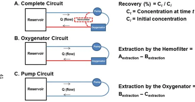
In order to determine the impact of each component on drug extraction, the experiment was designed with three circuit configurations (Figure 2.2). The Complete Circuit contained tubing, a pump, an oxygenator, and a hemofilter (Table 2.2). The Oxygenator Circuit was identical to the Complete Circuit except that the hemofilter was removed. The Pump Circuit was identical to the Oxygenator Circuit except that the oxygenator was removed. Any difference in extraction between the Complete and Oxygenator Circuits was due to the hemofilter. Similarly, any difference in extraction between the Oxygenator and Pump Circuits was due to the Oxygenator (Figure 2.2).
Circuit setup
26
solution: heparin sulfate (0.1 units), sodium bicarbonate (30mEq), and calcium
gluconate (6.5mg). Additional sodium bicarbonate and/or carbon dioxide were added to the system to maintain physiologic pH. To be consistent with standard practice at Duke, albumin was not routinely added to the circuit, and albumin concentrations were low (≤1.0 mg/dL). However, if a drug showed extraction in any of the circuit configurations, subsequent experiments were performed and human serum albumin was added to achieve two additional concentration levels: low-physiologic (2.2-2.8 mg/dL; typical for a child on ECMO) and physiologic (3.5-3.8 mg/dL). In order to examine the impact of albumin on extraction, extraction was compared between circuits with low, low-physiologic, and physiologic albumin concentrations.
Because there was no human connected to the circuit, the circuit was completed via a reservoir (plasmalyte IV bag). An ECMO heat exchanger was used to maintain a constant temperature of 36˚ C throughout the study. ECMO flow was set to 1L/min to simulate the clinical scenario for a 10kg child.
Control
Drug administration and sample collection
Drug was introduced into the circuit via a port downstream of the sampling port at time=0 and dosed to achieve a therapeutic concentration. Drug was dosed to achieve a comparable concentration in the control samples at time -5 minutes, sealed with a tight cap, and then gently mixed for 5 minutes until time=0. At time=0, the controls were returned to the water bath where they remained for the duration of the experiment, except at times of sample collection. Immediately prior to control sample collection, control tubes were removed from the water bath and gently inverted five times to ensure adequate mixing. In the initial set of experiments, samples were collected from the circuits and controls for 4 hours at the following time points: 1, 5, 15, and 30min, 1, 2, 3, and 4h. In order to better understand drug disposition over an entire dosing interval, subsequent experiments measured concentrations up to 24 hours by collecting additional samples at 8, 12, and 24h.
Analysis
28
following conditions: gas temperature 300ºC, gas flow 10 L/min, nebulizer pressure 50 psi, sheath gas temperature 345 ºC, sheath gas flow 11 L/min, capillary voltage 3500 V, nozzle voltage 500 V, Dynamic MRM scan type. The lower limit of quantification (LLOQ) was 0.01 mg/L with a calibration curve range of 0.01 to 10 mg/L. Intraday and interday precision (%CV) ranged from 1.4% to 8.5% and 2.8% to 5.8%, respectively. Micafungin concentrations were measured at the University of Texas Health System Fungal Testing Laboratory using HPLC with fluorescence detection. Plasma samples were acidified with phosphoric acid and precipitated with acetonitrile. Samples were centrifuged and diluted with 10mM ammonium acetate prior to injection into the HPLC system. Injection into the HPLC occurred under the following conditions: mobile phase with 10mM
intraday and interday precision ranged from 0% to 11.8% and 3.23 to 5.65%, respectively.
Drug recovery in circuits and controls was calculated at each sample time using the following equation:
% ∗ 100
where Ct is the concentration at time t and Ci is the initial concentration measured at time=1 minute. Data are reported as the mean and 95% confidence interval.
Ethics
The Duke University Medical Center Institutional Review Board provided a waiver of review because the protocol met the definition of research not involving human
subjects.
RESULTS
The total number of circuits studied, by configuration, are summarized for each drug in Table 2.3.
Fluconazole
30
97.8% (96.3, 99.3) at 4 hours. In the two Complete Circuits that were run for 24 hours, 95.2% (89.6, 100.9) of the initial concentration was recovered. When the hemofilter was removed to create the Oxygenator Circuit, 92.3% (83.1 101.5) of the initial fluconazole concentration was recovered at 4 hours (n=4). One Oxygenator Circuit was run for 24 hours and 98.4% of the initial concentration was recovered at 24 hours. When
fluconazole was administered to the Pump Circuit (n=1), 105.8% was recovered after 4 hours. Because no extraction was observed in the Complete or Oxygenator Circuits, additional Pump Circuits were not run. In the control samples, 100.2% (95.5, 104.9) of the initial fluconazole concentration was recovered at 4 hours (n=4) and 100.6% (98.5, 102.8) at 24 hours (n=3).
Micafungin
In the Complete Circuit (n=10), micafungin recovery was low at 4h (46.3% [35.3, 57.3]). However, when the hemofilter was removed (Oxygenator Circuit, n=4),
micafungin recovery at 4 hours was 91.1% (85.2, 97.0) (Figure 2.3.b). Similarly, in the Pump Circuit (n=1) and Control (n=4) 98.1% and 90.9% (86.8, 95.0), respectively, were recovered at 4 hours. By 24 hours, however, recovery of micafungin was low in the Complete (26.0% [15.0, 37.0]; n=9) and Oxygenator Circuits (42.6% [31.3, 53.9]; n=3) as well as in the Control samples (56.8% [49.2, 64.4]; n=3).
concentrations were recovered at 4 and 24 hours, respectively. At low physiologic albumin concentrations (2.2-2.8 g/dL; n=3) recovery was 58.4% (53.5, 63.3) and 35.7% (24.7, 46.7) at 4 and 24 hours, respectively. Similarly, when albumin concentrations were in the normal range (3.5-3.8 g/dL; n=3), micafungin recovery was 57.2% (54.8, 59.5) and 26.1% (20.4, 31.9) at 4 and 24 hours, respectively.
Because the hemofilter was responsible for micafungin extraction in the first 4 hours, micafungin concentrations were measured in the hemofiltrate for one circuit to determine if micafungin passively crossed the hemofilter membrane (hemofilter was only in-line and not actively filtering). Concentrations in the hemofiltrate at 1min, 1h, 2h, and 24h were only 0.07, 0.06, 0.21 and 0.16 mg/L, respectively. Concentrations in the plasma samples from the ECMO circuit at the same times were 15.5, 13.1, 11.6, and 8.1 mg/L, respectively. Concentrations in the hemofiltrate were <2% of concentrations in the circuit suggesting that micafungin does not cross the hemofilter membrane.
Amphotericin
32
Pump Circuits would not be expected. Therefore, additional circuit configurations were not evaluated for amphotericin. Of note, the concentrations of amphotericin in the control samples increased over the first 4 hours so that recovery was 143.8% (137.5, 150.1) of the initial concentration. Between 4 hours and 12 hours concentrations remained constant with recovery of 145.3% (140.4, 150.3) at 12 hours.
DISCUSSION
ECMO can alter drug pharmacokinetics (PK) directly and indirectly through a variety of mechanisms. Direct effects include 1) increased volume of distribution due to the addition of the large volume of exogenous blood required to prime the ECMO circuit, 2) hemofiltration, which is common in patients on ECMO, and 3) extraction of drug by components of the circuit.33 Ex vivo ECMO experiments such as those performed in this study provide insight into extraction via circuit-drug interactions. The degree of
interaction with the ECMO circuit is drug-dependent and likely influenced by the physicochemical properties of the drug and circuit components (e.g., oxygenator, hemofilter).23,30 Results of the present study demonstrate important differences in antifungal drug extraction by the ECMO circuit that can affect dosing recommendations in clinical practice.
Micafungin was highly extracted in the first 4 hours in a Complete Circuit but not in the other circuit configurations. This suggests that extraction was due to the
“trapped” the drug; or 3) direct adsorption by the hemofilter. Micafungin would not be expected to diffuse across the hemofilter membrane due to its high degree of protein binding (>99%) and the fact that the hemofilter was not actively filtering during the experiments. This was confirmed by collecting samples of hemofiltrate, which contained virtually undetectable concentrations of micafungin. Although areas of low flow can occur around the hemofilter, inconsistency in the degree of “trapping” and more
variability in recovery would be expected if this were the mechanism. Adsorption by the hemofilter appears to be the most likely explanation. This is supported by greater extraction at low albumin concentrations suggesting that the unbound fraction is
adsorbed by the hemofilter membrane. However, the polyethersulfone membrane used in these experiments was hydrophobic and had no net charge, making it less likely to interact with a hydrophilic anion such as micafungin. Further studies to understand the mechanism of drug loss to the hemofilter are needed.
These results are in contrast to studies of micafungin in continuous venovenous hemofiltration (CVVH) using similar hemofilters that showed no loss when micafungin concentrations were measured pre- and post-hemofilter.34,35 However, several
34
plasma. Differences in protein binding between the in vivo and ex vivo studies could also account for the differences. Albumin concentrations were higher in the in vivo CVVH studies. Because micafungin is highly protein bound, small changes in binding can substantially alter the amount of unbound drug available to be absorbed by the hemofilter.
More important than the hemofilter-related decrease in concentration over four hours was the observation that micafungin concentrations decreased over a 24 hour dosing interval in all circuit configurations and the controls. Non-specific binding to circuit materials should occur quickly (i.e., <4 hours).16,18-20 The continued extraction of micafungin over 24 hours in both circuits and controls suggests that drug degradation or plasma metabolism may have occurred. Micafungin is known to degrade in light.36 It is possible that light penetration of the ECMO tubing and control tubes caused sample degradation throughout the study. Clinically, ECMO circuits are exposed to light so that clinicians operating the circuit can monitor for clot accumulation in the components. As a result, a substantial amount of blood is exposed to light at any given time during ECMO support, and this may have important dosing implications for light-sensitive drugs. Alternatively, metabolism by plasma peptidases could result in continuous loss of micafungin. Although plasma metabolism is not described for micafungin, peptidases are a major elimination pathway for both caspofungin and anidulafungin, echinocandins with similar structure. Experiments are ongoing to determine the mechanism of
In the present study, fluconazole was not extracted by the ECMO circuit. The fluconazole results are consistent with a recent ECMO ex vivo study where fluconazole recovery at 24 hours in a system similar to the Oxygenator Circuit and Control used in this study were 96% and 102%, respectively.23 Based on physicochemical properties, fluconazole would not be expected to interact with the ECMO circuit. It is only slightly lipophilic (LogP 0.4) and should not undergo extensive hydrophobic binding to
polymers. The present findings also are supported by an ECMO ex vivo study that demonstrated minimal extraction of linezolid, a drug with similar physicochemical properties (LogP of 0.9, protein binding of 30%).23 Additionally, fluconazole has a neutral charge at physiologic pH so it would not be expected to undergo extensive ionic binding.
36
likely to explain the observed increase in concentration as most hemolysis occurred after four hours and a concomitant increase would have been expected in the circuit samples, as well. It is more likely that amphotericin did not distribute evenly in the control sample solution leading to sampling error. The deoxycholic acid component of amphotericin disperses very rapidly in plasma. In vitro studies show that amphotericin solubility is time dependent, and amphotericin is not completely solubilized even at one hour.39 It is possible that the amphotericin precipitated and was concentrated near the bottom of the control tubes. If samples were not adequately mixed, this could result in increasing concentrations until solubilization was complete. This phenomenon would be less likely in the ECMO circuit because it was flowing continuously. More work is
necessary to understand the disposition of amphotericin B deoxycholate in this system. Additionally, studies should be conducted to characterize the circuit interactions with other, more commonly used amphotericin forumulations (e.g. liposomal, lipid
complexed) that may result in interaction with the ECMO circuit.
both between coated and uncoated materials, and also between the different types of surface coatings.16,17 Many knowledge gaps remain. It is unknown if surface coatings on ECMO circuits change over time. Further, it is unknown to what extent endogenous materials (e.g., plasma proteins, platelets) compete for binding sites on circuit components. Future work should explore high-throughput systems to evaluate the interaction between ECMO circuit components and different drugs. Until that time, ex vivo experiments remain the best way to define specific circuit-drug interactions. The results from the present ex vivo experiments will be used to inform the ECMO
compartment of the fluconazole and micafungin PBPK models and translate those circuit-drug interactions into dosing recommendations in children on ECMO.
38
Table 2.1. Antifungal drug physicochemical properties and clearance pathways
Antifungal Charge LogP
Plasma Protein Binding (%)a
Molecular Weight (g/mol)
Primary Metabolic Pathway
Amphotericin Zwitterion 0.8 90 924 Renal
Fluconazole Neutral 0.4 11% 306 Renal
Micafungin Negative -0.4 99% 1270 Hepatic
aAmphotericin binds to plasma lipoproteins, albumin, and alpha-1-acid glycoprotein.40,41 Fluconazole
binds primarily to alpha-1acid-glycoprotein.42,43 Micafungin binds primarily to albumin.44
Table 2.2. ECMO circuit components
Component Manufacturer Model Material
Oxygenator Maquet Adult/Pediatric Quadrox iDa Polymethylpentane hollow fibers with
Biolineb coating
Hemofilter Sorin DHF0.2 Polyethersulfone
Pump Sorin Revolution Centrifugal Polycarbonate
Tubing Sorin Smart Tubing Phosphorylcholine coated polyvinylchloride
a Adult and pediatric oxygenators only differed in surface area (1.8m2 for adult and 0.8m2 for pediatric).
Because the oxygenator was not responsible for clinically significant extraction, and no difference was
observed between adult and pediatric oxygenator circuits, adult and pediatric oxygenators were assumed
to be equivalent for the purposes of these experiments.
Table 2.3. Number of circuits by configuration and drug
Configuration Fluconazole Micafungin Amphotericin
A. Complete Circuit
(hemofilter, oxygenator, pump, tubing) 3a 10d 3g
B. Oxygenator Circuit
(oxygenator, pump, tubing) 4b 4e -
C. Pump Circuit
(pump, tubing) 1 1 -
D. Control 4c 4f 3g
a 1 circuit run for 4 hours; 2 circuits run for 24 hours
b 3 circuits run for 4 hours; 1 circuit run for 24 hours
c 4 controls sampled for 4 hours; 3 controls sampled for 24 hours
d 10 circuits stratified by albumin level (g/dL): low (≤ 1; N=4), low physiologic (2.2-2.8; N=3); physiologic
(3.5-3.8; N=3). 1 circuit run for 4 hours (low albumin); 9 circuits run for 24 hours
e 2 circuits run for 4 hours; 2 circuits run for 24 hours
f 1 control sampled for 4 hours; 3 controls sampled for 24 hours
40
Figure 2.1. Molecular Structure of Antifungals
41
Figure 2.2. ECMO circuit configurations. A. Complete Circuit; B. Oxygenator Circuit; C. Pump Circuit
42
Figure 2.3. Recovery (mean [95% confidence interval]) by circuit configuration for each drug.
A Fluconazole – 4h Fluconazole – 24h
43
B Micafungin – 4h Micafungin – 24h
44
C Amphotericin B deoxycholate – 4h Amphotericin B deoxycholate – 12h
Figure 2.4. Micafungin recovery stratified by albumin concentration. Values are mean (95% confidence interval).
Control
46
REFERENCES
1. Amaker RD, DiPiro JT, Bhatia J. Pharmacokinetics of vancomycin in critically ill infants undergoing extracorporeal membrane oxygenation. Antimicrob Agents Chemother 1996;40:1139-42.
2. Buck ML. Vancomycin pharmacokinetics in neonates receiving extracorporeal membrane oxygenation. Pharmacotherapy 1998;18:1082-6.
3. Donadello K, Roberts JA, Cristallini S, et al. Vancomycin population pharmacokinetics during extracorporeal membrane oxygenation therapy: a matched cohort study. Crit Care 2014;18:632.
4. Mulla H, Pooboni S. Population pharmacokinetics of vancomycin in patients receiving extracorporeal membrane oxygenation. Br J Clin Pharmacol 2005;60:265-75.
5. Bhatt-Mehta V, Johnson CE, Schumacher RE. Gentamicin pharmacokinetics in term neonates receiving extracorporeal membrane oxygenation. Pharmacotherapy 1992;12:28-32.
6. Cohen P, Collart L, Prober CG, Fischer AF, Blaschke TF. Gentamicin pharmacokinetics in neonates undergoing extracorporal membrane oxygenation. Pediatr Infect Dis J
1990;9:562-6.
7. Munzenberger PJ, Massoud N. Pharmacokinetics of gentamicin in neonatal patients supported with extracorporeal membrane oxygenation. ASAIO Trans 1991;37:16-8. 8. Southgate WM, DiPiro JT, Robertson AF. Pharmacokinetics of gentamicin in neonates on
extracorporeal membrane oxygenation. Antimicrob Agents Chemother 1989;33:817-9. 9. Leuschen MP, Willett LD, Hoie EB, et al. Plasma fentanyl levels in infants undergoing
extracorporeal membrane oxygenation. J Thorac Cardiovasc Surg 1993;105:885-91. 10. Dagan O, Klein J, Bohn D, Koren G. Effects of extracorporeal membrane oxygenation on
morphine pharmacokinetics in infants. Crit Care Med 1994;22:1099-101.
11. Peters JW, Anderson BJ, Simons SH, Uges DR, Tibboel D. Morphine metabolite
pharmacokinetics during venoarterial extra corporeal membrane oxygenation in neonates. Clin Pharmacokinet 2006;45:705-14.
12. Peters JW, Anderson BJ, Simons SH, Uges DR, Tibboel D. Morphine pharmacokinetics during venoarterial extracorporeal membrane oxygenation in neonates. Intensive Care Med 2005;31:257-63.
13. Ahsman MJ, Hanekamp M, Wildschut ED, Tibboel D, Mathot RA. Population
pharmacokinetics of midazolam and its metabolites during venoarterial extracorporeal membrane oxygenation in neonates. Clin Pharmacokinet 2010;49:407-19.
14. Mulla H, McCormack P, Lawson G, Firmin RK, Upton DR. Pharmacokinetics of midazolam in neonates undergoing extracorporeal membrane oxygenation. Anesthesiology
15. Harthan AA, Buckley KW, Heger ML, Fortuna RS, Mays K. Medication adsorption into contemporary extracorporeal membrane oxygenator circuits. The journal of pediatric pharmacology and therapeutics : JPPT : the official journal of PPAG 2014;19:288-95. 16. Preston TJ, Hodge AB, Riley JB, Leib-Sargel C, Nicol KK. In vitro drug adsorption and
plasma free hemoglobin levels associated with hollow fiber oxygenators in the extracorporeal life support (ECLS) circuit. The journal of extra-corporeal technology 2007;39:234-7.
17. Preston TJ, Ratliff TM, Gomez D, et al. Modified surface coatings and their effect on drug adsorption within the extracorporeal life support circuit. The journal of extra-corporeal technology 2010;42:199-202.
18. Palmgren JJ, Monkkonen J, Korjamo T, Hassinen A, Auriola S. Drug adsorption to plastic containers and retention of drugs in cultured cells under in vitro conditions. European journal of pharmaceutics and biopharmaceutics : official journal of Arbeitsgemeinschaft fur Pharmazeutische Verfahrenstechnik eV 2006;64:369-78.
19. Unger JK, Kuehlein G, Schroers A, Gerlach JC, Rossaint R. Adsorption of xenobiotics to plastic tubing incorporated into dynamic in vitro systems used in pharmacological
research--limits and progress. Biomaterials 2001;22:2031-7.
20. Marchal-Heussler L, Barra J. Adsorption of Drugs. In: Hubbard AT, ed. Encyclopedia of Surface and Colloid Science. New York: Marcel Dekker, Inc.; 2002:294-306.
21. Shekar K, Fraser JF, Taccone FS, et al. The combined effects of extracorporeal
membrane oxygenation and renal replacement therapy on meropenem pharmacokinetics: a matched cohort study. Crit Care 2014;18:565.
22. Shekar K, Roberts JA, McDonald CI, et al. Sequestration of drugs in the circuit may lead to therapeutic failure during extracorporeal membrane oxygenation. Crit Care
2012;16:R194.
23. Shekar K, Roberts JA, McDonald CI, et al. Protein-bound drugs are prone to sequestration in the extracorporeal membrane oxygenation circuit: results from an ex vivo study. Crit Care 2015;19:164.
24. Lemaitre F, Hasni N, Leprince P, et al. Propofol, midazolam, vancomycin and cyclosporine therapeutic drug monitoring in extracorporeal membrane oxygenation circuits primed with whole human blood. Crit Care 2015;19:40.
25. Ahsman MJ, Wildschut ED, Tibboel D, Mathot RA. Pharmacokinetics of cefotaxime and desacetylcefotaxime in infants during extracorporeal membrane oxygenation. Antimicrob Agents Chemother 2010;54:1734-41.
26. van der Vorst MM, Wildschut E, Houmes RJ, et al. Evaluation of furosemide regimens in neonates treated with extracorporeal membrane oxygenation. Crit Care 2006;10:R168. 27. Wildschut ED, de Hoog M, Ahsman MJ, Tibboel D, Osterhaus AD, Fraaij PL. Plasma