The Neural Development of ‘Us and Them’
Jo
ao F. Guassi Moreira,
~
1,2Jay J. Van Bavel,
3and
Eva H. Telzer
2,41
Department of Psychology, University of California, Los Angeles, CA 90095, USA,
2University of Illlinois,
Urbana-Champaign IL 61820,
3Department of Psychology, New York University, New York City, NY 10003, USA
and
4Department of Psychology and Neuroscience, University of North Carolina, Chapel Hill, NC 27599, USA
Correspondence should be addressed to Eva H. Telzer, 235 East Cameron Ave, Chapel Hill, NC 27599, USA. E-mail: [email protected]
Abstract
Social groups aid human beings in several ways, ranging from the fulfillment of complex social and personal needs to the promotion of survival. Despite the importance of group affiliation to humans, there remains considerable variation in group preferences across development. In the current study, children and adolescents completed an explicit evaluation task of in-group and out-in-group members during functional neuroimaging. We found that participants displayed age-related increases in bilateral amygdala, fusiform gyrus and orbitofrontal cortex (OFC) activation when viewing in-group relative to out-group faces. Moreover, we found an indirect effect of age on in-group favoritism via brain activation in the amygdala, fusiform and OFC. Finally, with age, youth showed greater functional coupling between the amygdala and several neural regions when viewing in-group relative to out-group peers, suggesting a role of the amygdala in directing attention to motivation-ally relevant cues. Our findings suggest that the motivational significance and processing of group membership undergoes important changes across development.
Key words:group membership; social cognition; adolescence; social identity; development
Belonging to a group stretches beyond the satiation of immedi-ate social needs, fulfilling the overarching purpose of promoting survival (Tajfel and Turner, 1979; Brewer, 1991; Parrish and Edelstein-Keshet, 1999;Hogg, 2003). Aside from helping estab-lish a personal identity and boosting self-esteem, groups have long been thought to promote behavior aimed at achieving shared desired outcomes, facilitate information and resource sharing, and afford individuals greater protection from preda-tors (Allee, 1931; Tajfel and Turner, 1979; Brown et al., 1994; Spoor and Kelly, 2004;Bowles, 2006;Silk,et al., 2012). The fits of group membership confer such important survival bene-fits to humans that group affiliation and in-group preferences emerge very early in development and have been observed in every culture studied on earth (Brown, 1991; Aboud, 2003; Dunhamet al., 2011;Dunham and Emory, 2014). Althoughthe tendency for group aggregation, and subsequent importance of group membership, is not unique to humans (Parrish and Edelstein-Keshet, 1999), humans do display greater in-group fa-voritism than other non-human primates (Burkartet al., 2009). These findings imply that the significance of group membership in humans should be conserved cross-culturally and through-out the life-span. However, empirical evidence suggests
otherwise, revealing cultural variations and developmental fluctuations in the importance of group affiliation (Pfeiferet al., 2007;Ma-Kellamset al., 2011; Tantiet al., 2011; Dunham and Emory, 2014;Falket al.,2014;Baron and Dunham, 2015). These variations remain puzzling given the importance of groups to human survival. In thisstudy, we examine changes in neural sensitivity to group membership in childhood and adolescence to better understand the dynamic nature and shifting psycho-logical significance of social groups across development.
Developmental changes in the significance
of groups
Infancy and childhood
For young children and infants, groups help make sense of the different roles and categories that populate the social world, helping distinguish between friends and foes (Hirschfeld, 1995; Quinn et al., 2002; Bar-Haim et al., 2006; Kinzler et al., 2007; Wynn, 2008;Tayloret al., 2009;Hamlinet al., 2013). Children util-ize groups to facilitate future learning about social category con-cepts, supporting the premise that group membership allows
Received:29 June 2016;Revised:5 August 2016;Accepted:6 September 2016
VCThe Author (2016). Published by Oxford University Press. For Permissions, please email: [email protected]
184
doi: 10.1093/scan/nsw134
children to rapidly learn information crucial to navigating their social world (Baron and Dunham, 2015). Young children assume that out-groups are more likely to be hostile than friendly and become aware that in-group members are sources of support and nourishment (Kinzler and Spelke, 2011;Hamlinet al., 2013). This evidence, taken along with findings that young children are biased to remember threatening social stimuli (Kinzler and Shutts, 2008;Baltazaret al., 2012), suggests that children may display heightened vigilance towards outgroup members as a means to monitor threat.
In spite of out-group vigilance, infants and children also dis-play in-group preferences. Early conceptions of morality appear to be contingent upon group membership and are ostensibly driven by in-group biases (see Hamlin, 2014). Despite that in-fants normally favor those who exhibit prosocial behavior, they also prefer individuals who harm dissimilar others (Hamlin et al., 2007, 2010, 2011, 2013). Moreover, infants’ expectations are violated when in-group members fail to display pro-social be-havior to one another, such as when they hinder a fellow in-group member who needs assistance (Baillargeonet al., 2014, 2015). The trend of in-group favoritism persists throughout childhood as individuals endorse in-group favoritism and retain negative conceptions of out-group members (Bigleret al., 1997, 2001). Thus, group affiliation and its associated biases in infants and children influence their understanding of the world, im-parting them with information necessary for basic social func-tioning. Infants and children come to expect in-group members are readily available to provide help and may display increased vigilance towards out-group members to track potential sources of social threat.
Adolescence
Although group preferences may emerge at a very young age, evidence suggests the value and meaning of group belonging changes across development. Although individuals of all ages have demonstrated in-group favoritism—even within arbitrary groups—adolescents appear to be more sensitive to group affili-ations and their accompanying social identities than both chil-dren and adults (Tajfelet al, 1971;Brewer, 1979;Liebkind, 1983; Abramset al., 2003;Van Bavel et al., 2008, 2011;Pfeifer et al., 2009). Indeed, adolescents focus on the social aspects of their identity more so than children, and in some instances more than adults (Liebkind, 1983;Hartet al., 1993; Tarrantet al., 2001). For example, peer groups aid in establishing adolescents’ social and personal identity, with adolescents relying more on the opinions of others when constructing their self-construals (Brownet al., 1994; Pfeiferet al., 2009). Moreover, group member-ship offers an avenue of social support, conferring benefits to adolescents’ psychological and physiological health (Cacioppo and Cacioppo, 2014; Holt-Lunstad et al., 2015). Though group membership is important at all stages of development, in-groups become even more important for youths’ social identity upon reaching adolescence, suggesting that group identity is subject to psychological and motivational changes across development.
Furthermore adolescence in rodents, primates, and humans is marked by a social restructuring that renders increased orien-tation towards peers (Nelsonet al., 2016). This social reorienta-tion is thought to be mediated by alterareorienta-tions in brain development (Blakemore and Mills, 2014). In particular, neural regions involved in affective and salience processing [e.g. amyg-dala, ventral striatum, orbitofrontal cortex (OFC)] show height-ened activation to social stimuli among adolescents compared
with children or adults, suggesting that adolescents may be par-ticularly sensitive to socioemotional stimuli and may explain their unique attunement to social evaluation (Monket al., 2003; Nelson et al., 2005; Galvan et al., 2006; Hare et al., 2008). Moreover, neural regions considered part of the “social brain” that are involved in mentalizing or taking the perspective of others [e.g. medial prefrontal cortex (MPFC), temporoparietal junction (TPJ); Blakemore, 2008]show greater activation during adolescence compared to adulthood (Wanget al., 2006;Burnett et al., 2009;Blakemore, 2010; Gweonet al., 2011; van den Bos et al., 2011). Together, neuroscience research underscores how developmental changes in affective and social cognitive brain regions likely play an important role in directing adolescents’ attention towards social stimuli and increasing the salience of peer groups.
Neural correlates of social identity
Research has identified a network of brain regions implicated in social identity (see Cikara and Van Bavel, 2014 for a review). Specifically, the amygdala and fusiform gyrus are important in understanding the psychological significance of groups (e.g. Van Bavelet al., 2008, 2011). Originally considered to sit at the center of a neural network processing threat (Davis, 1992, 1994; LeDoux, 1996), the amygdala has been reconsidered to belong to a neural detection network that is sensitive to a broad range of salient stimuli (Vuilleumier and Brosch, 2009;Cunningham and Brosch, 2012). Evidence suggests that the amygdala may capture and direct attention towards noteworthy stimuli, especially emotional ones (Cunninghamet al., 2008;Anderson and Phelps, 2001; Cunningham and Brosch, 2012).
The amygdala has been consistently implicated in inter-group processes in both adult and developmental populations (Van Bavelet al., 2008; Telzeret al., 2013, 2015a). In particular, the amygdala appears to be sensitive to contextual differences that affect the motivational significance and salience of stimuli. For instance, the amygdala is sensitive to African American faces in adults when race is the emphasized and salient cat-egory (Liebermanet al., 2005). Yet, when adults are assigned to a mixed-race, novel group, the amygdala is sensitive to novel group members, irrespective of race. A similar phenomenon has also been documented across development wherein the amygdala responds preferentially to certain social categories (e.g. gender or race) as a function of their developmental signifi-cance (Telzeret al., 2013, 2015a). Thus, the amygdala is biased to respond to stimuli rendered motivationally significant by con-textual factors, including those that wax and wane in salience across development. Because the meaning and function of so-cial groups change across development, and given the amyg-dala’s sensitivity to contextual factors which influence the salience of social stimuli, we expect amygdala reactivity to in-and out-group members to vary with age depending on the meaning of groups.
positive feedback from peers (Guyeret al., 2011), suggesting that group membership facilitates deeper processing of faces.
Because individuals value group belonging and fellow in-group members, in-group membership also activates brain regions involved in reward valuation (Brewer, 1979, 1991; Baumeister and Leary, 1995). The ventral striatum and OFC, which encode for and represent subjective value (Kringelbach, 2005), tend to be activated when perceiving and favoring in-group members (Van Bavelet al., 2008;Telzeret al., 2015b). Further, adolescents show heightened ventral striatum activity both when receiving acceptance feedback from peers (Guyeret al., 2011) and when making risky decisions in their presence (Cheinet al., 2011). This research highlights the subjective value of fitting in and belong-ing to a group.
Since children use social group membership as a means to learn important information about the world, they may be more inclined to display increased vigilance towards out-group mem-bers as a means to monitor social threat, even though they still value in-group members. By contrast, adolescents, who are in the process of crafting an identity (Marcia, 1980), may be more concerned about others’ perspectives as they relate to their membership in a group. We propose that brain regions impli-cated in mentalizing and theory of mind processes may be sub-ject to developmental changes in their sensitivity to social group stimuli. Successfully navigating group environments re-quires at least an implicit ability to better recognize and attri-bute psychological agency and autonomy for in-group relative to out-group members. As such, mental state reasoning (i.e. mentalizing) and theory of mind may be recruited more when viewing in-group relative to out-group members (see Hackel et al., 2014). If children are concerned with monitoring social threat, it stands to reason that they may be more inclined to an-ticipate or infer the intentions and mental states of out-group members. Conversely, adolescents, who are highly sensitive to others opinions (Somervilleet al., 2013), are likely to be more inclined to infer the mental states of in-group members, espe-cially in a context in which group belongingness and a shared group identity are emphasized. Such mentalizing processes are facilitated by activation in the dorsomedial prefrontal cortex (DMPFC), especially when processing in-group relative to out-group targets (Mitchell et al., 2006; Rilling et al., 2008;
Molenberghs and Morrison, 2012). Moreover, structural connect-ivity between the TPJ and DMPFC predicts differences in inter-group bias (Baumgartner et al., 2015). Thus, neural regions implicated in perspective taking and mentalizing are robustly involved in intergroup processes, and we expect to see these re-gions come online in a developmentally appropriate fashion.
Methods
Participants
Participants included 56 children and adolescents (30 female), ages 8-16 years (Mage¼13.3 years, SD¼2.81 years). Power was determined using GPower (Faul and Erdfelder, 1992). Because such a study had not been previously conducted in children or adolescents, we used the conventional approach to assume a medium effect size. When using an estimated effect size of 0.5, an n of 55 would be needed to obtain statistical power (1b) of 0.9. Participants self-identified as White (n¼41), Black (n¼4), Asian (n¼3), Latino (n¼2) or mixed race (n¼6). Based on par-ental report, participants’ total family income ranged from less than $45,000 (n¼11) to greater than $90 000 (n¼29). Parents provided written consent and children provided written assent in accordance with the University of Illinois’ Institutional Review Board. Participants were compensated $50 for participating.
Establishment of novel in-group membership
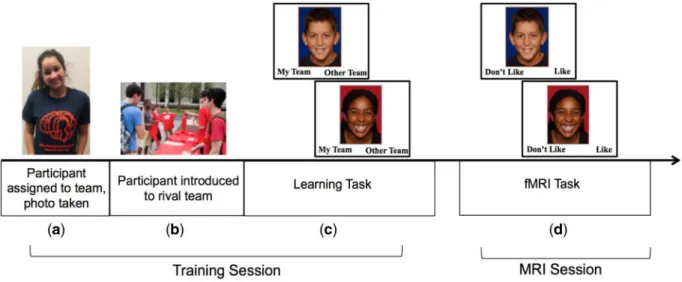
Participants arrived in the lab one at a time and were told that they would be on a team representing the ‘University of Illinois’ and that they would take part in a competition with research participants from the ‘Ohio State University’, a rival university. To make group membership salient, participants were given a t-shirt with the lab logo in their team’s colors (blue and orange), and a digital photograph was taken (Figure 1a). The researchers also wore the same t-shirt to increase the salience of team membership. Participants were shown a picture of rival univer-sity members receiving their t-shirts, which were scarlet and grey (Figure 1b). Notably, none of our participants were mem-bers of either university, helping ensure our results would not
Fig. 1.Group membership task.(a)participant is told they will be part of a competition between two research teams at different universities, is assigned to their team,
be driven by differences in preexisting university affiliation. During a learning task, participants were shown pictures of in-group and out-in-group team members (totaling 72 peers), who were described as participants who had already completed the study. Each face was displayed in random order, one at a time, with a label at the bottom indicating ‘my team’ and ‘other team’. Participants were instructed to press one of two buttons to indicate the correct team of each peer (Figure 1c). Photos were placed on blue (representing in-group) or red (representing out-group) backgrounds to provide a visual cue of team mem-bership. Participants also saw their own face two times on the colored background and categorized themselves into the appro-priate team in order to enhance their in-group identification.
The face stimuli were of children and adolescents ranging in age from 8 to 16 years. The faces were comprised of equal num-bers of males and females and split equally between White, Black and Asian. All faces were looking into the camera and smiling. Faces were taken from several databases including the National Institute of Mental Health Child Emotional Faces Picture Set (Egger et al., 2011). The faces of each race were matched based on pilot testing to ensure they were equally at-tractive (mean atat-tractiveness¼4.1 for each race on scale rang-ing from 1¼‘not at all’ to 7¼’very much’) and ranged equally in terms of perceived age (mean age¼5.7 for each race on a scale ranging from 1 to 9 (1¼‘5 or younger’, 5¼‘12 or 13’, 9¼ ‘20 or older’). Faces were randomly assigned to the teams ensur-ing equal representation of race, gender and age across the teams, and assignment was fully counterbalanced so that par-ticipants were equally likely to see each face as an in-group or out-group member. This ensured that any visual differences in the stimuli (e.g. attractiveness, luminance) could not account for observed differences between in-group and out-group members.
fMRI task
After completing the learning task, participants were placed in the scanner and completed an explicit evaluation task. For each trial of the task, participants were shown the same pictures as the learning task, this time with the instruction to indicate whether they ‘like’ or ‘dislike’ each person (Figure 1d). Participants pressed one of two buttons to indicate their re-sponse. The faces were presented on the color background repre-senting team membership. Participants completed 72 total trials, half of which were in-group members and half of which were out-group members. Each face was presented for 3 s with an inter-trial interval that was jittered randomly between 1.5 and3 s.
fMRI data acquisition and analysis
fMRI data acquisition. Imaging data were collected using a 3 Tesla Siemens Trio MRI scanner. The task included T2*-weighted echoplanar images (EPIs) [slice thickness¼3 mm; 38 slices; TR¼2 s; TE¼25 ms; matrix¼9292; FOV¼230 mm; voxel size 2.52.5 3mm3]. Structural scans consisted of a T2*weighted, matched-bandwidth (MBW), high-resolution, ana-tomical scan (TR¼4 s; TE¼64 ms; FOV¼230; matrix¼192 192; slice thickness¼3 mm; 38 slices) and a T1* magnetization-prepared rapid-acquisition gradient echo (MPRAGE; TR¼1.9 s; TE¼2.3 ms; FOV¼230; matrix¼256256; sagittal plane; slice thickness¼1 mm; 192 slices). The orientation for the MBW and EPI scans were oblique axial to maximize brain coverage.
fMRI data preprocessing and analysis. Neuroimaging data were preprocessed and analyzed using Statistical Parametric
Mapping (SPM8; Wellcome Department of Cognitive Neurology, Institute of Neurology, London, UK). Preprocessing for each par-ticipant’s images included spatial realignment to correct for head motion (no participant exceeded 3 mm of maximum image-to-image motion in any direction). The realigned func-tional data were co-registered to the high resolution MPRAGE, which was then segmented into cerebrospinal fluid, grey matter and white matter. The normalization transformation matrix from the segmentation step was then applied to the functional and T2 structural images, thus transforming them into standard stereotactic space as defined by the Montreal Neurological Institute and the International Consortium for Brain Mapping. The normalized functional data were smoothed using an 8 mm Gaussian kernel, full-width-at-half maximum, to increase the signal-to-noise ratio.
Statistical analyses were performed using the general linear model (GLM) in SPM8. Each trial was convolved with the canon-ical hemodynamic response function. High-pass temporal filter-ing with a cutoff of 128 s was applied to remove low-frequency drift in the time series. Serial autocorrelations were estimated with a restricted maximum likelihood algorithm with an autor-egressive model order of 1.
In each participant’s fixed-effects analysis, a GLM was cre-ated with 12 regressors of interest, modeled as events: in-group and out-group peers broken down by race (Black, White, Asian) and gender (Male, Female). Null events, consisting of the jittered inter-trial intervals, were not explicitly modeled and therefore constituted an implicit baseline. The parameter estimates re-sulting from the GLM were used to create linear contrast images comparing the conditions of interest (in-group >out-group). Random effects, group-level analyses were performed on all in-dividual subject contrasts using GLMFlex. GLMFlex corrects for variance-covariance inequality, partitions error terms, removes outliers and sudden activation changes in the brain, and ana-lyzes all voxels containing data (http://mrtools.mgh.harvard. edu/index.php/GLM_Flex). We conducted t-tests at the group level to examine overall differences in neural activation when processing group-status and race. In addition, we conducted whole brain regression analyses with age entered as the regres-sor to examine neural regions that showed increased activation as a function of age.
whole brain regression analyses to examine developmental changes in functional coupling between the conditions of interest.
To correct for multiple comparisons, we conducted a Monte Carlo simulation implemented using 3dClustSim in the soft-ware package AFNI (Ward, 2000). We used our group-level brain mask, which included only gray matter, and accounted for smoothing. Results of the simulation indicated a voxel-wise threshold ofP<0.005 combined with a minimum cluster size of 48 voxels for the whole brain, corresponding toP<0.05, False Wise Error corrected. We ran all analyses with mean response time (MRT) as a covariate. Adding this covariate ensures that our developmental effects are due to age differences and not to differences in psychomotor speed (see Supplemental Materials for behavioral results with MRT). We used the MarsBaR toolbox to extract parameter estimates from significant clusters in the group-level analyses.
Results
Behavioral ratings of in- and out-group peers
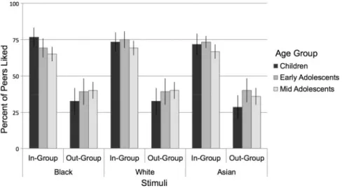
As a manipulation check, we first examined whether partici-pants would express in-group favoritism on self-reported liking. We conducted a three-way repeated-measures ANOVA on self-reported liking (percent liked) with two within subject factors representing the face stimuli (group status: in-group, out-group; race: Black, White, Asian) and age as a covariate. As predicted, we found a significant effect of group status [F(1,54)¼37.4,P<
0.0001,g ¼0.31], such that participants rated liking in-group peers (M¼70.3%, SE¼2.7%) more than out-group peers (M¼ 36.4%, SE¼3.5%). No other effects were significant (Ps>0.1). Thus, regardless of participants’ age or the race of the group member, participants reported liking in-group members signifi-cantly more frequently than out-group members. For descrip-tive purposes, we plotted the percent of peers who were rated as liked separated by group status, race, and age-group. We div-ided the sample into 3 age groups purely for descriptive pur-poses for plotting the behavioral effects (children: ages 8–10 years,n¼15; early adolescents: ages 11–14 years,n¼16; mid adolescents: ages 15–16 years,n¼25; Figure 2).
Developmental differences in the neural correlates of evaluating in-group relative to out-group members
First, we conducted a whole-brain analysis to examine neural activation when rating in-group relative to out-group members across the whole sample regardless of age. Results of the con-trast in-group>out-group revealed only one significant cluster of negative activation (i.e. greater activation to out-group rela-tive to in-group members) located in the right insula (xyz¼54, 14,5;k¼48,t¼3.16,P<0.005 corrected).
Next, we conducted whole brain regression analyses to test whether there are differential neural responses to in-group rela-tive to out-group members as a function of age. To this end, age was entered as a regressor on the contrast of in-group> out-group faces. We found significant effects in several regions, such that participants demonstrated greater activation to in-group>out-group members in the bilateral amygdala, bilateral fusiform gyrus, OFC, MPFC, MPPC and pSTS, as a function of age (Table 1). Age was not associated with greater activation to out-group>in-group members in any regions. Thus, we found de-velopmental increases in neural activation from childhood to adolescence in regions that code for emotional salience (amyg-dala), face processing (fusiform), subjective value (OFC) and so-cial cognition (MPFC, MPPC and pSTS) when rating in-group relative to out-group faces. For descriptive purposes, we plotted these individual differences (Figure 3). To this end, we extracted parameter estimates of signal intensity from each cluster of ac-tivation and plotted the age effects. Together, these neural ef-fects suggest that the salience of in-group members changes across development, such that younger children show relatively greater activation to out-group faces (as evidenced by param-eter estimates falling below the 0-point on the y-axis), and ado-lescents show relatively greater activation to in-group faces.
Linking neural correlates of group membership to behavioral biases favoring in-group members
Next, we examined how individual differences in behavioral in-group bias were associated with neural activation to in-in-group>
out-group faces. Behavioral biases were calculated as the differ-ence in the percent of in-group members who were liked minus
Fig. 2.Behavioral performance on the fMRI task. Participants rated liking more peers in their in-group than out-group, and this did not vary by the age of participants
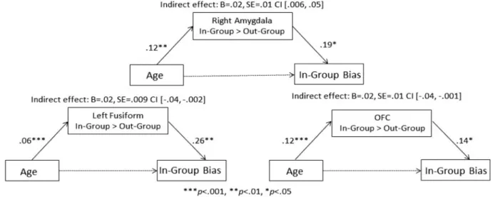
the percent of out-group members who were liked, such that higher scores represent a greater bias favoring the in-group. We regressed participants’ in-group bias score against whole brain activation for the in-group>out-group contrast while control-ling for age. As detailed in Table 2andFigure 4, in-group bias was significantly associated with increased activation to in-group relative to out-in-group members in the right amygdala, left fusiform, OFC, subgenual anterior cingulate cortex (sgACC) and bilateral TPJ. Importantly, the OFC, right amygdala, and left fusi-form clusters overlapped with those reported in the age-related analyses described earlier.
Neural reactivity mediates age differences and in-group bias
Given that similar neural patterns were found when examining age differences in neural activation as well as correlations with in-group bias, we examined whether age was associated with in-group bias via neural reactivity to in-groups. We extracted parameter estimates of signal intensity from the brain regions which showed overlap in activation to in-group relative to out-group members in the two sets of independent analyses (denoted by an asterisk in Table 2). We calculated the magni-tude and the significance of the indirect effects using the pro-cedures described by Preacher and Hayes (Preacher and Hayes, 2008), in which bootstrapping was performed with 1000 samples and a bias-corrected CI was created for the indirect effect. At a statistical threshold ofa¼0.05 (i.e. 95% CI), the indirect effects of age on in-group bias through neural activation were signifi-cant for the amygdala, fusiform and OFC (see Figure 5).
Age moderates the relationship between brain activation and in-group bias
In order to follow up and supplement our mediation analysis, we also ran moderation analyses in order to determine whether the association between neural reactivity to in-group members was conditional upon age. Using the same extracted beta values for the amygdala, OFC, and fusiform previously described in our
mediation analyses, we tested for moderation by age. We cen-tered age and neural activation, created an AgeBrain inter-action term, and then entered these terms into a multiple regression with bias scores as the predicted outcome. We ran separate moderation analyses for each brain region. Results re-vealed that our interaction term was significant for the amyg-dala (B¼0.166, SE¼0.067,b¼0.634,P¼0.016), suggesting that amygdala reactivity to ingroup faces is conditional upon age. For descriptive purposes only, we split our sample into three age groups as previously described and ran correlations be-tween bias scores and amygdala activation. Notably, only mid-dle adolescents displayed a significant correlation between bias and amygdala activation (r¼0.628,P¼0.001), whereas children (r¼0.351,P¼0.183) and young adolescents (r¼ .095,P>0.250) did not show a significant association. The interaction for the OFC and fusiform were not significant.
Neural connectivity with the amygdala
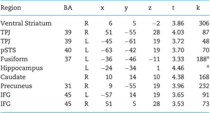
Finally, given our mediation results with the amygdala, we examined developmental changes in functional connectivity with the amygdala to in-group>out-group members. Social perception systems in the brain are widely distributed and thought to be organized in networks (Nelsonet al., 2005; Van Bavelet al., 2014). Moreover, the amygdala is thought to direct attention to important and noteworthy stimuli (Anderson and Phelps, 2001;Cunninghamet al., 2008; Cunningham and Brosch, 2012). Therefore, we conducted PPI analyses in order to examine the extent to which the amygdala co-activates with regions involved in face processing, reward value, and mentalizing, thereby allowing insight into the developmental processes that shape social perception and evaluation. In our whole-brain PPI analyses, we entered age as a regressor and found developmen-tal increases in connectivity between the amygdala and the ventral striatum, bilateral TPJ, MPPC and fusiform gyrus (Figure 6; Table 3). Thus, with age, youth showed greater functional coupling between the amygdala and these neural regions when viewing in-group relative to out-group peers, suggesting a role of the amygdala in directing attention to motivationally rele-vant cues.
Discussion
Groups are indispensible for survival to several species across the animal kingdom (Allee, 1931;Williams, 1964; Parrish and Edelstein-Keshet, 1999). Humans in particular show in-group fa-voritism (Burkartet al., 2009), which emerges very early in
devel-opment and persists from infancy through adulthood
(Baillargeon et al., 2014, 2015; Baron and Dunham, 2015). However, evidence also shows fluctuations and nuances in this phenomenon and further hints at the possibility that groups adopt different meanings across life (Tantiet al., 2011;Silket al., 2012;Dunham and Emory, 2014;Baron and Dunham, 2015). We found that brain regions implicated in affect, reward and social cognitive processes show developmental changes in neural sen-sitivity to novel peer in-groups, providing evidence for a striking developmental switch in the significance of groups from child-hood to adolescence. Moreover, developmental increases in neural activation mediated age differences in in-group favorit-ism. These neural and behavioral results reveal insight into the developmental changes that shape the shifting motivational importance of group membership across juvenile development.
From childhood to adolescence, participants showed linear increases in activation in the bilateral amygdala when rating in-Table 1.Neural regions which correlated with age during In-group>
Out-group ratings
Region BA x y z t K
Fusiform 37 R 32 52 8 4.27 48 Fusiform 37 L 30 34 26 4.72 1181a
pSTS 40 L 48 43 13 3.17 a
mPPC 31 L 9 46 4 3.46 a
Amygdala L 22 2 24 3.71 94 Amygdala R 24 1 29 3.49 472b
Temporal Pole 38 R 39 14 29 4.37 b
OFC 32/24 L 6 35 14 3.51 169c
IFG 45 L 30 44 14 3.79 c
Caudate R 21 10 22 3.72 105 Parahippocampus 27 R 18 31 14 3.56 84 Cuneus 17 L 9 85 5 3.55 127 Cerebellum L 6 58 47 3.48 51 Cerebellum R 36 67 35 3.56 61 Cerebellum R 15 61 50 3.20 51
group relative to out-group members. Although originally con-ceptualized as a threat detector (Davis, 1992, 1994), recent work has suggested that the function of the amygdala may instead be to detect and direct attention to motivationally relevant stimuli (Liebermanet al., 2005;Cunninghamet al., 2008;Vuilleumier and Brosch, 2009; Cunningham and Brosch, 2012). Importantly, the salience of different social identities may change in relevance depending on the context and developmental age of the individ-ual (e.g. Telzeret al., 2013, 2015b). Our findings provide evidence for a developmental shift in the salience of group membership, such that children displayed relatively greater amygdala activa-tion to out-group faces, as evidenced by the scatterplot showing activational patterns below zero in the youngest children, and adolescents showing relatively greater activation to in-group faces. This finding, coupled with research showing that children are biased to remember threatening social displays (Baltazar et al., 2012), substantiates the idea that out-groups may be more
salient to children by virtue of their perceived capacity for social threat. Incontrast, adolescents become increasingly motivated to learn about in-groups at a time in the lifespan when fitting in is of the utmost importance (Silket al., 2012). Thus, whereas young children may find out-groups salient, older adolescents may attend more to in-groups as a means of learning about an important social group. Indeed, the amygdala is involved in learning (Morriset al., 1998) in addition to attending to interest-ing, salient, and important stimuli (Canliet al., 2000; Hamann et al., 2002; Telzeret al., 2013, 2015a). Importantly, amygdala ac-tivation was associated with in-group bias, suggesting that the amygdala is detecting socially salient values and attitudes. Moreover, results from our moderation analyses show that only middle adolescents, but not young adolescents or children, dis-played a significant correlation between amygdala activation and in-group bias. This finding underscores adolescence as a particularly sensitive period for in-group biases, and further
Fig. 3With age, participants showed greater activation in the bilateral amygdala, left fusiform, OFC, mPPC, mPFC and pSTS to in-group relative to out-group faces. The
highlights the important role of the amygdala in intergroup behaviors.
Results of our functional connectivity analyses show devel-opmental increases in connectivity between the amygdala and the bilateral TPJ, fusiform and ventral striatum when partici-pants rated in-group relative to out-group peers. These are re-gions involved in social cognition, face processing, and reward processing, respectively (Kanwisher et al., 1997; Kringelbach, 2005;Frith and Frith, 2007). Thus, the amygdala may be involved in the detection of meaningful and important stimuli and then alerts and directs attention to relevant brain regions to process the faces in further depth (Van Bavelet al., 2011;Hackelet al., 2014). Because PPI analyses do not specify the direction of an ef-fect, another explanation is that brain regions involved in re-ward processing, face perception, and social cognitive processes first react to viewing novel in-group members and then trigger amygdala activation. Overall, these findings provide a novel
and unique perspective on the role of the amygdala as a social-salience-detector that communicates with other brain regions, co-activation that increases linearly across development. More broadly, this also serves in characterizing the developmental plasticity of the brain in modulating the ability to process social cognitive stimuli to accommodate ever changing social de-mands across juvenile development.
In addition, we found age-related increases in fusiform acti-vation when rating in-group relative to out-group peers. These findings are consistent with research on adults, which has shown that adults exhibit greater activation within the bilateral fusiform when viewing novel in-group relative to out-group faces (Van Bavelet al., 2008, 2011). Although the faces of all in-and out-group peers were matched in-and counterbalanced across participants to ensure neither group was visually more distinct than the other, the fusiform nevertheless showed strong differ-entiation between in- and out-group peers with age. Therefore, classifying faces along group boundaries may alter the depth with which faces are processed, and in-group belonging during adolescence may enhance encoding of in-group members, whereas out-group vigilance may contribute to enhanced pro-cessing of out-group members among younger children. These findings suggest that the amygdala may signal the importance of the social category, and the fusiform may come online to en-gage in deeper perceptual processing, individuating faces based on their psychological and motivational significance (Van Bavel et al., 2011).
Furthermore, we observed developmental increases in neu-ral regions that code for and represent subjective value. In par-ticular, youth showed developmental increases in OFC activation when rating in-group relative to out-group peers, and the ventral striatum showed developmental increases in func-tional coupling with the amygdala. Thus, viewing in-group members may activate brain regions involved in reward pro-cessing. This finding is consistent with prior work with adults, which has shown that individuals who favor novel in-group members show heightened OFC activity when viewing in-group relative to out-group members (Van Bavelet al., 2008), and re-warding in-group relative to out-group members engages the ventral striatum (Telzeret al., 2015b). We also observed heighted activation in the sgACC, TPJ, amygdala, fusiform and OFC as a function of individual differences in in-group favoritism. These results are consistent with prior work showing that a greater orientation towards one’s in-group is associated with heighted activation in networks involved in social perception (Van Bavel et al., 2008; 2011) and mentalizing (Cheonet al., 2011), suggesting that biases favoring one’s in-group are associated with richer encoding and more elaborate social cognition toward in-group faces.
At a time when the development of an identity is necessary for establishing an autonomous sense of self, groups become a source of social information for adolescents to sample from and build an identity, as evidenced by their reliance on other’s opin-ions and perspective in crafting their self-construals (Pfeifer et al., 2009). Indeed we also found developmental increases in activation of the social brain network (Blakemore and Mills, 2014; mPFC, mPPC, pSTS, TPJ) when viewing in-group relative to out-group faces. This neural recruitment highlights the psycho-logical shift in motivational differences of processing group membership between childhood and adolescence. Our results suggest teens may be keener than children to process social cues from in-groups, lending support to the notion that the psy-chological importance of groups is different between adoles-cents and children. The increased orientation towards group
Fig. 4.In-group bias is associated with greater activation in the right amygdala,
subgenual ACC, bilateral TPJ, left fusiform and OFC to in-group, relative to out-group, faces. These effects control for age and MRT.
Table 2.Neural regions which correlated with bias scores, while con-trolling for age, during In-group>Out-group ratings
Region BA x y z t k
Subgenual ACC 25 L 6 14 14 3.91 273a
OFC* 11 L 6 41 17 3.13 a
Amygdala* R 27 2 26 3.15 a
Fusiform* 37 L 63 49 11 3.81 80 TPJ 39 L 42 76 25 3.94 191b
Precuneus 31 L 3 61 31 3.29 b
TPJ 39 R 54 58 28 3.66 118 Precuneus 7 L 6 76 46 5.16 156 Cuneus 18 0 91 22 4.29 64 Cerebellum R 6 67 38 4.75 492c
Cerebellum L 15 64 35 3.75 c
Cerebellum L 39 64 41 3.48 50 Pallidum L 9 4 1 3.61 48
Fig. 6.Age-related increases in functional connectivity with the amygdala when rating in-group relative to out-group faces. Increased functional connectivity is found in the ventral striatum, TPJ and fusiform.
Fig. 5.Activation in brain regions showing responses to In-group>Out-group in both age and in-group bias regression analyses were found to significantly mediate
membership and enhanced social identity development in ado-lescence results in greater processing of in-group mental states and perspectives. We interpret our findings as supporting a de-velopmental shift in the meaning and salience of group mem-bership that occurs between childhood and adolescence. Yet, this is only one possible interpretation for the results reported here. An intriguing consideration for future research is whether these findings are indicative of an adolescent emergent or ado-lescent specific developmental transition (Casey, 2015). Although prior research has shown that adults also demon-strate heightened amygdala, fusiform, and OFC activation to in-group relative to out-in-group faces (Van Bavelet al., 2008), without adult comparisons in the same study, it is not clear whether adults’ neural sensitivity to in-groups is similar to, greater than, or less than that of adolescents. If adults display comparable patterns of neural activation in response to in-group faces, it would support the notion that the psychological importance of group belonging and its neural underpinnings remain stable after adolescence (i.e. adolescent emergent phenomenon), fur-ther supporting the notion of a developmental shift occurring between childhood and adolescence. Incontrast, if adults ex-hibit less neural sensitivity to in-group peers than adolescents, it would indicate that a potent orientation towards in-group peers is unique to adolescence, providing evidence for an adolescent-specific peak in the salience of group belonging. Future research with children and adolescents should include adult comparison samples in order to examine this question. Another consideration for future research is how the effects found here relate to actual behaviors. We only measured atti-tudes in this study and find that developmental changes in neu-ral processing of groups predicts biases favoring one’s ingroup. It is possible that such neural signals could have negative impli-cations for intergroup dynamics such as social exclusion or biased resource distribution.
Interestingly, we did not find age effects in our behavioral analyses of in-group favoritism. Both children and adolescents consistently reported liking in-group peers more than out-group peers, a trend that did not vary with age. This may have been due to the nature of how we required participants to evaluate group members. By having participants indicate a cat-egorical response (i.e. like/dislike) instead of rating likeability along a continuous scale, we alleviated task demands for our younger participants but also removed a source of variability
within the data. Thus, although we did not find increased in-group bias across development, we did find an indirect path-way. Our findings show that age was associated with greater neural biases (i.e. differentiation in a set of brain regions to in-group>out-group faces), and these neural biases were associ-ated with behavioral biases favoring the in-group. This suggests that although group membership is important for individuals of all ages (e.g. Dunhamet al., 2011; Van Bavelet al., 2008), it is likely that children and adolescents do not differ in who they like, but rather how much they like them and the psychological significance of that preference. This suggests there are import-ant age-related changes in behavioral biases being driven by maturation in the developing brain. All age groups in our study indicated liking in-group peers more than out-group peers, yet adolescents’ in-group preferences were differentiated from those of children by neural responses to social groups. Lastly, we note that although a direct effect from age to behavioral biases might be expected, this is not necessary for establishing statistically significant mediation, particularly in developmen-tal studies that focus on more disdevelopmen-tal processes (MacKinnon et al., 2000, 2002;Shrout and Bolger, 2002; Hayes, 2009; Rucker et al., 2011; Zhaoet al., 2010). This is noteworthy because it em-phasizes the role of the developing brain in shaping in-group biases. It implies that the functional architecture which sup-ports social cognitive processes is sensitive to changes in the social environment over a protracted period of time.
In conclusion, adolescence and childhood are periods marked as having distinct psychological interpretations of group belonging. In particular, childhood is characterized by the need to understand how and why the world works, whereas adolescence is marked by the increased importance of group af-filiation to fulfill developmental goals of establishing a social identity (Marcia, 1980;Pfeiferet al., 2009;Baron and Dunham, 2015). The latter occurs in tandem with a social reorientation of the teenage brain, a period of unique neural development dur-ing which brain regions involved in complex social processes undergo significant maturation (Nelsonet al., 2005; Blakemore and Mills, 2014). Together, our imaging data suggest a develop-mental shift in the psychological importance of groups across the first two decades of life and reveal the neurobiological sub-strates that underlie this process. As individuals develop nuanced conceptions about the world and engage in new
devel-opmental tasks, groups take on new meaning during
adolescence.
Acknowledgements
The authors would like to thank the members of the Developmental Social Neuroscience (University of Illinois) and Social Perception and Evaluation (New York University) Laboratories in addition to Renee Baillargeon for their in-sightful and helpful comments on this manuscript. In par-ticular, we thank Nicholas Ichien and Inge Karosevica for collecting the data. We greatly appreciate the assistance of the Biomedical Imaging Center. Responsibilities: J.V.B. and E.H.T. designed studies, J.F.G.M. and E.H.T. analyzed studies with input from J.V.B., and J.F.G.M. and E.H.T. wrote the art-icle with critical edits from J.V.B.
Funding
This paper was partially supported by grants from the National Science Foundation (no. 1459719 to E.H.T.; no. Table 3.Neural regions which were functionally coupled with the
amygdala and showed a correlation with age during In-group> Out-group ratings
Region BA x y z t k
Ventral Striatum R 6 5 2 3.86 306 TPJ 39 R 51 55 28 4.03 87 TPJ 39 L 45 61 19 3.72 48 pSTS 40 L 63 42 19 3.70 70 Fusiform 37 L 36 46 11 3.33 188a
Hippocampus L 24 34 1 4.46 a
Caudate R 10 14 10 4.38 168 Precuneus 31 R 9 55 19 3.96 232 IFG 45 L 57 14 19 3.65 91 IFG 45 R 51 5 28 3.53 73
1349089 to J.V.B.), the National Institutes of Health (R01DA039923 to E.H.T.), and generous funds from the Department of Psychology at the University of Illinois.
Supplementary data
Supplementary dataare available atSCANonline.
Conflict of interest. None declared.
References
Aboud, F.E. (2003). The formation of in-group favoritism and out-group prejudice in young children: Are they distinct attitudes? Developmental Psychology,39(1),48.
Abrams, D., Rutland, A., Cameron, L. (2003). The development of subjective group dynamics: Children’s judgments of norma-tive and deviant in-group and out-group individuals. Child Development,74(6), 1840–56.
Allee, W.C. (1931). Co-operation among animals.American Journal of Sociology,37(3), 386–98.
Anderson, A.K., Phelps, E.A. (2001). Lesions of the human amyg-dala impair enhancedperception of emotionally salient events.Nature,411(6835), 305–9.
Baillargeon, R., Setoh, P., Sloane, S., Jin, K., Bian, L. (2014). Infant social cognition: psychological and sociomoral reasoning. In: Gazzaniga, M. S., Mangun, G. R., editors. The Cognitive Neurosciences, 5th edn., pp. 7–14. Cambridge, MA: MIT Press. Baillargeon, R., Scott, R.M., He, Z.,et al. (2015). Psychological and
sociomoral reasoning in infancy. In: Mikulincer, M., Shaver, P. R., editors, Borgida, E., Bargh, J. A. Associate editors. APA Handbook of Personality and Social Psychology: Vol.1. Attitudes and Social Cognition, pp. 79–150. Washington, DC: American Psychological Association.
Baltazar, N.C., Shutts, K., Kinzler, K.D. (2012). Children show heightened memory for threatening social actions.Journal of Experimental Child Psychology,112(1), 102–10.
Bar-Haim, Y., Ziv, T., Lamy, D., Hodes, R.M. (2006). Nature and nurture in own-race face processing.Psychological Science,17(2), 159–63.
Baron, A.S., Dunham, Y. (2015). Representing ‘Us’ and ‘Them’: building blocks of intergroup cognition.Journal of Cognition and Development,16(5), 780–801.
Baumeister, R.F., Leary, M.R. (1995). The need to belong: desire for interpersonalattachments as a fundamental human motiv-ation.Psychological Bulletin,117(3), 497.
Baumgartner, T., Nash, K., Hill, C., Knoch, D. (2015). Neuroanatomy of intergroup bias: a white matter microstruc-ture study of individual differences.NeuroImage,122, 345–54. Bigler, R.S., Jones, L.C., Lobliner, D.B. (1997). Social categorization
and the formation of intergroup attitudes in children. Child Development,68(3), 530–43.
Bigler, R.S., Spears Brown, C., Markell, M. (2001). When groups are not created equal:Effects of group status on the formation of intergroup attitudes in children. Child Development,72(4), 1151–62.
Blakemore, S.J. (2008). The social brain in adolescence.Nature Reviews Neuroscience,9(4), 267–77.
Blakemore, S.J. (2010). The developing social brain: implications for education.Neuron,65(6), 744–7.
Blakemore, S.J., Mills, K.L. (2014). Is adolescence a sensitive period for sociocultural processing? Annual Review of Psychology,65, 187–207.
Bowles, S. (2006). Group competition, reproductive leveling, and the evolution of human altruism.Science,314(5805), 1569–72. Brewer, M.B. (1979). In-group bias in the minimal intergroup
situ-ation: a cognitive motivational analysis.Psychological Bulletin,
86(2), 307.
Brewer, M.B. (1991). The social self: On being the same and differ-ent at the same time.Personality and Social Psychology Bulletin,
17(5), 475–82.
Brown, D.E. (1991). Human Universals, pp. 118. New York: McGraw-Hill.
Brown, B.B., Mory, M.S., Kinney, D. (1994). Casting adolescent crowds in a relational perspective: caricature, channel, and context. In: Montemayor, R., Adams, G.R., Gullotta, T.P. editors. Personal relationships during adolescence, pp. 123–67. Thousand Oaks, CA: Sage Publications.
Burkart, J.M., Hardy, S.B., van Schaik, C.P. (2009). Cooperative breeding and human cognitive evolution. Evolutionary Anthropology,18(2009), 178.
Burnett, S., Bird, G., Moll, J., Frith, C., Blakemore, S.J. (2009). Development during adolescence of the neural processing of social emotion.Journal of Cognitive Neuroscience,21(9), 1736–50. Cacioppo, J.T., Cacioppo, S. (2014). Social relationships and
health: The toxic effects of perceived social isolation.Social and Personality Psychology Compass,8(2), 58–72.
Canli, T., Zhao, Z., Brewer, J., Gabrieli, J.D., Cahill, L. (2000). Event-related activation in the human amygdala associates with later memory for individual emotional experience.Journal of Neuroscience,20(19), RC99–1.
Casey, B.J. (2015). Beyond simple models of self-control to circuit-based accounts of adolescent behavior.Annual Review of Psychology,66, 295–319.
Chein, J., Albert, D., O’Brien, L., Uckert, K., Steinberg, L. (2011). Peers increase adolescent risk taking by enhancing activity in the brain’s reward circuitry. Developmental Science, 14(2), F1–10.
Cheon, B.K., Im, D.M., Harada, T.,et al. (2011). Cultural influences on neural basis of intergroup empathy. NeuroImage, 57(2), 642–50.
Cikara, M., Van Bavel, J.J. (2014). The neuroscience of intergroup relations: An integrative review. Perspectives on Psychological Science,9(3), 245–74.
Cunningham, W.A., Van Bavel, J.J., Johnsen, I.R. (2008). Affective flexibility evaluative processing goals shape amygdala activity. Psychological Science,19(2),152–60.
Cunningham, W.A., Brosch, T. (2012). Motivational salience amygdala tuning from traits, needs, values, and goals.Current Directions in Psychological Science,21(1), 54–9.
Davis, M. (1992). The role of the amygdala in fear and anxiety. Annual Review ofNeuroscience15(1), 353–75.
Davis, M. (1994). The role of the amygdala in emotional learning. International Review of Neurobiology,36, 225–66.
Dunham, Y., Baron, A.S., Carey, S. (2011). Consequences of “min-imal” group affiliations in children. Child Development,82(3), 793–811.
Dunham, Y., Emory, J. (2014). Of affect and ambiguity: The emer-gence of preference for arbitrary ingroups. Journal of Social Issues,70(1), 81–98.
Egger, H.L., Pine, D.S., Nelson, E.,et al. (2011). The NIMH Child Emotional Faces Picture Set (NIMH-ChEFS): a new set of chil-dren’s facial emotion stimuli.International Journal of Methods in Psychiatric Research,20(3), 145–56.
Falk, C.F., Heine, S.J., Takemura, K. (2014). Cultural variation in the minimal group effect.Journal of Cross-Cultural Psychology,
Faul, F., Erdfelder, E. (1992). GPOWER: A priori, post-hoc, and compromise power analyses for MS-DOS [Computer program]. Bonn, FRG: Bonn University, Department of Psychology.
Friston, K.J., Buechel, C., Fink, G.R., Morris, J., Rolls, E., Dolan, R.J. (1997). Psychophysiological and modulatory interactions in neuroimaging.Neuroimage,6(3), 218–29.
Frith, C.D., Frith, U. (2007). Social cognition in humans.Current Biology,17(16), R724–32.
Galvan, A., Hare, T.A., Parra, C.E.,et al. (2006). Earlier develop-ment of the accumbens relative to orbitofrontal cortex might underlie risk taking behavior in adolescents. The Journal of Neuroscience,26(25), 6885–92.
Guyer, A.E., Choate, V.R., Pine, D.S., Nelson, E.E. (2011). Neural circuitry underlying affective response to peer feedback in adolescence. Social Cognitive and Affective Neuroscience, nsr043.
Gweon, H., Young, L., Saxe, R.R. (2011). Theory of Mind for you, and for me: behavioral and neural similarities and differences in think-ing about beliefs of the self and other. In:Proceedings of the 33rd Annual Meeting of the Cognitive Science Society(pp. 2492–7).
Hackel, L.M., Looser, C.E., Van Bavel, J.J. (2014). Group member-ship alters the threshold for mind perception: The role of so-cial identity, collective identification, and intergroup threat. Journal of Experimental Social Psychology,52, 15–23.
Hamann, S.B., Ely, T.D., Hoffman, J.M., Kilts, C.D. (2002). Ecstasy and agony: activation of the human amygdala in positive and negative emotion.Psychological Science,13(2),135–41.
Hamlin, J.K. (2014). The origins of human morality: Complex socio-moral evaluations by preverbal infants. In:New Frontiers in Social Neuroscience, pp. 165–88. Springer International Publishing.
Hamlin, J.K., Wynn, K., Bloom, P. (2007). Social evaluation by pre-verbal infants.Nature,450(7169), 557–9.
Hamlin, J.K., Wynn, K., Bloom, P. (2010). Three-month-olds show a negativity bias in their social evaluations. Developmental Science,13(6), 923–9.
Hamlin, J.K., Wynn, K., Bloom, P., Mahajan, N. (2011). How in-fants and toddlers react to antisocial others.Proceedings of the National Academy of Sciences,108(50), 19931–6.
Hamlin, J.K., Mahajan, N., Liberman, Z., Wynn, K. (2013). Not like me ¼bad infants prefer those who harm dissimilar others. Psychological Science24(4),589–94.
Hare, T.A., O’Doherty, J., Camerer, C.F., Schultz, W., Rangel, A. (2008). Dissociating the role of the orbitofrontal cortex and the striatum in the computation of goal values and prediction errors.The Journal of Neuroscience,28(22), 5623–30.
Hart, D., Fegley, S., Chan, Y.H., Mulvey, D., Fischer, L. (1993). Judgments about personal identity in childhood and adoles-cence.Social Development,2(1), 66–81.
Haxby, J.V., Hoffman, E.A., Gobbini, M.I. (2002). Human neural systems for face recognition and social communication. Biological Psychiatry,51(1), 59–67.
Hayes, A.F. (2009). Beyond Baron and Kenny: statistical medi-ation analysis in the new millennium. Communication Monographs,76(4), 408–20.
Hirschfeld, L.A. (1995). Do children have a theory of race? Cognition,54(2), 209–52.
Hogg, M.A. (2003). Social identity. In: Leary, M. R. Tangney, J. P., editors. Handbook of Self and Identity, pp. 462–79. New York: Guilford Press.
Holt-Lunstad, J., Smith, T.B., Baker, M., Harris, T., Stephenson, D. (2015). Loneliness and social isolation as risk factors for mor-tality: a meta-analytic review. Perspectives on Psychological Science,10(2), 227–37.
Hugenberg, K., Young, S.G., Bernstein, M.J., Sacco, D.F. (2010). The categorization individuation model: an integrative ac-count of the other-race recognition deficit.Psychological Review,
117(4), 1168.
Kanwisher, N., McDermott, J., Chun, M.M. (1997). The fusiform face area: a module in human extrastriate cortex specialized for face perception. The Journal of Neuroscience,
17(11), 4302–11.
Kinzler, K.D., Shutts, K. (2008). Memory for “mean” over “nice”: The influence of threat on children’s face memory.Cognition,
107(2), 775–83.
Kinzler, K.D., Spelke, E.S. (2011). Do infants show social prefer-ences for people differing in race?Cognition,119(1), 1–9. Kinzler, K.D., Dupoux, E., Spelke, E.S. (2007). The native language
of social cognition. Proceedings of the National Academy of Sciences,104(30), 12577–80.
Kringelbach, M.L. (2005). The human orbitofrontal cortex: linking reward to hedonic experience. Nature Reviews Neuroscience,
6(9), 691–702.
LeDoux, J. (1996). Emotional networks and motor control: a fear-ful view.Progress in Brain Research,107, 437.
Lieberman, M.D., Hariri, A., Jarcho, J.M., Eisenberger, N.I., Bookheimer, S.Y. (2005). An fMRI investigation of race-related amygdala activity in African-American and Caucasian-American individuals.Nature Neuroscience,8(6), 720–2.
Liebkind, K. (1983). Dimensions of identity in multiple group alle-giance. In: Jacobson-Widding, A. editors.Identity: Personal and social-cultural, pp. 187–203. Atlantic Heights, N.J.: Humanities Press.
Ma-Kellams, C., Spencer-Rodgers, J., Peng, K. (2011). I am against us? Unpacking cultural differences in ingroup favoritism via dialecticism. Personality and Social Psychology Bulletin, 37(1), 15–27.
MacKinnon, D.P., Krull, J.L., Lockwood, C.M. (2000). Equivalence of the mediation, confounding and suppression effect. Prevention Science,1(4), 173–81.
MacKinnon, D.P., Lockwood, C.M., Hoffman, J.M., West, S.G., Sheets, V. (2002). A comparison of methods to test mediation and other intervening variable effects. Psychological Methods,
7(1), 83.
Maldjian, J.A., Laurienti, P.J., Kraft, R.A., Burdette, J.H. (2003). An automated method for neuroanatomic and cytoarchitectonic atlas-based interrogation of fMRI data sets.NeuroImage,19(3), 1233–9.
Marcia, J.E. (1980). Identity in adolescence.Handbook of Adolescent Psychology,9, 159–187.
McLaren, D.G., Ries, M.L., Xu, G.,et al. (2008). A method for im-proved sensitivity and flexibility of psychophysiological inter-actions in event-related fMRI experiments. In:Annual Meeting of the Organization for Human Brain Mapping.
Mitchell, J.P., Macrae, C.N., Banaji, M.R. (2006). Dissociable medial prefrontal contributions to judgments of similar and dissimi-lar others.Neuron,50(4), 655–63.
Molenberghs, P., Morrison, S. (2012). The role of the medial pre-frontal cortex in social categorization. Social Cognitive and Affective Neuroscience,9(3), 292–6.
Monk, C.S., McClure, E.B., Nelson, E.E.,et al. (2003). Adolescent immaturity in attention-related brain engagement to emo-tional facial expressions.Neuroimage,20(1), 420–8.
Morris, J.S.,Ohman, A., Dolan, R.J. (1998). Conscious and uncon-€ scious emotional learning in the human amygdala. Nature,
393(6684), 467–70.
on the process and its relation to psychopathology. Psychological Medicine,35(2), 163–74.
Nelson, E.E., Guyer, J.M., Guyer, A.E. (2016). Social re-orientation and brain development: An expanded and updated view. Developmental Cognitive Neuroscience,17, 118–27.
Parrish, J.K., Edelstein-Keshet, L. (1999). Complexity, pattern, and evolutionary trade-offs in animal aggregation. Science,
284(5411), 99–101.
Pfeifer, J.H., Ruble, D.N., Bachman, M.A., Alvarez, J.M., Cameron, J.A., Fuligni, A.J. (2007). Social identities and intergroup bias in immigrant and nonimmigrant children. Developmental Psychology,43(2),496.
Pfeifer, J.H., Masten, C.L., Borofsky, L.A., Dapretto, M., Fuligni, A.J., Lieberman, M.D. (2009). Neural correlates of direct and re-flected self-appraisals in adolescents and adults: When social perspective-taking informs self-perception.Child Development,
80(4), 1016–38.
Preacher, K.J., Hayes, A.F. (2008). Asymptotic and resampling strategies for assessing and comparing indirect effects in multiple mediator models. Behavior Research Methods, 40(3), 879–91.
Quinn, P.C., Yahr, J., Kuhn, A., Slater, A.M., Pascalis, O. (2002). Representation of the gender of human faces by infants: A preference for female.Perception,31(9), 1109–22.
Rhodes, G., Byatt, G., Michie, P.T., Puce, A. (2004). Is the fusiform face area specialized for faces, individuation, or expert indi-viduation?Journal of Cognitive Neuroscience,16(2), 189–203. Rilling, J.K., Dagenais, J.E., Goldsmith, D.R., Glenn, A.L., Pagnoni,
G. (2008). Social cognitive neural networks during in-group and out-group interactions.Neuroimage,41(4), 1447–61.
Rucker, D.D., Preacher, K.J., Tormala, Z.L., Petty, R.E. (2011). Mediation analysis in social psychology: Current practices and new recommendations. Social and Personality Psychology Compass,5(6), 359–71.
Shrout, P.E., Bolger, N. (2002). Mediation in experimental and nonexperimental studies: new procedures and recommenda-tions.Psychological Methods,7(4), 422.
Silk, J.S., Davis, S., McMakin, D.L., Dahl, R.E., Forbes, E.E. (2012). Why do anxious children become depressed teenagers? The role of social evaluative threat and reward processing. Psychological Medicine,42(10), 2095–107.
Spoor, J.R., Kelly, J.R. (2004). The evolutionary significance of af-fect in groups: Communication and group bonding. Group Processes and Intergroup Relations,7(4), 398–412.
Sporer, S.L. (2001). Recognizing faces of other ethnic groups: an integration of theories. Psychology, Public Policy, and Law,
7(1), 36.
Tajfel, H., Billig, M.G., Bundy, R.P., Flament, C. (1971). Social cat-egorization and intergroup behaviour.European Journal of Social Psychology,1(2), 149–78.
Tajfel, H., Turner, J.C. (1979). An integrative theory of intergroup conflict.The Social Psychology of Intergroup Relations,33(47), 74. Tanti, C., Stukas, A.A., Halloran, M.J., Foddy, M. (2011). Social
identity change: Shifts in social identity during adolescence. Journal of Adolescence,34(3), 555–67.
Tarrant, M., North, A.C., Edridge, M.D., Kirk, L.E., Smith, E.A., Turner, R.E. (2001). Social identity in adolescence. Journal of Adolescence,24(5), 597–609.
Taylor, M.G., Rhodes, M., Gelman, S.A. (2009). Boys will be boys; cows will be cows: Children’s essentialist reasoning about gen-der categories and animal species. Child Development, 80(2), 461–81.
Telzer, E.H., Flannery, J., Shapiro, M.,et al. (2013). Early experi-ence shapes amygdala sensitivity to race: an international adoption design.Journal of Neuroscience,33, 13484–88.
Telzer, E.H., Flannery, J., Humphreys, K.L.,et al. (2015a). “The Cooties Effect”: amygdala reactivity to opposite-versus same-sex faces declines from childhood to adolescence.Journal of Cognitive Neuroscience,27(9), 1685–96.
Telzer, E.H., Ichien, N., Qu, Y. (2015b). The ties that bind: Group membership shapes the neural correlates of in-group favorit-ism.NeuroImage,115, 42–51.
Van Bavel, J.J., Packer, D.J., Cunningham, W.A. (2008). The neural substrates of in-group bias a functional magnetic resonance imaging investigation.Psychological Science,19(11), 1131–9. Van Bavel, J.J., Packer, D.J., Cunningham, W.A. (2011).
Modulation of the fusiform face area following minimal expos-ure to motivationally relevant faces: evidence of in-group en-hancement (not out-group disregard). Journal of Cognitive Neuroscience,23(11), 3343–54.
Van Bavel, J.J., Xiao, Y.J., Hackel, L.M. (2014). Social identity shapes social perception and evaluation: Using neuroimaging to look inside the social brain. In: Derks, B., Scheepers, D., Ellemers, N. editors.The Neuroscience of Prejudice. New York, NY: Psychology Press.
Van den Bos, W., van Dijk, E., Westenberg, M., Rombouts, S.A., Crone, E.A. (2011). Changing brains, changing perspectives the neurocognitive development of reciprocity. Psychological Science,22(1), 60–70.
Vuilleumier, P., Brosch, T. (2009). Interactions of emotion and attention. In: Gazzaniga, M. editor.The Cognitive Neurosciences IV, pp. 925–34, Cambridge, MA: The MIT Press.
Wang, A.T., Lee, S.S., Sigman, M., Dapretto, M. (2006). Developmental changes in the neural basis of interpreting communicative intent. Social Cognitive and Affective Neuroscience,1(2), 107–21.
Ward, B.D. (2000). Simultaneous inference for fMRI data. Available at: http://afni.nimh.nih.gov/pub/dist/doc/manuals/ AlphaSim.pdf.
Williams, C.B. (1964). Patterns in the balance of nature and related problems in quantitative ecology. Theoretical and Experimental Biology,3(60), 324.
Wynn, K. (2008). Some innate foundations of social and moral cognition. In: Carruthers, P., Laurence, S., Stich, S., editors.The Innate Mind: Foundations and the Future. Oxford: Oxford University Press.