Selective single cell isolation for genomics using
microraft arrays
Joshua D. Welch
1,2,†, Lindsay A. Williams
3,4,†, Matthew DiSalvo
5,†, Alicia T. Brandt
6,
Raoud Marayati
3, Christopher E. Sims
7, Nancy L. Allbritton
3,5,7,8, Jan F. Prins
1,2, Jen
Jen Yeh
3,8,9,*and Corbin D. Jones
1,6,9,*1Curriculum in Bioinformatics and Computational Biology, The University of North Carolina at Chapel Hill, Chapel Hill,
NC 27599-3280, USA,2Department of Computer Science, The University of North Carolina at Chapel Hill, Chapel Hill, NC 27599-3280, USA,3iBGS––Integrative Program for Biological & Genome Sciences,3356 Genome Sciences Bldg, CB #7100 Chapel Hill, NC 27599-7100, USA,4Department of Epidemiology, The University of North Carolina at
Chapel Hill, Chapel Hill, NC 27599-3280, USA,5Joint Biomedical Engineering Program, The University of North
Carolina at Chapel Hill, Chapel Hill, NC 27599-3280, USA,6Department of Biology, Campus Box 3280, Coker Hall,
UNC-Chapel Hill, Chapel Hill, NC 27599-3280, USA,7Department of Chemistry, The University of North Carolina at
Chapel Hill, Chapel Hill, NC 27599-3280, USA,8Department of Pharmacology, The University of North Carolina at
Chapel Hill, Chapel Hill, NC 27599-3280, USA and9Department of Surgery, The University of North Carolina at
Chapel Hill, Chapel Hill, NC 27599-3280, USA
Received November 6, 2015; Revised July 28, 2016; Accepted July 29, 2016
ABSTRACT
Genomic methods are used increasingly to interro-gate the individual cells that compose specific tis-sues. However, current methods for single cell isola-tion struggle to phenotypically differentiate specific cells in a heterogeneous population and rely primar-ily on the use of fluorescent markers. Many cellular phenotypes of interest are too complex to be mea-sured by this approach, making it difficult to con-nect genotype and phenotype at the level of indi-vidual cells. Here we demonstrate that microraft ar-rays, which are arrays containing thousands of indi-vidual cell culture sites, can be used to select sin-gle cells based on a variety of phenotypes, such as cell surface markers, cell proliferation and drug re-sponse. We then show that a common genomic pro-cedure, RNA-seq, can be readily adapted to the sin-gle cells isolated from these rafts. We show that data generated using microrafts and our modified RNA-seq protocol compared favorably with the Fluidigm C1. We then used microraft arrays to select pancre-atic cancer cells that proliferate in spite of cytotoxic drug treatment. Our single cell RNA-seq data iden-tified several expected and novel gene expression changes associated with early drug resistance.
INTRODUCTION
A fundamental problem in modern biology is identify-ing genetic and genomic characteristics that determine the functional or phenotypic properties of individual tissues and cells in a multicellular organism. New genomics tech-niques, such as RNA-seq, ATAC-seq and Hi-C, have re-vealed hidden details about how the genome is organized and how that organization shapes gene expression to pro-duce phenotypes. These high-throughput techniques are in-dispensable tools, but they are most commonly performed on bulk tissue samples containing millions of cells. Such bulk analyses inherently blur the properties of individual cells within a tissue (e.g. (1)). An aggregate view may hide strong heterogeneity among cells within tissue, mask the effects of small, phenotypically distinct subpopulations of cells and drive a false impression of similarity across tissues. Targeting and genomic characterization of individual cells within a tissue resolves this problem and facilitates con-necting genotype and phenotype at the level of individual cells. Recently, several microfluidic methods have been de-veloped to enable isolation of dozens to tens of thousands of cells at once (2–5). The Fluidigm C1, for example, is a widely used microfluidic single cell sorting system that per-forms cell lysis, RNA isolation, and cDNA creation for 96 cells at once on a single chip (6). The C1 offers automated single cell isolation, but is unable to select specific cell types from a heterogeneous population, requiring the user to load a pre-selected set of cells. Pre-selection based on fluorescent
*To whom correspondence should be addressed. Tel: +1 919 962 4443; Fax: +1 919 962 1625; Email: [email protected]
Correspondence may also be addressed to Jen Jen Yeh. Tel: +1 919 962 4443; Fax: +1 919 962 1625; Email: jen jen [email protected]
†These authors contributed equally to the paper as first authors.
C
The Author(s) 2016. Published by Oxford University Press on behalf of Nucleic Acids Research.
markers can be performed using flow cytometry or similar approaches, but once cells enter the C1 chip, the user cannot determine which 96 cells will be captured from their starting pool.
In addition, even if a heterogeneous population of cells is pre-sorted based on fluorescence, many cellular pheno-types of interest are too complex to be captured by fluores-cent markers. These approaches cannot capture many im-portant cellular characteristics that can be measured only as ‘complex’ phenotypes. Complex phenotypes can involve a temporal component, such as proliferation, cell mobility, extracellular matrix invasion and drug resistance that can-not be characterized by fluorescent markers. This inability to select cells based on temporally or spatially varying phe-notypes limits the ability of existing single cell capture tech-nologies to fully define specific individual cell types and in-creases the risk that heterologous cells will be treated as a single population.
We have developed a novel protocol for single cell isola-tion and genomic analysis to address these limitaisola-tions and enable the linking of genotype to phenotype at the ual cell level. Our method allows for selection of individ-ual cells from a heterogeneous population based on com-plex phenotypes including cell surface markers, cell prolif-eration and drug response. This enables genomic character-ization at the single cell level by allowing the measurement of cellular phenotypes before cell isolation. We illustrate this approach by performing single cell RNA-seq on individual cells that were selected for specific phenotypes from a het-erogeneous population of cells. We focused on RNA-seq as it is particularly susceptible to the problems of bulk tissue analysis, it is currently one of the most commonly used sin-gle cell approaches and it is most readily comparable to the C1 technology (1,6).
MATERIALS AND METHODS
Cell line and culture conditions
CFPAC-1 pancreatic cancer cells were purchased from American Type Culture Collection (Manassas, VA, USA) and were used for all experiments. They were cul-tured in RPMI plus 10% fetal bovine solution and 1× penicillin/streptomycin. Prior to use for the sequenc-ing only experiments, CFPAC-1 cells were infected with mCherry lentivirus and flow cytometry sorted to enrich for the cells that highly express mCherry.
C1 single cell isolation and sample preparation for sequencing
C1 selection of single mCherry CFPAC-1 cells was per-formed according to the manufacturer’s recommended pro-tocol using a starting cell suspension of 10 000 cells/ml. Fol-lowing single cell isolation, the C1 chip was visualized un-der a confocal microscope using the Texas Red (546) filter to identify nests on the chip containing no cells, single cells and two or more cells. Following visualization, cDNA was cre-ated on the C1 chip using the manufacturer protocol and the ClonTech SMARTer Ultra Low Input kit. Next, RT-PCR was performed on a selection of five single cells, five double cells and five empty wells from the C1 chip using GAPDH (Hs02758991 g1) and EpCAM (hs00901888 g1) to ensure
proper samples were selected for subsequent analyses. Once confirmed, the RNA-sequencing libraries were created us-ing the manufacturer’s protocol with modifications specific to the C1 for lower input with the Nextera XT library prep kit.
C1 single cell isolation of healthy mouse blood spiked with mCherry CFPAC-1 cells
About 2 ml of healthy mouse blood was spiked with 2000 mCherry CFPAC-1 cells. Ficoll separation and mouse lin-eage and CD45 depletion was performed (Miltenyi Mouse Linage Cell Depletion Kit and Mouse CD45 Microbeads) using the MACS Separation Columns and MACS magnet. The depleted fraction was loaded onto the C1 per the manufacturer’s protocol. C1 chip was imaged as described above and Specific Target Amplification was used with the following TaqMan Probes (Life Technologies): (all CD probes are mouse specific) Cd19 (Mm0515420 m1), Cd11b (Mm00434455 m1), Cd2 (Mm00488928 m1), Cd41 (Mm00439741 m1), Cd123 (Mm00434273 m1), Cd45 (Mm01293577 m1), Cd235a (Mm00494848 m1), Cd62p (Mm0129531 m1), GAPDH (Hs02758991 g1), ACTIN B (Hs01060665 g1), EpCAM (human) (Hs00901888 g1). RT-PCR was performed on a subset of samples using 40 cycles.
Microfabrication of microraft arrays
Microraft arrays containing 44 100 microraft elements that were each 100 m square, 20 m deep and 15 m apart were fabricated as reported previously (7,8). Briefly, stan-dard UV photolithography was employed to create array templates in negative photoresist on glass. Polydimethyl-siloxane (PDMS) replica molding from these templates pro-duced PDMS microwell arrays, which were dip-coated in a solution of magnetic polystyrene. Due to discontinuous de-wetting, a bead of polystyrene remained within each mi-crowell and formed a microraft after baking off the sol-vent. The completed arrays were adhered to a polycarbon-ate cassette, oxygen plasma trepolycarbon-ated for 5 min, sterilized with ethanol, rinsed three times with 1×sterile phosphate buffered saline (PBS), and then coated with a thin layer of glucose to enhance surface wettability.
Microraft array single cell isolation similar to C1 isolation
the array using the needle device, collected using a mag-netic wand, rinsed once with sterile 1×PBS and then de-posited into a polymerase chain reaction (PCR) tube con-taining 4 l of sterile 1× PBS as the collection media, in which the cells were lysed without stripping them from the microraft culture surface. The entire release and col-lection process required 15–20 s to perform per sample, and sample tubes were immediately placed on dry ice and stored at−80◦C. The five single, five double, five empty mi-crorafts, two samples of ∼1000 mCherry CFPAC-1 cells, 200 pg of the SMARTer kit positive control and a wa-ter sample were processed for isolation and amplification of the mRNA using the ClonTech SMARTer Ultra Low Input Kit for Sequencing-V3 kit per the manufacturer’s recommendations. The resulting cDNA was analyzed with RT-PCR using GAPDH (Hs02758991 g1) and EpCAM (hs00901888 g1) probes to determine concentration along-side a standard curve of CFPAC-1 cDNA. Libraries were created using the C1 recommended Nextera XT kit with the C1 modifications for low concentration reactions.
Microraft array single cell isolation with RNase out and ERCC spike-ins
A second set of cells was isolated from the microraft ar-rays in the same manner as described above, with the ex-ception that RNase Out (Invitrogen) was added to the 1×PBS collection buffer at the recommended concentra-tion of 2 units/l. External RNA Controls Consortium (ERCC) controls (9) were added to each sample at a di-lution of 1:10 000 prior to RNA extraction. This didi-lution was selected based on RT-PCR using probes for ERCC 0130 (Ac03459943 a1) and ERCC 00170 (Ac03460062 a1) at various dilutions. The five selected single, five double, five empty microrafts, two samples of∼1000 mCherry CFPAC-1 cells, 200 pg of the SMARTer kit positive control and a water sample were processed for mRNA isolation and am-plification using the ClonTech SMARTer Ultra Low Input Kit for Sequencing-V3 kit. RT-PCR was performed to de-termine concentration of the cDNA products as described above. Libraries were then created using the standard Nex-tera XT library prep kit per ClonTech’s recommendations.
Gemcitabine treatment and phenotypic selection for single cells on microarrays
CFPAC-1 cells were stained with 10M CellTrace Far Red (DDAO-SE) (Invitrogen C34553) for 30 min at 37◦C prior to seeding. Four microraft arrays were seeded with∼10 000 cells each and incubated overnight in standard 10% FBS and 1×penicillin/streptomycin RPMI media containing no additional treatment, 0.9% saline (as vehicle), 2 nM gem-citabine and 5 nM gemgem-citabine. The arrays were imaged daily on an Olympus IX81 microscope using custom image acquisition software written using MATLAB and Micro-Manager (Vale Lab, UCSF) (10). This imaging step took place within a humidified, 37◦C, 5% CO2 stage incubator
on the microscope. Each day’s imaging run required ap-proximately 30 min to acquire images of all 44 100 micro-rafts on the array (15 minute focus scan + 5 min bright-field + 5 min fluorescence channel 1 + 5 min fluorescence
channel 2). After 72 h of culture, the cells were stained for EpCAM by incubating them in a 1:50 dilution of PE conjugated anti-CD326 (Life Technologies A15782) for 30 min at room temperature. In total, 31 proliferative and 31 non-proliferative colonies were identified manually by their specific location on the array using the arrays’ brightfield, CellTrace and anti-CD326 time-lapse image data. Prolifer-ative colonies were defined as colonies that grew to three or more cells starting from a single cell, while non-proliferative colonies were defined as those that remained as single cells for the entire 72 h culture period (see Supplementary Fig-ure S6). Specific microrafts of interest were isolated into 1× PBS plus RNase Out (2 units/l) as described above. The entire collection procedure required∼30 minutes and was performed at room temperature and atmosphere. RT-PCR was performed on all of the microraft samples prior to RNA sequencing in order to determine adequacy of RNA content for sequencing. RNA was extracted using the SMARTer kit and ERCC spike-ins were used as previously described.
RNA-seq data analysis
RNA-seq reads were aligned to hg19 using MapSplice (11) v. 2.1.4 and to the ERCC reference transcripts (9) using bowtie2 (12) v. 2.2.5. Genomic read distribution and gene body coverage plots were generated using RSeQC (13) v. 2.4 and UCSC hg19 known gene annotations. RSEM (14) v. 1.2.8 was run with UCSC hg19 known gene annotations to calculate gene expression levels and with the flag––estimate-rspd. Only samples with at least 1500 genes detected (detec-tion defined as at least 1 fragment per kilobase (FPKM)) were used in subsequent analyses. Gene set enrichment analysis of the C1 and microraft cells was performed us-ing GSEA (15) v. 2.2.0 run with default parameters and the Gene Ontology All gene set database (c5.all.v5.0.symbols). A one-tailed, homoscedastict-test atP=0.05 was used to determine differential expression; only genes with a mean expression level of at least 10 FPKMs in both proliferat-ing and non-proliferatproliferat-ing cells were tested. Enriched gene ontology terms were identified using DAVID (16) in back-ground list mode. Pearson correlations were computed ing the cor function in R. Heatmaps were generated us-ing the HeatMapImage module (v. 6) of the GenePattern (17) suite. We used the quasi-likelihood model in edgeR (glmQLFTest,18) to perform differential gene expression analysis of bulk versus single cell data.
RNA sequencing
All three sets of samples for each isolation (0, 1 and 2-cell nests or microrafts) were pooled and run on a single lane of a MiSeq for 50 pb paired end sequencing. Five MiSeq runs were completed for samples described above.
RESULTS
Overview of approach
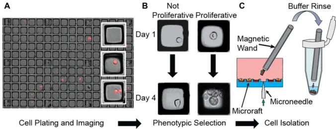
We developed a novel method for single cell selection, iso-lation and sequencing based on microraft arrays (Figure1, Supplementary Figure S1 and Supplementary File 5) (7,8). These arrays consist of thousands of individual square mag-netic polystyrene cell culture sites that are each contained within elastomeric microwells. Cells are plated on the mi-croraft array as a dilute suspension and settle onto indi-vidual microrafts. Once these cells have adhered, individ-ual microrafts chosen by the investigator can be dislodged from the underlying array using a motorized microneedle that pierces the array from below. Dislodged microraft cell carriers can then be magnetically manipulated into separate sample containers where the attached cells are lysed without being stripped from the microraft carrier.
Microraft arrays enable investigators to selectively isolate single cells of interest, even from small numbers of cells, based on a rich variety of cellular phenotypes, such as cel-lular morphology, proliferation and cell surface marker ex-pression levels. Individual cells on microrafts are isolated gently without the need to strip the cells from their sub-strate or perform microfluidic sorting. Furthermore, micro-raft arrays present an ideal environment for in vitro cul-ture, enabling examination of cell morphology and growth properties over extended periods of time or in response to treatment/conditions. Cell isolation performed using mi-croraft arrays yields highly viable cells that can be assayed or maintained in culture for other downstream applications.
Comparison of microrafts and the Fluidigm C1
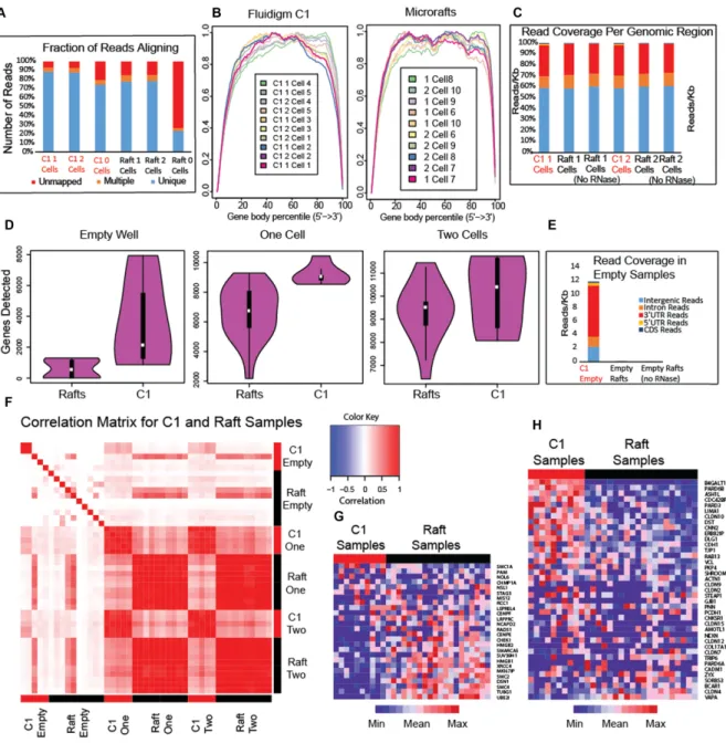
We compared RNA sequencing on microraft arrays and the Fluidigm C1 by isolating and sequencing pancreatic can-cer cells using both systems. We recognize that the two sys-tems utilized different techniques to isolate and sequence single cells and that there may be systematic technical vari-ation between the results from each system. It is also pos-sible that the process of cell isolation changes gene expres-sion levels. We investigated these effects by sequencing sam-ples containing 0, 1 and 2 cells from the same CFPAC-1 cell line using both the C1 and microrafts (Figure1A and Sup-plementary Figure S2). Overall, both approaches produced data of high quality, but we observed several notable dif-ferences. We compared the read coverage (Figure 2A), 3 bias (Figure2B), genomic read distribution (Figure2C) and number of genes detected (Figure2D) in the resulting RNA-seq data. The proportion of reads aligning to the genome, 3 bias and genomic read distribution for the C1 and mi-croraft data are highly similar. We noticed that the number of genes detected shows more variation for single microraft cells than single C1 cells, and the median number of genes detected using microrafts is smaller (Figure2D). Part of this difference reflects a difference in the multiplexing used for sequencing the two sets of samples as we were able to multi-plex more microraft samples per run than the C1. To show the relationship between number of genes detected and se-quencing depth, we plotted genes detected both as a func-tion of number of aligned reads, as well as genes detected per 100 000 reads (Supplementary Figure S3). We also
spec-ulate that this difference in genes detected could be due to the greater efficiency of reagent delivery in the microfluidic system, which allows the tiny amount of starting RNA to be concentrated in a microscale reaction volume. This differ-ence wasmuch smallerwhen comparing samples containing two cells (Figure2D), likely because of the larger amount of starting RNA generated by two cells.
Comparing the C1 empty samples with the microraft empty samples shows that the C1 empty samples contain a much higher proportion of reads mapping to the hu-man genome (Figure2E). These mapped reads from empty C1 samples are strongly enriched for coding regions of the genome, indicating that these reads come from RNA, not DNA contamination (Figure2E). The C1 empty samples show a much higher proportion of reads mapping to the 3 UTR (Figure2E), suggesting that the reads may origi-nate from broken or degraded transcripts. These reads may have originated from cells that lysed after being deposited in the C1, resulting in free-floating RNA in the cell sus-pension that subsequently was carried within the microflu-idics. We also performed qPCR on cells isolated on the C1 from a starting cell suspension mixture of mouse blood cells and mCherry CFPAC-1 cells, which were identified on the C1 chip under the Texas Red filter. We found mouse RNA in both empty nests and nests containing human cells (see Supplementary Table S1). Such cross-contamination was also observed in a similar recent evaluation of the Fluidigm C1 (2). In contrast, we observed few reads in the empty mi-croraft samples (Figure 2E), likely because we rinsed and deposited the microrafts into separate reaction wells before RNA extraction, removing any contaminating RNA.
It is difficult to assess how much contamination between wells on the C1 affects single cell RNA-seq results for two reasons. First, in the examples of contamination that we show in Supplementary Table S1, the CT values for the con-taminating RNA present in the empty wells are compara-ble to the CT values from wells containing single cells, in-dicating that the amount of contaminating RNA is signif-icant. Also, although computational filtering can be used to remove contaminating reads from another species, as in the example of mouse reads contaminating human RNA-seq data, there is no way of computationally disambiguat-ing RNA from two cells with the same genome. Thus, the real problem is potential mixing of RNA from different cells of the same species, blurring the identities of the individual cells under consideration.
Figure 1. Selective cell isolation using microraft arrays. (A) Composite fluorescence and bright-field microscopy of mCherry cells on microraft arrays. (B) Selection of cells based on a phenotype such as proliferation. (C) A microneedle dislodges microraft cell-carriers, which are manipulated through multiple buffer washes and into lysis buffer using a magnetic wand.
significant levels of additional technical variation. Because we could manually add spike-ins to each cell after isolation, we used two separate spike-in mixes with different concen-trations of the individual spike-in transcripts. This experi-ment is not possible on the Fluidigm C1, because spike-ins for all cells are loaded in a single batch. We used this infor-mation to show that the spike-in transcripts with different expression levels between the two mixes show the expected differential expression across the set of cells that we studied (Supplementary Figure S4).
We computed correlations between the gene expression profiles of cells and found that pairs of samples sequenced using the same platform showed strong concordance, indi-cating that both the microrafts and the C1 produce repro-ducible results across replicates (Figure2F and Supplemen-tary Figure S5). However, the correlations between pairs of samples from different platforms showed lower agreement (Figure2F and Supplementary Figure S5). In computing these correlations, we restricted the set of genes to those with an average expression level of at least 1 FPKM across both C1 and microraft samples to ensure that the effect was not simply due to differences in number of genes detected.
To further investigate the differences between C1 and mi-croraft samples, we performed gene set enrichment analysis (GSEA, 15) and found several gene sets with significantly higher transcript abundance (FDR: false discovery rate< 10%) in the microraft samples (Supplementary File 1). No gene sets show significant upregulation in the C1 samples after FDR correction (10% FDR), but several have signif-icant nominalP-values (P<0.05), which is suggestive of differences rather than conclusive and may reflect the fact there is less power for the C1 comparison because there are fewer C1 samples than microraft samples. An interesting theme emerges from the gene sets that show differences be-tween the two methods. Among the top gene sets upregu-lated in the raft cells are electron transport, mitochondrial protein, ribosome, nucleoside kinase, chromosome conden-sation (Figure 2G) and mitosis. All of these gene sets are required for active cell growth and division. Among the top gene sets showing up-regulation in the C1 cells, in contrast,
are cell junction (Figure2H), cell adhesion and cell migra-tion.
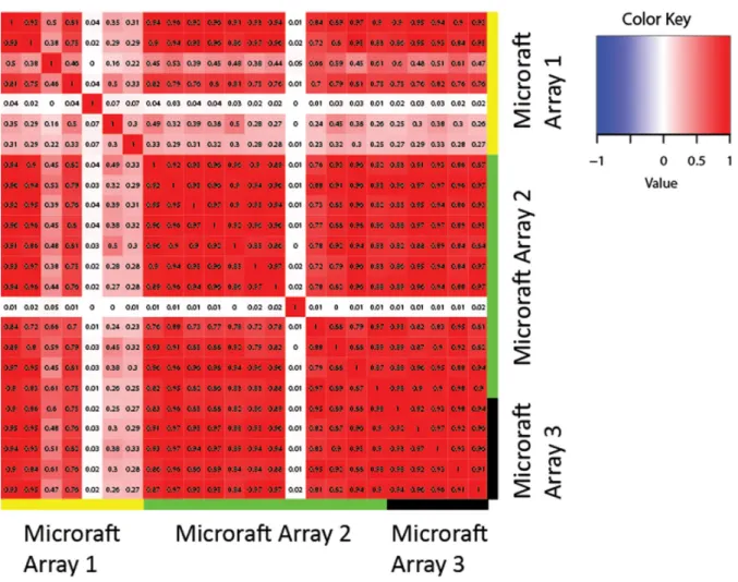
We confirmed the reliability of the microraft platform for single cell RNA-seq with two additional analyses that con-trol for artifacts unique to the microraft system. First, to control for possible variations in the microraft manufac-turing process, we compared RNA-seq results from cells grown on separate microraft arrays under identical condi-tions. Pairwise correlations among the cells showed strong concordance (Figure3and Supplementary Figure S6), indi-cating that systematic differences between microraft arrays are not a significant source of experimental variation. We also examined RNA-seq data from cells grown on the mi-crorafts and RNA-seq from bulk grown cells whose RNA was pooled and split into single cell volumes before se-quencing. Because the growing conditions are slightly dif-ferent and the RNA from bulk grown cells was pooled be-fore sequencing, we expect the gene expression profiles to be quite similar but not identical. Most genes appeared highly similar (Pearson ρ = 0.71, Spearman ρ = 0.70), with differences possibly caused by the slightly different grow-ing conditions for the bulk and sgrow-ingle cells. Differential pression analysis did, however, reveal 588 differentially ex-pressed genes (DEGs) (2% of exex-pressed genes), with only a handful of GO terms in common among genes upregulated in bulk versus microraft cells (Supplementary File 2). The only noticeable commonality among GO terms was trans-lation, with terms including ‘ribosome’, ‘ribosomal protein’ and ‘translational elongation’ appearing on the list (Supple-mentary File 2). These data show that for the CFPAC-1 cells characterized here in bulk and as single cells are largely sim-ilar, but that single cell resolution can identify subtle differ-ences between patterns inferred from bulk data versus single cells.
Selective cell isolation based on proliferation after gemc-itabine treatment
Figure 2.Comparing RNA-seq from cells isolated with microrafts and the Fluidigm C1. (A) Fraction of reads aligning to the human genome. (B) Gene body coverage plots measuring 3bias in read coverage. (C) Distribution of read coverage by genomic region. (D) Number of genes detected. (E) Read coverage for empty samples. (F) Heatmap of Pearson correlations among C1 and microraft cells. (G) Heatmap of chromosome condensation gene set, which shows strong upregulation in raft cells. (H) Heatmap of cell junction gene set, which shows slight upregulation in C1 cells.
cells, we performed a pilot study to evaluate the differences in proliferative capacity of single cancer cells in response to chemotherapy treatment. Proliferation is an important property of tumor cells that cannot be used as a basis for sorting using the C1 system. CFPAC-1 cells were plated onto a microraft array, treated with 2 or 5 nM gemcitabine and allowed to grow for 4 days. Control cells were plated and subjected to a placebo treatment and no treatment, re-spectively. We also stained the cells in this experiment for EpCAM, which illustrates the compatibility of microrafts and cell staining (Supplementary Figure S7). Tracking the cells on the array during the growth period showed that the placebo treatment had essentially no effect on cell growth, but the 5 nM gemcitabine treatment strongly restricted the
Figure 3.Reproducibility of microraft results. Heatmap shows the correlations among expression profiles of cells grown under identical conditions on three separate microraft arrays. The correlation among cells grown under the same conditions on either the same or different microraft array suggests strong reproducibility, although the correlation with some cells on array 1 is lower. With the exception of two cells, all of the cells on array 1 have<3000 genes detected, which partially reflects the multiplexing of array 1. In contrast, the smallest number of genes detected in any of the other cells shown is 5000. The correlations between cells on arrays 2 and 3 are all very high, although we observe some biological variation, as expected, among individual cells.
Figure 4. Quantification of CFPAC-1 colony growth. (A) Bulk cell mea-surements. (B) Single-colony measurements. Upper, middle and lower red lines indicate the 75th, 50th and 25th percentiles, respectively. The 50th percentile fold change was 4.46, 4.21, 3.95 and 2.58 for cells receiving no treatment, placebo, 2 and 5 nM drug, respectively.
not unreasonable given that each such microraft contains a clonal colony formed from a single cell.
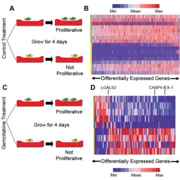
Figure 5. Linking gene expression changes to differences in proliferation after drug treatment. (A) Cells on no treatment and placebo treatment mi-croraft arrays are selected based on proliferation. (B) Heatmap of genes that change in dividing versus non-dividing cells from the no treatment and placebo treatment microraft arrays. (C) Cells on gemcitabine treat-ment microraft arrays are selected based on proliferation. (D) Heatmap of genes differentially expressed in cells that divide after gemcitabine treat-ment but not control cells.
observed lower expression ofIEX-1in cells dividing after gemcitabine treatment. Similarly, we observed downregula-tion ofCASP4in cells that proliferate after drug treatment. This result is consistent with prior reports ofCASP4 knock-down in pancreatic cells leading to decreased apoptosis af-ter bortezomib treatment (24). We also noted an example of up-regulation after drug treatment; LGALS3was pre-viously shown to induce cell proliferation and invasion in pancreatic cancer cells by activating Ras signaling (25). Fi-nally, several of our candidate genes overlapped with those seen in a somewhat similar bulk RNA-seq study looking at gemcitabine resistance (26).
Continued proliferation during gemcitabine treatment is also associated with DEGs functioning in secretion of pro-teins to the endoplasmic reticulum (ER) and, to a lesser ex-tent, mRNA surveillance and splicing (Supplementary File 3). Changes in ER stress response are known to allow tu-mors to survive chemotherapy treatment in some cases (27). Gemcitabine is a nucleoside analog that results in increased DNA damage, and response to DNA damage is known to be intertwined with ER stress response (27). We hypothesize that changes in the expression of this set of genes modifies ER stress response, which allows drug-treated cells to han-dle gemcitabine-induced damage.
Finally, we looked at the within-group gene expression differences among the cells from each treatment group, a comparison that highlights a key benefit of single cell res-olution. Given the small sample size and relatively low sequencing depth, our power to make conclusive state-ments is limited. Nevertheless, we observed several
inter-esting patterns. A set of genes showed much greater varia-tion in the gemcitabine-treated cells than the control cells (Supplementary Figure S8). Among such highly variable genes in the drug treated cells, we noticed DNA repair associated genes (NSMCE1,RAD23A) and/or oncogenes (RNF103-CHMP3). This indicates that gemcitabine treat-ment changed the expression levels of these genes, but pos-sibly only in a subset of the cells, hinting that in this ex-periment there are multiple mechanisms for continuing to proliferate in the presence of gemcitabine.
DISCUSSION
Microraft arrays provide two key advantages over exist-ing sexist-ingle cell isolation approaches for genomics: (i) selec-tive isolation without the need for a pre-selection step and (ii) the ability to sort cells based on complex temporal or spatial phenotypes, as well as by cellular markers. Here, we reported the application of this cellular isolation sys-tem to connect a proliferative pancreatic cancer cell phe-notype in the presence of chemotherapeutic to these cells’ altered transcriptome. We also showed that microrafts pro-duce data of comparable quality to the C1 but with less con-tamination between cells during the isolation process.
Microrafts promise to be broadly useful. To date more than 30 cell lines and a number of primary cell types have been successfully cultured on the arrays, which can be coated with any desired extracellular matrix to promote cell health and growth (7,28–31). Many dynamic pheno-types, such as cell mobility, invasiveness, morphology or biomarker expression, are of immense interest in basic re-search fields such as cell and developmental biology. Mi-crorafts are also useful for isolating rare cells, and in prin-ciple can be used to isolate cells from a suspension con-taining only a few dozen cells of interest. While the cur-rent report describes a manual protocol for cell isolation and analysis, the method has the potential to perform high-throughput cell isolation, because a single microraft array can be scaled to contain thousands to millions of microrafts and is amenable to incorporation in an automated plat-form. Indeed, work is ongoing to develop automated high-throughput instrumentation that will carry out the entire procedure of array scanning, target-cell identification, mi-croraft release, collection and transfer under temperature, humidity and CO2 controlled conditions at much faster
rates than is currently possible. By virtue of indexing on the array, cells in culture can also be used to provide time-series measurements of phenotypic phenomena as was done in the current work or to provide samples for multiple down-stream analyses.
There are also a number of potential applications for mi-crorafts in clinical research. For example, tumors often ex-hibit strong intratumoral gene expression and phenotypic heterogeneity. Microrafts could be used to make the link be-tween transcriptomic and functional heterogeneity within tumors. Some functional differences among tumor cells, such as the ability of cells to leave the solid tumor, have im-portant implications for patient outcomes. Microrafts pro-vide an invaluable tool for studying such phenomena by connecting gene expression with phenotypic measurements from individual cells.
DISCLOSURE DECLARATION
N.L.A. and C.E.S. are inventors of microraft arrays (US Patent #9,068155) and have financial interests in Cell Mi-crosystems, Inc.
SUPPLEMENTARY DATA
Supplementary Dataare available at NAR Online.
ACKNOWLEDGEMENTS
We are grateful for discussions with Erin Osborne, Sophia Tintori and Dan McKay regarding the nuances of single cell genomics. Thanks to Scott Magness and the Advanced An-alytics Core of the UNC Center for GI Biology and Disease for assistance with the Fluidigm C1.
FUNDING
NCBC [2013-MRG-1110, in part to C.D.J.]; UCRF (in part to C.D.J.); National Science Foundation (NSF) [ABI/EF0850237 to J.F.P.]; National Institutes of Health (NIH) [R43EB019752 to N.L.A.]; NSF Graduate Research Fellowship [DGE-1144081 to J.D.W.]; NIH BD2K Fellow-ship [T32 CA201159 to J.D.W.]. Funding for open access charge: NCBC [2013-MRG-1110].
Conflict of interest statement.N.L.A. and C.E.S. are inven-tors of microraft arrays (US Patent #9,068155) and have financial interests in Cell Microsystems, Inc.
REFERENCES
1. Saliba,A.-E., Westermann,A.J., Gorski,S.A. and Vogel,J. (2014) Single-cell RNA-seq: advances and future challenges.Nucleic Acids Res.,42, 8845–8860.
2. Macosko,E.Z., Basu,A., Satija,R., Nemesh,J., Shekhar,K.,
Goldman,M., Tirosh,I., Bialas,A.R., Kamitaki,N., Martersteck,E.M.
et al.(2015) Highly parallel genome-wide expression profiling of individual cells using nanoliter droplets.Cell,161, 1202–1214. 3. Klein,A.M., Mazutis,L., Akartuna,I., Tallapragada,N., Veres,A.,
Li,V., Peshkin,L., Weitz,D.A. and Kirschner,M.W. (2015) Droplet barcoding for single-cell transcriptomics applied to embryonic stem cells.Cell,161, 1187–1201.
4. Bose,S., Wan,Z., Carr,A., Rizvi,A.H., Vieira,G., Pe’er,D. and Sims,P.A. (2015) Scalable microfluidics for single cell RNA printing and sequencing.Genome Biol.,16, 120.
5. Kimmerling,R.J., Lee Szeto,G., Li,J.W., Genshaft,A.S., Kazer,S.W., Payer,K.R., de Riba Borrajo,J., Blainey,P.C., Irvine,D.J., Shalek,A.K.
et al.(2016) A microfluidic platform enabling single-cell RNA-seq of multigenerational lineages.Nat. Commun.,7, 10220.
6. Kolodziejczyk,A.A., Kim,J.K., Svensson,V., Marioni,J.C. and Teichmann,S.A. (2015) The technology and biology of single-cell RNA sequencing.Mol. Cell,58, 610–620.
7. Gach,P.C., Wang,Y., Phillips,C., Sims,C.E. and Allbritton,N.L. (2011) Isolation and manipulation of living adherent cells by micromolded magnetic rafts.Biomicrofluidics,5, 32002–3200212. 8. Wang,Y., Phillips,C., Xu,W., Pai,J.-H., Dhopeshwarker,R., Sims,C.E.
and Allbritton,N.L. (2010) Micromolded arrays for separation of adherent cells.Lab. Chip.,10, 2917–2924.
9. Jiang,L., Schlesinger,F., Davis,C.A., Zhang,Y., Li,R., Salit,M., Gingeras,T.R. and Oliver,B. (2011) Synthetic spike-in standards for RNA-seq experiments.Genome Res.,21, 1543–1551.
10. Edelstein,A., Amodaj,N., Hoover,K., Vale,R. and Stuurman,N. (2010) Computer control of microscopes using manager.Curr. Protoc. Mol. Biol., doi:10.1002/0471142727.mb1420s92.
11. Wang,K., Singh,D., Zeng,Z., Coleman,S.J., Huang,Y., Savich,G.L., He,X., Mieczkowski,P., Grimm,S.A., Perou,C.M.et al.(2010) MapSplice: accurate mapping of RNA-seq reads for splice junction discovery.Nucleic Acids Res.,38, e178.
12. Langmead,B. and Salzberg,S.L. (2012) Fast gapped-read alignment with Bowtie 2.Nat. Methods,9, 357–359.
13. Wang,L., Wang,S. and Li,W. (2012) RSeQC: quality control of RNA-seq experiments.Bioinformatics,28, 2184–2185. 14. Li,B. and Dewey,C.N. (2011) RSEM: accurate transcript
quantification from RNA-Seq data with or without a reference genome.BMC Bioinformatics,12, 323.
15. Subramanian,A., Tamayo,P., Mootha,V.K., Mukherjee,S., Ebert,B.L., Gillette,M.A., Paulovich,A., Pomeroy,S.L., Golub,T.R., Lander,E.S.et al.(2005) Gene set enrichment analysis: a
knowledge-based approach for interpreting genome-wide expression profiles.Proc. Natl. Acad. Sci. U.S.A.,102, 15545–15550.
16. Huang,D.W., Sherman,B.T. and Lempicki,R.A. (2009) Systematic and integrative analysis of large gene lists using DAVID
bioinformatics resources.Nat. Protoc.,4, 44–57.
17. Reich,M., Liefeld,T., Gould,J., Lerner,J., Tamayo,P. and Mesirov,J.P. (2006) GenePattern 2.0.Nat. Genet.,38, 500–501.
18. Robinson,M.D., McCarthy,D.J. and Smyth,G.K. (2010) edgeR: a bioconductor package for differential expression analysis of digital gene expression data.Bioinformatics,26, 139–140.
19. Treutlein,B., Brownfield,D.G., Wu,A.R., Neff,N.F., Mantalas,G.L., Espinoza,F.H., Desai,T.J., Krasnow,M.A. and Quake,S.R. (2014) Reconstructing lineage hierarchies of the distal lung epithelium using single-cell RNA-seq.Nature,509, 371–375.
20. Buettner,F., Natarajan,K.N., Casale,F.P., Proserpio,V., Scialdone,A., Theis,F.J., Teichmann,S.A., Marioni,J.C. and Stegle,O. (2015) Computational analysis of cell-to-cell heterogeneity in single-cell RNA-sequencing data reveals hidden subpopulations of cells.Nat. Biotechnol.,33, 155–160.
21. Hamidi,T., Alg ¨ul,H., Cano,C.E., Sandi,M.J., Molejon,M.I., Riemann,M., Calvo,E.L., Lomberk,G., Dagorn,J.-C., Weih,F.et al.
(2012) Nuclear protein 1 promotes pancreatic cancer development and protects cells from stress by inhibiting apoptosis.J. Clin. Invest.,
122, 2092–2103.
22. Sasada,T., Azuma,K., Hirai,T., Hashida,H., Kanai,M., Yanagawa,T. and Takabayashi,A. (2008) Prognostic significance of the immediate early response gene X-1 (IEX-1) expression in pancreatic cancer.Ann. Surg. Oncol.,15, 609–617.
23. Wu,M.X., Ustyugova,I. V, Han,L. and Akilov,O.E. (2013)
Immediate early response gene X-1, a potential prognostic biomarker in cancers.Expert Opin. Ther. Targets,17, 593–606.
24. Nawrocki,S.T., Carew,J.S., Pino,M.S., Highshaw,R.A., Dunner,K., Huang,P., Abbruzzese,J.L. and McConkey,D.J. (2005) Bortezomib sensitizes pancreatic cancer cells to endoplasmic reticulum stress-mediated apoptosis.Cancer Res.,65, 11658–11666.
25. Song,S., Ji,B., Ramachandran,V., Wang,H., Hafley,M., Logsdon,C. and Bresalier,R.S. (2012) Overexpressed galectin-3 in pancreatic cancer induces cell proliferation and invasion by binding Ras and activating Ras signaling.PLoS One,7, e42699.
26. Shen,Y., Pan,Y., Xu,L., Chen,L., Liu,L., Chen,H., Chen,Z. and Meng,Z. (2015) Identifying microRNA-mRNA regulatory network in gemcitabine-resistant cells derived from human pancreatic cancer cells.Tumour Biol.,36, 4525–4534.
27. Dicks,N., Gutierrez,K., Michalak,M., Bordignon,V. and
Agellon,L.B. (2015) Endoplasmic reticulum stress, genome damage, and cancer.Front. Oncol.,5, 11.
(2015) A high-throughput platform for stem cell niche co-cultures and downstream gene expression analysis.Nat. Cell Biol.,17, 340–349. 29. Attayek,P.J., Hunsucker,S.A., Wang,Y., Sims,C.E., Armistead,P.M.
and Allbritton,N.L. (2015) Array-based platform to select, release, and capture epstein-barr virus-infected cells based on intercellular adhesion.Anal. Chem.,87, 12281–12289.
30. Koh,J., Hogue,J.A., Wang,Y., DiSalvo,M., Allbritton,N.L., Shi,Y., Olson,J.A. and Sosa,J.A. (2016) Single-cell functional analysis of parathyroid adenomas reveals distinct classes of calcium sensing behaviour in primary hyperparathyroidism.J. Cell. Mol. Med.,20, 351–359.
31. Gordon,K.R., Wang,Y., Allbritton,N.L. and Taylor,A.M. (2015) Magnetic alignment of microelements containing cultured neuronal networks for high-throughput screening.J. Biomol. Screen.,20, 1091–1100.
32. Nagano,T., Lubling,Y., Stevens,T.J., Schoenfelder,S., Yaffe,E., Dean,W., Laue,E.D., Tanay,A. and Fraser,P. (2013) Single-cell Hi-C
reveals cell-to-cell variability in chromosome structure.Nature,502, 59–64.
33. Buenrostro,J.D., Wu,B., Litzenburger,U.M., Ruff,D., Gonzales,M.L., Snyder,M.P., Chang,H.Y. and Greenleaf,W.J. (2015) Single-cell chromatin accessibility reveals principles of regulatory variation.
Nature,523, 486–490.
34. Smallwood,S.A., Lee,H.J., Angermueller,C., Krueger,F., Saadeh,H., Peat,J., Andrews,S.R., Stegle,O., Reik,W. and Kelsey,G. (2014) Single-cell genome-wide bisulfite sequencing for assessing epigenetic heterogeneity.Nat. Methods,11, 817–820.
35. Rotem,A., Ram,O., Shoresh,N., Sperling,R.A., Goren,A., Weitz,D.A. and Bernstein,B.E. (2015) Single-cell ChIP-seq reveals cell