SPATIOTEMPORAL COORDINATION OF THE ACTIN CYTOSKELETON AND INTEGRIN ADHESION
Lindsay B. Case
A dissertation submitted to the faculty at the University of North Carolina at Chapel Hill in partial fulfillment of the requirements for the degree of Doctor of Philosophy in the Department of Cell and
Developmental Biology.
Chapel Hill 2014
iii ABSTRACT
Lindsay B. Case: Spatiotemporal coordination of the actin cytoskeleton and integrin adhesion during cell migration
(Under the direction of Dr. Clare Waterman)
Integrin-based adhesions mediate critical interactions between the cell and its external
environment. Integrins assemble into macromolecular “focal adhesions” (FAs) that contain hundreds of proteins and indirectly connect integrin cytoplasmic tails to the actin cytoskeleton. Forces transmitted across FAs drive tissue morphogenesis, cell movement, and extracellular matrix (ECM) remodeling. During cell migration, actin polymerization drives protrusion of the leading edge and integrins mediate adhesion to the ECM. However, efficient movement requires that these distinct processes are
spatiotemporally coordinated. Here we provide evidence that Arp2/3-mediated actin polymerization can stimulate integrin adhesion throughout the cell surface, while vinculin activation is spatiotemporally controlled by the macromolecular FA architecture to engage actin retrograde flow to growing FAs. We have identified and characterized “adhesive F-actin waves,” a novel integrin-mediated adhesion complex coupled to ventral actin polymerization. Integrins engage the ECM downstream of Arp2/3-mediated ventral F-actin waves in a variety of mammalian cell types. These adhesive F-actin waves require a cycle of integrin engagement and disengagement to the extracellular matrix for their formation and propagation, and exhibit morphometry and a hierarchical assembly and disassembly mechanism distinct from other integrin-containing structures. These results suggest that Arp2/3 activity, rather than the specific lamellipodium structure, is important for initiating integrin adhesion.
iv
v
In memory of my mother, Cynthia Case.
Your encouragement, love, and unwavering faith in my ability to succeed gave me the courage to aim for greatness.
vi
ACKNOWLEDGEMENTS
This work would not have been possible without the contribution and guidance of my wonderful colleagues and collaborators. I have been fortunate to be surrounded by amazing scientists at both the NIH and UNC who have provided me with an amazing education in cell biology.
First and foremost, I would like to thank my advisor, Clare Waterman, for giving me so many opportunities to further my research and my education. Clare took a chance on a quiet first-year graduate student and gave me a scientific home where I could flourish. Clare taught me to think critically and creatively, while also fostering my scientific independence by giving me the freedom to develop my ideas. She encouraged me to follow the science, even when my experiments took me down some unexpected paths. I am also extremely grateful for the opportunities Clare gave me outside of the lab. In addition to presenting my work at many conferences, I also participated in numerous courses in Woods Hole, MA as both a student and a teaching assistant. These “extracurricular” activities were instrumental in my education and growth as a scientist.
Many thanks to my thesis committee, Keith Burridge, Ken Jacobson, Jim Bear, and Sharon Campbell, for guidance, advice, and encouragement. My committee was truly a dream team of experts in the cytoskeleton, vinculin and microscopy, and I am extremely humbled to have spent so much time discussing my work with them.
vii
troubleshooting. Thanks to Ana Pasapera for teaching me how to immunoprecipitate and answering endless questions about western blots and immunostaining.
None of the superresolution experiments would have been possible without the help of our collaborators Harald Hess and Gleb Shtengel at Janelia Farm. I spent endless days (and sometimes nights) in their lab using their iPALM system, and they were always generous with giving me time on the microscope. Gleb patiently worked with me from the beginning as I learned how to use the machine and the software. Also, many thanks to Tony Kanchanawong, the iPALM pioneer, for sharing his knowledge and protocols with me before leaving the Waterman lab to start his own group.
In addition to Harold and Gleb, I had so many amazing collaborators who made a very ambitious project possible. Thank you to Sharon Campbell, Peter Thompson, and Caitlin Tolbert at UNC for many helpful discussions about vinculin biochemistry and a fruitful collaboration. Thanks in particular to Peter for performing biochemistry experiments and giving me advice on vinculin point mutations. Thanks to Michelle Baird and Michael Davidson at FSU for making so many DNA constructs. You are cloning rock stars! Thanks to Susan Craig at Johns Hopkins for sharing her vinculin FRET probe and for the many decades of meticulous vinculin biochemistry which provided the framework for my project.
viii
how to be a great microscopist. Special thanks to course director Robert Hard for first inviting me to be a teaching assistant and welcoming be back for so many years.
The support of administrators at UNC and NIH was essential for the completion of my dissertation and my graduate education. Thanks to Dorothee Honemond (NHLBI), Shwanna Thacker (NHLBI), Janice Warfford (UNC) and the NIH Office of Intramural Training and Education staff. Thanks also to Dorothee Honemond for sharing her office with me when there was no space in the lab!
Thank you to my undergraduate mentor Omar Quintero for introducing me to the cytoskeleton, cell migration, and microscopy and for sparking my interest in cell biology research. You were
instrumental in helping me start my journey toward graduate school and you continue to provide me with valuable advice and encouragement. Your dedication to undergraduate education and research is inspiring. Thanks to all my colleagues in the Laboratory of Cell Biology (NHLBI) and Cell and
Developmental Biology Department (UNC) for so many interesting discussions and seminars over the years. It has been an honor to work in two of the best cytoskeleton and cell biology communities in the world!
Finally, thank you to all of my family and friends who provide so much love and support and helped me bear my losses. You have always believed in me and continually remind me not to lose sight of what is truly important.
ix
TABLE OF CONTENTS
LIST OF TABLES ... xiii
LIST OF FIGURES ... xiv
LIST OF ABBREVIATIONS AND SYMBOLS ... xvi
CHAPTER 1: INTRODUCTION. INTEGRATION OF ACTIN DYNAMICS AND CELL ADHEION BY A THREE-DIMENSIONAL, MECHANOSENSAIVE MOLECULAR CLUTCH ... 1
1.1 Summary ... 1
1.2 The molecular clutch hypothesis: a brief history ... 1
1.3 Forces generated in the actin cytoskeleton drive actin retrograde flow ... 4
1.4 FAs are a three-dimensional macromolecular complex that physically connect the actin cytoskeleton to the ECM ... 4
1.5 The actin cytoskeleton is a master regulator of FAs ... 7
1.6 Forces at FAs can regulate protein-protein interactions and protein activity ... 9
1.7 Talin and vinculin regulate a mechanosensative FA clutch ... 10
1.8 The three-dimensional molecular clutch ... 12
1.9 Beyond FAs: molecular clutches at diverse adhesive interactions ... 13
CHAPTER 2: ADHESIVE F-ACTIN WAVES ARE A NOVEL INTEGRIN-MEDIATED ADHESION COMPLEX COUPLED TO VENTRAL ACTIN POLYMERIZATION ... 16
2.1 Summary ... 16
2.2 Introduction ... 16
x
Ventral F-actin waves are followed by integrin waves in mammalian cells ... 18
U2OS integrin waves are distinct from previously characterized integrin- containing structures ... 22
Ventral F-actin and integrin waves are ECM-dependent and require a cycle of integrin engagement to and disengagement from the ECM ... 25
Adhesive F-actin waves contain focal adhesion proteins that assemble and disassemble in a distinct stepwise order ... 29
Ventral F-actin waves are followed by integrin waves in mammalian cells ... 18
Ventral F-actin waves are followed by integrin waves in mammalian cells ... 18
2.4 Discussion ... 32
2.5 Experimental Procedures ... 36
Cell culture, Transfection, and Reagents ... 36
Microscopy ... 37
Analysis ... 38
CHAPTER 3: ADHESIVE F-ACTIN WAVES ARE INHIBITED BY THROMBIN-INDUCED MYOSIN CONTRACTILITY IN ENDOTHELIAL CELLS ... 39
3.1 Summary ... 39
3.2 Introduction ... 39
3.3 Results ... 41
Adhesive F-actin waves spontaneously form in resting endothelial cells ... 41
Adhesive F-actin waves are not contact-inhibited ... 43
Thrombin inhibits adhesive F-actin waves by the ROCK-dependent upregulation of myosinII contractility ... 43
Adhesive F-actin waves do not degrade the extracellular matrix ... 46
3.4 Discussion ... 48
xi
Cell culture, Transfection, and Reagents ... 49
Microscopy ... 49
Analysis ... 50
CHAPTER 4: USING INTERFEROMETRIC PHOTOACTIVATED LOCALIZATION MICROSCOPY (“iPALM”) TO STUDY FOCAL ADHESION NANOSCALE ARCHITECTURE ... 51
4.1 Introduction ... 51
4.2 Interferometric photoactivated localization microscopy ... 52
Achieving lateral resolution with single molecule localization ... 52
Achieving axial resolution with interferometry ... 53
Fiducial-based drift-correction, alignment and calibration ... 54
iPALM data processing ... 55
4.3 Application of iPALM to the study of focal adhesions ... 57
4.4 Conclusions ... 58
CHAPTER 5: MOLECULAR MECHANISM OF VINCULIN ACTIVATION AND NANO-SCALE SPATIAL ORGANIZATION IN FOCAL ADHESIONS ... 59
5.1 Summary ... 59
5.2 Introduction ... 59
5.3 Results ... 61
Vinculin is distributed throughout the three focal adhesion nano-domains ... 61
Vinculin is required to stabilize talin in an extended conformation in FA ... 62
Vinculin is oriented in FA with the tail above the head ... 64
Activation promotes association of vinculin with the FTL and ARL ... 66
Talin binding is required for vinculin activation in FA and promotes localization of active vinculin to the FTL and ARL ... 68
xii
Binding to phosphorylated paxillin targets vinculin to the ISL ... 73
Actin binding is required for activation but does not regulate vinculin nanoscale localization at FA ... 77
Vinculin exhibits a gradient in activation and axial position across single FA ... 80
Vinculin is recruited initially to the ISL and later to the FTL and ARL during myosin II-mediated FA maturation ... 81
5.4 Discussion ... 84
5.5 Experimental Procedures ... 87
Cell culture and transfection ... 87
Pharmacological treatments ... 88
Plasmids ... 88
Mutagenesis to generate vinculin and paxillin mutants ... 91
siRNA knockdown of endogenous protein ... 91
Western blot ... 91
Immunoprecipitation ... 92
TIRF Microscopy ... 92
FRET Imaging and Analysis ... 93
iPALM imaging ... 94
Analysis of processed iPALM data ... 96
Statistical analysis ... 96
CHAPTER 6: CONCLUSIONS AND FUTURE PERSPECTIVES ... 97
APPENDIX: iPALM DATA SUMMARY ... 104
xiii
LIST OF TABLES
xiv
LIST OF FIGURES
Figure 1.1. The molecular clutch hypothesis ... 2
Figure 1.2. Nano-scale architecture of the focal adhesion clutch ... 6
Figure 1.3. Molecular clutches may mediate diverse cell adhesive interactions ... 14
Figure 2.1. Ventral F-actin waves are followed by integrin waves in mammalian cells ... 19
Figure 2.2. Arp2/3-containing ventral F-actin waves are followed by integrin waves in U2OS cells ... 21
Figure 2.3. Integrin waves are distinct from podosomes and focal adhesions in U2OS cells ... 24
Figure 2.4. Integrin waves and ventral F-actin waves are visible by Interference Reflection Microscopy ... 26
Figure 2.5. Ventral F-actin and integrin waves require integrin engagement to extracellular matrix ... 28
Figure 2.6. Focal adhesion (FA) proteins assemble into integrin waves in a precise order ... 30
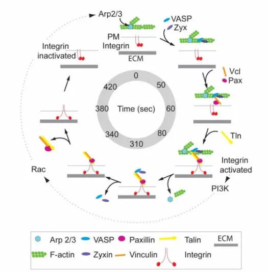
Figure 2.7. Speculative model of adhesive F-actin wave assembly and disassembly ... 34
Figure 3.1. HUVECs form adhesive F-actin waves containing integrin and paxillin ... 42
Figure 3.2. Adhesive F-actin waves are not contact inhibited ... 44
Figure 3.3. Thrombin inhibits adhesive F-actin waves through a RhoA-Rock pathway ... 45
Figure 3.4. Adhesive F-actin waves do not degrade the ECM ... 47
Figure 4.1. The iPALM system ... 56
Figure 5.1. Vinculin is distributed throughout the three focal adhesion nano-domains ... 63
Figure 5.2. Vinculin is required to maintain talin in an extended conformation ... 65
Figure 5.3. Vinculin is oriented in focal adhesions with the tail above the head, and activation promotes association of vinculin with the actin regulatory layer ... 67
xv
Figure 5.5. Talin binding is required for vinculin activation and localization
of active vinculin to the force transduction and actin regulatory layers ... 71
Figure 5.6. Paxillin is required for vinculin localization in the integrin
signaling layer but not for vinculin activation ... 74
Figure 5.7. Binding to phospho-paxillin targets vinculin to the integrin signaling layer ... 76
Figure 5.8. Actin binding is required for vinculin activation but does not regulate
vinculin position ... 78
Figure 5.9. Vinculin activation and nano-scale localization is spatio-temporally
regulated ... 82
Figure 5.10. Model of vinculin recruitment and activation during FA maturation ... 85
xvi
LIST OF ABBREVIATIONS AND SYMBOLS
ARL Actin regulatory layer
Au Gold
Bleb Blebbistatin
CCD Charge-coupled device
CO2 Carbon dioxide
Ctrl Control
Cyto Cyotplasm
CytoD Cytochalasin D
ECM Extracellular matrix
EGFP Enhanced green fluorescent protein
EM Electron microscopy
EMCCD Electron multiplying charge-coupled device
FA Focal adhesion
FAK Focal adhesion kinase
FRAP Fluorescence recovery after photobleaching
FRET Fluorescence resonance energy transfer
FTL Force transduction layer
xvii GFP Green fluorescent protein
HFF Human foreskin fibroblast
hr hour
HUVEC Human umbilical cord endothelial cell
ILK Integrin linked kinase
Inh Inhibitor
Int Integrin
IP immunoprecipitate
iPALM Interferometric photoactivated localization microscopy
IRM Interference reflection microscopy
ISL Integrin signaling layer
KD Knockdown
KPa kilo pascals
Kym Kymograph
LatA Latrunculin A
MEF Mouse embryonic fibroblast
min Minutes
mM millimolar
M micromolar
xviii MnCl2 Manganese chloride
mTurq mTurquoise
NA Numerical aperture
NeonGr NeonGreen
nm nanometer
Pa pascal
PALM Photoactivated localization microscopy
PFA Paraformaldehyde
PI3K Phosphoinositide-3-kinase
PIP2 Phosphatidylinositol 4,5-bisphosphate
PKC Protein kinase C
PMA Phorbol myristate acetate
pN Piconewton
PSF Point spread function
Pxn paxillin
ROCK Rho-associated protein kinase
ROI Region of interest
rpm Revolutions per minute
sec seconds
xix siRNA small interfereing RNA
tagRFP tag-red fluorescent protein
TIRFM Total internal reflection fluorescence microscopy
term terminus
Throm thrombin
Tln talin
TNF Tumor necrosis factor alpha
UV Ultraviolet
UTR Untranslated region
VBS Vinculin binding site
VEGF Vascular endothelial growth factor
Vcl vinculin
WT Wildtype
MAPK Mitogen activated protein kinase
1
CHAPTER 1: INTRODUCTION
INTEGRATION OF ACTIN DYNAMICS AND CELL ADHESION BY A THREE-DIMENSIONAL, MECHANOSENSATIVE MOLECULAR CLUTCH
1.1 Summary
Cell migration is important during embryonic development1,2, the immune response, and wound healing3, and the misregulation of cell migration can lead to inflammation and cancer metastasis4. Cell migration involves coordinated edge protrusion and adhesion to the extracellular matrix (ECM), usually in response to external chemical or physical guidance cues5. In order to move, cells must be able to apply forces to deform the extracellular environment and generate traction. The actin cytoskeleton is the major source of internally generated force that regulates cell shape and drives cell migration6. Actin
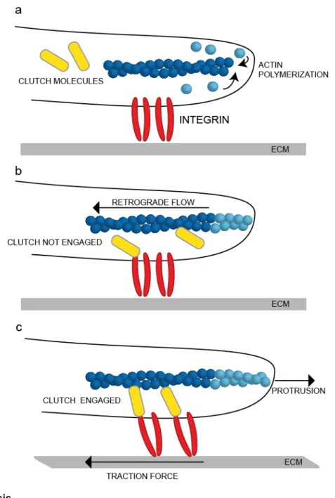
polymerization against the plasma membrane and myosin-II contraction of F-actin filaments generate a net rearward “retrograde flow” of the F-actin network relative to the direction of cell movement, but how does the cell convert rearward actin flow into forward cell movement? The molecular clutch hypothesis postulates that macromolecular focal adhesions (FAs) act as a clutch by mediating transient, indirect interactions between the retrograde moving actin cytoskeleton and ECM-bound integrins (Fig.1.1). Here we describe how forces originating in the actin cytoskeleton are transmitted to the ECM to generate the rearward traction forces that propel forward cell movement. We outline the growing body of evidence that force transduction is regulated by a three-dimensional molecular clutch consisting of the FA molecules talin and vinculin. We discuss how forces transmitted by the clutch impact the molecules within FAs and allow individual FAs to act as mechanosensors. Finally, we speculate about the generality of the
molecular clutch concept in driving dynamic cell interactions with other cells or their environment in critical physiological processes including the immune response, vascular function, and epithelial morphogenesis. 1.2 The molecular clutch hypothesis: a brief history
Cell movement has captivated scientists since the invention of the light microscope,7 and
2
Figure 1.1. The molecular
clutch hypothesis
3
In the early 1960s, Abercrombie proposed that protruding and ruffling membrane at the front of migrating chick fibroblasts was the main “locomotory organ” of the fibroblast8, and this leading edge was observed to undergo cycles of protrusion and retraction to result in net forward movement9. Abercrombie and Harris observed that marker particles derived from ink or resin moved centripetally from the leading edge along both the dorsal and ventral cell membranes at a constant rate and rearward direction relative to the direction of leading edge protrusion10,11. These first observations of “retrograde flow” were proposed to be related to forward edge protrusion and rearward traction forces, although the flow of particles was initially thought to be due to a moving plasma membrane10–12. However, it was later proposed by Wolpert and Allison that this rearward particle movement could be due to the movement of the filamentous network inside of the cell pulling proteins in a fluid plasma membrane12. Small and Abercrombie used electron microscopy to show that the leading edge is full of filamentous actin that is organized into the structurally distinct lamellipodia and lamella, and that actin within the lamellipodia is polarized with the growing barbed end facing the cell edge13,14. In 1985, Wang performed seminal FRAP experiments of
fluorescently-labeled actin to demonstrate that actin monomers were incorporated at the leading edge of the lamellipodia and then underwent a rearward movement away from the edge15. Forscher also
observed retrograde actin flow in neuronal growth cones and showed that retrograde actin flow was dependent on both actin polymerization and myosin-II contractility16,17. Theriot and Mitchinson demonstrated that actin polymerization in the lamellipodia was directly coupled to forward cell
movement18, and retrograde flow was observed to be inversely related to cell speed15,17,19,20. In 1988
Mitchison and Kirschner proposed that a “”molecular clutch” connected the retrograde moving actin cytoskeleton to extracellular matrix receptors in the plasma membrane, allowing tension to be exerted on the substrate21.
In the subsequent decades, the evidence supporting this molecular clutch hypothesis has grown; however, the basic principles remain unchanged (Fig. 1.1). Actin is rapidly polymerized in the
4
adhesion proteins to integrins, and thus retrograde flow slows down and forces from the actin cytoskeleton are propagated to the substrate, resulting in rearward traction and forward membrane protrusion. However, disengagement or slippage of the clutch results in faster retrograde flow, decreased traction forces, and cessation of membrane protrusion
1.3 Forces generated in the actin cytoskeleton drive actin retrograde flow
The retrograde movement of the actin cytoskeleton is the basis of the molecular clutch hypothesis. In adherent, migrating cells, the actin cytoskeleton is organized into two structurally and functionally distinct regions: the lamellipodium and the lamellum13,22,23. Rapid Arp2/3-mediated F-actin polymerization at the tip of the lamellipodium generates a pushing force against the plasma membrane and has been proposed to drive protrusion via a Brownian ratchet mechanism 24–26. F-actin
polymerization against the membrane also results in a counter-force that pushes the entire F-actin network rearward, and the majority of F-actin is depolymerized at the base of the lamellipodium 15,16,22,27. Therefore, although new actin monomers are continuously incorporated at the lamellipodium tip, the lamellipodial actin network exhibits treadmilling behavior and undergoes “retrograde flow” of ~0.5
micron/min22. The lamellum is located behind the lamellipodium (i.e. closer to the cell body) and contains
many distinct F-actin structures including dorsal stress fibers, transverse arcs, and ventral stress fibers28–
30. In the lamellum, myosin-II assembles into mini-filaments and contracts actin bundles to generate
forces that reorganize actin, disassemble actin, and drive a slower retrograde flow of ~0.25 micron/min
22,28,31,32. Thus, actin polymerization drives rapid retrograde flow in the lamellipodia, while myosin
contraction drives slower retrograde flow in the lamella.
1.4 FAs are a three-dimensional macromolecular complex that physically connect the actin
cytoskeleton to the ECM
There is evidence in diverse cell types that actin retrograde flow speed is inversely correlated to edge protrusion17,18,22,33, suggesting that actin retrograde flow provides the force for forward cell
5
Integrins are transmembrane heterodimers that can interact with different extracellular ligands such as fibronectin, collagen, and laminin37,38. Integrins have a large extracellular domain and a small cytoplasmic tail, and they must undergo a dramatic conformational change to become “activated” and competent to tightly bind ligand38–40. The β-integrin cytoplasmic tail binds several proteins including talin, α-actinin, and kindlin, but neither α-integrin or β-integrin can directly interact with actin 41–45. Therefore, the connection
between integrins and actin must be indirectly mediated by the assembly of the macromolecular FA structure. FAs can contain hundreds of different proteins including scaffolding proteins, structural proteins, kinases, and phosphatases, and their composition changes in response to diverse stimuli46–49. FAs form in the protruding lamellipodia as small puncta containing integrin, FAK, and paxillin 50–52. While
most of these “nascent” FAs have a lifetime of ~1 min, a subset of FAs are stabilized and undergo “maturation” when they reach the lamella51. FA maturation requires stress fiber assembly and myosin-II
activity51,53,54 as FAs elongate along an actin/α-actinin template in the direction of retrograde flow51 and undergo dramatic compositional changes47.
Although many proteins localize to FAs, these proteins are not homogenously distributed in the three-dimensional FA structure. Recent advances in light microscopy have allowed for the determination of protein localization in FAs at the nanoscale level, and has revealed that mature FAs are vertically stratified along the axis perpendicular to the ventral plasma membrane (Fig. 1.2)55–58. Paxillin and FAK associate with integrin cytoplasmic tails within ~30nm of the plasma membrane (i.e. low in the FA). In contrast, actin and the actin-associated proteins zyxin, vasp and α-actinin localize >50nm above the plasma membrane (i.e. high in the FA). Talin is a large protein that that forms the primary link between integrin and actin59. The talin head, which binds β-integrin tails, co-localizes with paxillin and FAK near
the plasma membrane, while the talin tail, which binds actin, localizes ~30nm higher42,57,60,61. Vinculin colocalizes with the talin rod in the intermediary layer57. This conserved layered organization of FA proteins is observed in diverse cell types, suggesting that it arises from the self-assembly of protein-protein interactions at FAs.
6
Figure 1.2. Nano-scale architecture of the focal adhesion clutch
7
the leading edge as the “distal tip”, while the tip closer to the cell body is the “proximal tip”. Actin stress fibers attach at the proximal tip of FAs, so actin-associated proteins are also concentrated at the proximal tip 50,57,62,63. In contrast, FAK-dependent phosphorylation of paxillin is highest at the distal tip of FAs, providing a mechanism for proteins to be concentrated in this region64. Talin molecules have been shown to be organized along the long axis of the FA such that the talin head is localized closer to the distal tip of the adhesion while the talin tail is stretched rearward in the direction of F-actin flow 65. It’s likely that other proteins are also organized along the long axis of the FA by actin retrograde flow.
Although FAs have a conserved nanoscale architecture, proteins within FAs are highly dynamic. Inactive integrins can freely diffuse in the plasma membrane at FAs, and activation triggers integrin immobilization 66,67. Most FA proteins, including paxillin, vinculin, α-actinin, talin, kindlin, FAK, zyxin, VASP, and ILK, rapidly exchange with the cytoplasmic pool (i.e. FRAP t1/2 measured to be less than 30
seconds) 62,68–70. Additionally, FRAP recovery at FAs is rarely approximated by a single exponential curve, suggesting that there are subpopulations of molecules within the FA with different dynamics. Indeed, both paxillin and vinculin have been measured to have at least four distinct sub-populations in the FA and surrounding cytoplasm, and paxillin and vinculin have different dynamics at the distal and
proximal tip of the FA 62. Thus, even when a FA appears stable for tens of minutes, the molecules within the adhesion are rapidly turning over. Furthermore, the types of interactions occuring within the FA can change over time. Fluorescence fluctuation correlation methods suggest that talin-vinculin complexes form prior to the integrin-talin complex in nascent FAs, while -actinin clusters periodically enter nascent FAs and transiently interact with integrins71. Fluoresecence cross-correlation spectroscopy studies have suggested that molecules can enter and leave the stable FA as preformed cytoplasmic complexes
corresponding to the different FA-layers. For example, paxillin and FAK co-localize in FA near the plasma membrane and diffuse together in the cytoplasm 72. Thus, it’s possible that the self-organization of protein-protein interactions dictating FA architecture may initiate from interactions in the cytoplasm. 1.5 The actin cytoskeleton is a master regulator of FAs
8
a master regulator of FAs. Actin polymerization, F-actin structural organization, and myosin-II contractility all contribute to the regulation of FAs in lamellipodia and lamella.
Actin retrograde flow has been proposed to directly contribute to integrin activation. The lateral force of retrograde flow potentially helps to drive integrin activation by separating α- and β- integrin cytoplasmic tails 76. Integrin activation can be initiated by binding of cytoplasmic proteins, such as talin, to
the integrin tails (“inside-out” activation) or by binding of integrins to their extracellular ligand (“outside-in” activation)40,77–79. Integrin activation involves a dramatic conformational change between a low-affinity
conformation with the extracellular domain folded close to the plasma membrane and a high-affinity conformation with the extracellular domain extended away from the plasma membrane40,79,80. However,
both inside-out and outside-in integrin activation correspond to a lateral separation of the α- and β-integrin cytoplasmic tails that can be measured by a loss in intermolecular FRET81. Furthermore, introducing an
artificial 14nm separation between the α5 and β1 cytoplasmic domains is sufficient to induce high-affinity binding to fibronectin in vitro, and molecular dynamics simulations suggest that the lateral force of actin retrograde flow could pull the tail away from the α tail to stabilize integrin heterodimers in an open, high-affinity conformation39,76. This force-dependent model of integrin activation predicts integrin cytoplasmic tails would open in the direction of retrograde flow, resulting in a polarized population of active integrins in FAs.
Actin polymerization also controls the formation of initial macromolecular nascent FAs. FA formation and stability in the lamellipodia requires active actin polymerization 51,75, and loss of Arp2/3 activity reduces FA assembly and results in disorganized, abnormal adhesions that do not support migration up a surface-bound gradient of ECM 82,83. Therefore, Arp2/3 activity is thought to be required for proper FA formation in the lamellipodium. The FA proteins FAK and vinculin have both been shown to bind directly to the Arp2/3 complex, suggesting a direct molecular link between Arp2/3 and FAs84,85. Although there is evidence that Arp2/3 activity could regulate adhesion formation through specific molecular interactions and indirect physical mechanisms, more research is needed to understand how Arp2/3 regulates nascent FA assembly.
9
lamellipodium-lamellum border 51,86. Thus, a row of maturing FAs spatially defines the
lamellipodium-lamellum border and contributes to the abrupt slowing of actin retrograde flow speeds in the lamella
22,73,75. During maturation, FAs undergo a compositional change as they grow and elongate in the
direction of retrograde flow 47,50,51,54,87. FAs grow at a rate proportional to actin flow, independent of specific molecular perturbations; thus, faster retrograde flow results in faster FA elongation88. This
suggests that FA growth, and therefore local integrin activation, is limited by the distance of actin retrograde movement, in agreement with the later force model of integrin activation76. FA maturation
requires tension to be applied across FAs, either from intracellular myosin contractility or extracellular pulling 89–94, and FA size correlates to the amount of applied force 74. During FA maturation, α-actinin is
recruited to cross-link actin filaments51. Mature FAs remain attached to actin stress fibers throughout their lifetime, and the maintenance of mature FAs requires association with contractile F-actin bundles 28,54,95.
Disruption of mDia2-generated dorsal stress fibers leads to abnormal FA morphology and dynamics,28,96 and several additional formin family members have been found in biochemically isolated FAs 47. While
actin regulates FAs both in the lamellipodia and the lamella, further work is needed to clarify the role of specific actin nucleators and F-actin structures in regulating the different stages of FA assembly, growth and disassembly.
1.6 Forces at FAs can regulate protein-protein interactions and protein activity
Forces generated in the actin cytoskeleton are transmitted across the macromolecular FA to generate traction on the extracellular substrate. Individual FAs have been measured to apply traction forces ranging from less than 1kPa to greater than 10kPa (1-10nN/µm2), although this is the cumulative forces distributed across many molecules in the FA74,97. The development of fluorescence- based
molecular tension sensors are allowing the direct measurement of forces applied across individual molecules98–100. The tension across individual vinculin molecules is estimated to be ~2.5pN when measured with a FRET biosensor98. Single-molecule integrin tension sensors based on FRET100 or quenching99 measured ~1-10 pN of tension across individual integrins at FAs. However, both vinculin
10
Proteins have evolved diverse mechanisms to respond to tension, and forces transmitted across FAs not only generate traction for cell migration but can also significantly alter protein localization and activity at FAs. Some molecules form catch-bonds, characterized by an increase in the dissociation lifetime upon increasing tensile force. For example, the lifetime of α5β1 integrin-fibronectin bonds is
significantly prolonged when 10-30pN of force is applied 102. Thus, increasing forces will stabilize the
integrin-fibronectin interaction. Other proteins, such as talin, undergo a force-dependent extension103. Although talin contains many vinculin binding sites (VBS) buried within the native structure, an
unstretched talin molecule contains a single available VBS 103. However, application of 2pN of force unmasks cryptic VBS allowing for the recruitment of additional vinculin molecules 103. Talin extension
occurs at forces >5pN, and binding of vinculin to talin prevents refolding of the talin rod in vitro104. However, at >25pN force, vinculin dissociates from talin 104. Force can also directly alter protein activity.
The application of a 1-3pN pulling force to the formin mDia2 bound to the end of an actin filament results in a significant increase in the actin polymerization rate 105,106.
1.7 Talin and vinculin regulate a mechanosensative FA clutch
If FAs are a molecular clutch, actin retrograde flow will be specifically engaged to the ECM at FAs. Thus, a key prediction of the molecular clutch hypothesis is that actin flow slows down locally at FAs as traction forces increase. Indeed, actin retrograde flow speeds have been experimentally observed to decrease at FAs, 53,67,75,88,107 and traction forces are observed to be highest at FAs53,74,108. Since actin does not directly interact with integrins, actin retrograde flow must be transmitted across a molecular “transmission interface” of actin and integrin binding proteins. Many molecules in FAs, including integrins, paxillin, FAK, -actinin, talin, and vinculin, have been observed to undergo retrograde flow at rates equal to or slower than the local actin retrograde flow 53,67,75,88,107,109, suggesting that retrograde flow is
transmitted across many of the molecules within FAs.
Molecules that directly bind integrins and/or actin are thought to be responsible for regulating force transmission across FAs by modulating the efficiency of the integrin-actin connection. When actin is tightly associated with integrins, there will be maximum force transmission at FAs. However, if there is “slippage” between the actin retrograde flow and the ECM, less force is expected to be transmitted 53,110–
11
to increase force transmission at FAs. Talin is one of the few molecules that can bind directly to both integrin tails and F-actin 42,60, and when talin is depleted cells have decreased traction forces and fail to stiffen in response to tension 113,114. Additionally, talin depletion causes slippage between actin
retrograde flow and the ECM, resulting in a much wider region of fast lamellipodial actin retrograde flow
114,115. Vinculin binds simultaneously to talin and F-actin, and helps to mechanically reinforce the
talin-actin linkage116,117. When vinculin is depleted cells have decreased traction forces, a widened
lamellipodia, and faster retrograde flow rates 88,97,117,118. Additionally, vinculin must interact with both talin and actin to restore WT retrograde flow and traction forces, suggesting that the talin-vinculin-actin linkage directly regulates FA clutch engagement88,117. Thus, recruitment of talin and vinculin to FAs engages actin retrograde flow and increases tractions forces on the ECM. The talin-vinculin interaction is highly sensitive to changes in force103,104, suggesting that clutch engagement has a positive feedback effect. As force transmission increases across talin, additional vinculin molecules are recruited to bind actin, slow retrograde flow, and increase force transmission. -actinin, which can bind integrin and actin, is also required for maximum force transmission across 3 integrins, and -actinin recruitment correlates with force generation119, suggesting that -actinin could regulate the molecular clutch in addition to
cross-linking actin.
Since the FA clutch can be dynamically regulated, force transmission could be variable over time
110. Furthermore, the amount of clutch engagement is predicted to be dependent on the stiffness of the
substrate110. Modeling predicts that if the cell is unable to significantly deform a stiff substrate, then there
will be stochastic, weak clutch engagement and constant, low traction forces110. However, on a softer substrate, cooperative engagement of many clutch molecules within FA can occur. As more molecules bind actin and integrins, retrograde flow slows and traction forces increase until the entire
macromolecular clutch breaks under the increased tension. Thus, on softer substrates cells are predicted to exhibit a “tugging” behaviour, with the position of peak traction and the magnitude of traction across individual FAs fluctuating over time97,110. Indeed, high-resolution traction force microscopy has shown
that traction forces fluctuate by several thousand Pa within individual FAs, and a single FA can undergo several transitions between low and high traction regimes each minute97. Furthermore, the ability for cells
12
regulating the FA clutch 97. FA “tugging” is mechanosensative, as it only occurs on soft substrates. On
stiff substrates individual FAs have stable, low traction forces, while on softer substrates individual FAs have fluctuating traction forces97. Fluctuating traction forces are required for cells to differentiate between soft and stiff substrates as they migrate97. Thus, individual FAs dynamically sample substrate stiffness by applying fluctuating pulling forces and act as mechanosensors to guide cell migration 97,110.
1.8 The three-dimensional molecular clutch
The three-dimensional organization of proteins in FAs likely imposes physical limitations on the molecular clutch. Since actin flows directionally from the distal tip to the proximal tip of FAs, forces applied to molecules within FAs are biased in this direction. As a result, molecules can become aligned within the FA. For example, talin molecules are oriented with the actin-binding tail domain higher and further from the leading edge than the integrin-binding head57,65. Actin retrograde flow likely polarizes
other molecules, such as integrins, and results in FA elongation away from the leading edge during maturation. There is also growing evidence that the clutch is differentially engaged along the length of the FA, as both actin retrograde flow speeds and traction forces are highest at the distal tip of mature FA53,97.
Since more than 50nm separates the actin cytoskeleton from integrin cytoplasmic tails, forces must be transmitted vertically down the FA. Forces are likely highest near the actin cytoskeleton and dissipate as they propagate toward the ECM, suggesting there would be differences in the retrograde flow of proteins in the different FA layers. Indeed, α-actinin has a fast retrograde flow speed of ~2.5um/min and co-localizes with actin >60nm above the plasma membrane57,107. In contrast, paxillin and integrin, which localize close to the plasma membrane, have retrograde flow speeds below 0.1um/min. Talin and vinculin, which localize in an intermediary layer between the plasma membrane and the actin
cytoskeleton, have flow speeds between 0.1-0.3 micron/min. Additionally, both the direction and speed of actin flow is most closely coupled with α-actinin flow, while vinculin and talin had slightly less coupling with actin107. Integrin and paxillin flow has the lowest observed coupling with actin flow speed and
13
In addition to the conserved three-dimensional organization of proteins in FAs, there are also spatial differences in retrograde flow and traction forces. Thus, within a single FA, there are three-dimensional nano-domains with varying protein composition and mechanical signatures (Fig. 1.2). Further work is needed to understand how this spatial organization influences protein activity at FAs. Perhaps the spatial compartmentalization of different groups of proteins helps to limit the types of interactions that can occur at FAs. As proteins move up, down or rearward with actin retrograde flow, they could come into contact with different groups of binding partners and varying magnitudes of mechanical tension.
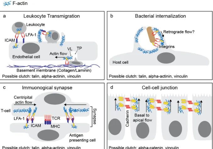
1.9 Beyond FAs: molecular clutches at diverse adhesive interactions
14
Figure 1.3. Molecular clutches may mediate diverse cell adhesive interactions.
(a) During leukocyte transmigration, initial cell-cell adhesion is mediated by LFA1-ICAM interactions. Transcellular migration occurs when the leukocyte migrates through a single endothelial cell. The migrating leukocyte extends invasive protrusions into the endothelial cell, while the endothelial cell forms a transmigratory cup around the leukocyte. Following successful transmigration, the transmigratory pore (TP) is closed by integrin-dependent ventral lamellipodia (VL) to restore endothelial barrier integrity. (b) Pathogens often seek entry into host cells by co-opting the integrin or cadherin adhesion machinery. Bacteria can bind to these adhesion receptors, stimulate actin polymerization, and activate clutch molecules to promote the formation of a phagocytic cup. (c) The T-cell immunological synapse requires centripetal actin flow to organize adhesion receptors into distinct domains. Rapid retrograde flow organizes and potentially activates LFA1 integrins in the actin rich regions. In contrast, the T-cell receptors (TCR) cluster in the actin-free center. (d) Cadherins mediate cell-cell adhesion and connect indirectly to the actin cytoskeleton through -catenin, -catenin and vinculin. Cadherins have been observed to undergo actin-dependent basal to apical flow that could generate force for epithelial
15
cadherin engages actin retrograde flow to promote neuronal growth cone migration126, and VE-cadherin
16
CHAPTER 2: ADHESIVE F-ACTIN WAVES ARE A NOVEL INTEGRIN-MEDIATED ADHESION COMPLEX COUPLED TO VENTRAL ACTIN POLYMERIZATION1
2.1 Summary
At the leading lamellipodium of migrating cells, protrusion of an Arp2/3-nucleated actin network is coupled to formation of integrin-based adhesions, suggesting that Arp2/3-mediated actin polymerization and integrin-dependentadhesion may be mechanistically linked. Arp2/3 also mediates actin
polymerization in structures distinct from the lamellipodium, in “ventral F-actin waves” that propagate as spots and wavefronts along the ventral plasma membrane. Here we show that integrins engage the extracellular matrix downstream of ventral F-actin waves in a variety of mammalian cell types. These “adhesive F-actin waves” require a cycle of integrin engagement and disengagement to the extracellular matrix for their formation and propagation, and exhibit morphometry and a hierarchical assembly and disassembly mechanism distinct from other integrin-containing structures. After Arp2/3-mediated actin polymerization, zyxin and VASP are co-recruited to adhesive F-actin waves, followed by paxillin and vinculin, and finally talin and integrin. Adhesive F-actin waves thus represent a previously
uncharacterized integrin-based adhesion complex associated with Arp2/3-mediated actin polymerization. 2.2 Introduction
Cell migration is a coordinated event involving protrusion, adhesion to the extracellular matrix (ECM), myosin II-driven contraction of the cell body, and adhesion disassembly at the cell rear. In the lamellipodium, protrusion of an Arp2/3-nucleated actin network is coupled to formation of integrin-based adhesions 51. Arp2/3-mediated actin polymerization and integrin-dependent adhesion may be
mechanistically linked, as the rate of adhesion assembly is directly correlated with the rate of lamellipodial protrusion 51, and the focal adhesion proteins vinculin and focal adhesion kinase (FAK) have been shown to interact with Arp2/3 84,85. While the Arp2/3-nucleated dendritic actin network is a defining characteristic
1 This chapter is adapted from work previously published as an article in PLoS ONE. The original citation
17
of the lamellipodium, dependent actin polymerization is not limited to this structure. Arp2/3-dependent actin polymerization is important for the formation of the immunological synapse, endocytosis and vesicle fusion, membrane ruffling, and ventral F-actin waves 128.
Ventral F-actin waves have been characterized in neutrophils, fibroblasts, and Dictyostelia129–131. In spite of their conservation across eukaryotic cells, the function of ventral F-actin waves is not well understood. In neutrophils, F-actin waves are induced by chemoattractant and are proposed to mediate cell migration 129, while in Dictyostelium, they are thought to be involved in phagocytosis 132. Ventral F-actin waves occur when F-actin spontaneously nucleates and polymerizes on the ventral, substrate-attached surface of cells, independently of the cell edge 131,133. This polymerizing actin can form discrete
spots, moving spots, or propagate in semicircular wave patterns 134.
Several studies have begun to characterize the mechanism of ventral F-actin wave formation and propagation. In Dictyostelia, myosin II does not localize to ventral F-actin waves and the formation and motion of ventral F-actin waves occurs in myosin II null cells 135. However, their sensitivity to actin
polymerization inhibitors and fluorescence recovery after photobleaching (FRAP) experiments indicate that ventral F-actin waves propagate by actin polymerization and treadmilling 129,135,136. Localization
studies have shown that ventral F-actin waves contain Arp2/3 and its activator, the WAVE complex, suggesting their involvement in stimulating actin treadmilling 129,131. Actin assembly by Arp2/3 in ventral F-actin waves may be mediated by a PI3K/Rac1 signaling cascade, since they are sensitive to the PI3K inhibitor LY294002, 132,137 and active Rac1 forms propagating wave patterns similar to ventral F-actin
waves 129. Together, these data suggest that PI3K and Rac1 promote WAVE- and Arp2/3-dependent actin treadmilling to form ventral F-actin waves and drive their propagation.
18
including podosomes and focal adhesions (FAs). Adhesive F-actin waves thus represent a previously uncharacterized integrin-based adhesion complex associated with Arp2/3-mediated actin polymerization. 2.3 Results
Ventral F-actin waves are followed by integrin waves in mammalian cells
Since Arp2/3-mediated actin polymerization is coupled to integrin adhesion in lamellipodia, we sought to determine if ventral actin waves were also coupled to integrin adhesion. To image ventral F-actin waves, we transfected cells with the F-F-actin-binding probe F-trF-actin-GFP (Inositol
1,4,5-Trisphosphate 3-Kinase A N66 actin binding domain fused to tdTomato 138), plated on 5µg/mL fibronectin, and imaged by Total Internal Reflection Fluorescence Microscopy (TIRFM). We observed spontaneous ventral F-actin waves in a variety of cell types including U2OS (human osteosarcoma cell line), B16-F10 (mouse melanoma cell line), and primary mouse embryonic fibroblasts (MEFs) (Figure 2.1a-f). We will refer to both moving spots of F-actin and propagating semicircular waves of F-actin as “ventral F-actin waves”. We measured ventral F-actin wave propagation velocities ranging from 1 -5 µm/min in the different cell types, comparable to the propagation speeds reported for Dictyostelium and mammalian cell ventral F-actin waves 130,133,135,139.
To test the hypothesis that ventral F-actin waves are coupled to integrin adhesion, we co-expressed F-tractin-GFP along with a fluorescent reporter for a fibronectin receptor, αV integrin-tagRFP
and untagged β3 integrin. Analysis by TIRFM showed that ventral F-actin waves were associated with αV
integrin waves in all cell types (Figure 2.1a-f). Dual color F-tractin and αV integrin kymographs revealed
19
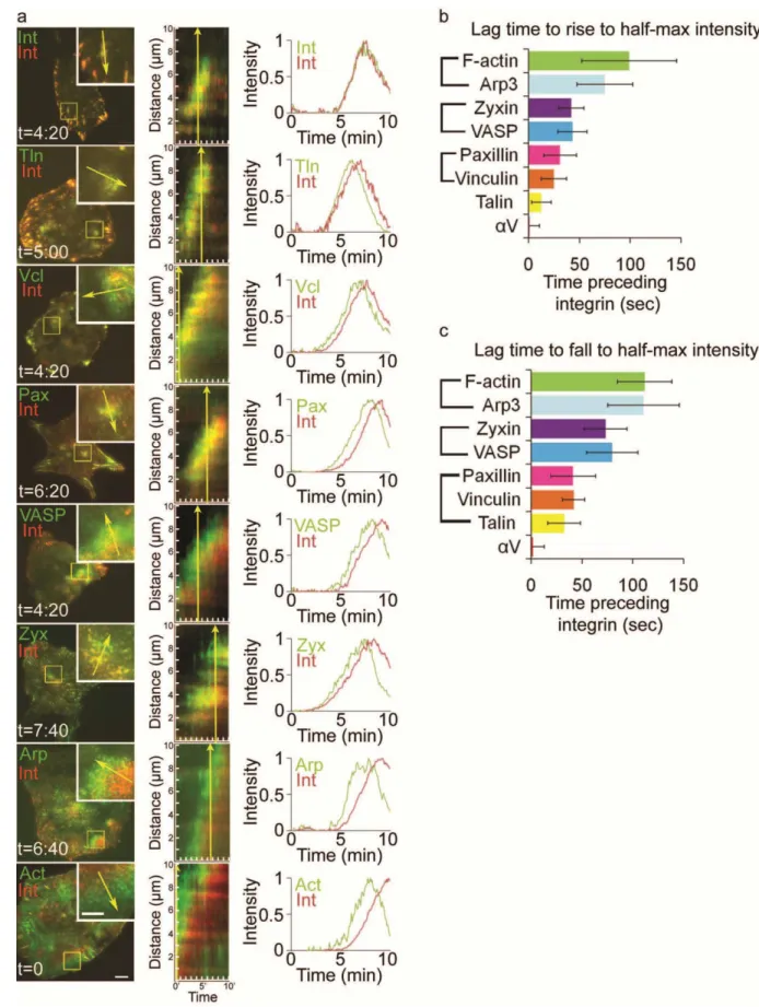
Figure 2.1. Ventral F-actin waves are followed by integrin waves in mammalian cells
(a,c,e) LEFT: Total internal reflection microscopy (TIRFM) image of a U2OS cell (a) B16-F10 cell (c) or a primary mouse fibroblast (d) co-expressing F-tractin-GFP to label actin filaments (green) and αV
20
t=8min(e)) demonstrates that F-tractin-GFP temporally precedes αV integrin-tagRFP in ventral F-actin
waves. (b,d,f) Normalized average intensity of F-tractin-GFP (green) and αV integrin-tagRFP (red) over
time. The average intensity in the green and red channels was measured in the region highlighted by a magenta circle in (a,c,e, left) and the values were normalized to the maximal intensity in the time series. To quantify assembly dynamics, we measured the lag time between when ventral F-actin and integrin waves reached half-maximal intensity. To quantify disassembly dynamics, we measured the lag time between when ventral F-actin and integrin waves fell from peak to half-maximal intensity. Lag times are labeled in (b,d,f). (g) Average lag times determined as described in (b,d,f) for the cell types noted, represented as mean ± SD, n= number of ventral F-actin waves analyzed.
waves (n=18) and that ventral F-actin waves fall from peak to half-maximal intensity an average of
135±46sec before αV integrin waves (n=16). Ventral F-actin and integrin waves in MEFs exhibited similar dynamics to U2OS cells, while B16 cells had slightly faster recruitment and disassembly of integrin waves (Figure 2.1g). However, in all cells observed, αV integrin was consistently recruited to ventral F-actin waves more than 20 seconds after ventral F-actin wave formation. We conclude that ventral F-actin waves are followed by integrin waves in mammalian cells.
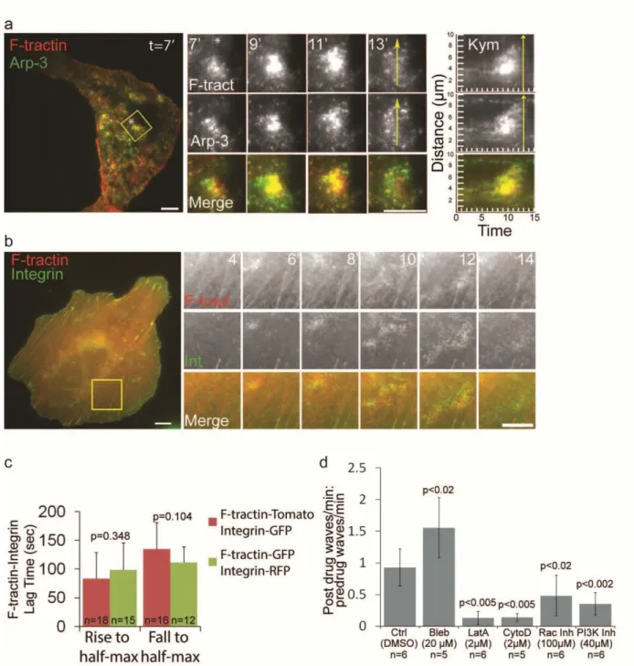
Ventral F-actin waves were most prevalent in U2OS cells, with ~60% of U2OS cells observed to have spontaneously forming ventral F-actin waves. In addition, co-transfection of F-tractin-tdTomato and Arp3-GFP confirmed that Arp2/3 co-localized with F-actin in U2OS ventral F-actin waves (Figure2.2a), similar to ventral waves reported in other cell types 135. U2OS ventral F-actin and integrin waves were also visible with both widefield epifluorescence (Figure 2.2b) and spinning disk confocal imaging (data not shown), suggesting that ventral waves represent a localized increase in the concentration of the proteins and are not a proximity artifact of TIRFM imaging. We confirmed that the measured lag times between actin and integrin waves were not due to differences in the fluorescent protein tag by analyzing ventral F-actin and V integrin waves in cells expressing F-tractin-tdTomato and V integrin-EGFP (Figure 2.2c).
Thus, U2OS cells serve as a good model for further characterization of integrin recruitment to spontaneously formed ventral F-actin waves.
21
Figure 2.2. Arp2/3-containing ventral F-actin waves are followed by integrin waves in U2OS cells
(a) LEFT: Total internal reflection fluorescence microscopy (TIRFM) image of a U2OS cell expressing F-tractin-tdtomato to label actin filaments (red) and Arp3-GFP (green). Scale bar = 10µm. The white asterisk is to aid in orientation of the region. CENTER: Images from a time-lapse series of the region highlighted by a yellow box, time in min shown. Scale bar = 10µm. The white asterisk is to aid in orientation of the region. RIGHT: Kymograph along the trajectory of ventral F-actin wave propagation (highlighted with a yellow arrow at t=13min) demonstrates that F-actin and Arp3 colocalize in space and time. Time in min is on the x-axis and distance in µm is on the y-axis. (b) RIGHT: Widefield
epifluorescent images of a U2OS cell expressing F-tractin tdTomato to label actin filaments (red) and αV
22
highlighted by a yellow box, time in min shown. Scale bar = 10µm. (c) Comparison of lag times with different fluorescent tags. Cells were co-transfected with either F-tractin-tdTomato and αV integrin-EGFP
or F-tractin-GFP and αV integrin-tagRFP. Graphs represent lag times between when ventral F-actin and
integrin waves reach half-maximal intensity (“Rise to half-max”) as well as when they decrease from peak to half-maximal intensity (“Fall to half-max”). Changing the fluorescent tags did not significantly change the lag time measurements. Values are represented as mean ± SD, n= number of ventral waves analyzed. P-values determined by Student’s t-test. (d) Effects of pharmacological perturbation on integrin waves (Bleb= blebbistatin; LatA=latrunculin A; CytoD=cytochalasin D; Rac Inh=NSC23766; PI3K Inh=LY 294002, concentrations below). Cells expressing either αV integrin-tagRFP or αV integrin-EGFP
were imaged by TIRFM during perfusion of drugs. We measured the number of waves per min
(“frequency”) before and after the drugs were added. We determined the effects of the drugs on integrin waves by dividing the post-drug frequency by the pre-drug frequency for each cell imaged. A value greater than one reflects an increase in wave frequency after drug addition, while a value less than one reflects a decrease in wave frequency after drug addition. n=number of cells analyzed. Data are represented as mean ± SD; P-values determined with Student’s t-test.
requirement of these activities for U2OS integrin wave formation. We imaged αV integrin-tagRFP or αV
integrin-EGFP during perfusion of Latrunculin A (to sequester actin monomers, 2µM), Cytochalasin D (to cap barbed actin filament ends, 2µM), LY294002 (to inhibit PI3K, 40µM), blebbistatin (to inhibit myosin II activity, 50uM), and NSC23766 (to inhibit Rac1, 100µM). We measured the number of waves per min (“frequency”) and then determined the effects of drugs on integrin waves by normalizing the post-drug frequency to the pre-drug frequency for each cell imaged. Latrunculin, Cytochalasin, NSC23766 and LY294002 all inhibited integrin wave frequency (Figure 2.2d). Blebbistatin treatment did not inhibit integrin wave frequency, but rather led to an increase in the number of integrin waves observed. This suggests that a decrease in myosin II contractility induces integrin wave formation. Together, these results show that Arp2/3- containing ventral F-actin waves are followed by integrin waves in U2OS cells, and that, similar to ventral F-actin waves, U2OS integrin waves require actin polymerization, PI3K activity and Rac1 activity, but not myosin II contractility, suggesting that ventral F-actin waves and integrin waves are coupled processes.
U2OS integrin waves are distinct from previously characterized integrin-containing structures We sought to determine if integrin waves were related to previously characterized integrin-containing structures including podosomes, invadopodia and FAs 140,141. Podosomes are peripheral adhesive structures with a core of F-actin surrounded by a small (~0.5-2µm) phase-dense ring of FA proteins 140–142. Phase contrast and spinning-disk confocal imaging of α
V integrin-EGFP revealed that
23
exhibited propagating movement, and no podosome-like structures were ever observed in U2OS cells (Figure 2.1a). Invadopodia are punctate, adhesive structures specialized for ECM degradation that are small (~0.50m), stable (>30 min lifetime), form in the cell center, and contain Arp2/3 and integrins 141. Although Arp2/3-containing puncta were sometimes observed in the center of U2OS cells, these
structures did not directly co-localize with integrins, and were short-lived and dynamic (data not shown). To verify that integrin waves did not degrade matrix, we plated U2OS cells on fluorescent gelatin for 8 hours, fixed cells, and immunostained for paxillin. Invadapodia and podosomes both degrade fluorescent gelatin, resulting in a decrease in fluorescence intensity appearing as dark holes in the substrate. We did not observe gelatin degradation beneath U2OS cells, even when the cells appeared to have paxillin-containing ventral wave structures (Figure 2.3b). Thus, in U2OS cells, integrin waves are not related to matrix-degrading podosomes or invadopodia.
To determine if integrin waves were similar to or precursors of FAs, we performed TIRFM imaging of αV integrin-EGFP and compared the morphometry and dynamics of integrin waves and FAs.
24
Figure 2.3. Integrin waves are distinct from podosomes and focal adhesions in U2OS cells
(a) LEFT: Spinning disk confocal and phase-contrast images of a U2OS cell expressing αV
integrin-EGFP. Scale bar = 10µm. RIGHT: Images from a time-lapse series of the region highlighted by a yellow box, time in min shown. The phase contrast image does not exhibit podosome structures as the integrin wave propagates across the ventral surface of the cell. Scale bar = 5 µm. (b) Spinning disk image of fixed U2OS cells plated on Oregon Green-gelatin (left) and stained for paxillin (right). Yellow arrow highlights possible paxillin-containing waves. (c) Distance from the cell edge of integrin waves (wave) and focal adhesions (FA) measured in U2OS cells expressing αV integrin-EGFP. Values are represented
as mean ± SD; P-values determined with Student’s t-test. (d) Area of waves and FAs measured in U2OS cells expressing αV integrin-EGFP. Values are represented as mean ± SD; P-values determined with
Student’s t-test. (e) Fluorescence density of waves and FAs measured in U2OS cells expressing αV
integrin-EGFP. Values are represented as mean ± SD; P-values determined with Student’s t-test.
(f) Lifetimes of waves and FAs measured in U2OS cells expressing αV integrin-EGFP. FA lifetimes longer
than 45min were recorded as 45min. Values are represented as mean ± SD; P-values determined with Student’s t-test. (g) LEFT: TIRFM image of a cell expressing αV integrin-EGFP. Scale bar = 10µm.
25
substrate (Figure 2.3g), similar to actin in ventral F-actin waves, which propagate by actin polymerization and treadmilling 131. This differed from FAs, in which integrin speckles moved coherently relative to the substrate as FAs released from the ECM and slid (Figure 2.3g and 107). Furthermore, integrin speckles moved significantly faster in FAs as compared to in waves (Figure 2.3h). Together, these results support the notion that U2OS integrin waves are distinct from podosomes, invadopodia and FAs.
Ventral F-actin and integrin waves are ECM-dependent and require a cycle of integrin engagement to and disengagement from the ECM
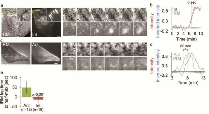
Next, we sought to determine if integrins in ventral waves were interacting with the ECM. To determine if integrin appearance in ventral waves corresponded to the membrane contacting the ECM, we performed Interference Reflection Microscopy (IRM) and TIRFM imaging of cells expressing αV
integrin-mCherry. Dark areas in IRM images indicate regions of the plasma membrane in very close (~10-30 nm) proximity to the coverslip 144. This showed that integrin waves co-localized with regions of local IRM darkening (Figure 2.4a-b). To determine if ventral F-actin waves were coupled to adhesion in cells without expression of exogenous integrin, we performed IRM and TIRFM of cells expressing F-tractin-GFP (Figure 2.4c-d). This revealed that ventral F-actin waves were followed by IRM darkening, similar to the dynamics of αv integrin relative to those of F-actin. Indeed, analysis of when αv integrin or
F-actin and inverse IRM reached half-maximal intensities in the region of a wave showed that dark areas in IRM appeared simultaneous with integrin appearance (-13.7 ±16.4sec after integrin, n=19) and followed F-actin appearance (48 ± 30sec after F-actin, n=12) (Figure 2.4e). These results suggest that when integrins appear after ventral F-actin waves, they bring the membrane into close proximity to the substrate, and that integrin waves are not an artifact of αVβ3 integrin over-expression.
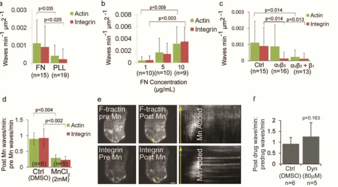
We next sought to determine if ventral F-actin and integrin wave formation and/or propagation require integrin-ECM engagement. To test the requirement of ECM, we plated cells transfected with F-tractin-GFP and αV integrin-tagRFP on either 5µg/mL fibronectin or 0.01% poly-L-lysine-coated coverslips
26
Figure 2.4. Integrin waves and ventral F-actin waves are visible by Interference Reflection Microscopy
(a) LEFT: Interference reflection microscopy (IRM) and total internal reflection (TIRF) images of a U2OS cell expressing αV integrin-mCherry. Inset (yellow box) shows focal adhesion morphology in IRM and
TIRF. Scale bar = 10µm. RIGHT: Images from a time-lapse series from the regions highlighted by a magenta box, time in min shown. Scale bar = 5µm. (b) Normalized average intensity of αV
integrin-mCherry (red) and inverted IRM intensity (blue) over time in the regions highlighted by a magenta in (a, left). (c) LEFT: Interference reflection microscopy (IRM) and total internal reflection (TIRF) images of a U2OS cell expressing F-tractin-GFP to label actin filaments. Scale bar = 10µm.
RIGHT: Images from a time-lapse series from the regions highlighted by a magenta box, time in min shown. Scale bar = 5µm. (d) Normalized average intensity of F-tractin-GFP (green) and inverted IRM intesnsity (blue) over time from the region highlighted in (c, left). (e) IRM lag time to rise to half-maximal inverted intensity, relative to rise to half maximal intensity of F-tractin and Integrin. The lag time between when αv integrin or F-tractin reached half-maximal intensity and when IRM reached half-maximal intensity
27
requirement of ECM for ventral F-actin and integrin waves, we plated cells expressing F-tractin-GFP and αV integrin-tagRFP on increasing concentrations of fibronectin. We found that both ventral F-actin and
integrin wave frequency increased in a dose-dependent manner (Figure 2.5b). These results suggest that formation of ventral F-actin and integrin waves is ECM-dependent.
We next plated cells on fibronectin in the presence or absence of the function-blocking LM609 antibody to αVβ3 integrin (Figure 2.5c 145). αV integrin-tagRFP did not localize to paxillin-EGFP labeled adhesions in cells plated on fibronectin in the presence of 20µg/mL LM609, confirming that 20µg/mL of LM609 is sufficient to block αVβ3 interaction with fibronectin (data not shown). Analysis of ventral F-actin
wave frequency showed that integrin waves, but not ventral F-actin waves, were inhibited in cells plated in the presence of 20µg/mL LM609 (n=16) compared with control cells (n=15). To determine if the ventral F-actin waves observed in the presence of LM609 were dependent on β1 integrins, we plated cells in the presence of LM609 and the function-blocking P4C10 antibody to β1 integrin146. This resulted in fewer cells attached to the coverslip, likely due to adhesion defects (data not shown). However, analysis of wave frequency in cells that adhered and spread showed that both ventral F-actin and αV integrin waves
were inhibited in the presence of LM609 and P4C10 (n= 13) compared with control cells (n= 15) (Figure 2.5c). Thus, ventral F-actin and integrin waves require integrin activation.
Since integrin waves require integrin engagement to the ECM and appear to propagate by treadmilling engagement to the ECM (Figure 2.3g), we sought to determine if they also required integrin disengagement from the ECM. Mn2+ binds to the extracellular metal ion binding sites of integrin inducing
conformational changes correlated with higher affinity binding to ligand and is commonly used to induce integrin activation 147. We analyzed ventral actin and integrin wave frequency in cells expressing
F-tractin-GFP and αV integrin-tagRFP and treated with 2mM MnCl2 to induce integrin activation (Figure
2.5d). This revealed that MnCl2 significantly inhibited both ventral F-actin and integrin waves.
Furthermore, imaging cells during perfusion revealed that MnCl2 produced an immediate effect on ventral
28
Figure 2.5. Ventral F-actin and integrin waves require integrin engagement to extracellular matrix
(a) Ventral F-actin and integrin waves require ECM. U2OS cells expressing F-tractin-GFP to label actin filaments and αV integrin-tagRFP were plated on either 5µg/mL fibronectin (FN) or 0.01% poly-L-lysine
(PLL). In A, B, and C, cells were imaged for 10 min and the average number of waves per min per µm2 was measured. P-values determined with Student’s t-test. n=number of cells analyzed. (b) Ventral F-actin and integrin waves are sensitive to FN concentration. U2OS cells expressing F-trF-actin-GFP and αV
integrin-tagRFP were plated on increasing concentrations of FN (1µg/mL, 5µg/mL and 10µg/mL). (c) Integrin waves require integrin engagement to ECM. U2OS cells expressing F-tractin-GFP and αV
integrin-tagRFP were plated on 5µg/mL FN in the presence of 20µg/ml LM609 antibody to block αVβ3
binding to FN (“αVβ3”) or 20µg/ml LM609 antibody + P4C10 (1:20 dilution) to block αVβ3 and β1 binding to
FN (“αVβ3 + β1”). (d) Effect of MnCl2 on ventral F-actin and integrin waves. Cells expressing
F-tractin-GFP and αV integrin-tagRFP were imaged 15min prior to and 30min after perfusion of 2mM MnCl2. We
measured the number of waves per min (“frequency”) before and after MnCl2 was added. We determined
the effects of MnCl2 on waves by dividing the post-drug frequency by the pre-drug frequency for each cell
imaged. A value greater than one reflects an increase in wave frequency after drug addition, while a value less than one reflects a decrease in wave frequency after drug addition. n=number of cells analyzed. Data are represented as mean ± SD; P-values determined with Student’s t-test. (e) Total internal reflection fluorescence microscopy (TIRFM) images of a U2OS cell expressing F-tractin-GFP and αV integrin-tagRFP immediately prior to (LEFT) and after (CENTER) perfusion of 2 mM MnCl2 addition.
RIGHT: Kymograph along the trajectory of ventral F-actin wave propagation (highlighted with yellow arrows). MnCl2 stops the propagation of both F-actin and integrin waves. Scale bar = 10µm. (f) Effect of
Dynasore hydrate (Dyn) on integrin waves. Cells expressing either αV integrin-EGFP were imaged by
29
waves and require actin polymerization, these results further suggest a positive feedback loop between integrin adhesion and Arp2/3-mediated actin polymerization in ventral F-actin waves.
To determine if U2OS integrin waves required endocytic recycling of integrins148, we imaged αV
integrin-tagRFP during perfusion of Dynasore hydrate (to inhibit dynamin GTPase activity, 80 M). Dynasore hydrate did not affect integrin wave frequency, suggesting that integrin waves do not require dynamin-mediated endocytosis of integrins for their turnover (Figure 2.5f). These results suggest that integrin waves do not require recycling of integrins from the plasma membrane, but rather require a cycle of activation and inactivation of integrins already at the cell surface. We conclude that ventral F-actin waves are adhesive structures that consistently recruit and activate integrins to engage with the ECM. We will henceforth refer to these actin- and integrin- containing ventral wave structures as “adhesive F-actin waves”.
Adhesive F-actin waves contain focal adhesion proteins that assemble and disassemble in a distinct stepwise order
Since actin does not directly bind integrin, we sought to determine what proteins may be involved in coupling actin polymerization to integrin adhesion in adhesive F-actin waves. We co-expresssed fluorescently tagged FA adapter and actin-binding proteins together with αV -integrin-tagRFP and imaged
by TIRFM to identify proteins that localized to adhesive F-actin waves. This revealed that zyxin-EGFP, VASP-venus, paxillin-EGFP, vinculin-EGFP and talin-EGFP all localized to αV integrin-tagRFP containing
adhesive F-actin waves (Figure 2.6a, left). However, kymograph analysis of time-lapse images revealed that many of these proteins did not temporally coincide with the appearance of αV integrin, but, similar to
F-actin, preceded αV integrin appearance in ventral F-actin waves (Figure 2.6a, center). To confirm that
FA protein expression did not promote association with ventral F-actin waves, we immunolocalized endogenous paxillin and F-actin in fixed U2OS cells and found that endogenous paxillin was associated with ventral F-actin wave-like structures (data not shown). However, similar to fluorescently tagged proteins, F-actin and endogenous paxillin did not perfectly overlap; rather, they were separated into a region of F-actin, followed by a region of paxillin and F-actin colocalization, followed by a region of paxillin.
To determine the order of protein assembly into adhesive F-actin waves, we analyzed the
30