THE ADAPTOR PROTEIN SHC IS A CRITICAL REGULATOR OF
ANGIOGENIC AND SHEAR STRESS SIGNALING IN
ENDOTHELIAL CELLS
Daniel Timothy Sweet
A dissertation submitted to the faculty of the University of North Carolina at Chapel Hill in partial fulfillment of the requirements for the degree of Doctor of
Philosophy in the Curriculum in Genetics and Molecular Biology
Chapel Hill 2012
Approved by: Ellie Tzima PhD
ABSTRACT
DANIEL TIMOTHY SWEET: The Adaptor Protein Shc is a Critical Regulator of Angiogenic and Shear Stress Signaling in Endothelial Cells
(Under the direction of Ellie Tzima)
Endothelial cells (ECs), which form the lining of blood vessels, actively participate in many aspects of cardiovascular development and pathologies such as cancer and atherosclerosis. Vascular ECs are unique in the diverse array of signals that they are capable of sensing from soluble growth factors, immobile extracellular matrix (ECM) proteins and mechanical forces. Studying EC
responses to this array of signals will enhance our understanding of the etiology of prevalent diseases such as cancer and atherosclerosis and lead to improved treatments.
Shc is an evolutionarily conserved adaptor protein that mediates signaling cascades downstream of activated receptors and is essential for development of the cardiovascular system. This dissertation focuses on defining the roles that Shc plays in EC responses to angiogenic cues and mechanical force.
zebrafish, we found that Shc is required for sprouting angiogenesis in vivo. Shc
mediates signaling from integrins and VEGF receptors which is required for haptotaxis, survival and sprouting. Interestingly, Shc integrates VEGF and ECM signaling as VEGF-induced survival requires Shc specifically on fibronectin.
Fluid shear stress, the frictional force from blood flowing over ECs, regulates EC function and allows vessels to respond to changes in tissue
physiology but also contributes to vessel pathogenesis such as atherosclerosis. We have shown that Shc is required for transducing shear stress signaling directly downstream of the ‘mechanosensory complex’. Shc is required for
induction of the inflammatory response that is activated by disturbed shear stress and underlies the development of atherosclerotic plaques. Additionally, Shc is required in mice for shear-induced collateral artery remodeling and arterial specification during arteriogenesis.
ACKNOWLEDGEMENTS
With the completion of my PhD requirements and dissertation, I am grateful to several people who have provided guidance and support which was essential to my development as a scientist and personally.
First, I would like to sincerely thank my mentor Ellie Tzima for her
guidance, patient instruction and daily example of how to be a good scientist. As Ellie’s first graduate student, I took a risk joining her lab as she had no track record of mentoring students but she has greatly exceeded any expectations I had for a mentor. Ellie does an excellent job of being available and easy to talk to about data and planning future experiments. She treats her students as peers and values their opinions, while still teaching and pushing them to reach their full potential as scientists. While many graduate students become disillusioned and uninterested by their 5th year of research, my excitement for cardiovascular research has grown due to Ellie’s enthusiasm for the research and constant discussion about exciting new ideas and techniques. Ellie’s mentoring has been crucial to my scientific development and success and I thank her for all that she has done to help me along the way.
member of my committee has been willing to provide reagents and protocols to aid in my experiments. I would like to particularly thank Vicki Bautch who has been extremely supportive in providing me with feedback at our labs’ joint lab meetings and allowing me to learn several techniques in her lab which were used in Chapter 2. This collaboration was essential for the revisions of my Blood paper and I thank her for being eager to help whenever we asked. Similarly, I thank Keith Burridge for a collaboration in which we examined the role of Shc in shear-induced RhoA regulation. This project is still incomplete so it was not included in this dissertation but I thank Dr. Burridge for providing reagents and experimental support for this unpublished research.
I would also like to thank all members of the Tzima lab: Maggie
McCormick, Caitlin Collins and Zhongming Chen. Everyone has provided useful comments at lab meeting as well as help with experiments on a day-to-day basis. We each have our own areas of expertise and each person is willing to help the others with new experiments, paper writing, slides for talks and general
brainstorming. The Tzima lab is a great place to work and I am grateful to Maggie, Caitlin and Zhongming for being so easy to get along with.
understand it. I cannot thank my parents enough for all they’ve done in raising me and supporting me throughout my life.
Last but not least I would like to thank my fiancé Kristen Tamburro. Kristen has always been in my corner and has been my ‘science-fluent’ support system in success and failure. Kristen is my biggest fan and on many occasions has helped me figure out problems with my research and even contributed reagents and protocols from time to time. She is understanding of working long hours to finish an experiment and has kept me sane through the last five years by helping me develop a nice life outside of the lab. Kristen is my partner and I am eternally thankful to have her in my life. Also I’d like to thank Kristen’s parents, Robert and Laurie Tamburro who live nearby and have accepted me into their family and made me feel at home in North Carolina.
TABLE OF CONTENTS
LIST OF FIGURES... ... xi
LIST OF ABBREVIATIONS AND SYMBOLS ... xiii
CHAPTER I. INTRODUCTION ... 1
PREFACE ... 1
SIGNAL TRANSDUCTION IN ECs ... 1
ADAPTOR PROTEIN SHC ... 2
ANGIOGENESIS ... 4
MECHANOTRANSDUCTION: ECs RESPOND TO MECHANICAL FORCE ... 5
ARTERIOGENESIS ... 7
RESEARCH PRESENTED IN THIS DISSERTATION ... 8
REFERENCES ... 12
II. THE ADAPTOR PROTEIN SHC INTEGRATES GROWTH FACTOR AND ECM SIGNALING DURING POSTNATAL ANGIOGENESIS ... 19
PREFACE ... 19
OVERVIEW ... 19
INTRODUCTION ... 21
Zebrafish Morpholino Injection ... 22
Mice ... 23
X-gal Stain of R26R Tissue ... 23
Matrigel Plug Assay ... 24
Mouse Lung Endothelial Cell Isolation ... 24
Fibrin Gel Bead Assay ... 25
Endothelial Cell Culture & Lentivirus Infection ... 26
Cell Spreading Assay ... 26
Cell Migration Assays ... 27
EC Survival Assay ... 27
Retinal EC Proliferation Assay ... 28
EC Adhesion Assay ... 28
VEGF Treatments & Western Blot Analysis ... 29
Quantification & Statistical Analysis ... 30
RESULTS ... 30
Angiogenesis in Zebrafish Requires Shc ... 30
Endothelial Shc is Required for Proper Angiogenesis in vivo ... 32
Endothelial Shc is Required for Tube Assembly & Sprouting in vitro ... 34
Shc is Required for Integrin- Mediating EC Signaling. ... 35
Shc is Required for VEGF- Mediated EC Signaling ... 36
Shc Mediates Akt Activation Downstream
of VEGF & Integrin Activation ... 38
DISCUSSION ... 38
REFERENCES ... 54
III. SHC MEDIATES THE ENDOTHELIAL RESPONSE TO SHEAR STRESS IN VITRO & DURING ARTERIOGENESIS IN VIVO ... 59
PREFACE ... 59
OVERVIEW ... 60
INTRODUCTION ... 62
METHODS ... 65
BAEC Culture, Transfections & Shear Stress ... 65
Immunoprecipitation, Western Blot & Antibodies ... 65
Immunofluorescence Microscopy ... 67
Leukocyte Adhesion Assay ... 67
Animals ... 68
Unilateral Hindlimb Ischemia ... 68
Laser Doppler Imaging ... 69
Morphometry ... 69
Immunohistochemistry ... 70
Leukocyte Density ... 71
Capillary Density ... 71
iMLEC Cell Culture & Shear Stress ... 71
Statistical Analysis ... 73
RESULTS ... 73
Shc is Activated by Shear Stress ... 73
Shc Associates with Components of the ‘Mechanosensory Complex’ in Response to Shear Stress ... 74
Shear Stress-Induced Shc Association with Integrin αvβ3 is Dependent on ECM Composition ... 75
Shc Mediates the Flow-Induced Inflammatory Response ... 76
Endothelial Shc is Required for Plantar Perfusion Recovery Following Femoral Artery Ligation ... 77
Shc is Required for Collateral Artery Remodeling & Angiogenesis ... 78
Shc is Required for Flow-Induced NF-κB Activation & Inflammation in Collaterals ... 79
Notch-Dependent Collateral EC Arterial Specification Requires Shc ... 80
DISCUSSION ... 82
REFERENCES ... 94
IV. CONCLUSIONS & PERSPECTIVES ... 99
OVERVIEW OF FINDINGS ... 99
CHAPTER II: THE ADAPTOR PROTEIN SHC INTEGRATES GROWTH FACTOR AND ECM SIGNALING DURING POSTNATAL ANGIOGENESIS ... 100
CHAPTER III: SHC MEDIATES THE ENDOTHELIAL RESPONSE TO SHEAR STRESS IN VITRO AND DURING ARTERIOGENESIS IN VIVO ... 106
LIST OF FIGURES
Figure 1.1: ECs integrate diverse environmental signals
from multiple cell-surface receptors ... 11 Figure 2.1: Shc is required for intersegmental vessel sprouting
angiogenesis in zebrafish ... 43 Figure 2.2: Endothelial Shc knockout causes
defective angiogenesis in vivo ... 44
Figure 2.3: Endothelial Shc is required for tube
assembly & sprouting in vitro ... 46
Figure 2.4: Shc is required for integrin-mediated spreading
& haptotaxis on fibronectin but not collagen ... 47 Figure 2.5: Shc is required for VEGF-induced EC survival but not
migration toward VEGF or proliferation ... 49 Figure 2.6: Survival requires integration of VEGF &
integrin signaling through Shc ... 51 Figure 2.7: Shc is required for specific signal transduction pathways
downstream of integrins & VEGF ... 52 Figure 3.1: Shc is activated by shear stress ... 85 Figure 3.2: Shear stress induces ECM-independant Shc
association with the ‘mechanosensory complex’
& ECM-dependant Shc association with integrins ... 86 Figure 3.3: Shc is required for shear stress-induced NF-κB
activation & leukocyte adhesion to the endothelium ... 88 Figure 3.4: Shc signaling regulates plantar perfusion
Figure 3.5: Shc is required for pre-existing collateral
remodeling & angiogenesis ... 91 Figure 3.6: Shc is required for collateral EC
proliferation & inflammation ... 92
Figure 3.7: Shc regulates shear stress-induced collateral EC arterial specification via activation of the Notch pathway ... 93
Figure 4.1: The adaptor protein Shc is a critical regulator of
LIST OF ABBREVIATIONS AND SYMBOLS
BAEC Bovine Aortic Endothelial Cell bFGF basic Fibroblast Growth Factor CH1/CH2 Collagen Homology domain 1/2
CL Collagen
DLAV Dorsal Longitudinal Anastomotic Vessel Dll-1, 4 Delta-like 1, Delta-like 4
ECM Extracellular Matrix ECs Endothelial Cells
EGF Epidermal Growth Factor
eNOS endothelial Nitric Oxide Synthase ERK Extracellular-Related Kinase 1/2
FN Fibronectin
HUVEC Human Umbilical Vein Endothelial Cell ICAM-1 Intercellular Adhesion Molecule-1 ISV Intersegmental Vessel
kDa kilodaltons
LM Laminin
MAPK Mitogen Activated Protein Kinase MLEC Mouse Lung Endothelial Cell
MO Morpholino
NF-κB Nuclear Factor kappa-light-chain-enhancer of activated B cells
NICD Notch Intracellular Domain NO Nitric Oxide
PDGF Platelet Derived Growth Factor PI3K Phosphoinositide 3-Kinase
PTB Phospho-Tyrosine Binding domain ROS Reactive Oxygen Species
RTK Receptor Tyrosine Kinase SH2 Src Homology-2
Shc Src homology-2 domain containing shRNA short hairpin Ribonucleic Acid SMC Smooth Muscle Cell
TNF-α Tumor Necrosis Factor-alpha
VCAM Vascular Cell Adhesion Molecule VEGF Vascular Endothelial Growth Factor
CHAPTER I.
INTRODUCTION
PREFACE
Parts of this chapter were adapted from a previously published review article 1. I wrote the original manuscript and Ellie Tzima wrote and edited the final version of the manuscript.
SIGNAL TRANSDUCTION IN ECs
Endothelial cells (ECs) are a specialized cell type that form the inner lining of blood vessels and are actively involved in many aspects of cardiovascular development and pathology. Vascular ECs are quite unique in the diverse array of signals that they are exposed to. The constant flow of blood over the
endothelium brings a myriad of soluble growth factors and cytokines that bind to receptors on the EC surface while also imparting mechanical forces that are sensed by the endothelium. Signals from the surrounding ECM are sensed by integrins that form specific cell-ECM adhesions. These signaling cascades are ______________________________________
required for important physiological EC functions, such as wound healing and inflammation, but also pathological EC functions, such as tumor angiogenesis and the chronic inflammation associated with atherosclerosis. This dissertation has aimed to examine signal transduction pathways in ECs that are activated by angiogenic growth factors, mechanical force and the ECM. By studying signal transduction in ECs, we will improve upon our current understanding of the causes of and potential treatments for prevalent diseases such as cancer and atherosclerosis. My research has focused on the adaptor protein Shc, which we have found is an important mediator of EC signaling in response to angiogenic cues and mechanical force.
ADAPTOR PROTEIN SHC
The mammalian ShcA gene encodes three Shc isoforms of 46, 52 and 66
kDa – all of which originate from the same mRNA either through alternative RNA splicing or translation initiation sites 1, 2. The isoforms only differ in the length of their N-terminal CH2 domain and all three include the SH2 and PTB domains important in phospho-tyrosine receptor binding as well as the CH1 domain which houses three important tyrosine phosphorylation sites (see 3 for review). Shc is ubiquitously expressed in adults 4 and has homologues in
Drosophila and C. elegans, indicating an important evolutionarily conserved role for the protein 5, 6.
Factor (EGF) 11, Platelet-Derived Growth Factor (PDGF) 12, 13, Insulin 14-17, and basic Fibroblast Growth Factor (bFGF) 18, 19. When Shc binds activated RTKs, Shc itself is phosphorylated and then associates with secondary signaling molecules such as Grb2, which lead to activation of Ras 3. Not only is Shc important in signaling from a variety of growth factor RTKs, but Shc also
associates with and mediates signaling from a subset of integrins. Activation of integrins α6β4 (laminin receptor), α1β1 (collagen/laminin receptor), 5 1
(fibronectin) and αvβ3 (vitronectin/fibronectin) induces phosphorylation of Shc and Shc:integrin association. Conversely, ligation of α2β1 (collagen/laminin), α3β1 (promiscuous), α6β1 (laminin) and β2 integrins does not 20-22. Shc has an
established role in Ras signaling in non-EC tissues, but its role in EC signaling is less well defined.
Shc is critical for cardiovascular development, as Shc knockout mice are embryonic lethal at embryonic day 11.5 23 due to defects in heart development, as well as cell-cell contacts and mural cell coverage of the blood vessels. Further genetic studies revealed that Shc expression specifically in cardiac myocytes is sufficient for embryonic cardiovascular development and adult heart function 24, 25. While a role for Shc in heart development and function has been established, relatively little is known about the role of Shc in the function of ECs. Shc has been implicated in signaling downstream of some EC-specific RTKs in
vitro. For example, Vascular Endothelial Growth Factor (VEGF) stimulation of
ECs caused Shc to associate with VEGF Receptor-2 (VEGFR-2) and
that are required in ECs for proper angiogenesis 21, 28-30. Because Shc associates with integrins and RTKs that are critical for angiogenesis and
mechanotransduction, we hypothesized that Shc may act as a signaling hub in ECs.
ANGIOGENESIS
Blood vessels are formed through two sequential processes:
vasculogenesis and angiogenesis. Vasculogenesis is the initial differentiation of endothelial cells from precursors and assembly into vessels whereas
angiogenesis is a process of new vessel growth by sprouting from the existing vasculature 31-33. Angiogenesis is important during embryonic development for proper patterning of the vascular tree as well as in adults for wound healing and during tumorigenesis. Angiogenesis is a highly coordinated sprouting and
remodeling process that is controlled by several signaling pathways such as Tie2 and Notch, but the VEGF pathway is the principal master regulator of
angiogenesis. VEGF-A (referred to as simply VEGF) is a soluble growth factor whose expression is upregulated in hypoxic or cancer tissue to stimulate
nascent vessel sprouts from the parent vessel, the underlying ECM is remodeled and integrins α5β1 and αvβ3 are upregulated in angiogenic ECs. While the role of these integrins is controversial, it is clear that blocking either of these integrins
can impede the angiogenic process 29 therefore proper function of integrins α5β1 and αvβ3 is necessary in angiogenic ECs.
MECHANOTRANSDUCTION: ECs RESPOND TO MECHANICAL FORCE
Vascular ECs are constantly exposed to hemodynamic forces due to blood flowing over them. Fluid shear stress, the frictional drag force from blood flow, is crucial in determining the shape, cytoskeletal organization and function of ECs and allowing vessels to respond to changes in tissue physiology but also contributing to vessel pathogenesis 36-38. Although systemic risk factors such as smoking, diabetes, and high plasma levels of cholesterol and lipoproteins are associated with the development of atherosclerosis, atherosclerosis is a focal disease and forms preferentially at vessel bifurcations and curvatures- sites where blood flow is disturbed or turbulent 39. In regions of arteries where flow is low (<5 dynes/cm2) and disturbed, atherosclerosis is promoted because
atherogenic or atheroprotective EC phenotype, which underlies the development, or lack, of atherosclerotic plaque formation and therefore studying how ECs sense and respond to shear stress is a critical health care priority.
Shear stress is detected by a variety of molecular force sensors located throughout the EC membrane 39, 42. Forces from the apical (luminal) surface are transmitted through the cytoskeleton to points of attachment that resist shear stress 41. In that regard, both cell-cell and cell-ECM adhesions have been
implicated in shear stress signal transduction. At cell-cell adhesions, our lab has previously reported a ‘mechanosensory complex’ comprised of PECAM-1, VE-Cadherin and VEGFR-2 which is necessary and sufficient for a subset of cellular responses to shear stress 43. Indeed, PECAM-1 is required for cytoskeletal alignment to flow as well as atherosclerosis and flow-mediated vessel remodeling
in vivo 43-46. At the basal EC surface, integrins have also been implicated in
mechanotransduction. Shear stress stimulates the conversion of integrins to a high affinity state, followed by their binding to the ECM and subsequent activation of multiple signaling pathways downstream of integrin activation 47-49.
Interestingly, the activation of the inflammatory transcription factor NF-κB by flow
is dependent upon the ECM composition – as it is activated on fibronectin (FN) but prevented from being activated in cells growing on collagen (CL) 50, 51.
EC responses to shear stress have been well characterized in vitro and
conformational activation and then these high-affinity integrins bind to their respective ligand in the underlying ECM 48. On a time scale of minutes to hours, shear stress activates Rho family GTPases and initiates the process of
cytoskeletal remodeling 48, 57-59, stimulates tyrosine phosphorylation of proteins such as MAPKs 60, release of Reactive Oxygen Species (ROS) 61 and activates transcription factors such as NF- B which are critical for initiating the
inflammatory response to shear stress 62. Slower responses that occur on a time scale of hours to days include altered expression of shear-responsive genes encoding Kruppel-like factor 2 (KLF2) 63, 64, eNOS, and cell adhesion molecules E-selectin, Intercellular Cell Adhesion Molecule (ICAM)-1 and Vascular Cell Adhesion Molecule (VCAM)-1 65, 66. The hallmark of the long-term adaptive EC response to laminar shear stress is the rearrangement of actin microfilaments and microtubules and their elongation in the direction of flow 67-69, while long-term disturbed flow induces chronic inflammation and causes leukocyte adhesion and transmigration into the vessel wall 67, 70.
ARTERIOGENESIS
Shear stress regulates vessel size (caliber) whereas pressure determines vessel wall thickness 71. Shear force is dependent on vessel diameter, so
arteries dynamically respond to changes in shear stress by remodeling inward or outward to compensate for the new shear environment 72, 73. Following an
arteriogenesis 74. Arteriogenesis describes the formation of mature arteries from pre-existent interconnecting arterioles after an arterial occlusion 75. Artery-to-artery connections, called collateral arteries, arise during development and act as a natural bypass mechanism to circumvent circulation in the case of a large artery occlusion 74, 76. Following an arterial occlusion, blood is re-routed into pre-existing collaterals, dramatically increasing shear stress and inducing outward remodeling, allowing the vessel to carry more flow and restore
circulation to downstream ischemic tissue. The sudden increase in collateral flow is sensed by ECs and activates several signaling pathways that are critical for vessel remodeling including proliferation of ECs and smooth muscle cells, vessel dilation via NO production, and inflammation and leukocyte transmigration into the vessel wall via the NF- B pathway 74, 77. Arteriogenesis is critical to prevent tissue death following myocardial infarction or other arterial occlusion, and understanding this process will help in treatment and prevention of
cardiovascular disease, the number one cause of death in industrialized nations such as the United States.
RESEARCH PRESENTED IN THIS DISSERTATION
Chapter II.
Determine the role of the adaptor protein Shc in EC signaling during
angiogenesis. Previous studies have shown that Shc can bind to activated
VEGFR-2 or integrins αvβ3 and α5β1. Because these transmembrane receptors have been implicated in controlling angiogenesis, I hypothesized that Shc plays a role in angiogenesis by mediating signaling from one or both of these signaling hubs. Using loss-of-function studies in mouse and zebrafish, I first assessed a possible role for Shc in angiogenesis in vivo. Next, I performed a series of
functional assays in vitro to test which EC functions are regulated through Shc
signaling. Finally, I uncovered the molecular mechanism by examining the role of Shc in mediating signaling pathways that are known to be required for
angiogenesis.
Chapter III.
Determine the role of Shc in EC responses to shear stress. Our lab recently
reported a ‘mechanosensory complex’ in ECs that is necessary and sufficient for conferring cells the ability to respond to shear stress. I hypothesized that Shc mediates signaling downstream of this ‘mechanosensory complex’ and therefore is required for mechanotransduction in ECs. First, I performed in vitro
experiments in ECs to test the role of Shc in mediating signaling from the
arteriogenesis was examined, focusing on inflammation and arterial specification, which underlie collateral vessel remodeling during arteriogenesis.
Chapter IV.
Synthesize the findings of the research chapters and assess the impact of
these findings on the field of vascular biology. The findings of this
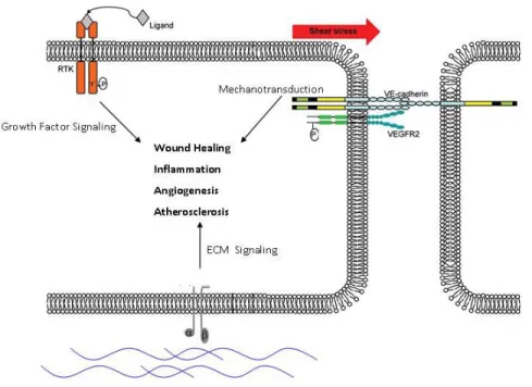
Figure 1.1: ECs integrate diverse environmental signals from multiple
cell-surface receptors
Endothelial Cells (ECs) use cell-surface receptors to sense a diverse array of signals which direct specific EC responses. Soluble growth factors and cytokines secreted from surrounding tissue are sensed by Receptor Tyrosine Kinases (RTKs). Also, mechanical force such as shear stress from blood flowing over the EC layer is sensed by a ‘Mechanosensory Complex’ comprised of VE-Cadherin and VEGFR2 located at cell-cell junctions. Third, the composition of the
REFERENCES
1. Pelicci G, Lanfrancone L, Grignani F, McGlade J, Cavallo F, Forni G, Nicoletti I, Pawson T, Pelicci PG. A novel transforming protein (SHC) with an SH2 domain is implicated in mitogenic signal transduction. Cell. 1992;70:93-104.
2. Migliaccio E, Mele S, Salcini AE, Pelicci G, Lai KM, Superti-Furga G, Pawson T, Di Fiore PP, Lanfrancone L, Pelicci PG. Opposite effects of the p52shc/p46shc and p66shc splicing isoforms on the EGF receptor-MAP kinase-fos signalling pathway. EMBO J. 1997;16:706-716.
3. Ravichandran KS. Signaling via shc family adapter proteins. Oncogene.
2001;20:6322-30.
4. Pelicci G, Dente L, De Giuseppe A, Verducci-Galletti B, Giuli S, Mele S, Vetriani C, Giorgio M, Pandolfi PP, Cesareni G, Pelicci PG. A family of shc related proteins with conserved PTB, CH1 and SH2 regions. Oncogene.
1996;13:633-641.
5. Lai KM, Olivier JP, Gish GD, Henkemeyer M, McGlade J, Pawson T. A drosophila shc gene product is implicated in signaling by the DER receptor tyrosine kinase. Molecular & Cellular Biology. 1995;15:4810-8.
6. Luzi L, Confalonieri S, Di Fiore PP, Pelicci PG. Evolution of shc functions from nematode to human. Curr Opin Genet Dev. 2000;10:668-674.
7. Rozakis-Adcock M, McGlade J, Mbamalu G, Pelicci G, Daly R, Li W, Batzer A, Thomas S, Brugge J, Pelicci PG. Association of the shc and Grb2/Sem5 SH2-containing proteins is implicated in activation of the ras pathway by tyrosine kinases. Nature. 1992;360:689-92.
8. Egan SE, Giddings BW, Brooks MW, Buday L, Sizeland AM, Weinberg RA. Association of sos ras exchange protein with Grb2 is implicated in tyrosine kinase signal transduction and transformation. Nature. 1993;363:45-51.
9. Li N, Batzer A, Daly R, Yajnik V, Skolnik E, Chardin P, Bar-Sagi D, Margolis B, Schlessinger J. Guanine-nucleotide-releasing factor hSos1 binds to Grb2 and links receptor tyrosine kinases to ras signalling. Nature. 1993;363:85-88.
11. Ruff-Jamison S, McGlade J, Pawson T, Chen K, Cohen S. Epidermal growth factor stimulates the tyrosine phosphorylation of SHC in the mouse. J Biol Chem.
1993;268:7610-7612.
12. Gelderloos JA, Rosenkranz S, Bazenet C, Kazlauskas A. A role for src in signal relay by the platelet-derived growth factor alpha receptor. J Biol Chem.
1998;273:5908-5915.
13. Yokote K, Mori S, Hansen K, McGlade J, Pawson T, Heldin CH, Claesson-Welsh L. Direct interaction between shc and the platelet-derived growth factor beta-receptor. J Biol Chem. 1994;269:15337-15343.
14. Skolnik EY, Batzer A, Li N, Lee CH, Lowenstein E, Mohammadi M, Margolis B, Schlessinger J. The function of GRB2 in linking the insulin receptor to ras signaling pathways. Science. 1993;260:1953-1955.
15. Skolnik EY, Lee CH, Batzer A, Vicentini LM, Zhou M, Daly R, J. MM,Jr, Backer JM, Ullrich A, White MF. The SH2/SH3 domain-containing protein GRB2 interacts with tyrosine-phosphorylated IRS1 and shc: Implications for insulin control of ras signalling. EMBO Journal. 1993;12:1929-36.
16. Pronk GJ, de Vries-Smits AM, Buday L, Downward J, Maassen JA, Medema RH, Bos JL. Involvement of shc in insulin- and epidermal growth factor-induced activation of p21ras. Mol Cell Biol. 1994;14:1575-1581.
17. Pronk GJ, McGlade J, Pelicci G, Pawson T, Bos JL. Insulin-induced phosphorylation of the 46- and 52-kDa shc proteins. J Biol Chem.
1993;268:5748-5753.
18. Eswarakumar VP, Lax I, Schlessinger J. Cellular signaling by fibroblast growth factor receptors. Cytokine Growth Factor Rev. 2005;16:139-149.
19. Klint P, Kanda S, Claesson-Welsh L. Shc and a novel 89-kDa component couple to the Grb2-sos complex in fibroblast growth factor-2-stimulated cells. J Biol Chem. 1995;270:23337-23344.
20. Mainiero F, Pepe A, Wary KK, Spinardi L, Mohammadi M, Schlessinger J, Giancotti FG. Signal transduction by the alpha 6 beta 4 integrin: Distinct beta 4 subunit sites mediate recruitment of Shc/Grb2 and association with the
cytoskeleton of hemidesmosomes.[erratum appears in EMBO J 2000 oct 16;19(20):5585]. EMBO Journal. 1995;14:4470-81.
21. Wary KK, Mainiero F, Isakoff SJ, Marcantonio EE, Giancotti FG. The adaptor protein shc couples a class of integrins to the control of cell cycle progression.
22. Mainiero F, Murgia C, Wary KK, Curatola AM, Pepe A, Blumemberg M, Westwick JK, Der CJ, Giancotti FG. The coupling of alpha6beta4 integrin to ras-MAP kinase pathways mediated by shc controls keratinocyte proliferation. Embo J. 1997;16:2365-75.
23. Lai KM, Pawson T. The ShcA phosphotyrosine docking protein sensitizes cardiovascular signaling in the mouse embryo. Genes & Development.
2000;14:1132-45.
24. Hardy WR, Li L, Wang Z, Sedy J, Fawcett J, Frank E, Kucera J, Pawson T. Combinatorial ShcA docking interactions support diversity in tissue
morphogenesis. Science. 2007;317:251-6.
25. Vanderlaan RD, Hardy WR, Kabir MG, Pasculescu A, Jones N, Detombe PP, Backx PH, Pawson T. The ShcA phosphotyrosine docking protein uses distinct mechanisms to regulate myocyte and global heart function. Circ Res. 2010.
26. Laramee M, Chabot C, Cloutier M, Stenne R, Holgado-Madruga M, Wong AJ, Royal I. The scaffolding adapter Gab1 mediates vascular endothelial growth factor signaling and is required for endothelial cell migration and capillary formation. J Biol Chem. 2007;282:7758-7769.
27. Zanetti A, Lampugnani MG, Balconi G, Breviario F, Corada M, Lanfrancone L, Dejana E. Vascular endothelial growth factor induces SHC association with vascular endothelial cadherin: A potential feedback mechanism to control
vascular endothelial growth factor receptor-2 signaling. Arterioscler Thromb Vasc Biol. 2002;22:617-22.
28. Hynes RO. Cell-matrix adhesion in vascular development. J Thromb Haemost. 2007;5 Suppl 1:32-40.
29. Stupack DG, Cheresh DA. Integrins and angiogenesis. Curr Top Dev Biol.
2004;64:207-238.
30. Brooks PC, Clark RA, Cheresh DA. Requirement of vascular integrin alpha v beta 3 for angiogenesis. Science. 1994;264:569-571.
31. Carmeliet P. Angiogenesis in life, disease and medicine. Nature.
2005;438:932-936.
32. Poole TJ, Coffin JD. Vasculogenesis and angiogenesis: Two distinct morphogenetic mechanisms establish embryonic vascular pattern. J Exp Zool.
33. Coffin JD, Poole TJ. Embryonic vascular development: Immunohistochemical identification of the origin and subsequent morphogenesis of the major vessel primordia in quail embryos. Development. 1988;102:735-748.
34. Ferrara N, Gerber HP, LeCouter J. The biology of VEGF and its receptors.
Nat Med. 2003;9:669-76.
35. Herbert SP, Stainier DY. Molecular control of endothelial cell behaviour during blood vessel morphogenesis. Nat Rev Mol Cell Biol. 2011;12:551-564.
36. A. GM,Jr, Topper JN, Nagel T, Anderson KR, Garcia-Cardena G. Endothelial dysfunction, hemodynamic forces, and atherogenesis. Annals of the New York Academy of Sciences. 2000;902:230-9; discussion 239-40.
37. Caro CG, Fitz-Gerald JM, Schroter RC. Arterial wall shear and distribution of early atheroma in man. Nature. 1969;223:1159-60.
38. Hahn C, Schwartz MA. Mechanotransduction in vascular physiology and atherogenesis. Nat Rev Mol Cell Biol. 2009;10:53-62.
39. Chatzizisis YS, Coskun AU, Jonas M, Edelman ER, Feldman CL, Stone PH. Role of endothelial shear stress in the natural history of coronary atherosclerosis and vascular remodeling: Molecular, cellular, and vascular behavior. J Am Coll Cardiol. 2007;49:2379-93.
40. Davies PF. Flow-mediated endothelial mechanotransduction. Physiol Rev.
1995;75:519-60.
41. Davies PF, Mundel T, Barbee KA. A mechanism for heterogeneous
endothelial responses to flow in vivo and in vitro. J Biomech. 1995;28:1553-60.
42. Lehoux S, Castier Y, Tedgui A. Molecular mechanisms of the vascular responses to haemodynamic forces. J Intern Med. 2006;259:381-92.
43. Tzima E, Irani-Tehrani M, Kiosses WB, Dejana E, Schultz DA, Engelhardt B, Cao G, DeLisser H, Schwartz MA. A mechanosensory complex that mediates the endothelial cell response to fluid shear stress. Nature. 2005;437:426-31.
44. Chen Z, Tzima E. PECAM-1 is necessary for flow-induced vascular remodeling. Arterioscler Thromb Vasc Biol. 2009;29:1067-1073.
46. Harry BL, Sanders JM, Feaver RE, Lansey M, Deem TL, Zarbock A, Bruce AC, Pryor AW, Gelfand BD, Blackman BR, Schwartz MA, Ley K. Endothelial cell PECAM-1 promotes atherosclerotic lesions in areas of disturbed flow in ApoE-deficient mice. Arterioscler Thromb Vasc Biol. 2008.
47. Jalali S, del Pozo M, Chen KD, Miao H, Li YS, Schwartz MA, Shyy JY, Chien S. Integrin-mediated mechanotransduction requires its dynamic interaction with specific extracellular matrix (ECM) ligands. Proc Natl Acad Sci U S A.
2001;98:1042-1046.
48. Tzima, E., del Pozo, M.A., Shattil, S.J., Chien, S. and Schwartz,M.A. Activation of integrins in endothelial cells by fluid shear stress mediates rho-dependent cytoskeletal alignment. EMBO J. 2001;20:4639-47.
49. Katsumi A, Orr AW, Tzima E, Schwartz MA. Integrins in
mechanotransduction. Journal of Biological Chemistry. 2004;279:12001-4.
50. Orr AW, Sanders JM, Bevard M, Coleman E, Sarembock IJ, Schwartz MA. The subendothelial extracellular matrix modulates NF-kappaB activation by flow: A potential role in atherosclerosis. Journal of Cell Biology. 2005;169:191-202.
51. Orr AW, Hahn C, Blackman BR, Schwartz MA. p21-activated kinase signaling regulates oxidant-dependent NF-kappa B activation by flow. Circ Res.
2008;103:671-679.
52. Busse R, Fleming I. Regulation of NO synthesis in endothelial cells. Kidney Blood Press Res. 1998;21:264-6.
53. Fleming I, Fisslthaler B, Dixit M, Busse R. Role of PECAM-1 in the shear-stress-induced activation of akt and the endothelial nitric oxide synthase (eNOS) in endothelial cells. J Cell Sci. 2005;118:4103-11.
54. Osawa M, Masuda M, Harada N, Lopes RB, Fujiwara K. Tyrosine
phosphorylation of platelet endothelial cell adhesion molecule- 1 (PECAM-1, CD31) in mechanically stimulated vascular endothelial cells. Eur J Cell Biol.
1997;72:229-37.
55. Jin ZG, Ueba H, Tanimoto T, Lungu AO, Frame MD, Berk BC. Ligand-independent activation of vascular endothelial growth factor receptor 2 by fluid shear stress regulates activation of endothelial nitric oxide synthase. Circulation Research. 2003;93:354-63.
57. Tzima E, Del Pozo MA, Kiosses WB, Mohamed SA, Li S, Chien S, Schwartz MA. Activation of Rac1 by shear stress in endothelial cells mediates both
cytoskeletal reorganization and effects on gene expression. Embo J.
2002;21:6791-800.
58. Tzima E. Role of small GTPases in endothelial cytoskeletal dynamics and the shear stress response. Circ Res. 2006;98:176-85.
59. Wojciak-Stothard B, Ridley AJ. Shear stress-induced endothelial cell
polarization is mediated by rho and rac but not Cdc42 or PI 3-kinases. Journal of Cell Biology. 2003;161:429-39.
60. Tseng H, Peterson TE, Berk BC. Fluid shear stress stimulates mitogen-activated protein kinase in endothelial cells. Circ Res. 1995;77:869-78.
61. Hsieh HJ, Cheng CC, Wu ST, Chiu JJ, Wung BS, Wang DL. Increase of reactive oxygen species (ROS) in endothelial cells by shear flow and involvement of ROS in shear-induced c-fos expression. J Cell Physiol. 1998;175:156-62.
62. Khachigian LM, Resnick N, A. GM,Jr, Collins T. Nuclear factor-kappa B interacts functionally with the platelet-derived growth factor B-chain shear-stress response element in vascular endothelial cells exposed to fluid shear stress. J Clin Invest. 1995;96:1169-75.
63. Lin Z, Hamik A, Jain R, Kumar A, Jain MK. Kruppel-like factor 2 inhibits protease activated receptor-1 expression and thrombin-mediated endothelial activation. Arterioscler Thromb Vasc Biol. 2006;26:1185-9.
64. Dekker RJ, van Soest S, Fontijn RD, Salamanca S, de Groot PG, VanBavel E, Pannekoek H, Horrevoets AJ. Prolonged fluid shear stress induces a distinct set of endothelial cell genes, most specifically lung kruppel-like factor (KLF2).
Blood. 2002;100:1689-98.
65. Nagel T, Resnick N, F. DC,Jr, A. GM,Jr. Vascular endothelial cells respond to spatial gradients in fluid shear stress by enhanced activation of transcription factors. Arterioscler Thromb Vasc Biol. 1999;19:18a25-34.
66. Sampath R, Kukielka GL, Smith CW, Eskin SG, McIntire LV. Shear stress-mediated changes in the expression of leukocyte adhesion receptors on human umbilical vein endothelial cells in vitro. Ann Biomed Eng. 1995;23:247-56.
67. Malek AM, Alper SL, Izumo S. Hemodynamic shear stress and its role in atherosclerosis. Jama. 1999;282:2035-42.
69. Girard PR, Nerem RM. Shear stress modulates endothelial cell morphology and F-actin organization through the regulation of focal adhesion-associated proteins. J Cell Physiol. 1995;163:179-93.
70. Orr AW, Helmke BP, Blackman BR, Schwartz MA. Mechanisms of mechanotransduction. Dev Cell. 2006;10:11-20.
71. Holtz J, Forstermann U, Pohl U, Giesler M, Bassenge E. Flow-dependent, endothelium-mediated dilation of epicardial coronary arteries in conscious dogs: Effects of cyclooxygenase inhibition. J Cardiovasc Pharmacol.
1984;6:1161-1169.
72. Langille BL. Remodeling of developing and mature arteries: Endothelium, smooth muscle, and matrix. J Cardiovasc Pharmacol. 1993;21 Suppl 1:S11-7.
73. Langille BL, O'Donnell F. Reductions in arterial diameter produced by chronic decreases in blood flow are endothelium-dependent. Science. 1986;231:405-7.
74. Schaper W. Collateral circulation: Past and present. Basic Res Cardiol.
2009;104:5-21.
75. Scholz D, Cai WJ, Schaper W. Arteriogenesis, a new concept of vascular adaptation in occlusive disease. Angiogenesis. 2001;4:247-57.
76. Fulton WF. Anastomotic enlargement and ischaemic myocardial damage. Br Heart J. 1964;26:1-15.
CHAPTER II.
THE ADAPTOR PROTEIN SHC INTEGRATES GROWTH FACTOR
AND ECM SIGNALING DURING POSTNATAL ANGIOGENESIS
PREFACE
This work was previously published in Blood in early 2012 1. My role in
this project included initiating the study and designing the experiments. I
performed all experiments and data analysis, wrote the manuscript and prepared the figures. Zhongming Chen provided technical assistance with tissue staining. David M. Wiley and Victoria L. Bautch provided the protocol, reagents and
assistance with the Fibrin Bead assay (Figure 2.3). Ellie Tzima was the principal investigator of the study and designed the experiments, analyzed the data and wrote the manuscript.
OVERVIEW
Angiogenesis requires integration of cues from growth factors, ECM proteins and their receptors in endothelial cells. Here, we show that the adaptor _______________________________________
1 Sweet DT, Chen Z, Wiley DM, Bautch VL, Tzima E. The adaptor protein Shc integrates growth factor and ECM signaling during postnatal angiogenesis.
protein Shc is required for angiogenesis in zebrafish, mice, and in cell culture models. Shc knockdown embryos show defects in intersegmental vessel sprouting in the zebrafish trunk. Shc flox/flox; Tie2-Cre mice display reduced
angiogenesis in the retinal neovascularization model and in response to VEGF in the Matrigel plug assay in vivo. Functional studies reveal a model whereby Shc
is required for integrin-mediated spreading and migration specifically on
INTRODUCTION
Angiogenesis, the sprouting and growth of new blood vessels from pre-existing vasculature, is critical for wound healing and in diseases such as
rheumatoid arthritis, diabetes and cancer 1. Angiogenesis is a highly coordinated tissue remodeling process activated by proangiogenic growth factors, such as VEGF, whose expression is upregulated in hypoxic or cancer cells. VEGF receptors expressed on the EC surface become activated when bound to the VEGF ligand, and initiate signaling cascades that lead to EC proliferation, migration, survival and tube formation 2. Basement membrane deposition and mechanical cues from the extracellular matrix transmitted via integrins also participate to coordinate vessel sprouting and remodeling in conjunction with the VEGF signaling pathway 3. Given their transmembrane structure, ability to form associations with adaptor molecules and ability to bind to extracellular ligands, VEGF receptors and integrins are well positioned to serve as functional hubs during the angiogenic process 4.
8, 9. Global knockout of
Shc1 in mice causes embryonic lethality at E11.5 10.
These embryos exhibit severe defects in the cardiovascular system, including defective heart development and vessel remodeling. More detailed gene
targeting work has shown that expression of the PTB domain of Shc specifically in cardiomyocytes is critical for mid-gestational heart development and
embryonic life 11. Conditional knockout strategies have shown that Shc is also important for the proper development/function of other organs such as skeletal muscle11, brain 12 , cardiomyocytes 13 and thymocytes 14, as tissue specific deletion of Shc resulted in living, but mis-developed mice. To address the role of Shc in angiogenesis in vivo, we studied loss of Shc function using morpholino
antisense technology in zebrafish. Additionally, we used the Tie2-Cre transgene to generate mice null for Shc in ECs and some hematopoietic cells 15.
Surprisingly, these mice survive through development, thus enabling us to
investigate the role of Shc in postnatal angiogenesis. Here, we show that Shc is required for proper angiogenesis in vivo in both the zebrafish and mouse.
Mechanistically, Shc is required for transmitting signals downstream of two major angiogenic signaling hubs, VEGFR-2 and integrins.
METHODS
Zebrafish Morpholino Injection
Two splice-blocking morpholinos targeting the zebrafish ortholog of Shc
5’-ATAAAGAATTGGAAACCTTTCTCCT -3’. ShcMO2 resulted in better Shc
knockdown and was used for experiments. Shc or Standard Control morpholinos were injected into one-cell-stage Tg(kdrl:egfp) zebrafish embryos at 8 ng (2x) or
16ng (4x) per embryo. Embryos were scored and imaged at 24 hpf stage by embedding in 1% agarose solution with 0.016% tricaine to inhibit movement. Z-stacks were taken using 5x and 20x objectives on a Zeiss Pascal confocal microscope. Control 4x MO n=118; Shc 2x MO n=72; Shc 4x MO n=105 embryos. Numbers indicate all fish counted from 3 independent experiments.
Mice
Shc floxed mice were a kind gift from Dr. Kodi Ravichandran at University of Virginia16. Tie2-Cre (B6.Cg-Tg(Tek-cre)12Flv/J) and R26R (B6.129S4-Gt(ROSA)26Sortm1Sor/J) mice were purchased from Jackson Labs. All
housing, breeding and experimental procedures using mice were in accordance with national guidelines and regulations and were approved by the Institutional Animal Care and Use Committee at the University of North Carolina- Chapel Hill.
X-gal Stain of R26R Tissue
The Rosa26 Reporter mice (Jackson Labs) were used to monitor expression of Tie2-Cre. Male Shcflox/flox; Tie2-Cre+ were mated with female R26R mice to
produce Shcflox/+; R26R+; Tie2-Cre+ and Shcflox/+; R26R+ offspring. Four
overnight with 1mg/ml X-Gal and then counterstained with Nuclear Fast Red. Slides were imaged using the 4x, 10x and 40x objectives on an Olympus BX61 light microscope. Representative images are shown.
Matrigel Plug Assay
4-6 week old littermates were used to assay angiogenesis from subcutaneous tissue into Growth Factor Reduced Matrigel (BD Biosciences). Cold matrigel was mixed with heparin (50 U/ml; Sigma) and VEGF (250 ng/ml) or vehicle (ddH2O). Mice were lightly anaesthetized using isofluorane and cold matrigel (0.5 ml) was injected into the abdominal subcutaneous tissue along the peritoneal mid-line. After 7 days, mice were euthanized and matrigel plugs were removed and fixed in 4% paraformaldehyde, embedded in paraffin, sectioned and H&E stained. Blood vessel infiltration into the plug was quantified in 4 sections per plug, each 100μm apart, by counting number of cells per mm2 using ImageJ software. Images were obtained on an Olympus BX61 light microscope and expressed as mean values of 5-6 mice per condition.
Mouse Lung Endothelial Cell Isolation
Mouse Lung Endothelial Cells (MLECs) were isolated from 6-9 day old
Shcflox/flox and Shcflox/flox; Tie2-Cre+ littermate mice. Lungs were dissected
Dynabeads (Invitrogen). PECAM-positive cells were plated in tissue culture flasks coated with 10μg/mL fibronectin and cultured in EGM-2 (Lonza). ECs
were immortalized by transduction with Polyoma Middle T antigen expressing retrovirus, and selected using G418. Four clones per genotype were isolated and validated for expression of endothelial markers VEGFR-2, VE-Cadherin and PECAM-1. Cells were cultured in EGM-2 (10% FBS).
Fibrin Gel Bead assay
Fibrin Gel Bead Assays were performed following the previously published protocol17. In short, MLECs were incubated with dextran-coated Cytodex 3 microcarriers (Amersham Pharmacia Biotech) at a concentration of 400 cells per bead in 1.5 ml of EGM-2 medium (Lonza) for 4 h at 37°C. The following day, cell-coated beads were washed with EGM-2 and resuspended in 2 mg/ml Fibrinogen (Sigma) solution plus 0.15 U/ml aprotinin (Sigma) at a concentration of 500 cell-coated beads/ml in 2.5 mg/ml. 500 microliters of fibrinogen/bead solution was added to 0.625 units of thrombin (Sigma) per well of a glass bottom 24-well tissue culture plate. Cultures were grown for 3 days in EGM-2 (10% FBS) and fixed in 4% PFA on day 3. Cells were stained with Phalloidin (FITC) to visualize actin and DRAQ5 to mark EC nuclei and imaged using an Olympus FLV500 inverted confocal microscope using the 10x and 40x objectives. Sprouts were quantitated by counting # sprouts per bead and # nuclei per sprout.
Endothelial Cell Culture & Lentivirus Infection
Human umbilical vein endothelial cells were purchased from Lonza and maintained in M199 supplemented with 10% Fetal Bovine Serum, 30ug/mL Endothelial Cell Growth Supplement, 100ug/mL Heparin and 1x Pen/Strep (all purchesd from Sigma). HUVECs were starved in M199 media with 0.5% FBS and 1x Pen/Strep for 4 hours before using in experiments unless noted. HUVEC were used between passage 2 and 9.
shRNAs against Shc or non-specific control were designed by Dharmacon and subcloned into pLentiLox 5.0 vector for production of lentivirus in 293T cells as previously described18. shShc target sequence
GGGGAGGAGTAACCTGAAA targets all 3 isoforms of the human Shc1 gene.
Control shNS sequence GATCGACTTACGACGTTAT has no match in the human, mouse or rat genomes.
Cell Spreading Assay
HUVECs were detached with trypsin, washed with PBS and kept in suspension for 30 minutesin the starvation medium. An equal number of cells were plated in Starvationmedium on glass coverslips coated in 10ug/ml fibronectin, 10ug/ml collagen (Millipore) or PBS. After 25 minutes,cells were fixed with 2%
are mean +/- SEM (n=2 independent experiments, >100 cells counted per condition per experiment).
Cell Migration Assays
Haptotaxis and chemotaxis to VEGF assays were performed using Boyden Chambers (Transwells– Corning) with 8 μm pores.
Haptotaxis experiments – The underside of the Transwell filter was coated in either 10ug/ml fibronectin or collagen or PBS for 2 hr at 37◦ C.
Chemotaxis experiments – Both the top and bottom of the filters were coated with 10ug/ml fibronectin for 2 hr at 37 C.Filters were washed in PBS and then blocked in PBS+3% BSA for 1 hr at 37◦ C and then washed again. HUVECs were FACS sorted to obtain a pure population of GFP-positive cells, and 10,000 cells were loaded into the upper portion of each chamber. After 4 hr incubation, filters were washed 2x in cold PBS and non-migrating cells were removed from top of chamber with a cotton swab. Filters were fixed in 2% formaldehyde and migration was quantitated by blind counting the number of migratory cells on the lower surface of the membrane using an inverted Zeiss Axiovert S100
microscope. Five random fields were imaged per filter. Values shown are mean +/- SEM (n=3 independent experiments, 2 filters per condition per experiment).
EC Survival Assay
supplemented with 100ng/ml VEGF (Millipore) or vehicle control to induce apoptosis. After 24 hr, media was removed and cells were washed 1x with PBS to remove floating cell debris and lysed for western blot. Values shown are mean +/- SEM (n=4 independent experiments)
Retinal EC Proliferation Assays
Proliferating cells were marked using the Click-iT EdU kit (Invitrogen) following manufacturer instructions. 100 mg/g EdU was injected intraperitoneally into each P5 pup. Two hours later, pups were anaesthetized with isofluorane and
euthanized by injection of 2% Paraformaldehyde into the left ventricle to fix vessels. Retinas were dissected out and fixed in 4% PFA and then stained with Isolectin B4 (Sigma) to mark ECs, DAPI to mark nuclei and EdU to mark
proliferative nuclei. Flatmounted retinas were imaged using a Zeiss LSM 710 confocal microscope – 20x and 60x objectives. Images were analyzed using ImageJ (NIH) to count either number of branchpoints per 100 μm2 or % of proliferative nuclei by dividing the number of EdU-positive, isolectin-positive nuclei by the total number of isolectin positive nuclei. For branchpoint analysis,
Shcflox/flox n= 17; Shcflox/flox; Tie2-Cre+ n= 15 mice. For proliferation analysis, Shcflox/flox n= 5; Shcflox/flox; Tie2-Cre n= 8 mice.
EC Adhesion Assay
spun down to pellet cells and resuspended in HUVEC starvation media. Equal number of cells were seeded on dishes coated in FN or CL (10μg/mL) and
allowed to adhere for 15 min (ERK activation) or 30 min (Akt activation) in incubator. Non-adherent cells were washed away with PBS and adherent cells were lysed and processed for western blot.
VEGF Treatments & Western Blot Analysis
Lentivirus infected HUVEC were grown to confluence on FN coated dishes and starved for 4 hours. Media was removed from all dishes and replaced with fresh starvation media supplemented with 100ng/ml VEGF (Millipore) or vehicle control. Cells were incubated for 5 minutes, then washed 1x in PBS and were lysed in buffer containing 1% Noniodet P-40, 150 mM NaCl, 50 mM Tris-HCL (pH 7.8), 2 mM EDTA, 10 mM NaF, 10 mM Na2P2O7, 2mM Na3VO4 10 μg/mL leupeptin, 4 μg/mL pepstatin and 0.1 U/mL aprotinin. Lysates were cleared by spinning at max speed in a tabletop centrifuge and supernatant was combined with 10x Lamelli Sample Buffer and boiled briefly. Lysate was loaded into a 4-12% NuPage Bis-Tris gel and run according to manufacturer instructions using the Licor Odyssey system.
Antibodies used: α -Shc (BD Transduction), α -GAPDH (Millipore), α -
Cleaved Caspase 3 (Cell Signaling). Intensity of bands was quantified using ImageJ and protein intensity was divided by GAPDH to normalize for total protein concentration. Quantification is shown as the mean of >3 experiments.
Quantification & Statistical Analysis
Band intensity of immunoblots was quantified using the ImageJ program.
Each experimental group was analyzed using single factor analysis of variance. P-values were obtained by performing two-tailed Student’s t test using
Excel. Statistical significance was defined as P < 0.05.
RESULTS
Angiogenesis in Zebrafish Requires Shc
A role for Shc in patterning of the vascular system and sprouting
angiogenesis was assayed by depleting Shc protein from zebrafish embryos. Sprouting of Intersegmental Vessels (ISVs) dorsally from the aorta is a VEGF-driven process that can easily be visualized in situ using transgenic zebrafish 19.
Shc protein was depleted from Tg(kdrl:egfp) zebrafish embryos at the 1 cell
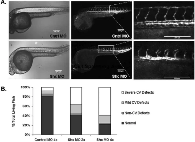
morphants showed impaired ISV formation at 30 hpf (Figure 2.1A). At high concentration of Shc-MO (16 ng/embryo) as well as low concentration
(8ng/embryo), 73% and 55% of Shc-MO fish displayed a cardiovascular defect compared to only 12% of fish injected with high concentration of Standard Control-MO (Figure 2.1B). The predominant vascular phenotype observed was defective growth of ISVs dorsally and improper Dorsal Longitudinal Anastamotic Vessel (DLAV) formation, while other defects ranged from complete loss of ISV sprouts or abnormal overall ISV patterning (termed “Severe CV Defects”) to dilated Caudal Ventral Vein and partial connection of ISVs to DLAV (“Mild CV Defects”). In all conditions, a small percentage of zebrafish exhibited non-cardiovascular (CV) defects such as gross defects in head, eye, fin or tail morphology. We believe this is a non-specific effect of accidentally injuring the embryo with the micropipette during morpholino injection. No obvious defects were observed in heartbeat, aorta morphology or overall zebrafish patterning, indicating a specific role for Shc in angiogenesis during zebrafish development. These results are consistent with the phenotype of the global Shc knockout mouse, which exhibited cardiovascular development defects 10. In contrast to the mouse, Shc-MO zebrafish did not display increased lethality compared to
Control-MO injected fish. This apparent discrepancy may be due to the unique ability of zebrafish to survive significantly longer than mice without a functional heart or vascular system 20 and/or the incomplete depletion of Shc protein in the Shc-MO zebrafish. The zebrafish genome is known to have undergone
escaped this duplication, and only a single Shc gene exists in zebrafish. The morpholinos used here target unique sequences at the locus LOC563639 and nowhere else in the zebrafish genome. While we cannot rule out the existence of another un-anottated Shc-like gene playing a redundant or unique role in
zebrafish development, nobody has reported such a gene.
Endothelial Shc is Required for Proper Angiogenesis in vivo
To specifically inactivate the Shc1 gene in ECs, female mice carrying
floxed alleles of Shc exons 1 and 216 were intercrossed with male transgenic mice expressing Cre recombinase under the control of the Tie2 promoter which is expressed specifically in ECs and some hematopoietic cells 15. In this cross, the Tie2-Cre allele was always donated from the father to minimize leakage of Cre expression into other tissues, which can occur when Tie2-Cre is donated by the mother. Surprisingly, Shcflox/flox; Tie2-Cre+ were born at the expected
Mendelian ratio and these animals display no gross anatomic abnormalities or decrease in fertility compared to Shcflox/flox controls. To verify tissue specific
Cre/loxP recombination in our mice, we crossed Shcflox/flox; Tie2-Cre+ mice to
revealed a complete reduction in all three Shc isoforms in the Shcflox/flox;
Tie2-Cre+ animals (data not shown).
To determine whether angiogenesis is affected in Shcflox/flox; Tie2-Cre+
animals, we used two in vivo models: neonatal retinal neovascularization and the
Matrigel plug assay. Vascularization of the murine retina commences after birth, as the vessels originating at the optic nerve spread radially over the inner surface of the retina on the pre-existing template of astrocytes, guided by a gradient of VEGF-A to form a two-dimensional vascular plexus 21. At postnatal day 5, retinas were isolated and stained with Isolectin B4 to mark ECs. Shcflox/flox;
Tie2-Cre+ mice exhibited a less dense primitive plexus at the vascular front
compared to both Shcflox/flox and Shc wt/wt; Tie2-Cre+ controls (Figure 2.2A).
Vascular density in the retina was quantified by counting the number of branchpoints per 100 μm2 area as well as % vascular area, both of which
revealed a significant decrease in Shcflox/flox; Tie2-Cre+ compared to littermate
controls (Figure 2.2A, lower). Both genotypes of control mice, Shcflox/flox and
Shc wt/wt; Tie2-Cre+, showed equal retinal vascular density, indicating Tie2-Cre
expression itself if not responsible for the phenotype, so only Shcflox/flox
littermate controls were used in the remaining experiments.
impaired in Shcflox/flox; Tie2-Cre+ littermates (Figure 2.2B). Collectively, these
data suggest that endothelial Shc is required for proper postnatal angiogenesis in
vivo.
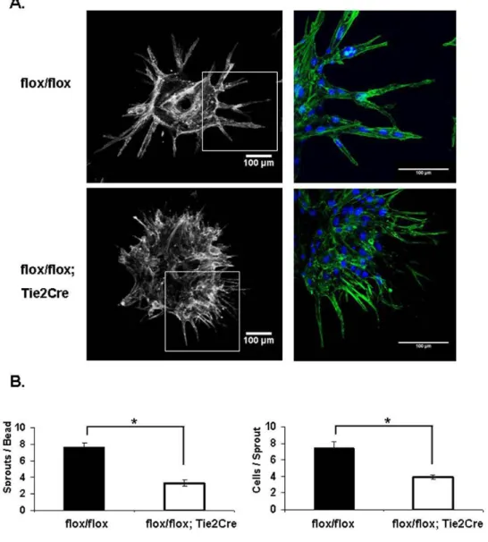
Endothelial Shc is Required for Tube Assembly & Sprouting in vitro
To further determine the role of Shc in the EC angiogenic response, we performed the Fibrin Gel Bead assay. The Fibrin Gel Bead assay was preferred over other available in vitro angiogenesis assays because this assay involves
actual sprouting of ECs off of the bead over a period of a few days so EC proliferation, migration and survival are required; whereas standard ‘tube
formation’ assays largely involve assembly of ECs into capillary-like tubes over a timecourse of a few hours, so the processes of proliferation, survival and
sprouting are less important. Secondly, fibrin gel is more applicable because it is made of digested fibrinogen, and Shc has previously been shown to bind to fibrinogen-binding integrins such as α5β1 and αvβ3 whereas the standard Matrigel is a complex mixture of several ECM proteins such as collagen, laminin and others. ECs isolated from the lungs of Shcflox/flox; Tie2-Cre+ and Shcflox/flox
mice (MLECs) were coated on beads and embedded in Fibrin gel. Control
Shcflox/flox ECs sprouted outward off the bead and lumenized to form
capillary-like vessels in the 3D Fibrin matrix, as is typically seen using HUVEC.
Interestingly, Shcflox/flox; Tie2-Cre+ MLECs displayed a striking defect in both
number and size of sprouts (Figure 2.3A). While Shcflox/flox; Tie2-Cre+ MLECs
rate, these filopodia failed to develop into full sprouts and the tip cells remained stuck on the bead. Quantification revealed a significant reduction in number of sprouts per bead as well as number of cells per sprout, indicating an important role for Shc in EC sprouting (Figure 2.3B).
Shc is Required for Integrin-Mediated EC Signaling
To understand the mechanism underlying the role for Shc in angiogenesis, we examined signaling downstream of two major angiogenic receptors: integrins and VEGFR-2 in ECs. Previous work has shown that Shc binds to a subset of activated integrins and mediates signaling. Upon outside-in integrin activation by ligation to its ECM ligand, Shc is phosphorylated and recruited to integrins α5β1 (fibronectin receptor) and αvβ3 (fibronectin/vitronectin), but not to α2β1 (collagen) or α6β1 (laminin) 22. We therefore tested the role of Shc in integrin-dependent angiogenic responses. For the following experiments, Human Umbilical Vein Endothelial Cells (HUVECs) were infected with lentivirus that expresses either Shc (shShc) or non-specific (shNS) shRNA in order to deplete Shc protein (data not shown). The role of Shc in integrin-mediated cell spreading on ECM was tested by seeding equal numbers of ECs on fibronection (FN) or collagen (CL) and measuring the cell area. While there was no difference in spreading on CL in the absence of Shc, Shc-depleted ECs showed impaired spreading on FN compared to control ECs (Figure 2.4A), suggesting that Shc is required
Similar to the spreading experiments, EC migration toward FN was impaired in Shc-depleted ECs, while migration toward CL occurred independent of Shc (Figure 2.4B) These results indicate that Shc is required for integrin-mediated spreading and migration towards FN, therefore suggesting that Shc selectively mediates angiogenic signaling downstream of FN-binding integrins.
Shc is Required for VEGF-Mediated EC Signaling
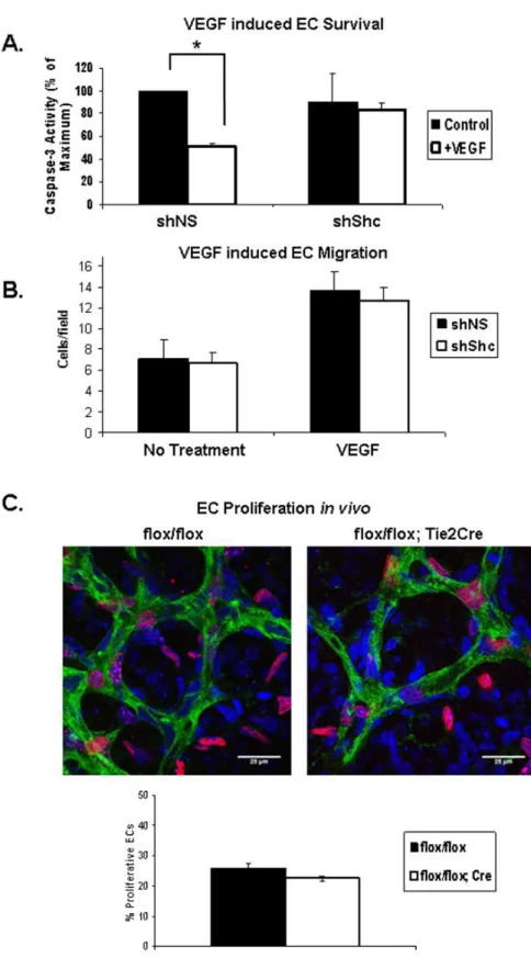
VEGF induces Shc phosphorylation and its association with VEGFR-2 and VE-Cadherin 23, 24. However, the role of Shc, if any, in signaling downstream of VEGF remains unexplored. We assayed the role of Shc in VEGF-induced EC survival and migration in vitro, as well as proliferation of retinal ECs in vivo. EC
survival was assayed by inducing apoptosis in ECs in the presence or absence of VEGF. Apoptosis was quantified by measuring the level of cleaved caspase 3 present in the cell lysates. VEGF treatment resulted in a 50% decrease in
analyzed EdU (5-ethynyl-2’-deoxyuridine) incorporation into endothelial nuclei 2 hours after injection into the P5 mice (Figure 2.5C). Retinas were stained to mark ECs green (Isolectin), all cell nuclei blue (DAPI) and proliferating cells red (EdU). By comparing the number of cells that stained positive for all three markers divided by the total number of ECs, we found the number of
EdU-positive EC nuclei was slightly lower in Shcflox/flox; Tie2-Cre+ mice compared to
controls (Figure 2.5C), but these differences did not reach statistical significance. Thus, Shc does not play a significant role in EC proliferation. Together, these data are consistent with a model in which Shc function is important for survival signaling in response to VEGF, but not for VEGF-induced migration or
proliferation.
Integration of VEGF & integrin signaling via Shc
Our data show a role for Shc in processes downstream of both VEGF and integrins. We hypothesized that Shc mediates crosstalk between these two receptors. To test this hypothesis, we performed survival experiments on ECs plated on FN vs CL. VEGF treatment resulted in a ~50% decrease in apoptosis in shNS control cells grown on either FN or CL, similar to what was seen in Figure 2.5A. Interestingly, shShc ECs grown on CL showed similar
Shc Mediates Akt Activation Downstream of VEGF & Integrin Activation
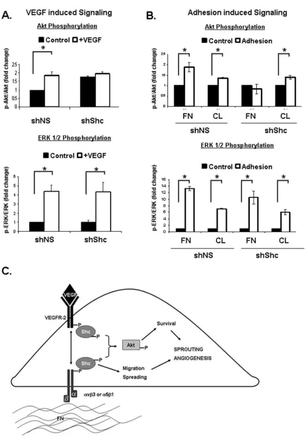
To further delineate the signaling pathways that are mediated by Shc downstream VEGF and integrin signaling, we assayed activation of two key signaling cascades, Akt and ERK 1/2. shShc ECs treated with VEGF failed to activate Akt, whereas VEGF-induced ERK 1/2 activation was similar to shNS control ECs (Figure 2.7A). Interestingly, the requirement for Shc in the activation of Akt was specific to VEGF, as Epidermal Growth Factor (EGF) induced robust activation of Akt in both shNS and shShc ECs (data not shown).
Similarly, Akt activation by adhesion of ECs to FN was impaired in shShc ECs, whereas shShc ECs plated on CL could activate Akt normally (Figure 2.7B), indicating that Shc mediates Akt activation specifically downstream of FN binding integrins. In contrast, ERK 1/2 was activated similarly in both shNS and shShc ECs on both FN and CL, indicating that Shc is not important for ERK 1/2
activation downstream of either FN or CL binding integrins. Together, these data are consistent with a model in which Shc function is important for Akt signaling which promotes survival downstream of VEGF specifically on FN, whereas Shc is dispensable for ERK 1/2 activation and EC proliferation.
DISCUSSION
morphant zebrafish embryos show defects in ISV sprouting in the trunk, while
Shcflox/flox; Tie2-Cre+ mice display impaired angiogenesis in the retina and in
the Matrigel plug assay in vivo. Using an in vitro model of angiogenesis, we
show that Shc is required for sprouting and tube formation. Mechanistically, Shc integrates signaling downstream of integrins and VEGF. Shc is required for integrin-mediated spreading and migration specifically on FN, as well as survival in response to VEGF. Importantly, Shc integrates VEGF and integrin signaling, as VEGF-induced survival on FN requires Shc, whereas survival in ECs on CL does not. Activation of the Akt, but not ERK1/2, pathway in response to both VEGF and integrin activation depends on Shc. Combined, these processes are critical for angiogenesis and provide a mechanism by which Shc integrates signals from VEGF and integrins to mediate angiogenesis (Figure 2.7C). The observation that Shc is required for VEGF-induced EC survival is reminiscent of the reported role for VE-Cadherin in this process 51. Indeed, it is likely that Shc is involved in signaling downstream of this VE-Cadherin:VEGFR-2 complex that leads to Akt activation and cell survival because the same lab later showed that VEGF treatment of ECs induces a Shc association with both VE-Cadherin and VEGF-2 23. It is unknown whether Shc mediates assembly of the VE-Cadherin: VEGFR-2 complex or merely signals downstream of this complex after it is formed, but this will be an area of further research.
represent central signaling axes during angiogenesis. Activation of either VEGFR-2 or αvβ3 induces physical association of the two receptors, which is important for VEGFR-2 phosphorylation 26. Function of both receptors is required for proper signaling, as inhibition of αvβ3 or VEGFR-2 function decreases
VEGFR-2 activation and complex formation27, 28. Our work here shows that Shc is required for mediating and integrating angiogenic responses downstream of both integrins (FN-binding integrins specifically) and VEGF, thus coordinating the angiogenic process as a whole.
Shc was originally described as an oncogene, and mutation of Shc attenuates tumor growth in mice 29. Shc overexpression in fibroblasts causes transformation 9 and Shc is required for cellular transformation in
ErbB2-overexpressing breast cancer cells 30, as well as in mammary tumors induced by Polyoma Middle T expression 31. In humans, clinical studies have associated Shc activation with poor patient prognosis 29. These data, combined with our current findings, suggest that Shc is critical for many steps of tumorigenesis, including cellular transformation of tumor cells themselves, as well as
angiogenesis in ECs that feed the tumor and enable its growth. Therefore, Shc may be an interesting target for cancer treatment at multiple levels.
for embryonic development, but it is required postnatally for angiogenesis. Induction of Tie2-Cre expression has been reported at E9.515, which precedes
embryonic lethality of the global Shc knockout at E11.5, so mis-timing of Shc gene excision does not appear to be the reason for Shcflox/flox; Tie2-Cre+
mouse survival. Emerging research has set precedence for the idea that conditional gene knockout using the Tie2-Cre transgene can result in mice that
initially develop a normal vasculature while exhibiting defective angiogenic capacity. Tie2-Cre mediated conditional knockout of genes such as
Endothelin-132, 33, TFPI34, ADAM1735, PPARγ 36 and Dicer 37 yield viable mice with cardiovascular defects, while the corresponding global knockout animal is embryonic lethal. Thus, genes such as Shc and others appear to have differing roles in developmental vs. postnatal angiogenesis. This hypothesis is
strengthened in light of the literature on proteins that interact with Shc. Our results indicate that Shc is required for signaling downstream of FN-binding integrins such as αvβ3 and/or α5β1, which are upregulated during angiogenesis. Surprisingly, endothelial knockout of αv38,β339, or α540 results in viable mice, while antagonism of either of these integrins using blocking antibodies results in a defect in angiogenesis41-43. We also show that Shc mediates a subset of
signaling responses downstream of VEGF. In particular, Shc is required for EC survival but not proliferation in response to VEGF.
both mouse and fish. Flow also promotes hematopoetic cell development 48, 49
in
vivo and atheroprotective laminar flow inhibits HUVEC tubule formation and
migration in vitro50. Therefore, a role for Shc in flow-driven angiogenesis is an
attractive idea. Integration of VEGF- and flow dependant signaling was recently reported during zebrafish vascular remodeling 46, and future experiments are aimed at understanding the role of Shc in these processes. Angiogenesis