THE CLASP FAMILY REGULATES MICROTUBULE DYNAMICS BY USING AN ARRAY OF TOG-LIKE DOMAINS
Jonathan B. Leano
A dissertation submitted to the faculty of the University of North Carolina at Chapel Hill in partial fulfillment of the requirements for the degree of Doctor of Philosophy in the Department of Biochemistry and Biophysics.
Chapel Hill 2014
ABSTRACT
Jonathan B. Leano: The CLASP family regulates microtubule dynamics by using an array of TOG-like domains
(Under the direction of Dr. Kevin C. Slep)
ACKNOWLEDGEMENTS
First and foremost, I would like to express my gratitude to my advisor, Dr. Kevin Slep, for his patient, motivating, and encouraging support throughout my graduate studies. Through his guidance, I was able to mature into the scientist I am today. I could not have imagined a better advisor and I am fortunate to have worked in your laboratory.
I would also like to thank my thesis committee: Dr. Drew Lee, Dr. Richard Cheney, Dr. Hengming Ke, and Dr. Stephen Crews. Your thoughtful insights and challenging questions into my work motivated me to critically evaluate and improve my research.
In addition to my advisor and thesis committee, I would also like to thank Dr.
Stephen Rogers and the members of his laboratory for helping me to develop and perform the cell-based assays in my work. I would like to thank Dr. Barry Lentz and the Molecular and Cellular Biophysics Program, Dr. Michael Miley at the UNC Crystallography Core, Dr. Ashitosh Tripathy at the UNC Macromolecular Interactions Facility, and Dr. Ashalla Freeman and the Initiative for Maximizing Student Diversity Program.
I would like to thank my fellow lab mates in the Slep Lab – it has been a pleasure to work and collaborate with every one of you.
PREFACE
The work in Chapter 2 is a manuscript that was published in the journal, Structure, in 2013, under the direction of Dr. Kevin Slep and with the support of Dr. Stephen Rogers’ reagents, equipment, and advice. This work focused on characterizing CLASP1 TOG2 using structural, biochemical, and cellular studies. Kevin Slep crystallized TOG2 and collected diffraction data, while I built the model and refined the structure. After the work was published, I solved another crystal structure of TOG2 at resolution of 1.8Å. The citation to the manuscript is: Leano, J.B., Rogers, S., Slep, K.C. (2013). A Cryptic TOG Domain with a Distinct Architecture Underlies CLASP-Dependent Bipolar Spindle Formation. Structure. 21(6):939-50.
The work in Chapter 3 is an ongoing project attempting to crystallize TOG1
(CLASP1 1-257) and TOG3 (843-1092). TOG1 crystals diffract to 2.0Å, but failed attempts in SAD phasing and molecular replacement has prevented us from obtaining a crystal structure. TOG3 crystals have been obtained and will be screened for higher resolution diffraction in future studies. Trent Wei, an undergraduate student I mentored for two years, designed constructs for TOG3 and performed initial expression and purification of these constructs.
The work in Chapter 4 is also an ongoing project characterizing the interaction between CLASP C-terminal and CLIP-170 coiled coil domain using structural and
CLIP-170 coiled coil domain. After screening these constructs for binding, CLASP 1253-1522 and CLIP-170 331-456 were used for biochemical studies and crystallization. CLASP 1253-1522 and CLIP-170 331-456 formed a complex under SEC-MALS and ITC experiments. Crystals of these proteins alone and in complex with each other were obtained, but did not diffract or diffracted to low-resolution.
TABLE OF CONTENTS
LIST OF FIGURES ... xii
LIST OF TABLES ... xiv
LIST OF ABBREVIATIONS AND SYMBOLS ... xv
CHAPTER 1: INTRODUCTION ... 1
Microtubules are dynamic and physically robust polymers that are essential for a host of cellular processes ... 1
Microtubule associated proteins (MAPs) and plus-end tracking proteins (+TIPs) regulate MT dynamics ... 8
MAPs and +TIPs play important roles in the formation and maintenance of the mitotic bipolar spindle ... 14
XMAP215 promotes MT polymerization through a pentameric array of TOG domains ... 18
CLASP is a conserved family of +TIPs that promotes MT stability ... 22
References ... 28
CHAPTER 2: A Cryptic TOG Domain with a Distinct Architecture Underlies CLASP-Dependent Bipolar Spindle Formation ... 39
Summary ... 39
Introduction ... 40
Experimental Procedures ... 42
Cloning and Expression ... 42
Crystallization ... 44
Drosophila S2 Cell Expression Plasmids ... 45
Cell Culture and Transfection ... 45
RNAi ... 46
Western Blots ... 46
Immunofluorescence Microscopy and Mitotic Spindle Classification ... 47
Circular Dichroism ... 48
Light Scattering Tubulin Polymerization Assay ... 48
Microscopic Analysis of Tubulin Polymerization in Vitro ... 49
The Intra-HEAT Loops Carry Canonical TOG Domain Tubulin-Binding Determinants ... 53
Results ... 47
The CLASP1 TOG2 Structure Adopts a Bent Conformation that Contrasts with XMAP215 Family TOG Domains ... 54
The CLASP1 TOG2-Tubulin Interaction Is Predicted to Be Dramatically Different from XMAP215 Family TOG-Tubulin Interactions ... 55
TOG2 is Required for CLASP-Mediated Bipolar Spindle Formation ... 60
TOG2 Promotes Microtubule Lattice Association In Vivo ... 63
TOG2 Increases Microtubule Polymerization Rates In Vitro ... 65
Discussion ... 67
REFERENCES ... 69
Tables ... 73
CHAPTER 3: Expression, Purification, and Crystallization of
TOG1 and cryptic-TOG3 Domains from Human CLASP1 ... 79
Summary ... 79
Experimental Procedures ... 80
Cloning and Expression ... 80
Protein Purification ... 80
Crystallization ... 81
Heavy-atom and halide soaks ... 83
Data Collection and Structure Determination Attempts ... 83
Results ... 84
Expression, Purification, and Crystallization of CLASP1 TOG1 ... 84
Data collection and attempts at obtaining phasing information for CLASP1 TOG1 ... 84
Secondary structure prediction, purification and crystallization of cryptic TOG3 ... 87
Discussion ... 91
References ... 85
Supplemental Figures ... 99
CHAPTER 4: CLASP C-terminal Domain interacts with CLIP-170 Coiled-coil Domain ... 100
Summary ... 100
Experimental Procedures ... 101
Cloning and Expression ... 101
Protein Purification ... 101
Isothermal Titration Microcalorimetry ... 103
Size Exclusion Chromatography and Multi-angle Light Scattering ... 104
Results ... 104
Cloning, Expression, and Purification of CLASP1 C-terminal domain and CLIP-170 coiled-coil domain ... 104
Binding analysis of CLASP1 C-terminal domain and CLIP-170 coiled-coil domain ... 107
Crystallization of CLASP1 C-terminal domain and CLIP-170 coiled-coil domain ... 110
Discussion ... 111
References ... 114
Supplemental Figures ... 115
CHAPTER 5: DISCUSSION AND FUTURE WORK ... 117
CLASP uses an array of structurally distinct TOG domains to regulate microtubules ... 117
The C-terminal domain of CLASP promotes dimerization and interaction with CLASP-associating factors ... 123
Concluding remarks ... 126
References ... 128
LIST OF FIGURES
Figure 1-1. Microtubules are polymers of αβ-tubulin ... 2
Figure 1-2. Overview of microtubule dynamics ... 4
Figure 1-3. αβ-tubulin binds guanine nucleotides at two sites ... 5
Figure 1-4. Structures of αβ-tubulin are observed in different conformations ... 7
Figure 1-5. MAPs bind along the microtubules lattice to alter microtubule dynamics ... 10
Figure 1-6. Plus-end tracking proteins (+TIPs) interact in a network to modulate MT plus-end dynamics ... 13
Figure 1-7. The mitotic bipolar spindle is a microtubule-based machine that segregates chromosomes ... 15
Figure 1-8. Microtubule poleward flux ... 17
Figure 1-9. TOG domains are tubulin-binding domains ... 20
Figure 1-10. TOG domains interact with αβ-tubulin ... 21
Figure 1-11. CLASP and ch-TOG/XMAP215 family uses an array of TOG domain to regulate microtubule dynamics ... 23
Figure 1-12. CLASP’s C-terminal domain (CTD) interacts with various factors to promote specific functions of CLASP at different cellular locations ... 25
Figure 2-1. CLASP Contains a Conserved, Cryptic TOG Array ... 51,52 Figure 2-2. CLASP1 TOG2 Has a Bent Structure ... 57
Figure 2-3. CLASP1 TOG2 and/or αβ-Tubulin Is Likely to Undergo Conformational Change upon Binding ... 58
Figure 2-4. MAST Depletion Causes Bipolar Spindle Defects that Can Be Partially Rescued in Trans ... 59
Figure 2-5. Point Mutations across the TOG2 Tubulin-Binding Surface Fail to Rescue MAST-Mediated Bipolar Spindle Formation ... 62
Figure 2-6. MAST TOG2 Determinants Mediate MT-Lattice Binding ... 64
Figure 2-S1. CLASP family members are composed of an array of conserved cryptic TOG domains ... 76 Figure 2-S2. CLASP TOG2 Protomers Align Well and Form Minimal Contacts with Symmetry Mates ... 77 Figure 2-S3. Second crystal structure of CLASP1 TOG2 at 1.8Å resolution ... 78 Figure 3-1. Expression, purification, and crystallization of CLASP1 TOG1
(residues 1-257) ... 85 Figure 3-2. Predicted secondary structure of CLASP1 TOG3 ... 88 Figure 3-3. Expression, purification, and crystallization of CLASP1 TOG1
(residues 843-1092) ... 90 Figure 3-S1. CLASP1 TOG3 HR C is predicted to consist of
anti-parallel helices ... 99 Figure 4-1. Various constructs of CLASP C-terminal domain and
CLIP-170 coiled-coil domain ... 105 Figure 4-2. Expression, purification, and crystallization of CLASP1 CTD
(residues 1253-1522) ... 106 Figure 4-3. Expression, purification, and crystallization of CLIP-170
coiled coil domain constructs ... 107 Figure 4-4. CLASP C-terminal domain forms weak interactions with
CLIP-170 coiled-coil domain ... 109 Figure 4-5. Size exclusion chromatography and multiangle light scattering
(SEC-MALS) analysis of CLASP CTD and CLIP-170 CC ... 110 Figure 4-S1. CLASP CTD and CLIP-170 CC display interaction
through native gel electrophoresis ... 115 Figure 4-S2. Larger construct of CLIP-170 (residues 331-588) forms
a dimer under SEC-MALS ... 116 Figure 5-1. CLASP promotes MT pause and MT growth ... 127
LIST OF TABLES
Table 2-1. Data Collection and Refinement Statistics for human
CLASP1 TOG2 ... 73 Table 2-S1. Mitotic spindle morphology in Drosophila S2 cells
transfected with MAST constructs ... 74 Table 2-S2. Data Collection and Refinement Statistics for human
CLASP1 TOG2 solved to 1.8Å resolution. ... 75 Table 3-1. Data collection on native and multi-derivative crystals
of CLASP1 TOG1. ... 98
LIST OF ABBREVIATIONS and symbols
Å Angstrom
β-ME beta-mercaptoethanol
Br bromide
CaCl2 calcium chloride
CD circular dichroism
CENP-E Centromere-associated protein E
ch-TOG colonic and hepatic tumor-overexpressed gene CLASP CLIP170 associated protein
CLIP170 cytoplasmic linker protein 170 Cu(II)Cl2 copper(II) chloride
CuSO4 copper sulfate
Da Dalton
DARPin designed ankryin repeat protein DMSO dimethyl sulfoxide
dsRNA double-stranded RNA
DTACC Drosophila transforming acidic coiled-coil DTT dithiothreitol
EB1 end binding protein 1
eGFP enhanced green fluorescent protein FBS fetal bovine serum
GFP green fluorescent protein
GMPCPP guanosine-5’-[(α,β)-methyleno]triphosphate GTP guanosine triphosphate
HCl hydrochloric acid
HEAT Huntingtin, elongation factor 3, protein phosphatase 2A, target of rapamycin 1
Hg mercury
HR HEAT repeat
IPTG isopropyl β-D-1-thiogalactopyranoside KCl potassium chloride
kDa kilo Dalton
kMTs kinetochore microtubules KOH potassium hydroxide L liter v. leucine
LL5β Pleckstrin homology-like domain family member 2
M molar
MAP microtubule associated protein
mg milligram
MgCl2 magnesium chloride
min minute
ml milliliter
ML maximum-likelihood
MPa megapascal Msps Minispindles
MT microtubule
MWCO molecular weigh cut off NaCl sodium chloride
NaN3 sodium azide
Ni2+-NTA nickel nitrilotriacetic acid
nm nanometer
PAGE polyacrylamide gel electrophoresis PBS phosphate buffer saline
PEG polyethylene glycol
PIP3 phosphatidylinositol (3,4,5)-trisphosphate
rmsd root-mean-square deviation
res residues
RNAi RNA interference rpm rotations per minute
SAD single-wavelength anomalous diffraction SASA solvent accessible surface area
SDS sodium dodecyl sulfate
SEC-MALS size exclusion chromatography coupled with multi-angle light scattering SeMet selenomethionine
µg microgram
µl microliter
µM micromolar
µm micron
UTR untranslated region UV ultraviolet
WT wild type
Xkid Xenopus kinesin-like protein kif22a
CHAPTER 1: INTRODUCTION
Microtubules are dynamic and physically robust polymers that are essential for a host of cellular processes
Microtubules (MTs) are dynamic components of the cellular cytoskeleton that facilitate cell-shape change, cell migration, intracellular transport, and mitotic spindle formation. (Desai and Mitchison, 1997). Microtubules are tubular polymers composed of heterodimers of α- and β-tubulin (55 kDa each) (Figure 1-1A). These αβ-tubulin
heterodimers polymerize in a head-to-tail fashion to form the protofilament (Figure 1-1B), and approximately thirteen protofilaments associate laterally to form the microtubule (Figure 1-1C) (Mandelkow and Mandelkow, 1989) (Akhmanova and Steinmetz, 2008). The αβ -tubulin heterodimer is made up of two different 55-kDa proteins that assemble into a specific, asymmetric orientation. As a consequence of this asymmetry, αβ-tubulin is intrinsically polar. This polarity is propagated along the microtubule lattice to form
microtubules ends with two different polymerization rates. The microtubule end that consists of the exposed β-tubulin is the fast-growing “plus” end while α-tubulin is oriented towards the slow-growing “minus” end (Figure 1-1C) (Allen and Borisy, 1974) (Desai and
Figure 1-1. Microtubules are polymers of αβ-tubulin. (A) Cartoon representation of the crystal structure of αβ-tubulin (1JFF). (B) Tubulin heterodimers interact in a head-to-tail fashion to form the protofilament. (C) Thirteen protofilaments interact laterally to form the hollow, tubular structure of the microtubule. The asymmetric orientation of αβ-tubulin gives microtubules its inherent polarity. The end that comprises of exposed α-tubulin is referred to as the “minus-end” while the end that comprises of exposed β-tubulin is referred to as the “plus-end.” Figure adapted from (Akhmanova and Steinmetz, 2008).
The most essential and fundamental property of microtubules is dynamic instability, the ability of microtubules to stochastically switch between states of growth
[polymerization], pause, and shrinkage [depolymerization] (Mitchison and Kirschner, 1984) (Figure 1-2). Cryo-EM images that capture growing microtubules display a sheet of MT protofilaments forming at MT ends (Figure 1-2A), while shrinking microtubules display frayed ends (Figure 1-2B) (Simon and Salmon, 1990) (Mandelkow et al., 1991). Events that characterize abrupt, stochastic transitions between states of growth and shrinkage are called
Į
β
Į
β
-
tubulin
Protofilament
Microtubule
Plus-end
Minus-end
A
B
rescue and catastrophe (Figure 1-2). The transition from growth to rapid shrinkage is referred to as “catastrophe,” while the transition from shrinkage to a state of growth is called “rescue” (Walker et al., 1988). To transition between states of growth and shrinkage, the microtubule goes through a kinetically metastable intermediate state called the “pause” state, in which the microtubule is neither growing nor shrinking (Tran et al., 1997). Mechanical and
UV-microbeam severing of self-assembled microtubules and axoneme-nucleated microtubules produces stable MT ends that do not undergo states of polymerization and depolymerization (Walker et al., 1989) (Tran et al., 1997). Cryo-electron microscopy (Cryo-EM) images that captured different populations of dynamic microtubules in vitro showed a large population of blunt MT ends along with MT sheets and frayed ends. These blunt MT ends suggest they are occupying a metastable intermediate “pause” state between growing and shrinking
microtubules (Figure 1-2C) (Simon and Salmon, 1990) (Mandelkow et al., 1991) In addition, the blunt MT ends can switch to phases of growth or shortening, suggesting that dynamic MTs must occupy the pause state to undergo rescue or catastrophe (Arnal et al., 2000).
Figure 1-2. Overview of microtubule dynamics. (A) GTP-tubulin (red and green spheres) adds to pre-existing sheets of protofilaments on growing microtubules. The transition from shrinking to growing microtubules is referred to as rescue. (B) GDP-tubulin (purple and green spheres) adopts a “curved” conformation, resulting in frayed ends that promote shrinking microtubules. The transition from growing to shrinking microtubules is referred to as catastrophe. (C) The blunt-ended MT is displaying the pause state, in which the MT is neither growing nor shrinking. Eventually, the paused MT will undergo growing or shrinking phases. Figure adapted from (Akhmanova and Steinmetz, 2008).
Catastrophe
Rescue
Growing MT
Shrinking MT
Paused MT
A
B
Figure 1-3. αβ-tubulin binds guanine nucleotides at two sites. Cartoon representation of the crystal structure of αβ-tubulin (1JFF). Sphere representation of GDP molecule that occupies the nucleotide exchange site (E-site), which is located on β-tubulin. Sphere representation of GTP molecule that occupies the non-exchangeable site (N-site), which is located on α-tubulin.
(Figure 1-2A) (Simon and Salmon, 1990; Akhmanova and Steinmetz, 2008). Tubulin hydrolyzes GTP to GDP at the E-site during or shortly after polymerization (Hyman et al., 1992). Consequently, GDP-bound tubulin (GDP-tubulin) comprises most of the microtubule lattice (Figure 1-1C and 1-2). However, GTP hydrolysis promotes tubulin to adopt a curved conformation that disrupts lateral contacts between protofilaments (Müller-Reichert et al., 1998). This causes the protofilaments to “peel” away from the MT lattice, creating frayed ends (Figure 1-2B). These frayed ends will disassemble to into protofilament rings or non-polymerized tubulin subunits (Mandelkow et al., 1991). Lateral and longitudinal contacts
Į
β
Į
β
-
tubulin
GDP (E-site)
between GDP-tubulin within the MT lattice force GDP-tubulin to remain in a “straight” conformation. This constraint builds tension, causing protofilaments to peel away and promote MT depolymerziation. To prevent microtubule depolymerization, studies have proposed that a thin layer of GTP-bound tubulin (GTP cap) is sufficient to stabilize the microtubule plus end and facilitate polymerization (Figure 1-2A) (Mitchison and Kirschner, 1984) (Voter et al., 1990) (Walker et al., 1991). Similar studies involving the addition of tubulin bound to GMCPMP, a non-hydrolyzable analog of GTP, forms a GMCPMP cap that is one or two tubulin heterodimers deep (longitudinally) and is sufficient to prevent
depolymerization (Drechsel et al., 1994) (Caplow et al., 1996).
As mentioned previously, microtubule assembly involves converting tubulin between curved and straight conformations. Structural studies observing both non-polymerized tubulin and polymerized microtubules reveal insights into straight-bent conformational changes. Electron crystallography structures of zinc-induced sheets of tubulin bound to taxol reveals tubulin protofilaments in a straight conformation, which mimics the state of tubulin found in microtubules (Figure 1-4A) (Nogales, et al., 1998) (Löew et al., 2001) (Nettles et al., 2004). The structures of incorporated MTs contain only GDP-tubulin. In contrast, X-ray crystallography structures of non-polymerized tubulin bound to depolymerizing factors such as the stathmin homologue RB3 (Ravelli, et al., 2004) and a designed ankryin repeat protein (DARPin) (Figure 1-4 B and C) (Pecqueur et al., 2012), show the tubulin heterodimers in a curved conformation. In these structures, either GDP (Figure 1-4B) or GTP (Figure 1-4C) was bound to β-tubulin indicating that non-polymerized tubulin adopts a curved
Figure 1-4. Structures of αβ-tubulin are observed in different conformations. (A) Crystal structure of αβ-tubulin (1JFF) in a “straight” conformation that mimics tubulin incorporated into the microtubule lattice. Sphere representation of a GDP molecule occupies the E-site located on β-tubulin. (B) Crystal structure of αβ-tubulin (4FFB) captured in a “curved” conformation by a DARPin molecule (not shown). GDP molecule occupies the E-site in a curved tubulin, which emulates the depolymerizing state of tubulin. (C) Crystal structure of
αβ-tubulin (4DRX) also captured in a “curved” conformation by a DARPin molecule (not shown), but containing GTP molecule at the E-site, which emulates a state of tubulin that is primed for polymerization.
only changes from a curved to straight conformation during MT assembly when lateral interactions between tubulin heterodimers force tubulin to adopt the straight conformation (Peng et al., 2014). These structures reveal that tubulin adopts different conformations depending on the state of MT assembly/disassembly.
GDP
GDP
GTP
Į
β
Į
Straight Tubulin
Curved Tubulin
A
B
C
1JFF 4FFB 4DRX
Incoporated into microtubule lattice
Depolymerizing microtubules
Microtubule associated proteins (MAPs) and plus-end tracking proteins (+TIPs) regulate MT dynamics
Most of the research on microtubule structure and dynamics explained above were characterized through in vitro experiments that reconstituted microtubule dynamics using purified or isolated tubulin, guanine nucleotides, and microtubule-binding proteins. While microtubules are inherently dynamic in vitro, microtubule-associated proteins (MAPs) are a diverse family of proteins that regulate intracellular microtubule dynamics to ensure proper assembly and disassembly of microtubule at specific locations within the cell and precise times during the cell cycle (Amos and Schliper, 2005). Their ability to regulate microtubule dynamics in accordance with temporal and spatial cues controls fundamental processes such as mitosis, cell shape and differentiation, and intracellular trafficking. Defects in MAPs have been implicated in pathological conditions including cancer, cardiovascular, and
neurodegenerative diseases (Akhmanova and Steinmetz, 2008). MAPs associate with non-polyermized tubulin, the microtubule minus end, the microtubule lattice, and the microtubule plus end (Halpain and Dehmelt) (Akhmanova, Steinmetz, 2008) to alter rates of
polymerization, depolymerization, and pause.
Although MAPs can associate with the minus end and plus end of microtubules, classical MAPs bind along the microtubule lattice and non-polyermized tubulin to stabilize or destabilize microtubules (Halpain and Dehmelt, 2006). MAPs that stabilize microtubules are referred to as structural MAPs and include doublecortin and the Tau/MAP2 family of
microtubule-binding repeats (DCX1/R1 and DCX2/R2) that bind and stabilize microtubules (Figure 1-5A) (Taylor, K.R., et al., 2000). Mutations of doublecortin result in misguided neuronal migration, leading to brain developmental disorders, including the formation of a “double cortex.” (Amos and Schlieper 2005). One of the most well characterized structural MAPs is the Tau/MAP2 family of microtubule stabilizers. Tau is an important stabilizer of axonal microtubules and is primarily expressed in the nervous system. Tau binds
microtubules through short microtubule-binding motifs that repeat two or four times depending on the isoform (Figure 1-5B) (reviewed by Butner and Kirschner, 1991). Hyperphosphorylation of Tau detaches Tau from microtubules, resulting in a loss of microtubule stability and eventually leads to the aggregation of Tau into neurofibrillary tangles (NFTs). These tau-containing NFTs are one of the hallmarks for Alzheimer’s disease (Mandelkow and Mandelkow, 1998). The multiple microtubule-binding repeats that
comprises these structural MAPs enables it to decorate the microtubule lattice and crosslink tubulin subunits along the microtubule, providing overall microtubule stability and
promoting microtubule growth.
In opposition to structural MAPs, microtubule destabilizers reduce the rate of
Figure 1-5. MAPs bind along the microtubules lattice to alter microtubule dynamics. Various MAPs (associated proteins) bind to microtubule using a binding domains or repeats (purple boxes). (A) Doublecortin uses two tandem microtubule-binding repeats (DCX1 and DCX2) and a serine-proline rich region (S/P rich) (red box) to bind and stabilize microtubules. (B) Tau uses 2-4 short microtubule-binding repeats (R1-4) and a basic proline-rich region (red box) to bind microtubules. Tau contains an N-terminal projection domain that extends from the outer surface of the microtubule when Tau is bound to microtubules. (C) The Katanin p60 subunit is a 60kDa ATPase subunit that binds microtubule along its microtubule-binding domain, in which it severs microtubules using its AAA ATPase domain (green box). Katanin’s C-terminal domain (brown box) is used to oligomerize with the regulatory Katanin p80 subunit. (D) RB3/Stathmin 4 sequesters tubulin subunits using its stathmin-like domain (SLD) which contains a coiled-coil region.
Doublecortin
MT binding AAA CTD
Katanin p60 subunit
DCX1 DCX2 S/P-rich
Tau
P-rich R1 R2 R3 R4
Projection domain
RB3/Stathmin 4
SLD
100 A.A
coiled-coil
A
B
C
through phosphorylation (Marklund et al., 1996). Stathmin overexpression is found in many malignant cancers including breast, lung, and ovarian cancer (Bièche, et al., 1998; Chen et al., 2003; Price et al., 2000). Stathmin sequesters free tubulin through a stathmin-like domain (SLD) domain, preventing microtubule incorporation and thereby inhibiting microtubule polymerization (Figure 1-5D). Biochemical studies reveal that one molecule of stathmin tightly binds two tubulin heterodimers to form a T2S complex (Jourdain et al. 1997). Crystal
structures of the T2S complex showed a long alpha-helical domain of stathmin interacting
with a pair of tubulin heterodimers and capping the α-tubulin end, preventing longitudinal contacts with another tubulin heterodimer (Gigant et al., 2000) (Ravelli et al. 2004). Microtubule destabilizers such as katanin, and stathmin facilitate the rearrangement of the microtubule cytoskeleton in accordance with temporal and spatial cues. In summary, classical MAPs including microtubule stabilizers (structural MAPs) and destabilizers coordinate with each other to maintain the critical balance between microtubule
polymerization/depolymerization and to alter the microtubule cytoskeleton in accordance with temporal and spatial cues.
Plus-end tracking proteins (+TIPs) are a diverse group of specialized MAPs that accumulate at plus ends to modulate MT dynamics (Schuyler and Pellman, 2001). +TIPs were initially discovered by time-lapse video microscopy of green fluorescent protein (GFP) fusion construct of CLIP-170 displaying “comet-like” trajectories that coincided with the growing tips of microtubules (Perez, et al. 1999). Additional +TIPs have since been
2007). The colonic and hepatic tumor overexpressed gene (ch-TOG) protein belongs to a family of proteins that include Xenopus member XMAP215 and Drosophila member
minispindles, which functions as microtubule polymerases to enhance the rate of microtubule growth (Brouhard et al., 2008). End-binding proteins (EB) promote microtubule growth and suppresses catastrophe. +TIPs, including the ones mentioned previously, function in a vast network to accumulate at the MT plus end and alter its dynamics.
Figure 1-6. Plus-end tracking proteins (+TIPs) interact in a network to modulate MT plus-end dynamics. Different types of +TIPs interact with the MT plus-end. (A) End-binding protein 1 (EB1) can automatously track growing microtubule plus-ends (red arrow). EB1 contains an N-terminal calponin homology domain (orange circle) to interact with microtubules, a coiled-coil region (light blue), an EB homology domain (yellow box), and an EEY/F-like motif at its C-terminus. (B) CLASP’s SxIP motif (vertical black line), which is located within a basic serine-rich linker region, directly binds with the EB homology domain on EB1 (green arrow). (C) CLIP-170’s directly binds EB1 using CLIP-170’s CAP-gly domains (purple boxes) to interact with EEY/F-like motifs (red boxes) on EB1 (blue arrow). CLIP-170 contains a coiled-coil domain (light blue) and a EEY/F-like motifs at its C-terminus, similar to that of EB1. (Figure adapted from van der Vaart et al., 2011)
SxIP MT Plus End
CLASP
EB1
CLIP-170
A
B
a focus of this thesis. Another interacting mode of EB1 is the CAP-Gly domain, which is characterized by highly conserved glycine and hydrophobic residues. +TIPs, including CLIP-170, may contain single or multiple copies of CAP-Gly domains (Akhmanova and Steinmetz, 2008). CAP-Gly domains contain a conserved GKNDG motif that interacts with the EEY-motif located at the C-terminal tail of the EBH domain (Figure 1-6C) (Weisbrich et al., 2007). Although the ch-TOG family does not contain either a SxIP motif or a CAP-Gly domain, the SxIP-motif-containing protein SLAIN2 binds both ch-TOG and EB1, thereby conferring ch-TOG plus end localization. SLAIN2 is required for ch-TOG-dependent
persistent MT growth (van der Vaart et al., 2011) (Kumar and Wittman, 2012). Because EB1 is a critical microtubule regulator and the central hub of the +TIP network, loss-of-function mutations or depletion in EB1 results in a decrease of microtubule dynamics.
MAPs and +TIPs play important roles in the formation and maintenance of the mitotic bipolar spindle
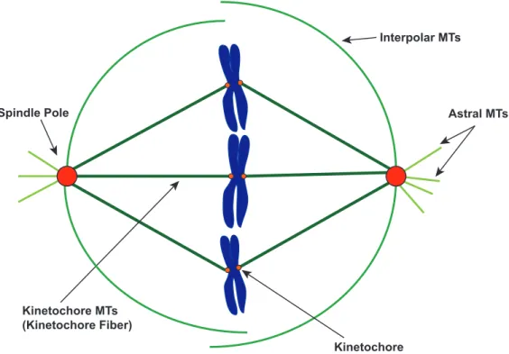
Figure 1-7. The mitotic bipolar spindle is a microtubule-based machine that segregates chromosomes. A simplified model of the bipolar spindle during metaphase. Microtubules nucleate from two spindle poles (red circle) located on opposite sides. Astral microtubules (light green lines) extend towards the cell cortex. Interpolar microtubules (medium green lines) grow towards the central region of the spindle and connect with other interpolar MTs nucleated from the opposite spindle pole. Dozens of kinetochore microtubules (dark green lines) attach to the kinetochore (small orange circles) located on chromosomes (blue) and bundle with each other to form the kinetochore fiber.
astral microtubules, and 3) interpolar microtubules. Kinetochore microtubules (kMTs) attach to specialized protein structures on the chromosomes, called kinetochores. Astral
microtubules grow with their plus ends extending towards the cell cortex. Interpolar microtubules grow towards the central region of the spindle and interconnect with microtubules from the opposite spindle pole (Figure 1-7) (Glotzer, 2009).
Dozens of kMTs bundle to form the physically robust, kinetochore fiber. The forces required for chromosome segregation are generated from the kinetochore fibers (Kitamura et
Spindle Pole Astral MTs
Interpolar MTs
Kinetochore Kinetochore MTs
al., 2009). Poleward microtubule flux is one of the major forces that powers chromosomal motion during mitosis. This force occurs when microtubules polymerize at the kinetochore, the kMT lattice continuously moves towards the spindle pole, and tubulin subunits are removed at the spindle pole (Figure 1-8) (Margolis et al., 1978). Consequently, controlled regulation in balancing polymerization/depolymerization rates of the fluxing kMTs at the kinetochore and spindle pole is required to coordinate the position and movement on chromosomes through mitosis (Buster et al., 2007). During metaphase, the bipolar spindle reaches a steady-state of poleward microtubule flux, in which the rate of polymeration at the kMT plus end is matched by deplymerization at the kMT minus end. This balance is essential in maintaining the proper positioning of the chromosomes at the spindle center or metaphase plate (Mitchison et al., 1989; Rogers et al., 2005; Jaqaman et al., 2010). During anaphase, kMT polymerization stops and poleward MT flux slows to approximately half the metaphase rate (Rogers et al., 2005). This decreases the length of the kinetochore fiber and promotes the proper separation of sister chromatids into daughter cells.
+TIPs are required to attach microtubules to the cellular cortex to control spindle positioning and to attach microtubules to the kinetochore to coordinate chromosome segregation (Maiato et al., 2004; Tamura and Draviam, 2012). EB1 also acts as a major regulator of MT plus end dynamics during mitosis, interacting with the dynamic network of +TIPs to differentially regulate mitotic MT plus ends (Steinmetz and Akhmanova, 2008). Depletion of EB1 using double-stranded RNA interference (RNAi) in Drosophila S2 cells produces several defects including astral microtubule reduction, defocused spindle poles, and misalignment of the bipolar spindle, away of the cell center (Rogers et al., 2002). The
Figure 1-8. Microtubule poleward flux. During microtubule poleward flux kinetochore microtubules continuously grow at the kinetochore (orange ellipse). CLASP (green box) is an essential component of the outer kinetochore that promotes MT polymerize at the kinetochore. At the spindle pole (red circle), kinetochore microtubules continuously depolymerize. The MT growth at the kinetochore coordinated with the MT depolymerize at the spindle pole generates MT poleward flux that is directs towards the spindle pole. Controlling the growth/shrinkage rates of fluxing kinetochore microtubules determines the rate of poleward flux.
increases the rate of microtubule polymerization (Brouhard et al., 2008). XMAP215 and ch-TOG proteins promote MT growth and play an essential role in stabilizing spindle
microtubules (Matthews et al. 1998; Tournebize et al. 2000; Popov et al. 2001). The
Drosophila member of the CLASP family, MAST/Orbit, stabilizes spindle microtubules and incorporates tubulin subunits into the fluxing kinetochore fibers (Maiato et al., 2005).
CLASP interacts with EB1 and LL5β to attach and stabilize MT plus end to the cell cortex (Mimori-Kiyosue et al., 2005; Lansbergen et al, 2006). In C. elegans embryos, the three C. elegans CLASP homologs (CLS-1, CLS-2, CLS-3) function redundantly to regulate astral microtubule attachment to the cell cortex. In a one-cell C. elegans embryo, depleting CLS-2
C L A S P
MTs depolymerize
at spindle pole MTs polymerize
at kinetochore
in combination with either CLS-1 and CLS-2 results in defects in nuclear rotation, spindle displacement, and spindle length maintenance (Espiritu et al., 2013). CLIP-170 plays an essential role during early mitosis, facilitating kinetochore-microtubule attachment and chromosome alignment (Tanenbaum et al., 2006; Amin et al., 2014). A diverse network of +TIPs, including EB1, ch-TOG, CLASP, and CLIP-170, interact with one another to form and maintain the mitotic bipolar spindle. Their precise regulation of MT plus end dynamics is required to form microtubule-kinetochore attachments, generate poleward microtubule flux, and maintain correct spindle length and positioning, all required to coordinate the faithful segregation of chromosomes into daughter cells.
ch-TOG promotes MT polymerization through a pentameric array of TOG domains The ch-TOG (colonic and hepatic tumor overexpressed gene) family is a conserved family of MT polymerases that accelerates MT growth rates (Kinoshita et al., 2002;
Brouhard et al., 2008). The ch-TOG gene was identified through the characterization of a cDNA clone amplified from human brain tumor tissue and was found to be overexpressed in colon and hepatic tumor cells, leading to name the gene ch-TOG [colonic and hepatic tumor-overexpressed gene] (Charrasse et al., 1995). The Xenopus member, XMAP215 was
Members of this conserved family of +TIPs and mitotic MT regulators include S. pombe
Dis1 (fission yeast), S. cerevisiae Stu2 (budding yeast), Drosophila minispindles , and human ch-TOG (Ohkura et al., 1988; Wang et al., 1997; Cullen et al., 1999).
In addition to regulating microtubule plus end, ch-TOG family members were found to play essential roles in forming and maintaining the mitotic bipolar spindle. Dis1 was discovered as a cold-temperature sensitive mutant that blocks mitotic chromosome separation (Ohkura et al., 1988). Stu2 localizes to spindle pole bodies (SPB), along spindle microtubules and kinetochores (Wang et al., 1997; He et al., 2001). Ch-TOG/XMAP215 promotes MT growth and plays an essential role in stabilizing spindle microtubules (Matthews et al. 1998; Tournebize et al. 2000; Popov et al. 2001). Minispindles (msps) mutants disrupt spindle structural integrity, resulting in small or abnormally organized spindles and short astral MTs (Cullen et al., 1999). Confirming support was produced from a full-genome RNAi screen in
Drosophila S2 cells in which msps RNAi resulted in a shortened metaphase spindle
phenotype (Goshima et al., 2007). Msps localizes to spindle microtubules and is also highly concentrated at the centrosome (Cullen et al., 1999). Msps interacts with the centrosomal protein D-TACC to stabilize microtubule minus ends growing out of the centrosome/spindle pole (Lee et al., 2001). TACC is a centrosome-associated protein family, conserved across metazoans that uniformly recruit ch-TOG members to the centrosome during mitosis (Lee et al., 2001). The ch-TOG family plays an essential role in promoting microtubule growth and stabilizing spindle microtubules. Depletion or mutation of ch-TOG family members results in blocked chromosome segregation and leads to aberrant phenotypes including shortened metaphase spindle and additional spindles.
Figure 1-9. TOG domains are tubulin-binding domains. Cartoon representation of Stu2 TOG1 (PDB: 4FFB). TOG (tumor overexpressed gene) domains are comprised of six HEAT (huntingtin, elongation factor 3, the PR65/A subunit of protein phosphatase 2A and the lipid kinase Tor) repeats (HR A-F). The face that is composed of intra-HEAT loops (face containing red-highlighted residues) forms the binding surface. Conserved tubulin-determinant tubulin-determinants (highlighted red) located within the intra-HEAT loops of HR A-E are essential in promoting TOG-tubulin interactions.
structure analysis of ch-TOG revealed an N-terminal repeating unit referred to as TOG domains. These TOG domains are composed of ~200 residues that repeat in an array of five units (Ohkura et al., 2001). Higher eukaryotic members of the XMAP215 family contain an array of five TOG domains, while lower eukaryotic yeast members have two tandem TOG domains and a homodimerization coiled-coil domain (Wang et al., 1997; Spittle et al., 2000; Nakaseko et al., 2001; Slep, 2010).
TOG domains bind tubulin heterodimers and recruit them to MTs (Al-Bassam et al. 2011). TOG domains are composed of 12 helices that consecutively pair into a series of
N
C
W23 V69 R116 K150K151 R200
Figure 1-10. TOG domains interact with αβ-tubulin. (A) Cartoon representation of Stu2 TOG1 (PDB: 4FFB) (grey-white) in complex with αβ-tubulin (α-tubulin: light green) (β -tubulin: light blue). (B) Sphere representation of same structure shown on (A). The face composed of intra-HEAT loop regions on Stu2 TOG1 form extensive contacts with αβ -tubulin. HR A-D forms contacts with β-tubulin, while HR E-F interacts with α-tubulin.
six Huntingtin, elongation factor 3, protein phosphatase 2A, target of rapamycin 1 (HEAT) repeats (HR) (Al-Bassam et al., 2007; Slep and Vale, 2007). HEAT repeats consists of a helix-loop-helix motif and are found in a number of proteins including Huntingtin and
protein phosphate 2A to mediate protein-protein interactions (Neuwald and Hirano, 2000). In the case of TOG domains, six HEAT repeats linearly align to form an oblong, paddle-like structure that binds tubulin (Figure 1-9) (Slep et al., 2009). Intra-HEAT loops define one face of the domain and contain conserved tubulin-binding determinants that interact with the αβ -tubulin heterodimer (Figure 1-10) (Ayaz et al., 2012). These -tubulin-binding determinants are a combination of conserved hydrophobic and positively charged residues (Figure 1-9) (Slep et al., 2007). Mutating a conserved solvent-exposed tryptophan on the first intra-HEAT loop ablated the ability of a Msps TOG1-2 construct to interact with αβ-tubulin over gel filtration
(Slep et al., 2007).
The pentameric TOG domain array underlies the ability of ch-TOG members to promote MT plus end polymerization. One of the proposed mechanisms for MT
polymerization is that the TOG domain array serves as a template for multiple tubulin heterodimers to form on the MT plus end (Gard and Kirschner, 1987; Slep et al., 2010). The crystal structure showing Stu2 TOG1 interacting along the side of the tubulin heterodimer that is exposed on the outside of the MT, arranged with the TOG domain’s long axis aligned with the MT longitudinal axis, suggest that the array may template tubulin to assemble in a linear fashion, forming a short protofilament (Ayaz et al., 2012).
CLASP is a conserved family of +TIPs that promotes MT stability
Human CLASP was first identified as a CLIP-family-associated protein. It was identified as having two family members, CLASP1 and CLASP2. CLASP1 ubiquitously expresses across human tissue, while CLASP2 primarily expresses in neurons and testes tissue (Akhmanova et al., 2001). CLASP shares homology with two previously identified proteins: Saccharomyces cerevisiae Stu1 (a suppressor of a β-tubulin mutation)
and Drosophila multiple asters (MAST)/orbit/chromosome bows, named after its mutant monopolar spindle phenotypes (Pasqualone and Huffaker, 1994; Lemos et al., 2000; Inoue et al., 2000; Fedorova et al., 1997). Homologous members have since been identified across eukaryotic species, forming the CLASP family of conserved microtubule regulators (Hannak and Heald, 2006; Bratman and Chang, 2007).
EB1-dependent, mediated through a central EB1-binding SxIP motif (Mimori-Kiyosue et al., 2005). At the MT plus end, CLASP modulates MT dynamics, promoting MT pause, stabilization, and rescue during interphase (Akhmanova et al., 2001; Sousa et al.,
2007; Drabek et al., 2006). RNAi depletion of the Drosophila homologue MAST/Orbit in
Drosophila S2 cells resulted in a lower frequency of MT pause events, suggesting that MAST/Orbit promotes the pause state. During mitosis, CLASP localizes to kinetochores and promotes tubulin incorporation into fluxing kinetochore fibers (Maiato et al., 2003, 2005; Cheeseman et al., 2005; Pereira et al., 2006). Inhibiting CLASP function in mitosis, either through RNAi or deletion of CLASP’s C-terminal domain, induces spindles to collapse into a monopolar spindle and cause abnormal chromosome congression (Maiato et al., 2002)
(Maiato et al., 2003). Overall, CLASP is a key spatial regulator of MT dynamics as well as mitotic spindle structure and dynamics.
Figure 1-11. CLASP and ch-TOG/XMAP215 family uses an array of TOG domain to regulate microtubule dynamics. Ch-TOG/XMAP215 family is a microtubule polymerase that consists of five arrayed TOG domains (purple boxes) and a conserved C-terminal domain (brown box). CLASP is a microtubule stabilizer that consists of an N-terminal TOG domain (green box; TOG1) and C-terminal domain (orange box) that associates with other binding partners. Two central conserved regions are predicted to be cryptic TOG-like domains (green box; crTOG2 and crTOG3), suggesting that CLASP contains an array of TOG domains similar to ch-TOG/XMAP215.
1 TOG1 CTD 1538
CLASP (microtubule stabilizer)
crTOG2 crTOG3
1 TOG1 TOG2 TOG3 TOG4 TOG5 CTD 2074
CLASP has a distinct domain architecture compared to other microtubule regulators and plus-end tracking proteins. CLASP was originally annotated as having a conserved C-terminal domain used to bind CLIP-170 as well as an N-C-terminal TOG domain (Figure 1-11) (Akhmanova et al., 2001). This TOG array works as a multivalent-tubulin binding platform, collectively modulating MT dynamics (Widlund et al., 2011; Ayaz et al., 2012). It was thus surprising that CLASP would only have a single TOG domain, because ch-TOG/XMAP215 activity requires an array. When the structure of ch-TOG/XMAP215 family TOG domains were determined from Drosophila and yeast, the authors noted discontinuous determinants in two conserved central regions of CLASP that bore sequence similarity to TOG domain intra-HEAT loops (Slep and Vale, 2007). Secondary structure predictions of the two central conserved regions in CLASP showed 12 alpha helices that corresponded to the six arrayed HEAT repeats found in previously solved crystal structures of ch-TOG/XMAP215 TOG domains. The authors hypothesized that these two central conserved regions were cryptic TOG-like domains, whose discontinuous tubulin-binding determinants were retained, but the intervening regions had diverged, specifically the composition of the inter-HEAT loops and the lengths and surface-exposed residues of the HEAT-repeat helices. Other HEAT-repeat containing proteins are made up of degenerate HEAT-like sequences, suggesting that they form a structural scaffold that facilitates protein-protein interactions despite diverging in amino acid sequences (Takano and Gusella, 2002). The discontinuous sequence similarity with ch-TOG/XMAP215 TOG domains prevented standard BLAST searches from
identifying CLASP’s cryptic TOG domains as TOG domains and required structure-based insight. This cryptic TOG domain hypothesis suggested that CLASP, like
work characterizing the yeast CLASP member Stu1 mapped β-tubulin binding activity to the region encompassing the first predicted cryptic TOG domain (Yin et al., 2002). Subsequent work with Drosophila, Schizosaccharomyces pombe, and Xenopus CLASP members has implicated this region as a key mechanistic determinant in CLASP function (Slep and Vale, 2007; Al-Bassam et al., 2010; Patel et al., 2012).
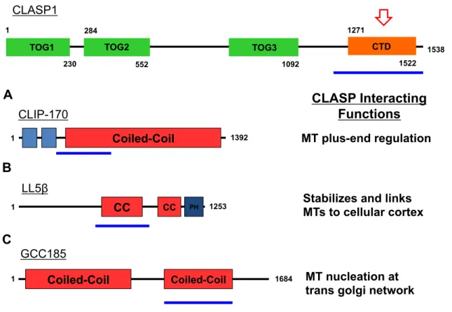
Figure 1-12. CLASP’s C-terminal domain (CTD) interacts with various factors to promote specific functions of CLASP at different cellular locations. CLASP’s CTD (red arrow) interacts with coiled-coil regions (blue lines) of known CLASP-associating factors. (A) CLASP interacts with CLIP-170 to regulate MT plus-end dynamics. (B) CLASP interacts with LL5β to stabilize and attach microtubules to the cellular cortex during interphase. (C) CLASP interacts with GCC185 to nucleates microtubules at the trans golgi network (TGN) independent of centrosomes.
CLASP’s C-terminal domain (CTD) mediates CLASP dimerization and interacts with other factors to recruit CLASP to different locations in the cell. Secondary structure analysis of the CLASP CTD predicts a short coiled-coil domain, suggesting that this domain may
1
230
TOG1
284
552 1092
1271
1522 1538
CTD
TOG2 TOG3
1 Coiled-Coil 1392
CLIP-170
MT plus-end regulation
1253
1 CC CC PH
LL5!
Stabilizes and links MTs to cellular cortex
1684
1 Coiled-Coil Coiled-Coil
GCC185
MT nucleation at trans golgi network
mediate CLASP dimerization. Biochemical analysis of purified full-length CLASP indicates identified a monomer and a homodimer population (Patel et al., 2012). The CLASP CTD binds directly to the coiled-coiled domains of CLASP-interacting proteins (Figure 1-12). The coiled-coiled domain of CLIP-170 directly binds the CLASP CTD (Figure 1-12A)
(Akhmanova et al., 2001) and forms a complex with EB1 to regulate MT plus-end dynamics (Mimori-Kiyosue, et al., 2005). CLASP also forms a complex with ELKS and the
phosphatidylinositol-3,4,5-triphosphate (PIP3) binding protein, LL5β, to attach and stabilize MT plus ends to the cell cortex (Figure 1-12B) (Lansbergen et al., 2006). The trans-Golgi-network (TGN) protein GCC185 recruits CLASP to the TGN in order to nucleate non-centrosomal MTs at the TGN (Figure 1-12C) (Efimov et al., 2007). In mitosis, CLASP interacts with kinetochore-associated protein CENP-E to tether MT plus ends to kinetochores and mechanistically promote bipolar spindle assembly and chromosome segregation (Hannak et al., 2006). The common feature that these CLASP-interacting proteins share is that a section of their coiled-coil domain is required to interact with CLASP’s CTD. Predicted secondary structure elements similar to cryptic TOG2 and TOG3 were found in CLASP CTD, such as the six HEAT repeats of a TOG domain. However, the tubulin-binding
determinants that mediate TOG-tubulin interactions are not present in the intra-HEAT loops, suggesting that the CLASP CTD may retain the structural identity of a TOG domain but does not have TOG-like tubulin-binding activity. Interestingly, two short coiled-coil regions were predicted to exist in CLASP CTD, which may underlie CLASP homodimerization and its interaction with the coiled-coil of its binding partners. Whether the CLASP CTD requires the structural elements of a divergent TOG-like domain or its two coiled-coil regions for
My thesis work involves establishing the presence of a TOG array in CLASP and investigating the role of these TOG domains in differentially promoting microtubule pause and growth. I am also investigating the role of CLASP’s C-terminal domain in CLASP dimerization and its interaction with known CLASP-associating factors. My work employs a combination of X-ray crystallography, biochemical, and cellular assays on human CLASP and its Drosophila homologue, MAST/Orbit. We present the crystal structure of the first cryptic TOG-like domain from human CLASP1 that we designate as TOG2. CLASP1 TOG2 has a bent architecture not observed in TOG domain structures determined to date. The bent TOG architecture has concomitant implications for the conformation of αβ-tubulin in complex with CLASP and potentially underlies CLASP-dependent MT pause and rescue events. We support our structural investigation with analysis of TOG2 structural
determinants and their key role in bipolar mitotic spindle formation, in vitro MT
polymerization, and in vivo MT lattice association. We also have purified TOG1 and the second cryptic TOG-like domain (TOG3) of human CLASP1. We are currently optimizing crystallization conditions that will produce high-resolution crystal structures of each
REFERENCES
1. Akhmanova, A., Hoogenraad, C.C., Drabek, K., Stepanova, T., Dortland, B., Verkerk, T., Vermeulen, W., Burgering, B.M., De Zeeuw, C.I., Grosveld, F., et al.
(2001). Clasps are CLIP-115 and -170 associating proteins involved in the regional regulation of microtubule dynamics in motile fibroblasts. Cell 104, 923-935.
2. Akhmanova, A., and Steinmetz, M.O. (2008). Tracking the ends: a dynamic protein network controls the fate of microtubule tips. Nature Reviews Molecular Cell Biology
9, 309-322.
3. Al-Bassam, J., and Chang, F. (2011). Regulation of microtubule dynamics by TOG-domain proteins XMAP215/Dis1 and CLASP. Trends in Cell Biology 21, 604-614. 4. Al-Bassam, J., Larsen, N.A., Hyman, A.A., and Harrison, S.C. (2007). Crystal
structure of a TOG domain: conserved features of XMAP215/Dis1-family TOG domains and implications for tubulin binding. Structure 15, 355-362.
5. Al-Bassam, J., van Breugel, M., Harrison, S.C., and Hyman, A. (2006). Stu2p binds tubulin and undergoes an open-to-closed conformational change. The Journal of Cell Biology 172, 1009-1022.
6. Allen, C., and Borisy, G.G. (1974). Structural polarity and directional growth of microtubules of Chlamydomonas flagella. Journal of Molecular Biology 90, 381-402. 7. Amin, M.A., Itoh, G., Iemura, K., Ikeda, M., and Tanaka, K. (2014). CLIP-170 is
required to recruit PLK1 to kinetochores during early mitosis for chromosome alignment. Journal of Cell Science.
8. Amos, L.A., and Schlieper, D. (2005). Microtubules and maps. Advances in Protein Chemistry 71, 257-298.
9. Arnal, I., Karsenti, E., and Hyman, A.A. (2000). Structural transitions at microtubule ends correlate with their dynamic properties in Xenopus egg extracts. The Journal of Cell Biology 149, 767-774.
10.Ayaz P, Ye X, Huddleston P, Brautigam CA, Rice LM. (2012). A TOG:alphabeta-tubulin complex structure reveals conformation-based mechanisms for a microtubule polymerase. Science. 337, 857–860.
11.Baas, P.W., and Buster, D.W. (2004). Slow axonal transport and the genesis of neuronal morphology. Journal of Neurobiology 58, 3-17.
CLIP-170 tracks growing microtubule ends by dynamically recognizing composite EB1/tubulin-binding sites. Journal of Cell Biology 183, 1223-33.
13.Belmont, L., Mitchison, T., and Deacon, H.W. (1996). Catastrophic revelations about Op18/stathmin. Trends in Biochemical Sciences 21, 197-198.
14.Belmont, L.D., Hyman, A.A., Sawin, K.E., and Mitchison, T.J. (1990). Real-time visualization of cell cycle-dependent changes in microtubule dynamics in cytoplasmic extracts. Cell 62, 579-589.
15.Bièche, I., Lachkar, S., Becette, V., Cifuentes-Diaz, C., Sobel, A., Lidereau, R., and Curmi, P.A. (1998). Overexpression of the stathmin gene in a subset of human breast cancer. British Journal of Cancer 78, 701-709.
16.Bratman SV, Chang F. Stabilization of overlapping microtubules by fission yeast CLASP. (2007). Developmental Cell. 13, 812–827.
17.Brouhard, G.J., Stear, J.H., Noetzel, T.L., Al-Bassam, J., Kinoshita, K., Harrison, S.C., Howard, J., and Hyman, A.A. (2008). XMAP215 is a processive microtubule polymerase. Cell 132, 79-88.
18.Buey, R.M., Mohan, R., Leslie, K., Walzthoeni, T., Missimer, J.H., Menzel, A., Bjelic, S., Bargsten, K., Grigoriev, I., Smal, I., et al. (2011). Insights into EB1 structure and the role of its C-terminal domain for discriminating microtubule tips from the lattice. Molecular Biology of the Cell 22, 2912-2923.
19.Buey, R.M., Sen, I., Kortt, O., Mohan, R., Gfeller, D., Veprintsev, D., Kretzschmar, I., Scheuermann, J., Neri, D., Zoete, V., et al. (2012). Sequence determinants of a microtubule tip localization signal (MtLS). The Journal of Biological Chemistry 287, 28227-28242.
20.Buster, D.W., Zhang, D., and Sharp, D.J. (2007). Poleward tubulin flux in spindles: regulation and function in mitotic cells. Molecular Biology of the Cell 18, 3094-3104. 21.Butner, K.A., and Kirschner, M.W. (1991). Tau protein binds to microtubules through
a flexible array of distributed weak sites. The Journal of Cell Biology 115, 717-730. 22.Caplow, M., and Shanks, J. (1996). Evidence that a single monolayer tubulin-GTP
cap is both necessary and sufficient to stabilize microtubules. Molecular Biology of the Cell 7, 663-675.
24.Chen, G., Wang, H., Gharib, T.G., Huang, C.-C., Thomas, D.G., Shedden, K.A., Kuick, R., Taylor, J.M.G., Kardia, S.L.R., Misek, D.E., et al. (2003). Overexpression of oncoprotein 18 correlates with poor differentiation in lung adenocarcinomas. Molecular & Cellular Proteomics 2, 107-116.
25.Cheeseman I.M., MacLeod I., Yates J.R., 3rd, Oegema K., Desai A. (2005) The CENP-F-like proteins HCP-1 and HCP-2 target CLASP to kinetochores to mediate chromosome segregation. Current Biolology 15, 771–777.
26.Cimini, D., and Degrassi, F. (2005). Aneuploidy: a matter of bad connections. Trends in Cell Biology 15, 442-451.
27.Cullen, C.F., Deák, P., Glover, D.M., and Ohkura, H. (1999). Mini spindles: A gene encoding a conserved microtubule-associated protein required for the integrity of the mitotic spindle in Drosophila. The Journal of Cell Biology 146, 1005-1018.
28.Desai A, Mitchison TJ. (1997). Microtubule polymerization dynamics. Annual Review of Cell and Developmental Biology 13, 83-117.
29.Drechsel, D.N., Hyman, A.A., Cobb, M.H., and Kirschner, M.W. (1992). Modulation of the dynamic instability of tubulin assembly by the microtubule-associated protein tau. Molecular Biology of the Cell 3, 1141-1154.
30.Drabek K., van Ham M., Stepanova T., Draegestein K., van Horssen R., Sayas C.L., Akhmanova A., Ten Hagen T., Smits R., Fodde R, et al. (2006). Role of CLASP2 in microtubule stabilization and the regulation of persistent motility. Current Biology
16, 2259–2264
31.Drechsel, D.N., and Kirschner, M.W. (1994). The minimum GTP cap required to stabilize microtubules. Current Biology 4, 1053-1061.
32.Drewes, G., Ebneth, A., and Mandelkow, E.M. (1998). MAPs, MARKs and microtubule dynamics. Trends in Biochemical Sciences 23, 307-311.
33.Dumont, S., and Mitchison, T.J. (2009). Compression regulates mitotic spindle length by a mechanochemical switch at the poles. Current Biology : CB 19, 1086-1095. 34.Efimov, A., Kharitonov, A., Efimova, N., Loncarek, J., Miller, P.M., Andreyeva, N.,
Gleeson, P., Galjart, N., Maia, A.R.R., McLeod, I.X., et al. (2007). Asymmetric CLASP-dependent nucleation of noncentrosomal microtubules at the trans-Golgi network. Developmental Cell 12, 917-930.
36.Fedorova S.A., Chubykin V.L., Gucachenko A.M., Omel’ianchuk L.V. (1997). Mutation chromosome bows (chb-v40), inducing the abnormal chromosome spindle in Drosophila melanogaster. Genetika 33, 1502–1509.
37.Gadde, S., and Heald, R. (2004). Mechanisms and molecules of the mitotic spindle. Current Biology 14, R797-805.
38.Gard, D.L., and Kirschner, M.W. (1987). A microtubule-associated protein from Xenopus eggs that specifically promotes assembly at the plus-end. The Journal of Cell Biology 105, 2203-2215.
39.Gigant, B., Curmi, P.A., Martin-Barbey, C., Charbaut, E., Lachkar, S., Lebeau, L., Siavoshian, S., Sobel, A., and Knossow, M. (2000). The 4 A X-ray structure of a tubulin:stathmin-like domain complex. Cell 102, 809-816.
40.Gleeson, J.G., Lin, P.T., Flanagan, L.A., and Walsh, C.A. (1999). Doublecortin is a microtubule-associated protein and is expressed widely by migrating neurons. Neuron
23, 257-271.
41.Glotzer, M. The 3Ms of central spindle assembly: microtubules, motors and MAPs. (2009). Nature Reviews Molecular Cell Biology 10, 9–20.
42.Goshima, G., Wollman, R., Goodwin, S.S., Zhang, N., Scholey, J.M., Vale, R.D., and Stuurman, N. (2007). Genes required for mitotic spindle assembly in Drosophila S2 cells. Science316,417-421.
43.Halpain, S., and Dehmelt, L. (2006). The MAP1 family of microtubule-associated proteins. Genome Biology 7, 224.
44.Hannak, E., and Heald, R. (2006). Xorbit/CLASP links dynamic microtubules to chromosomes in the Xenopus meiotic spindle. The Journal of Cell Biology 172, 19-25.
45.Hayashi, I., and Ikura, M. (2003). Crystal structure of the amino-terminal microtubule-binding domain of end-binding protein 1 (EB1). The Journal of Biological Chemistry 278, 36430-36434.
46.He, X., Rines, D.R., Espelin, C.W., and Sorger, P.K. (2001). Molecular analysis of kinetochore-microtubule attachment in budding yeast. Cell 106, 195-206.
47.Heald, R., Nogales, E. (2002). Microtubule dynamics. Journal of Cell Science 115, 3-4.
48.Holmfeldt, P., Brattsand, G., and Gullberg, M. (2003). Interphase and monoastral-mitotic phenotypes of overexpressed MAP4 are modulated by free tubulin
49.Honnappa, S., John, C.M., Kostrewa, D., Winkler, F.K., and Steinmetz, M.O. (2005). Structural insights into the EB1-APC interaction. The EMBO Journal 24, 261-269. 50.Hyman, A.A., Salser, S., Drechsel, D.N., Unwin, N., and Mitchison, T.J. (1992). Role
of GTP hydrolysis in microtubule dynamics: information from a slowly hydrolyzable analogue, GMPCPP. Molecular Biology of the Cell 3, 1155-1167.
51.Inoue Y.H., do Carmo Avides M., Shiraki M., Deak P., Yamaguchi M., Nishimoto Y., Matsukage A., Glover D.M. (2000). Orbit, a novel microtubule-associated protein essential for mitosis in Drosophila melanogaster. Journal of Cell Biology 149,153– 166.
52.Jaqaman, K., King, E.M., Amaro, A.C., Winter, J.R., Dorn, J.F., Elliott, H.L., McHedlishvili, N., McClelland, S.E., Porter, I.M., Posch, M., et al. (2010). Kinetochore alignment within the metaphase plate is regulated by centromere
stiffness and microtubule depolymerases. The Journal of Cell Biology 188, 665-679. 53.Jourdain, L., Curmi, P., Sobel, A., Pantaloni, D., and Carlier, M.F. (1997). Stathmin:
a tubulin-sequestering protein which forms a ternary T2S complex with two tubulin molecules. Biochemistry 36, 10817-10821.
54.Kinoshita, K., Habermann, B., and Hyman, A.A. (2002). XMAP215: a key
component of the dynamic microtubule cytoskeleton. Trends in Cell Biology 12, 267-273.
55.Kitamura, E., Tanaka, K., Komoto, S., Kitamura, Y., Antony, C., and Tanaka, T.U. (2010). Kinetochores generate microtubules with distal plus ends: their roles and limited lifetime in mitosis. Developmental Cell 18, 248-259.
56.Kline-Smith, S.L., and Walczak, C.E. (2002). The microtubule-destabilizing kinesin XKCM1 regulates microtubule dynamic instability in cells. Molecular Biology of the Cell 13, 2718-2731.
57.Kumar, P., and Wittmann, T. (2012). +TIPs: SxIPping along microtubule ends. Trends in Cell Biology 22, 418-428.
58.Lansbergen, G., Grigoriev, I., Mimori-Kiyosue, Y., Ohtsuka, T., Higa, S., Kitajima, I., Demmers, J., Galjart, N., Houtsmuller, A.B., Grosveld, F., et al. (2006). CLASPs attach microtubule plus ends to the cell cortex through a complex with LL5beta. Developmental Cell 11, 21-32.
60.Lemos C.L., Sampaio P., Maiato H., Costa M., Omel’yanchuk L.V., Liberal V., Sunkel C.E. (2000) Mast, a conserved microtubule-associated protein required for bipolar mitotic spindle organization. EMBO J. 19, 3668–3682.
61.Li, H., DeRosier, D.J., Nicholson, W.V., Nogales, E., and Downing, K.H. (2002). Microtubule structure at 8 A resolution. Structure 10, 1317-1328.
62.Löwe, J., Li, H., Downing, K.H., and Nogales, E. (2001). Refined structure of alpha beta-tubulin at 3.5 A resolution. Journal of Molecular Biology 313, 1045-1057. 63.Maiato, H., Fairley, E.A.L., Rieder, C.L., Swedlow, J.R., Sunkel, C.E., and Earnshaw,
W.C. (2003). Human CLASP1 is an outer kinetochore component that regulates spindle microtubule dynamics. Cell 113, 891-904.
64.Maiato, H., Khodjakov, A., and Rieder, C.L. (2005). Drosophila CLASP is required for the incorporation of microtubule subunits into fluxing kinetochore fibres. Nature Cell Biology 7, 42-47.
65.Maiato, H., Sampaio, P., Lemos, C.L., Findlay, J., Carmena, M., Earnshaw, W.C., and Sunkel, C.E. (2002). MAST/Orbit has a role in microtubule-kinetochore attachment and is essential for chromosome alignment and maintenance of spindle bipolarity. The Journal of Cell Biology 157, 749-760.
66.Maiato, H., Sampaio, P., and Sunkel, C.E. (2004). Microtubule-associated proteins and their essential roles during mitosis. International Review of Cytology 241, 53-153.
67.Mandelkow, E., and Mandelkow, E.M. (1989). Microtubular structure and tubulin polymerization. Current Opinion in Cell Biology 1, 5-9.
68.Mandelkow, E., and Mandelkow, E.M. (1995). Microtubules and microtubule-associated proteins. Current Opinion in Cell Biology 7, 72-81.
69.Mandelkow, E.M., and Mandelkow, E. (1998). Tau in Alzheimer's disease. Trends in Cell Biology 8, 425-427.
70.Mandelkow, E.M., Mandelkow, E., and Milligan, R.A. (1991). Microtubule dynamics and microtubule caps: a time-resolved cryo-electron microscopy study. The Journal of Cell Biology 114, 977-991.
71.Margolis, R.L., Wilson, L., and Keifer, B.I. (1978). Mitotic mechanism based on intrinsic microtubule behaviour. Nature 272, 450-452.
73.Matthews, L.R., Carter, P., Thierry-Mieg, D., and Kemphues, K. (1998). ZYG-9, a Caenorhabditis elegans protein required for microtubule organization and function, is a component of meiotic and mitotic spindle poles. The Journal of Cell Biology 141, 1159-1168.
74.McNally, F.J., and Vale, R.D. (1993). Identification of katanin, an ATPase that severs and disassembles stable microtubules. Cell 75, 419-429.
75.Mimori-Kiyosue, Y., Grigoriev, I., Lansbergen, G., Sasaki, H., Matsui, C., Severin, F., Galjart, N., Grosveld, F., Vorobjev, I., Tsukita, S., et al. (2005). CLASP1 and CLASP2 bind to EB1 and regulate microtubule plus-end dynamics at the cell cortex. The Journal of Cell Biology 168, 141-153.
76.Mitchison, T., and Kirschner, M. (1984). Dynamic instability of microtubule growth. Nature 312, 237-242.
77.Mitchison, T.J. (1989). Polewards microtubule flux in the mitotic spindle: evidence from photoactivation of fluorescence. The Journal of Cell Biology 109, 637-652. 78.Müller-Reichert, T., Chrétien, D., Severin, F., and Hyman, A.A. (1998). Structural
changes at microtubule ends accompanying GTP hydrolysis: information from a slowly hydrolyzable analogue of GTP, guanylyl (alpha,beta)methylenediphosphonate. Proceedings of the National Academy of Sciences of the United States of America 95, 3661-3666.
79.Nakaseko, Y., Goshima, G., Morishita, J., and Yanagida, M. (2001). M phase-specific kinetochore proteins in fission yeast: microtubule-associating Dis1 and Mtc1 display rapid separation and segregation during anaphase. Current Biology 11, 537-549. 80.Nettles, J.H., Li, H., Cornett, B., Krahn, J.M., Snyder, J.P., and Downing, K.H.
(2004). The binding mode of epothilone A on alpha,beta-tubulin by electron crystallography. Science 305, 866-869
81.Neuwald, A.F., and Hirano, T. (2000). HEAT repeats associated with condensins, cohesins, and other complexes involved in chromosome-related functions. Genome Research 10, 1445-1452.
82.Nogales, E., Wolf, S.G., and Downing, K.H. (1998). Structure of the alpha beta tubulin dimer by electron crystallography. Nature 391, 199-203.