©2017 Steven Jae Doo Kim ALL RIGHTS RESERVED
ABSTRACT
Steven Jae Doo Kim: Functional analysis of rs3811046 and rs3811047 variants in Caucasians (Under the direction of Steven Offenbacher)
Inflammation is an essential for the survival of the host, not only to fight off pathogens but also for immunotolerance to avoid autoimmunity. Turning off the inflammation is as important as turning them on, because lack thereof will lead to uncontrolled chronic inflammation, unnecessary structural damage, and compromised or delayed wound healing which do not benefit the host.
Periodontal disease is one of the example of such uncontrolled chronic inflammation.
Periodontitis is a combined result of pathogenic bacteria establishing themselves in the biofilm‐gingival interface, and the excessive and uncontrolled immune response to the bacteria and their byproducts. It is actually the host immune response that destroys the periodontal attachment apparatus, and not the direct destructive action of the pathogens.
Some individuals are more predisposed to inflammation. It can be due to genetic predisposition, others could be due to systemic conditions. We describe one of such example where single nucleotide polymorphisms can lead to pro‐inflammatory profiles in Caucasian subjects. In chapter 1, we describe how we honed in onto a number of SNPs that are on the coding region of a cytokine called IL‐37. Chapter 2, we created human recombinant IL‐37b and observe the effects of variants on its anti‐ inflammatory function. In chapter 3 we obtained human samples from known genotypes and observed pro‐inflammatory profiles in the subjects with the SNP variance. Finally in chapter 4, we discuss its
To my parents who always believed in me
and shaped me into what I am today.
I could not have done this without their loving support.
ACKNOWLEDGEMENTS
It was one of the many days ever since I started working as a periodontitis/general dentist. The pay was not good, and after each repetitive tiring day I was zoned out in front of a television set, with little energy left for anything except staring in front of me. I was not even understanding what was going on in the program. For some reason I snapped out of it and was looking around trying to understand where I am, what I have been doing, and perhaps more importantly, what I will be probability doing from now on… Then it dawned on me: This is what my life is going to me like for the rest of my career until I retire! Not like this, I needed to make a change. I went back to my alma mater, consulted Dr. Majorie Jeffcoat at University of Pennsylvania School of Dentistry, about career options and a prospect of studying for a PhD degree. She said, “What makes you lose sleep at night? If you are not overly enthusiastic about it, then don’t bother because you are not going to like it.”
At that time I was cavalier about that comment and brushed it aside. I better make a leap of faith while I still had any mental capacity and brawn. Anywhere else but here please, I thought, and left the clinic without looking back. I was fortunate enough to be accepted into the Oral and Craniofacial Biomedicine (then called Oral Biology) PhD program at UNC at Chapel Hill.
became building blocks for me to become a solid researcher. Dr. Sompop Bencharit I first met
fortuitously at a Taekwondo martial art training school. He was a wonderful training partner, a reliable coworker as a clinician, a mentor in the lab who kept bringing in fresh perspectives, and finally, a great thesis committee member who helped me achieve my PhD degree. Dr. Ricardo Teles looked after me ever since he joined Offenbacher lab. His incisive analyses always startled me and made me try a tad harder. Dr. Zhi Liu was polite and reserved, but when he made comments they were to the point and very pertinent. Many thanks to Dr. Ceib Phillips, for directly and indirectly helping me wrap my project up, and looking after my interest during my vulnerable time.
Additionally, I had a lot of help from the post docs. Dr. Shaping Zhang would go above and beyond his call of duty and often drop by to give me critical pieces of advice on pertinent occasions, sometimes even before I realized I needed them. Dr. Julie Marchesan was there not only as a post doc but as a friend, and she never hesitated in giving me great pieces of life advice for a budding researcher. She knew fully well what I acquired during this malleable time would affect the rest of my research career. Dr. Yizu Jiao impressed me with his vast array of abilities and his meticulous approach to any given projects. I have learned a lot, and I still have more to learn from him. He is currently my role model as an ideal post doc. I would also like to extend my sincere gratitude to Dr. William “Todd” Seaman from Dr. Jennifer Webster‐Cyriaque lab, who was more than willing to help me at any given moment.
Whenever I had random questions popping up in my head, he was the first person I sought council.
Mr. Russel Levy, who were instrumental in keeping a tight well‐oiled ship, which really helped our lab running smoothly and getting results quickly.
And last but not least, my older sister Gloria, who gave me tons of criticisms when I was not ready for them yet. But let’s face it, she was indeed looking after me.
Dr. Jeffcoat, as a clinician researcher I will continuously strive to become one of the best in my field, and hopefully I will also reach a place where I not only instill enthusiasm to my students in their research, but also make them understand what is required of them to go down this extremely
TABLE OF CONTENTS
LIST OF TABLES ... x
LIST OF FIGURES ... xi
LIST OF ABBREVIATIONS ...xiii
CHAPTER 1: INTRODUCTION ... 1
1.1. Figures ... 10
CHAPTER 2: Observe the effect of variants on IL‐37 anti‐inflammatory function with human recombinant IL‐37b ... 18
2.1. Material and Methods ... 18
2.1.1. Generation of human recombinant IL‐37b using Escherichia coli ... 18
2.1.2. In vitro caspase‐1 cleavage experiment, direct gel staining ... 19
2.1.3. In vitro caspase‐1 cleavage experiment, Western blot with Km and Vmax calculation ... 19
2.1.4. Making better tools: EF.CMV. RFP vector for eukaryotic cells ... 20
2.1.5. In vivo caspase‐1 cleavage experiment in transfected HEK293T cells ... 20
2.1.6. LPS stimulation of HEK293T, THP‐1 co‐culture system ... 20
2.1.7. LPS stimulation of human recombinant IL‐37b pretreated RAW263.7 cell line ... 21
2.2. Results ... 22
2.3. Conclusions ... 25
CHAPTER 3: Observe the effect of variant of IL‐37 in Caucasian human samples... 34
3.1. Material and Methods ... 34
3.1.1. Screening process ... 34
3.1.2. Blood collection and sample processing: Whole blood experiment ... 35
3.1.3. Blood collection and sample processing: Dendritic cell differentiation and LPS stimulation .. 36
3.1.4. GCF collection and inflammatory mediator assessment ... 37
3.1.5. Gingival tissue biopsy, isoform expression preference ... 37
3.2. Results ... 39
3.3. Conclusions ... 41
3.4. Tables ... 42
3.5. Figures ... 43
CHAPTER 4: DISCUSSION ... 49
ACKNOWLEDGEMENTS ... 55
APPENDIX ... 56
REFERENCES ... 57
LIST OF TABLES
Table 3.4.1. Genotyping results ... 40
LIST OF FIGURES
Figure 1.1.1. GWAS results leading to 7 SNPs of interest ... 9
Figure 1.1.2a. Population genetics of rs3811046 and rs3811047 based on 1000 genome project ... 10
Figure 1.1.2b. Population genetics of rs2708943, rs2723183, rs2723187, rs2708947, and rs2723192 based on 1000 genome project ... 11
Figure 1.1.3a. Gene sequence of IL‐37b and SNP locations ... 12
Figure 1.1.3b. Amino acid sequence of pro IL‐37b and missense mutations ... 13
Figure 1.1.4. PolyPhen‐2 predictions of the effect of SNPs on IL‐37 function ... 14
Figure 1.1.5. 5 possible isoforms of IL‐37b ... 15
Figure 1.1.6. IL‐37b extracellular and intracellular pathways ... 16
Figure 2.4.1. Generating human recombinant pro IL‐37b from E. coli ... 25
Figure 2.4.2. Direct gel staining of caspase‐1 treated pro IL‐37b, and densitometry analysis ... 26
Figure 2.4.3a. Western blot of in vitro pro IL‐37b cleavage experiment and its densitometry analysis .... 27
Figure 2.4.3b. Km and Vmax calculation based on densitometry of Western blot ... 28
Figure 2.4.4. IL‐37b transfection of eukaryotic cell lines, and confirmation of IL‐37b productions via Western blot ... 29
Figure 2.4.5 IL‐37b maturation by caspase‐1 in vivo, through NLRP3 constitution experiment on HEK293T cell line ... 30
Figure 2.4.6 HEK293T and THP‐1 co‐culture experiment ... 31
Figure 2.4.7 RAW246.7 cells, E. coli (strain O111:B4) LPS stimulation after pro IL‐37b pretreatment ... 32
Figure 3.5.1. Pyrosequencing of human saliva DNA for genotyping ... 41
Figure 3.5.2. Blood experiments of the genotyped participants ... 42
Figure 3.5.3. Whole blood experiment. IL‐1β, IL‐6, and TNF‐α expression levels. ... 43
compared among the 1.1, 1.2, and 2.2 genotypes ... 45 Figure 3.5.6. Isoform expression preference in human gingival tissues ... 46 Supplemental Figure. IL‐37b mRNA expression of transiently transfected HEK293T cells ... 54
LIST OF ABBREVIATIONS
ARIC Atherosclerosis Risk in Communities
CD25 Cluster of differentiation 25, alpha chain of IL‐2 receptor
CD36 Cluster of differentiation 36
DAMP Damage associated molecular signal
DARIC Dental Atherosclerosis Risk in Community Study
Foxp3 Forkhead box P3
GAPDH Glyceraldehyde‐3 phosphate
GCF Gingival crevicular fluid
G‐CSF Granulocyte colony‐stimulating factor
GM‐CSF Granulocyte macrophage colony‐stimulating factor
GWAS Genome‐wide association study
IFN‐γ Interferon gamma
IL‐1 Interleukin 1
IL‐1β Interleukin 1 beta
IL‐1ra Interleukin 1 receptor antagonist
IL‐2 Interleukin 2
IL‐4 Interleukin 4
IL‐6 Interleukin 6
IL‐8 Interleukin 8
IL‐10 Interleukin 10
IL‐17 Interleukin 17
IL‐18 Interleukin 18
IL‐18BP Interleukin 18 binding protein
IL‐18Rα Alpha subunit of IL‐18 receptor
IL‐19 Interleukin 19
IL‐20 Interleukin 20
IL‐22 Interleukin 22
IL‐24 Interleukin 24
IL‐26 Interleukin 26
IL‐27 Interleukin 27
IL‐28 Interleukin 28
IL‐29 Interleukin 29
IL‐30 Interleukin 30
IL‐35 Interleukin 35
IL‐37 Interleukin 37
LPS Lipopolysaccaride
MAF Minor allele frequency
MIP‐1α Macrophage Inflammatory Protein 1 alpha, CCL3
PAMP Pathogen‐associated molecular pattern
PCR polymerase chain reaction
qRT‐PCR Real time PCR
RANTES Regulated on Activation, Normal T Cell Expressed and Secreted, CCL5
SIGIRR Single Ig IL‐1‐related receptor, IL‐1R8
SMAD Homologs of mothers against decapentaplegic protein and Caenorhabditis elegans protein SMA
SNP Single nucleotide polymorphism
TGF‐β Transforming growth factor‐beta
TLR Toll‐like receptor
TNF Tumor necrosis factor
Treg Regulatory T cell
TFH Follicular helper T cell
TH1 Type 1 helper T cell
TH2 Type 2 helper T cell
CHAPTER 1: INTRODUCTION
Based on 2009‐2010 National Health and Examination Survey in the Unites States population, 47 percent of adults aged 30 years and older have periodontitis, and that number increases to 70.1 percent for 65 years and older (Eke, Dye et al. 2012). Early on, clinicians had noticed there was a familial
predisposition to this widespread disease (Benjamin and Baer 1967, Jorgenson, Levin et al. 1975, Beaty, Boughman et al. 1987), and its genetic basis needed to be investigated. The classic way of investigating genetic effects on any disease are twin studies: Between 38 to 82% of the population variance of the periodontal measures of disease was contributed to genetic factors (Michalowicz, Aeppli et al. 1991). Periodontal disease does not follow Mendel’s law of inheritance, as demonstrated in monozygotic and dizygotic twin studies (Corey, Nance et al. 1993). It became clear that a collection of risk variants
contribute to the onset of the seemingly common form of periodontal disease. To observe their collective roles it became necessary to conduct large population based studies such as genome‐wide association study (GWAS).
Our 4910 Caucasian GWAS data was re‐analyzed. We used the upper quartile of IL‐1b
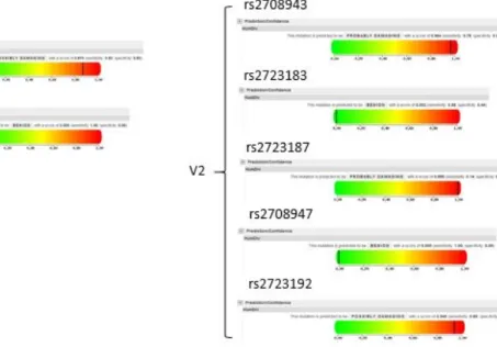
concentration in gingival crevicular fluid (GCF) to define the pro‐inflammatory phenotype. Biomarkers in GCF is known to correlate well with periodontal disease progression (Engebretson, Grbic et al. 2002, Zhong, Slade et al. 2007, Khalaf, Lonn et al. 2014, Kinney, Morelli et al. 2014). Looking into 2.5 million SNPs in 22 somatic chromosomes, we identified a number of single nucleotide polymorphisms (SNPs) that stood out in chromosome 2 [Figure 1.1]. Of particular interest, with the most significant association (p values of less than 1x10 ) and minor allele frequency (MAF) above 5%, were SNPs rs3811046 and rs3811047. They were in strong linkage disequilibrium to each other, and caused missense mutation on a gene coding for a cytokine called IL‐37 [Figure 1.1]. A second, less common locus was also noted, with a p value of 4.2x10 . This time there were 5 SNPs (rs2708943, rs2723183, rs2723187, rs2708947, and rs2723192) in strong linkage disequilibrium, all causing missense mutations on the IL‐37 gene as well [Figure 1.1].
(G/A, D218N) all also lead to missense mutations at their respectable amino acid locations [Figure 1.3a and 1.3b].
No such SNP associations were observed when GWAS was conducted on our African American population of 776 subjects (data not shown), indicating the effects of the 7 SNPs are ethnicity specific.
A web‐based simulator was used to predict the effects of the aforementioned polymorphisms on the IL‐37 protein. PolyPhen‐2 is a bioinformatics tool used to predict protein damage caused by missense mutations. It calculates a score from 0 to 1, with 0 being “benign” and 1 being “possibly damaging”. The algorithm is based on eight sequence‐based and three structure‐based predictive features (Adzhubei, Schmidt et al. 2010). The program predicted that the mutation caused by rs3811046 is possibly damaging with a score of 0.870. Polymorphisms in rs2708943, rs2723187 are probably damaging to the protein it is coding for (scores of 0.964 and 0.999 respectively). Lastly, the missense mutation caused by rs2723192 SNP is predicted to be possibly damaging (0.940) [Figure 1.4].
With so many SNPs of interest coding for the same IL‐37 gene at different exon locations, it becomes necessary to know the normal function of IL‐37 before understanding how those functions could be adversely affected. It was formerly known as IL‐1F7, and its presence was first predicted through in silico research in 2000 (Dunn, Sims et al. 2001). Nold et al. first showcased its anti‐inflammatory functions
atrial fibrillation (Li, Li et al. 2017). On the other hand, downregulation of IL‐37 mRNA and protein levels was noted in tissue samples taken from individuals with Behcet’s disease (Ye, Wang et al. 2014), or
individuals showing aggravation of intervertebral disc degeneration (Wan, Sun et al. 2014). Clearly, IL‐37 is involved in a wide variety of diseases involving chronic uncontrolled inflammation. But correlation alone is insufficient to elucidate the role of IL‐37, especially when it is increased in some diseases and decreased in others.
Regulatory cytokines are essential in turning off inflammation once it is not needed any more. Lack of such regulation can not only lead to uncontrolled chronic inflammatory diseases, but also autoimmunity. Immune tolerance is achieved through central tolerance in the thymus, as well as peripheral tolerance where regulatory T (Treg) cells prevent the activation of autoreactive T cells (Josefowicz, Lu et al. 2012). The Treg cells suppresses immune responses through direct cell contact and
cell factor dependent mechanisms, such as consumption of IL‐2 or production of IL‐10, IL‐35, and TGF‐β (Vignali, Collison et al. 2008, Shevach 2009, Yamaguchi, Wing et al. 2011). IL‐2, IL‐10, and TGF‐β are considered the classic triad of the anti‐inflammatory cytokines, and IL‐35 is amongst the newly discovered anti‐inflammatory cytokines besides IL‐27 and IL‐37 (Banchereau, Pascual et al. 2012).
When IL‐2 was discovered through its ability to induce in vitro growth of activated T cells (Malek and Castro 2010), it was first predicted that IL‐2 was pro‐inflammatory, and its deficiency would lead to immunodeficiency. However, IL‐2 deletion in mice caused them to die prematurely, from activated T cells with autoimmune anemia and inflammatory bowel disease (Kundig, Schorle et al. 1993). The discovery of Treg cells with high density of CD25 (alpha chain of the IL‐2 receptor) corrected such initial misconception, and now IL‐2 is considered an anti‐inflammatory mediator (Sakaguchi, Sakaguchi et al. 2011). IL‐2 is critical for maintenance of Treg cells in the periphery, and neutralization of IL‐2 results in autoimmunity
inhibits follicular helper T cell (TFH) development without affecting already differentiated TFH cells (Ballesteros‐Tato, Leon et al. 2012). Overall, IL‐2 can prevent uncontrolled expansion of immune responses and limit overall inflammation.
IL‐10 superfamily includes IL‐10, IL‐19, IL‐20, IL‐22, IL‐24, IL‐26, IL‐28, and IL‐29. This IL‐10 superfamily is highly pleiotropic, not all members of the superfamily are anti‐inflammatory. While some members mediate immune suppression and promote self‐tolerance, others enhance antibacterial, antiviral or antitumor activity (Commins, Steinke et al. 2008). IL‐10 in particular, limits immune response and prevents immune system mediated damage to the host (Li and Flavell 2008). IL‐10 synthesis is a characteristic of almost all leukocytes (Wolk, Kunz et al. 2002), but the main sources are mainly monocytes, macrophages, and T helper cells (Seki, Osada et al. 1998, Roers, Siewe et al. 2004, Murai, Turovskaya et al. 2009). IL‐10 affects all three key functions of monocyte/macrophages: Release of immune mediators, antigen presentation, and phagocytosis (Sabat, Grutz et al. 2010). IL‐10 inhibits the release of pro‐inflammatory mediators such as TNF‐α, IL‐1β, IL‐6, IL‐8, G‐CSF, and GM‐CSF from
monocyte/macrophages (de Waal Malefyt, Abrams et al. 1991, Fiorentino, Zlotnik et al. 1991). Other anti‐ inflammatory mediators such as IL‐1 receptor antagonist and soluble TNF‐α receptor release are
enhanced by IL‐10 (Jenkins, Malyak et al. 1994, Joyce, Gibbons et al. 1994, Hart, Hunt et al. 1996). Independent of its inhibitory effects on antigen presenting cells, IL‐10 inhibits both the proliferation and the cytokine synthesis of CD4+ T cells (Del Prete, De Carli et al. 1993, Groux, Bigler et al. 1996). It should be noted that IL‐10 is not always inhibitory, as it has a potent effect of the growth and differentiation of B cells (Defrance, Vanbervliet et al. 1992, Rousset, Garcia et al. 1992). Overproduction is as harmful as underproduction, as excessive amounts of IL‐10 are associated with systemic lupus erythematosus, melanoma, leishmaniasis and tuberculosis (O'Garra, Barrat et al. 2008).
2017). TGF‐β1 inhibits Th1 cells, Th2 cells, and cytotoxic T cells, while inducing differentiation of Treg cells and TH17 cells (Banchereau, Pascual et al. 2012). Working with IL‐10 or IL‐21, TGF‐β1 also induces CD40‐ activated B cells to switch isotypes from IgM+, IgD+ to IgA+ B cells, playing a pivotal role in mucosal immunity (Banchereau, Pascual et al. 2012). TGF‐β1 deficient mice develop early fatal inflammatory disease, which starts before any major challenge with microbes. Such phenotype can be rescued with depletion of either CD4+ or CD8+ T cells (Shull, Ormsby et al. 1992). Unlike IL‐10, TGF‐β is expressed in most tissues and seems to have a role in immune homeostasis (Li, Mai et al. 2012). TGF‐β is essential for induction of Foxp3 in naïve CD4+ T cells, leading to Treg Cells (Chen, Jin et al. 2003, Dardalhon, Awasthi et
al. 2008). TGF‐β also induces the differentiation of naïve T cells into TH17 cells, while inhibiting the generation of TH1 and TH2 cells (Li, Wan et al. 2007). Consequently, the gut shows enrichment of Foxp3+ Treg cells and TH17 cells, and the balance between the two populations are tightly controlled (Dullaers, Li
et al. 2009). TGF‐β is first translated as a dimeric pre‐pro‐ TGF‐β, then is cleaved to form a latent TGF‐β (LTGF‐β) complex composed of LAP (latency‐associated peptide) that wraps around a homodimeric mature TGF‐β (Shi, Zhu et al. 2011). A second proteolytic cleavage generates three forms of TGF‐β: 1) small latent form of LTGF‐β, 2) a larger latent form, composed of LTGF‐β linked to a binding protein, and 3) LTGF‐β bound to a membrane protein (Tran, Andersson et al. 2009). Lastly, another proteolytic processing finally frees up the active TGF‐β component.
IL‐27 is produced mainly by macrophages and dendritic cells (DCs). Just like IL‐2, IL‐27 was first thought to be pro‐inflammatory, as it was initially described to promote TH1 response (Pflanz, Timans et
al. 2002). Such misconception was corrected when it was observed that mice deficient of IL‐27 specific receptors had intact TH1 responses, yet succumbed to CD4+ T cell‐mediated pathology when infected with
(Wojno and Hunter 2012). Additionally, IL‐27 was shown to induce T cells to produce the anti‐
inflammatory cytokine IL‐10 (Awasthi, Carrier et al. 2007, Fitzgerald, Zhang et al. 2007, Stumhofer, Silver et al. 2007). The therapeutic use of IL‐27 needs caution, as IL‐27 also suppresses IL‐2 thereby hampering the growth of Treg cells (Wojno, Hosken et al. 2011). Through this mechanism, IL‐27 can induce of colitis in
mice (Cox, Kljavin et al. 2011). IL‐27 is composed of IL‐27p28 (also called IL‐30) and Epstein Barr‐induced virus 1 subunit (EBI3) (Pflanz, Timans et al. 2002). IL‐27p28, when acting alone, can act as an antagonist of signal‐transducing receptor gp130 (Stumhofer, Tait et al. 2010), leading to blockage of signaling mediated by IL‐6, IL‐11, and even IL‐27. Additional studies are required to fully understand the physiological and pathological role of IL‐27 and its individual subunits.
IL‐35 is also a member of the IL‐12 family, just like IL‐27. EBI3 and IL‐12p35 subunits form the IL‐ 35 heterodimer (Collison, Workman et al. 2007). IL‐35 is not constitutively expressed in tissues (Li, Mai et al. 2012), and is mainly produced by Treg cells (Hamano, Himeno et al. 2003). IL‐35 is peculiar in that it is capable of transforming CD4+ effector T cells into a novel Foxp3 negative Treg cell population, which in turn can also produce IL‐35 (Collison, Chaturvedi et al. 2010). IL‐35 stimulated Treg cells can protect against collagen‐induced arthritis through IL‐10 production (Kochetkova, Golden et al. 2010). Ectopic expression of IL‐35 in pancreatic beta cells can also prevent auto‐immune diabetes (Bettini, Castellaw et al. 2012).
(Kumar, McDonnell et al. 2000). There are two reports of N‐terminal sequencing results on the cleavage location: One paper showed that amino acid 20 was the cleavage site and caspase‐1 was the enzyme responsible (Kumar, Hanning et al. 2002), while another reported that the cleavage location was at amino acid 45 but the enzyme responsible was not elucidated (Pan, Risser et al. 2001) [Figure 1.3b]. Both pro IL‐ 37b and mature IL‐37b can act on NK cells to reduce INF‐γ production, but the mature IL‐37b is more efficient (Bufler, Azam et al. 2002). IL‐37b is known to act intracellularly and extracellularly. Only the mature form of IL‐37b can enter the nucleus (Sharma, Kulk et al. 2008, Ross, Grimmel et al. 2013) by binding to phosphorylated SMAD3 (Nold, Nold‐Petry et al. 2010), and affects transcription of
inflammatory mediators (Sharma, Kulk et al. 2008). On the other hand, both pro and mature forms of IL‐ 37b are secreted. Extracellular IL‐37b is known to bind to receptor complexes composed of an alpha subunit of IL‐18 receptor (IL‐18Rα) and a Single Ig IL‐1‐related receptor (SIGIRR, also known as IL‐1R8) (Li, Neff et al. 2015, Lunding, Webering et al. 2015) [Figure 1.5]. The anti‐inflammatory role of IL‐37 described by the aforementioned Nold group was based on the following observations: 1) pro‐inflammatory
cytokines were suppressed with IL‐37 in macrophages, peripheral blood mononuclear cells, and epithelial cells, 2) silencing IL‐37 increased pro‐inflammatory cytokines, 3) IL‐37 transgenic mice were protected from LPS induced shock (Nold, Nold‐Petry et al. 2010). Since then, other studies confirmed IL‐37 to have anti‐inflammatory functions in other experimental models. IL‐37 transgenic mice experienced less colitis (McNamee, Masterson et al. 2011). IL‐37 reduced concanavalin A‐induced hepatitis and Lipopolysaccaride (LPS)‐induced sepsis in mice (Bulau, Fink et al. 2011). IL‐37 played a protective role against myocardial ischaemia/reperfusion injury by inhibiting toll‐like receptor (TLR)‐4 expression and increasing IL‐10 levels (Wu, Meng et al. 2014). Our lab also demonstrated that recombinant IL‐37 injected mice experienced less alveolar bone loss in experimental periodontitis (in preparation).
observe this in in vitro experiments and in vivo cell line experiments. In chapter 3, we will collect human samples and observe the effects of the SNP variants on the primary cell responses.
1.1. Figures
Figure 1.1.1. GWAS results leading to 7 SNPs of interest
When upper quartile of GCF IL‐1β concentration was used as the phenotype for GWAS,
statistically significant SNPs were found on the chromosome #2. Zoomed in diagram of the Manhattan plot indicates a number of SNPs highly significant, which caused missense mutation on IL1F7 (IL‐37). 7 of
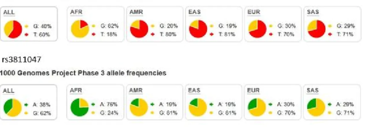
Figure 1.1.2a. Population genetics of rs3811046 and rs3811047 based on 1000 genome project
1000 genome project result shows that both rs3811046 and rs3811047 differ in their minor allele frequency according to ethnicity of the subjects. Of particular note is the difference between Africans and
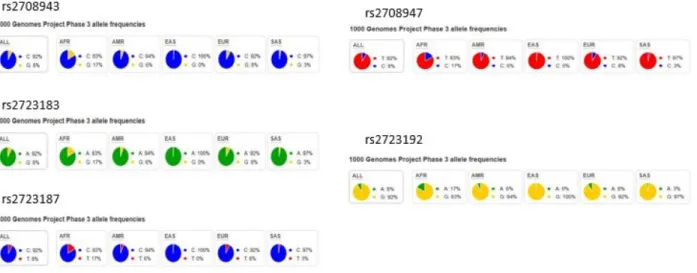
Figure 1.1.2b. Population genetics of rs2708943, rs2723183, rs2723187, rs2708947, and rs2723192 based on 1000 genome project
Population genomics of SNPs from the 2nd haplotype. Despite minor variations, the minor alleles
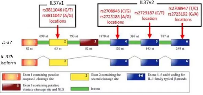
Figure 1.1.3a. Gene sequence of IL‐37b and SNP locations
Illustration of genomic DNA of IL‐37 and exons comprising the IL‐37b isoform. SNPs of the 1st
haplotype are all located on the exon #2, while SNPs of the 2nd haplotype are dispersed on exons 4, 5, and
6. These three exons code for the β‐strands typical to the IL‐1 family. Figure adopted and modified from Boraschi et al. 2011.
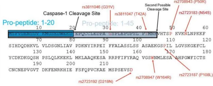
Figure 1.1.3b. Amino acid sequence of pro IL‐37b and missense mutations
Amino acid sequence of pro IL‐37b, and critical locations of interests. The sequence was pulled from UniProt website (http://www.uniprot.org/uniprot/Q9NZH6). There are two different reports describing the pro‐peptide length. 20 amino acid or 45. The enzyme that cleaves at location #20 is known to be caspase‐1. Amino acid substitutions are illustrated in red. The mutations caused by rs3811046 and rs3811047 are close to the cleavage sites. Caution is needed in interpreting this UniProt data because they assumed the ancestral amino acid to be the wild type. In Caucasians, that is not the case at two sites: The WT has valine (V) at amino acid #31 location, and alanine (A) at amino acid #42 location.
Figure 1.1.4. PolyPhen‐2 predictions of the effect of SNPs on IL‐37 function
Figure 1.1.5. 5 possible isoforms of IL‐37b
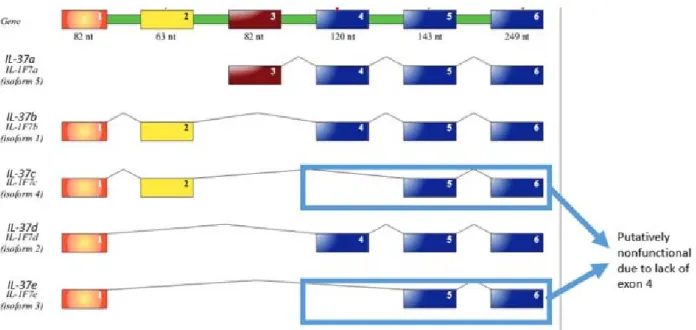
Illustration of 5 isoforms of IL‐37. IL‐37d (isoform 4) and IL‐37e (isoform 3) lack exon 4, which is part of the three exons that code for the β‐strands. Therefore, IL‐37d and IL‐37e are predicted to be nonfunctional. Figure was adopted from the work of Boraschi et al. 2011.
Figure 1.1.6. IL‐37b extracellular and intracellular pathways
Extracellular and intracellular pathways of IL‐37b. A number of agonists cause increase of pro IL‐ 37b production. Caspase‐1 matures the pro IL‐37b. Mature IL‐37b can bind to phosphorylated SMAD3 and translocate into the nucleus. Both pro and mature IL‐37b are secreted extracellularly, and its presence can be detected by a receptor complex composed of IL‐18Rα subunit and orphan receptor SIGIRR subunit (also known as IL‐1R8). The interaction between the receptor complex and IL‐37b is affected by IL‐18 binding protein (IL‐18BP). Both extracellular and intracellular pathways inhibits innate immune response of the cell. Diagram was constructed based on papers published by Bufler et al. 2002, Boraschi & Dinarello 2006, Nold et al. 2010, Bulau et al. 2013, Bulau et al. 2014, Li et al. 2014, Wu et al. 2014, Li et al. 2015, and Lundig et al. 2015.
CHAPTER 2: Observe the effect of variants on IL‐37 anti‐inflammatory function with human
recombinant IL‐37b
2.1. Material and Methods
2.1.1. Generation of human recombinant IL‐37b using
Escherichia coli
at amino acid #42) and mutant (V1, G at amino acid #31 and T at amino acid #42) human recombinant pro IL‐37b proteins were generated this way [Figure 2.4.1].
2.1.2.
In vitro
caspase‐1 cleavage experiment, direct gel staining
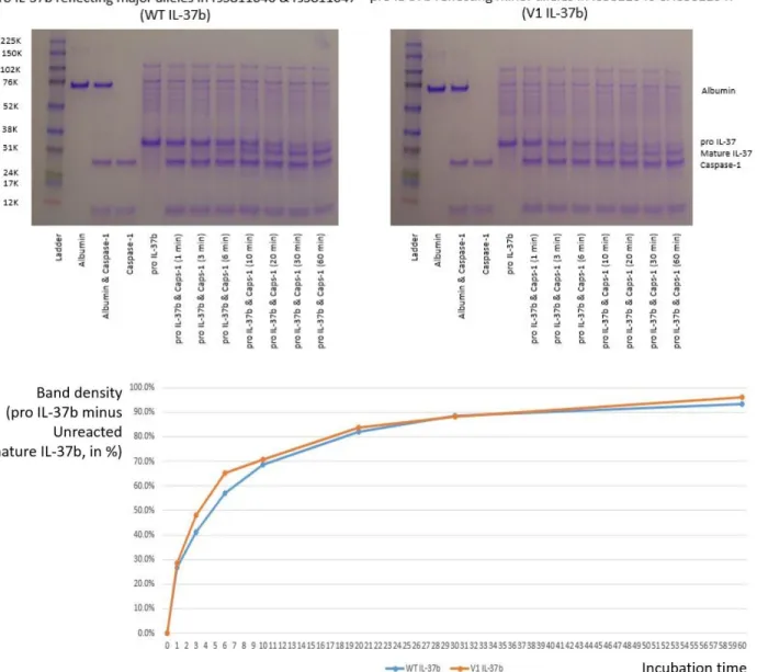
Human recombinant active caspase‐1 was purchased from Enzo Life Sciences (NY, USA). The enzyme came in concentration of 100 U/μL in a solution that was composed of the following: 50 mM HEPES, pH7.4, 100mM sodium chloride, 0.5% CHAPS, 1mM EDTA, 10% glycerol, and 10 mM DTT. The solution was used directly without dilution. Aliquots were created for each time points: 300 units of active caspase‐1 enzyme and 0.5 μg of human recombinant pro IL‐37b were mixed into each, 5 μL reaction mix. The individual aliquots were immediately incubated at 37 degree Celsius for their corresponding minutes (1, 3, 6, 10, 20, 30, and 60), then its reaction was terminated by adding 4x loading buffer and further denaturing the protein mix in 100 degree Celsius. Such experiment was done firstly with the human recombinant WT pro IL‐37b, and secondly with the human recombinant V1 pro IL‐37b. Samples were run through electrophoresis and the gel was directly stained using Coomassie blue (R‐250 dye). The protein stains now visible in the gels were digitally captured and ImageJ program (National Institutes of Health. MA, USA) was used for densitometry analysis. The amount of shift in the band from pro IL‐37b to mature IL‐37b was compared between WT and V1.
2.1.3.
In vitro
caspase‐1 cleavage experiment, Western blot with K
mand V
maxcalculation
Burk plot was used to derive Km and Vmax values for caspase‐1 cleavage reactions when substrates were either the wild (WT) or mutated form (V1) [Figure 2.4.3b].
2.1.4. Making better tools: EF.CMV. RFP vector for eukaryotic cells
EF.CMV.RFP vector for eukaryotic cell use was designed, where the transfected cells would constitutively express pro IL‐37b (by CMV promoter) and Red Fluorescent Protein (by EF promoter). 4 variations of pro IL‐37b gene inserts were used: WT, reflecting the 7 major alleles observed in Caucasians; V1, reflecting Caucasian minor alleles in the first haplotype (rs3811046 and rs3811047); V2 reflecting Caucasian minor alleles in the second haplotype (rs2708943, rs2723183, rs2723187, rs2708947, and rs2723192); V1V2, reflecting Caucasian minor alleles in both first and second haplotypes (rs3811046, rs3811047, rs2708943, rs2723183, rs2723187, rs2708947, and rs2723192). Clonal expansion and confirmation through sequencing was done in similar manner as in the previous description. Both
transient transfection (HEK293T, human embryonic kidney cell line) and permanent transfection (MPC11, human plasma cell line) were confirmed by IL‐37b and RPF bands in Western blots.
2.1.5.
In vivo
caspase‐1 cleavage experiment in transfected HEK293T cells
With collaboration with Dr. Jenny Ting’s lab, NLRP3 reconstitution experiment was conducted on pro IL‐37b transfected HEK293T cells. Caspase‐1 cleavage inside the cells, and amount of secreted IL‐37b were measured through Western blot of the cell lysate and supernatant. Transfection efficiency was evaluated through microscope as well as RPF band strength of the lysate through Western blot.
2.1.6. LPS stimulation of HEK293T, THP‐1 co‐culture system
25 ng/mL E. coli (strain O111:B4) LPS was added and 6 hours later the spent media, HEK293T cells (attached), and THP‐1 cells (floating) were collected. IL‐1β levels in the spent media was measured through ELISA in triplicates. IL‐37 levels in the supernatant and intracellular RFP and IL‐37 levels of HEK293T cells were measured through Western blots.
2.1.7. LPS stimulation of human recombinant IL‐37b pretreated RAW263.7 cell line
RAW263.7 cells were pretreated with 1, 10, and 100 pg/mL human recombinant pro IL‐37b for 30 minutes. 10 ng/mL of E. coli (strain O111:B4) LPS was added and spent media was collected at 12 hour post stimulation. Pro‐inflammatory cytokine levels were measured through ELISA in triplicates.
2.2. Results
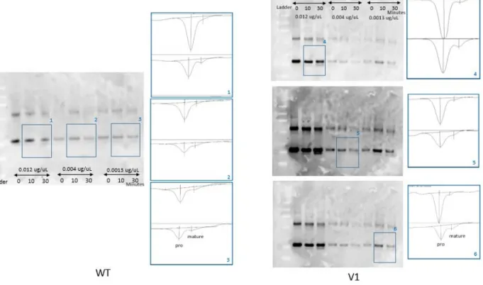
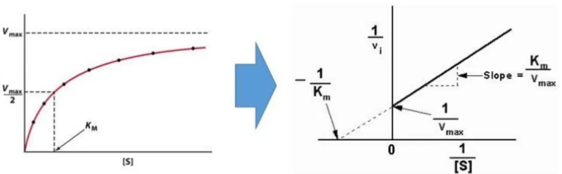
Densitometry analysis of the stained gels seemed to indicate that the variant pro IL‐37b is more readily cleaved by caspase‐1 during the first 30 minutes [Figure 2.4.2]. This result proved that the caspase‐ 1 digestion experiment with human recombinant pro IL‐37b was a viable experiment. However, protein stains digitally captured from a 1mm thick translucent gel resulted in a blurry out of focus image, which compromised the accuracy needed for finer analysis. The Coomassie blue stain was not sensitive enough to detect substrates in low concentrations, such as 0.0013 μg/μL. Pierce Silver staining (Thermo Scientific. MA, USA) of the gels was attempted, but despite its enhanced sensitivity, the very narrow detection range was not conducible for our study (data not shown). Western blot was the next best alternative besides ELISA. ELISA was not possible because antibodies specific enough to tell the pro or mature IL‐37b apart were not available. Western stain of in vitro caspase‐1 experiment was analyzed through densitometry, and through the Lineweaver‐Burk plot. The Michaelis‐Menten constant, Km, of WT pro IL‐37b was 1.74
10 , whereas that of V1 pro IL‐37b was 7.22 10 . This meant the mutated V1 substrate had 4.25 times less affinity to the caspase‐1 enzyme. Vmax of WT pro IL‐37b was 0.5 10 mol / (L sec), and V1 pro IL‐37b was 1 10 mol / (L sec). Although the mutated V1 substrate may have less affinity to the caspase‐1 enzyme, when the substrates are saturated, V1 can overtake the WT reaction because it has 2 times higher maximum reaction velocity [Figure 2.4.3b].
The EF.CMV.RFP vector was successfully used to transfect the pro IL‐37b gene both transiently (HEK293T) and permanently (MPC11). Confirmation was done with Western blots [Figure 2.4.4].
This meant Km and Vmax cannot be derived with this system. It also became clear that despite same levels of transfection (equivalent density of RFP bands), IL‐37b proteins were not produced at same levels: More pro WT was being produced than pro V1 intracellularly to begin with, and this lead to more of mature IL‐ 37b detected on the supernatant [Figure 2.4.5]. Additionally, it was found out that caspase‐1 could be self‐activated if the transfection amount was 30 ng/well in the 24‐well plate (data not shown).
Simplification of the experiment was possible as this meant ASC and NLRP3 gene transfections were not necessary.
We realized that the differing amount of mature IL‐37b needs to be reflected in our experiments. The HEK293T & THP‐1 co‐culture experiment was designed with this in mind. When equivalently
transfected HEK293T cells, co‐cultured with THP‐1 cells were stimulated with E. coli (strain O111:B4) LPS, the IL‐1β levels in the WT transfected system were less than that of non‐transfected control. HEK293T only culture did not show any IL‐1β response to LPS, so the result was contributed by THP‐1 only. The results reconfirmed the anti‐inflammatory action of IL‐37b (p=0.001). More importantly, The V1
transfected system exhibited higher IL‐1β levels compared to WT (p<0.001), indicating compromised anti‐ inflammatory function, despite equivalent transfection levels. Western blot indicated that again, there were differing amounts of mature IL‐37b in the supernatants: WT had more mature IL‐37b compared to the V1 counterpart [Figure 2.4.6].
Lastly, we wanted to see if the mutated IL‐37b had compromised anti‐inflammatory functions on its own. RAW264.7 cells were pretreated with the same controlled amount of WT or V1 pro IL‐37b, then E. coli (strain O111:B4) LPS stimulation was done for 12 hours. IL‐6 in the spent media was used as a
created through E. coli was functional, as it could reduce the IL‐6 response. On the other hand, the mutated V1 IL‐37b failed to reduce IL‐6 response of RAW264.7 cells [Figure 2.4.7].
2.3. Conclusions
The IL‐37b first haplotype variant (V1) has less affinity to caspase‐1 but will reach higher reaction velocity.
V1 causes less production and secretion of IL‐37b in both transient and permanently transfected cells.
The decreased V1 environment causes LPS stimulated HPT‐1 cells to overproduce IL‐6 compared to wild type IL‐37b.
V1 IL‐37b exhibit compromised anti‐inflammatory ability in LPS stimulated RAW264.7 cells.
2.4. Figures
Figure 2.4.1. Generating human recombinant pro IL‐37b from E. coli
Human recombinant pro IL‐37b was produced using pET30(a)+ as the vector. Gene insert was created with PCR with addition of Xhol and Ndel sites. Empty vector, and vector with gene insert was cloned with DH5α first, then the proteins were produced in BL21(DE3) cells. After lysing away the cells with a sonicator, Ni‐NTA agarose beads were used to purify the proteins. After two overnight dialyses the recombinant proteins were ready for experiments, such as in vitro caspase‐1 cleavage. Our human recombinant pro IL‐37b has two segments at the C‐terminal that do not exist in the natural form:
Figure 2.4.2. Direct gel staining of caspase‐1 treated pro IL‐37b, and densitometry analysis
Figure 2.4.3a. Western blot of in vitro pro IL‐37b cleavage experiment and its densitometry analysis
Densitometry analysis of Western blot. 3 different concentrations of substrates (human recombinant pro IL‐37b) were incubated for 3 different times (0, 10, and 30 minutes). For the V1 experiment, the exposure time had to be adjusted to capture the band strength where the faint mature IL‐37b band could be well detected, while the stronger pro IL‐37b band was not overly exposed. This ensured that the digitally captured band density still had linear relationship with the detected IL‐37
Figure 2.4.3b. Km and Vmax calculation based on densitometry of Western blot
With the known reaction time and substrate concentrations, reaction velocity could be calculated. Lineweaver‐Burk plot allowed us to derive the maximum reaction velocity (Vmax) and Michaelis‐Menten constant (Km) of each reactions for comparison.
Figure 2.4.4. IL‐37b transfection of eukaryotic cell lines, and confirmation of IL‐37b productions via Western blot
IL‐37b gene was inserted into EF.CMV.RFP vector, and the plasmids were transiently transformed into HEK293T cell lines, permanently transfected into MPC11 plasma cell lines. Transformation success was confirmed via Western blots of RFP and IL‐37b proteins in the cell lysates.
Figure 2.4.5 IL‐37b maturation by caspase‐1 in vivo, through NLRP3 constitution experiment on HEK293T cell line
NLRP3 constitution through transfecting HEK293T cells with ASC, NLRP3, and pro caspase‐1 genes was successful. When pro IL‐37 genes were introduced into the same cells, the system secreted mature IL‐ 37 into the supernatant. Pro IL‐1β was used as positive control for caspase‐1 action.
Figure 2.4.6 HEK293T and THP‐1 co‐culture experiment
HEK293T cells were transfected with pro capase‐1 and pro IL‐37b genes, then co‐cultured with THP‐1 cells. After 10 hours of E. coli (strain O111:B4) LPS stimulation, IL‐1β levels of the spent media was used as a surrogate to measure the innate immune response of THP‐1 cells. Mature IL‐37b levels were measured with Western blot of the spent media, transfections were confirmed by the RFP band strengths of the HEK293T cell lysates.
Figure 2.4.7 RAW246.7 cells, E. coli (strain O111:B4) LPS stimulation after pro IL‐37b pretreatment
RAW246.7 cells were pretreated with set concentrations of either WT or V1 pro IL‐37b for 30 minutes and stimulated with E. coli (strain O111:B4) LPS. Experiment was done in triplicates and IL‐6 levels in the spent media were measured in triplicates via ELISA.
CHAPTER 3: Observe the effect of variant of IL‐37 in Caucasian human samples
3.1. Material and Methods
3.1.1. Screening process
The inclusion criteria for the human sample collection were as follows:
1) Caucasian ethnicity
2) Between the ages of 18 and 65 years old
3) Has minimum of 20 natural teeth, excluding third molars 4) Has at least 3 teeth in the posterior sextant
5) Able and willing to follow study procedures and instructions 6) Read, understood, and signed informed consent form
Exclusion criteria were:
1) Participant has chronic disease with oral manifestations, including diabetes 2) Participant is a smoker, or a previous smoker within the past 2 years 3) Participant has gross oral pathology other than periodontal disease
4) Participant had been treated with antibiotics for any medical or dental condition within 1 month of the screening exam
5) Participant had been treated for two weeks or more with any medication that is known to affect periodontal status within 1 month of the screening exam
7) Participant has any significant organ disease or bleeding disorder 8) Participant has infectious disease such as hepatitis, HIV, or tuberculosis 9) Participant has anemia or other blood dyscrasias
10)Participant is on anticoagulant therapy
11)Participant has dental or medical conditions that is likely to require antibiotic treatment during the study period
12)Participant is pregnant, expecting to be pregnant, or nursing
13)Participant has anything that would place him/her at increased risk or preclude the individual’s full compliance with or completion of the study
2 mL of saliva was collected from potential participants using OG‐500 Oragene DNA collection kit (DNA genotek. ON, USA). DNA was purified from the samples using PT‐L2P‐5 solution according to the manufacturer’s instructions.
Custom forward and reverse primers were designed to amplify the region of interest from the genomic DNA (biotin labelled forward primer: TGCTAACCTCACTGCGTCTGAC; reverse primer:
ATCACCTCACCCCGAGGC; sequencing primer: CCTTACTTGTGTGAACAAA). The forward primer was biotin labeled at its 5’ end for downstream processing required by the pyrosequencer. The host DNA was PCR amplified using the custom primers, and genotype discerned with the sequencing primer using a pyrosequencer (PyromarkMD from QIAGEN. Hilden, Germany) [Figure 3.5.1]. The now genotyped participants were contacted, and when the participants showed interest in, and consented for, further participation, appointments were made for blood draws via venipuncture.
3.1.2. Blood collection and sample processing: Whole blood experiment
experiment. E. coli (strain O111:B4) LPS was added to 2 mL of aliquoted whole blood to create a final concentration of 0, 0.01, and 0.1 μg/mL. The mix was incubated at room temperature with gentle undulation for 2 hours as described by Offenbacher et al. (Barros, Wirojchanasak et al. 2010). mRNA was purified from each aliquots using QIAamp RNA Blood Mini Kit (QIAGEN. Hilden, Germany). IL‐1β, IL‐6, and TNF‐α expression levels were measured and compared between WT homozyogote and V1 homozygote groups. GAPDH (glyceraldehyde‐3‐phosphate) was used as internal control for ∆∆Ct calculation [Figure 3.5.2].
3.1.3. Blood collection and sample processing: Dendritic cell differentiation and LPS stimulation
3.1.4. GCF collection and inflammatory mediator assessment
Eight GCF samples (2 per quadrant) will be collected from the mesio‐buccal and mesio‐
lingual sites of each of the 1
stmolars. If a first molar was missing, the collection was done on
mesio‐buccal and mesio‐lingual of the 2
ndmolar. When both the first and second molars were
not present, mesio‐buccal and mesio‐lingual sites of the 2
ndpremolar was used for GCF
collection. GCF was collected with PerioPaper strips, and Periotron 8000 device (Oraflow Inc. NY,
USA) was used to measure its volume. The samples were kept in liquid nitrogen until they were
ready to be read. The frozen strips were thawed to room temperature and eluted out with
diluent. Luminex Multiplex assay was performed using Bio‐Plex 200 system (Bio‐Rad
Laboratories. CA, USA) to measure the amount of 6 mediators: IL‐1β, IL‐6, IL‐8, TNF‐α, G‐CSF, and
MIP‐1β. The concentrations were calculated, and stratified according to the participant’s
genotypes. We had data of 36 such subjects, and Dental Atherosclerosis Risk in Community Study
(DARIC) data of 107 subjects were added to this. A total of 143 subjects with genotypes 1.1
(homozygous major alleles in the first haplotype, n=65), 1.2 (heterozygous for the major and
minor alleles in the first haplotype, n=66), 2.2 (homozygous minor alleles in the first haplotype,
n=12) were compared. PROC mixed model was used for analysis, with p<0.05 as defined to be
statistically significant.
3.1.5. Gingival tissue biopsy, isoform expression preference
mRNA was extracted with RNeasy Mini Kit (QIAGEN. Hilden, Germany). Real time PCR result was used to create expression ratio amongst the 5 isoforms in the gingival tissues. The ratio was compared among the 3 genotype groups.
3.2. Results
Three hundred twenty seven people were screened, and they were genotyped into 1.1
(homozygous for the major allele in the 1st haplotype), 1.2 (heterozygous for the major and minor alleles
in the 1st haplotype), and 2.2 (homozygous for the minor alleles in the 1st haplotype) groups [Table 3.4.1].
Whole blood was collected from 68 subjects for whole blood stimulation experiment. A comparison of pro‐ IL‐1β, IL‐6, and TNF‐α expression between 4 individuals with 1.1 genotype and 4 from 2.2 genotype showed general trend of increased pro‐inflammatory cytokines in the presence of SNP variants, with TNF‐ α expression being statistically significant [Figure 3.5.3].
IL‐1β was used as surrogate measure of inflammatory response of DCs to E. coli (strain O111:B4) LPS. For each subjects, 1 hour or 6 hour expression levels were the highest, with notable variance within each groups. Such variance made it impossible to choose a single time point (e.g. 1 hour or 6 hour) for overall comparison. Additionally, the pilot experiment showed that the IL‐37 is expression level was highest at 12 hour after LPS stimulation [Figure 3.5.2]. This meant the IL‐1β levels at 12 and 24 hours should be included in the analysis, as our purpose was to compare of the effects of IL‐37 on the IL‐1β expression levels. Area under the curve (AUC) i.e., the sum of all fold values from 0, 1, 6, 12, and 24 hours, was used as representative measure of individual immune response, and that value was compared between 1.1 and 2.2 genotypes. A statistically significant difference between the genotype groups could be observed, with 2.2 group having higher IL‐1β expression levels [Figure 3.5.4].
When the isoform expression ratio was measured from the gingival biopsies, we observed increased ratio of the putative nonfunctional isoforms (IL‐37c and IL‐37e) in the 1.2 and 2.2 genotype groups compared to 1.1 group.
3.3. Conclusions
Human whole blood from the V1 genotype subjects showed tendency of hyper‐inflammatory profiles in terms of IL‐1β, IL‐6 and TNF‐α expression when stimulated.
Dendritic cells differentiated from V1 genotype subjects also demonstrated hyper‐inflammatory profile in IL‐1β expression.
There was a general trend of increased pro‐inflammatory cytokine concentrations in the GCF, when the subjects had minor alleles.
Gingival tissues collected from subjects with the minor allele had less active IL‐37 isoforms in terms of their ratio.
3.4. Tables
Table 3.4.1. Genotyping results
Total of 327 subjects were screened for genotyping. 68 of them consented and participated in blood draw and whole blood experiments. 41 of such subjects we conducted dendritic cell differentiation of their peripheral monocytes. A smaller group, 8 of each genotypes we collected gingival biopsies.
3.5. Figures
Figure 3.5.1. Pyrosequencing of human saliva DNA for genotyping
Host DNA was purified from the saliva, DNA region of interest was PCR amplified, and genotyped based on the rs3811046 and rs3811047 alleles (1st haplotype) through pyrosequencing. Principles of
pyrosequencing and sample analysis examples are illustrated.
Figure 3.5.2. Blood experiments of the genotyped participants
Whole blood was collected with venipuncture. 6 mL of the whole blood was stimulated directly with E. coli (strain O111:B4) LPS and RNA was isolated to measure pro‐inflammatory cytokine expression. The remaining blood were used to purify monocytes. IL‐4 and GM‐CSF stimulation for 7 days
differentiated the monocytes further to dendritic cell phenotypes, but it was the LPS stimulation that fully differentiated them. It was during this LPS stimulation where we followed the cells up and collected RNA and spent media at 0, 1, 6, 12, and 24 hours. The time points were based on pilot experiments, which
Figure 3.5.3. Whole blood experiment. IL‐1β, IL‐6, and TNF‐α expression levels.
Whole blood was stimulated at final concentration of 0, 0.01, and 0.1 μg/mL E. coli (strain
O111:B4) LPS for 2 hours at room temperature. Pro‐inflammatory cytokine mRNA expression was derived through 2 ∆∆ calculation. GAPDH was used as internal control. Error bars indicates standard errors.
Figure 3.5.4. DC stimulation with E. coli, IL‐1β expression comparison between 1.1 and 2.2 genotypes
6 subjects from the WT group (1.1 genotype in the 1st haplotype) were compared against 6
subjects form the V1 group (2.2 genotype in the 1st haplotype). Their differentiated dendritic cells (derived
from peripheral blood) were stimulated with 0.1 μg/mL E. coli (strain O111:B4) LPS and followed up from 0, 1, 6, 12, and 24 hours. IL‐1β expression was measured via qRT‐PCR in triplicates in a single 384 reaction plate. GAPDH was used as internal control. Fold expression values were individually normalized to 0 hour non‐stimulated controls within each subjects. Area under the curve (AUC) was compared between WT (1.1) and V1 (2.2) groups and was found to be statistically significantly different (p < 0.05).
Figure 3.5.5. Inflammatory mediator concentration in human GCF, compared among the 1.1, 1.2, and 2.2 genotypes
The GCF levels of IL‐1β, IL‐6, IL‐8, TNF‐α, G‐CSF, and MIP‐1β were measured by immunobead multiplexing for genotyped subjects in the DARIC population (n=107), supplemented with 36 subjects who were genotyped for the 1st haplotype locus by pyrosequencing to enrich the population for the minor allelic
variant. A total of 143 subjects with genotypes 1.1 (n=65), 1.2 (n=66), 2.2 (n=12) are shown with Z scores for each mediator, normalized to the mean GCF cytokine concentration levels of 1.1 genotype. Error bars indicate standard errors.
Figure 3.5.6. Isoform expression preference in human gingival tissues
Human gingival tissues were collected from 4 individuals each, from 1.1, 1.2, or 2.2 genoytpe groups. Isoform cDNA specific primers were designed and used to compare 5 different isoform expressions. qRT‐PCR results were plotted in ratios for each genotype groups.
CHAPTER 4: DISCUSSION
The anti‐inflammatory role of the IL‐37b is being actively elucidated since it discovery, yet we know very little on how polymorphisms in single nucleotides can affect those functions. Literature search on SNP variants on IL‐37 resulted in very few hits: Pei et al. had investigated the effects of rs3811047 and reported they did not observe increased susceptibility of rheumatoid arthritis in Chinese Han population, and they observed lower swollen joint count, swollen joint index, rest pain, and health assessment questionnaire score in the 1.2 and 2.2 genotype groups compared to 1.1 group. Their data suggested that the minor alleles were actually protective, and not destructive to the host (Pei, Xu et al. 2013). These results should be interpreted with caution, because the findings are from observation on different ethnicity. Ethnicity plays a crucial part in the study of SNP variants, as our GWAS showed statistically significant association with a phenotype (upper quartile of GCF IL‐1β concentration) in one ethnicity (Caucasians) but not another (African Americans). Contrary to their report on ethnic Han Chinese, our data on Caucasian samples suggests minor alleles in rs3811046 and rs3811047 (considered as one unit, or haplotype, as the SNPs showed strong linkage equilibrium) causes disruption of IL‐37b function, and leads to hyper‐inflammatory profile of the host.
Out of 7 SNPs of interest found through GWAS, we focused on two SNPs in the 1st haplotype, as
their minor allele frequency (MAF) was reported to be around 0.40. The other 2nd haplotype (including
rs2708943, rs2723183, rs2723187, rs2708947, and rs2723192), the MAF was too low (0.08) for us to investigate their effects in human subjects within the allotted schedule of our project. It would have required considerably longer recruitment time for genotype screening due to its rarity.
As previously mentioned, the IL‐37b is first produced in a precursor form, only to be matured afterwards by removing a pro‐peptide region at its N‐terminal (Kumar, McDonnell et al. 2000). It has been shown that a single mutation introduced at the amino acid #20 site totally abolished the caspase‐1 cleavage, and therefore its maturation (Kumar, Hanning et al. 2002). The SNPs rs3811046 and rs3811047 are not located at the cleavage site (amino acid locations are on 31 and 42) and therefore expected not to totally abolish the reaction. However, they are close enough to warrant an investigation if mutations at those sites cause change in its maturation efficiency. On the other hand, the 5 SNPs of the second haplotype are located on the 3 exons that make up the β‐strands typical to the IL‐1 family. Therefore, it was suspected that they will affect the affinity of IL‐37b to its receptor complex, and not caspase‐1 cleavage.
With the premise that the caspase‐1 cleavage reaction of pro IL‐37b meets three assumptions (steady‐state approximation, free ligand approximation, and rapid equilibrium approximation), Michaelis‐ Menten kinetics can be applied. In an in vitro experiment, where the substrate concentration, enzyme amount, and reaction time is under control, the Michaelis‐Menten constant (Km) and maximum reaction velocity (Vmax) could be calculated and compared. The Km is defined as the concentration of the substrate
substrate concentration increases. If the same phenomenon happens in vivo, that difference in the enzyme affinity (4.25 times higher to capase‐1 with the WT substrate compared to V1 substrate) may be caught up by the V1 reaction because it is capable of reaching twice the maximum reaction velocity. But, this scenario may only happen in systems with constant pro IL‐37b overproduction and therefore, substrate saturation (such as in transfected cell lines with constitutive CMV promoter), and not in cells in a natural state. That said, it may be enough to compare WT vs. V1 caspase‐1 reaction with just Km values only: WT pro IL‐37b is more readily matured than V1 pro IL‐37b, and therefore subjects with WT genotype will have more mature IL‐37b in their system to regulate inflammation.
The production of recombinant proteins has revolutionized biochemistry. E. coli is one of the organisms of choice for production for the following reasons: The bacteria has fast growth kinetics with doubling time of 20 minutes (Sezonov, Joseleau‐Petit et al. 2007), high cell density cultures can be easily achieved (Lee 1996), rich complex media can be made from readily available and inexpensive
components, and transformation with exogenous DNA is fast and easy (Pope and Kent 1996). Despite such convenience, the prokaryotic system is not without its faults. Post‐translational modification, such as protein glycosylation will not be possible with this system. So far we have not found evidence that IL‐37b goes through post‐translational modification, and numerous papers have been describing IL‐37b functions based on recombinant IL‐37b created with E. coli (Li, Neff et al. 2015, Cavalli, Koenders et al. 2016, Liu, Xue et al. 2016, Zhu, Sun et al. 2016, Li, Zhai et al. 2017, Zeng, Song et al. 2017). Our experiments also demonstrated that recombinant pro IL‐37b created through such prokaryotic system retained its anti‐ inflammatory functions.