THE ROLE OF ANKYRIN REPEAT AND SOCS BOX CONTAINING PROTEIN 4 (ASB4) IN TROPHOBLAST DIFFERENTIATION AND PSEUDOVASCULOGENESIS
WH Davin Townley-Tilson
A dissertation submitted to the faculty of the University of North Carolina at Chapel Hill in partial fulfillment of the requirements for the degree of Doctor of Philosophy in the
Department of Cell and Developmental Biology.
Chapel Hill 2014
ii ©2014
iii ABSTRACT
WH Davin Townley-Tilson: The Role Of Ankyrin Repeat And SOCS Box Containing Protein 4 (ASB4) In Trophoblast Differentiation And Pseudovasculogenesis (Under the
direction of Cam Patterson)
Vascularization of the placenta is a critical developmental process that ensures fetal viability. The vascular health of the placenta affects both maternal and fetal well being; however, relatively little is known about the early stages of placental vascular development. The ubiquitin ligase Ankyrin repeat, SOCS box-containing 4 (ASB4) promotes embryonic vascular lineage commitment and is highly expressed early in placental development. The transcriptional regulator Inhibitor of DNA binding 2 (ID2) negatively regulates trophoblast differentiation during development and is a target of many ubiquitin ligases. Due to their contrasting effects during differentiation, we investigated whether ASB4 mediates vascular differentiation through its ligase activity on ID2 in the placenta. Placentas from Asb4-/-mice exhibited myriad vascular differentiation defects, including abnormal overexpression of ID2, and pregnant Asb4-/- mice phenocopied human pre-eclampsia. We determined that ASB4 directly interacted with ID2 in trophoblast cells, leading to ID2’s ubiquitination and
iv
This work is dedicated to mother, who wasn’t able to see me complete my goal; my father, who instilled the value of education in me; my brother, who has always been smarter than
v
ACKNOWLEDGEMENTS
I would firstly like to thank my advisor, Dr. Patterson, for giving me the opportunity to work in his laboratory. His advice, wisdom, and mentorship have been invaluable
throughout my time working for him.
I would like to thank my committee for pushing me to work faster, harder, and smarter. Your suggestions, availability, and direction have guided and molded this work; it couldn’t have been completed without your help.
vi
TABLE OF CONTENTS
LIST OF FIGURES ... viii
LIST OF ABBREVIATIONS ... ix
CHAPTER 1: GENERAL INTRODUCTION ...1
Vasculogenesis, pseudovasculogenesis, and trophoblast differentiation ...1
ASB4 as a ubiquitin ligase ...4
ID family of proteins ...8
Pre-eclampsia and pathologies of the placenta ...11
Hypothesis...14
CHAPTER 2: ASB4 PROMOTES TROPHOBLAST DIFFERENTIATION THROUGH THE DEGRADATION OF ID2 ...22
Introduction ...22
Materials and Methods ...23
Mouse generation, blood pressure, and proteinuria ...23
In situ hybridization, immunofluorescence, and immunohistochemistry ...24
Cell culture and immunoblotting ...25
In vitro ubiquitination assay...26
Placental cell differentiation assays ...27
Statistical analysis ...28
Results ...28
vii
ASB4 negatively regulates ID2 expression through
polyubiquitination and proteasome dependant degradation ...31
ASB4 mediates placental cell differentiation and function in vitro ...33
Asb4-/-mice phenocopy human patients with pre-eclampsia ...36
Discussion ...37
CHAPTER 3: GENERAL DISCUSSION ...55
The role of ASB4 in vascular commitment and patterning ...55
ASB4 as an E3 ligase ...58
The role of ASB4 in placental pathologies and pre-eclampsia ...61
viii
LIST OF FIGURES
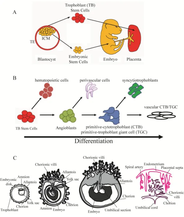
Figure 1.1 Placental development and differentiation ...16
Figure 1.2 ASB4 as an E3 ligase ...18
Figure 1.3 ID proteins regulates bHLH-mediated transcription ...19
Figure 1.4 Vascular remodeling during placental development ...20
Figure 2.1 Asb4 expression in the developing placental vasculature...41
Figure 2.2 Asb-/- placentas express markers of undifferentiated vasculature and TB cells ...43
Figure 2.3 ID2 expression increases in placentas that lack Asb4 ...44
Figure 2.4 ASB4 negatively regulates ID2 expression through polyubiquitination and associates with ID2 in JAR cells ...45
Figure 2.5 ASB4 promotes JAR cell-mediated endothelial apoptosis and stabilization of endothelial cell networks ...47
Figure 2.6 ASB4 promotes TB cell differentiation in vitro ...48
Figure 2.7 Pregnant Asb4-/- mice display symptoms of pre-eclampsia ...49
Figure S2.1 Diminished mature vasculature in Asb4-/- placentas is not due to increased apoptosis or abnormal proliferation ...50
Figure S2.2 Asb4 deletion induces vascular dysfunction and mislocalization of blood vessels in the placenta ...51
Figure S2.3 N-terminally tagged ID2 is resistant to ASB4-mediated degradation ...52
Figure S2.4 ASB4 degrades ID2 in a proteasome-dependant manner, and does not affect ID2 cellular location ...53
ix
LIST OF ABBREVIATIONS
APC/C Anaphase promoting complex/cyclosome
ASB4 Ankyrin repeat and SOCS containing protein 4
bHLH Basic helix-loop-helix
cKit Kit oncogene, a/k/a stem cell growth factor receptor
CTB Cytotrophoblast
dbcAMP N(6),2'-O-dibutyryladenosine 3':5' cyclic monophosphate
D-box Destruction box motif
DR Degradation-resistant
DS Degradation-sensitive
E Embryonic days post fertilization
E1 Ubiquitin activating enzyme
E2 Ubiquitin conjugating enzyme
E3 Ubiquitin ligase
ELISA Enzyme-linked immunosorbent assay
EPO Erythropoetin
ES Embryonic stem (cell)
FIH Factor inhibitin HIF
GAPDH Glyceraldehyde 3-phosphate dehydrogenase
hCG Human chorionic gonadotropin
HELLP Hemolysis, elevated liver enzymes, low platelet count
HIF Hypoxia inducible factors
x
HRP Horseradish peroxidase
ID2 Inhibitor of DNA binding 2
IUGR Intrauterine growth restriction
JAR Choriocarcinoma cell line
K Lysine
KDM1 Lysine (K)-specific demethylase 1
LL Lysine-less
MEK1 Mitogen-activated protein kinase kinase 1
MMP Matrix metalloproteinase
PECAM Platelet endothelial cell adhesion molecule
PlGF Placental growth factor
Rb Retinoblastoma gene
RING-finger Really interesting new gene domain that binds zinc cations ROC1 A RING-finger E3 ligase encoded by Rbx
SCF Stem cell factor ligand
SCFR Mast/stem cell growth factor receptor, a/k/a cKit sFlt1 Soluble-Fms-like tyrosine kinase 1
SOCS Suppressor of cytokine signaling
TB Trophoblast
TBSC Trophoblast stem cell
TGC Trophoblast giant cell
1 CHAPTER 1
GENERAL INTRODUCTION
Vasculogenesis, pseudovasculogenesis, and trophoblast differentiation1
Blood vessel formation is classically divided into two categories: vasculogenesis and angiogenesis. Vasculogenesis is the formation of new blood vessels from the de novo
production of endothelial cells. Angiogenesis refers to the formation of new blood vessels via extension or remodeling of existing blood vessels. Angiogenesis occurs throughout
development and in adulthood, whereas vasculogenesis is generally thought to occur during a limited period early in development.
Vasculogenesis is further divided into two categories: extraembryonic, that occurs in the yolk sac (which functions as the source of early blood cell formation in the
developmental blood circulatory system) and allantois, (which gives rise to the umbilical vasculature) and embryonic, i.e. restricted to the embryo itself [1]. Extraembryonic blood vessel formation precedes embryonic vasculogenesis and provides communication between the fetal circulation and the yolk sac to facilitate the transfer of nutrients and blood gases to the developing embryo, and ultimately gives rise to the placenta [2]. In mammals, blood islands assembling within the mesodermal layer of the yolk sac are the first occurrence of vasculogenesis. Blood islands are foci of hemangioblasts that differentiate in situ, forming a
2
loose inner mass of hematopoietic precursors and an outer luminal layer of angioblasts. Blood islands eventually coalesce into a functional vascular network that constitutes the vitelline circulation, which is adapted to transfer nutrients from the yolk sac to the embryo proper [2]. The chorion is the outermost layer of extraembryonic mesoderm and
trophectoderm, surrounding the embryo and all other components of the conceptus. The chorionic villi, which arise from the chorion, are small projections that intercalate the uterine tissue, maximizing the surface area in contact with maternal blood [3]. Extraembryonic vasculogenesis in Eutherians (placental mammals) also supplies the allantois with primitive vessels in preparation for chorion fusion and umbilical vessel formation, thus initiating the vascular connection between the fetal and maternal placental tissues [4]. This allantois-chorion fusion is what is responsible for the placenta proper, whereby the allantois and umbilical vasculature joins the chorion, which has now intertwined with the maternal vasculature, forming a network of vessels that will supply the fetus with nutrients and oxygen, and carry away waste from the fetus into the maternal blood stream where it can be eliminated [3]. This vascularization of the early placenta is crucial for the health and viability of not only the fetus, but also the mother (Figure 1.1) [5-7].
Embryonic vasculogenesis (sometimes referred to as intraembryonic vasculogenesis) occurs throughout most of the embryonic mesoderm. The endocardium and great vessels are the first embryonic endothelial structures formed during development [2]. Parallel with heart development, vasculogenesis is initiated within the aortic primordia, a collection of
3
precursor cells and the pro-epicardium make contact with the developing heart tube and quickly spread over the entire heart, giving rise to the capillaries, veins, and arteries of the coronary vasculature, smooth muscle cells and pericytes [9]. Lastly, the capillary plexi of endothelial cells are remodeled into a unidirectional circuit to allow proper blood circulation throughout the embryo, forming the mature vasculature. To accomplish this vascular
specification, endothelial cells are specified into an arterial or venous fate; then, as blood flow commences, vascular smooth muscle cells are recruited to the endothelium to provide structural support and elasticity to the now mature vasculature [10].
The junction of embryonic, extraembryonic, and maternal vasculature is the placenta. Though the placenta is the only transient organ in the body, it requires a great deal of
specification for its development, especially within the context of the placental vasculature, during its short window of development to ensure fetal and maternal wellness. Placental vasculogenesis is similar compared with that of embryonic and extraembryonic
vasculogenesis in that endothelial cells must differentiate from progenitor cells to form the vasculature. However, the initial progenitor cell reservoir is different, and many of the
differentiating endothelial cell characteristics are unique to the placenta. These characteristics include different cell adhesion molecules [11] and intracellular markers of differentiation [12].
4
trophoblasts that form and remodel the placental vasculature [13]. Villous trophoblasts have endothelial cell functions in the chorionic villi and also fuse into syncytiotrophoblasts, which are the epithelial covering of these villi that penetrate the uterus [14]. Extravillous
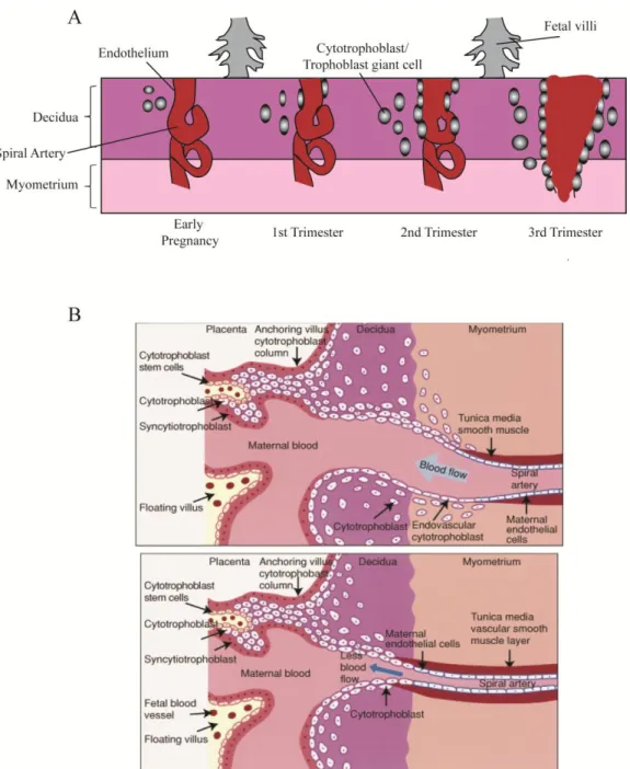
trophoblasts invade and migrate through the junctional zone of the placenta into the maternal decidua, where they replace the endothelial cells that line the spiral arteries in a process called “pseudovasculogenesis” [6]. These differentiating CTBs “switch” integrin expression profiles from one expression pattern that allows for cell motility and extracellular matrix degradation during migration and invasion to an endothelial-like integrin profile of differentiated cells that form tight junctions in the arteries, creating high capacity, low resistance blood vessels that allow for the exchange of blood gasses and nutrients from the mother to the developing fetus [11,15,16]. Many of these same processes are conserved in mice [17] in that cells originating from a trophoblast (TB) stem cell progenitor migrate and invade the maternal arteries, but in mice, these are thought to derive from trophoblast giant cell intermediaries, rather than cytotrophoblast lineages [18].
Prior work in this lab uncovered the ankyrin repeat, SOCS box-containing 4 (ASB4) protein as a mediator of embryonic stem cell to endothelial differentiation [19]. Further, ASB4 was found to be highly expressed in the extraembryonic vasculature including the allantois, yolk sac, and most notably, the developing placenta [19]. Therefore, we decided to investigate the role of ASB4 in placental vascular development.
ASB4 as a ubiquitin ligase
5
The SOCS box binds to an elongin-B/elongin-C/cullin/ROC complex of proteins whose function as components of a ubiquitin ligase complex has been well characterized [21]. The N-terminal protein interaction motif binds to substrate protein(s) to optimally position them for ubiquitination and subsequent degradation [22]. Thus, SOCS proteins are the substrate-receptors for an ubiquitin ligase complex that controls steady-state levels of substrate
proteins. Because of this essential function, SOCS proteins like ASB4 are carefully regulated both at the transcriptional and post-translational level to tightly control substrate protein levels.
The central role of all E3 ligases is targeting substrate proteins for ubiquitination. The well characterized ubiquitination process involves three enzymes that catalyze the activation and transfer of the 7 kDa ubiquitin protein to the targeted substrate protein. Briefly, the E1 ubiquitin activating enzyme covalently binds to the ubiquitin protein via an ATP-dependent process then transfers it to the E2 ubiquitin conjugating enzyme. From there, the ubiquitin molecule is either transferred directly to the substrate protein, due to the resulting structural proximity modulated by the E3 ligase (where the E3 ligase never directly interacts with ubiquitin itself), or is step-wise transferred to the E3 ligase, and further conjugated to the substrate protein (Figure 1.2) [23].
There are three forms of ubiquitination: mono-, multi- (or multi-mono-), and
polyubiquitination. Mono-ubiquitination is where a single ubiquitin moiety is conjugated to a single ε-NH2 group of an internal lysine residue of the target substrate. Proteins can also
undergo multi-ubiquitination, whereby a single ubiquitin is conjugated to multiple internal lysine residues on the target protein. Lastly, polyubiquitination occurs when multiple
6
(or a free α- NH2 group of the N-terminal residue) on the target substrate. These different
ubiquitination reactions result in very different consequences for the substrate protein. Typically, mono- and multi-ubiquitination are thought to be involved in nonproteolytic, reversible, signaling events such as endocytosis, membrane trafficking, DNA repair, and gene silencing [24]. Polyubiquitination, in contrast, is generally thought of as being
responsible for the proteasome-dependent degradation of a target protein. However, adding yet another layer of complexity to the ubiquitination process, ubiquitin chains can link to one of seven different lysine residues (K6, K11, K27, K29, K33, K48, and K63) within the ubiquitin molecule, leading to different signaling outcomes. Lysine 48-linked ubiquitin chains are by far the most widely studied and best characterized, and proteins with these K48 side chains are targeted for proteolysis [25]. K63-linked polyubiquitin chains instead lead to protein interactions that are involved in endocytic trafficking, inflammatory response, protein translation, and DNA repair [26]. The other five homotypic polyubiquitination chains (K6, K11, K27, K29 and K33) have been observed in cells; however, their roles are still emerging [27].
7
of ASB4. However, areas of high energy consumption (e.g., testes, heart, and brain) in adult mice have ASB4 ligase activity [31-33], reinforcing our hypothesis that ASB4 regulates vascular development and differentiation [34,35]. Further, ASB4 is abundantly expressed in the developing placenta and is highly upregulated during the differentiation of embryonic stem (ES) cells into endothelial cell lineages [19]. In addition, Asb4 transcription decreases when endothelial cells are challenged by laminar shear stress [36], highlighting the
importance of ASB4 in the vasculature.
Interestingly, many of the few reports describing ASB4 illustrate the epigenetic regulation of Asb4, specifically as an imprinted gene [37-40]. In the case of Asb4, only the maternal allele is expressed, while the paternal allele is silenced. Though genomic imprinting has been found in all Eutheria, less than one percent of all genes are imprinted [41]. Though not thoroughly understood, imprinting is thought as of a result of an evolving “disagreement” between maternal and paternal genes over the allocation of maternal resources to offspring, known as the Genetic Conflict Theory, which was originally identified by David Haig over twenty years ago [42]. Briefly, this theory states that genes in offspring are predicted to demand more resources from the mother than the mother is selected to provide, and the optimal level of demand on maternal resources may differ for the two alleles depending on the parent of origin. The genetic outcome of this parental antagonism predicts preferential expression from one of the parental alleles [43,44].
8
imprinted genes showing conserved activity between mouse and human placental function and growth [46]. In addition to morphological differences, recent evidence points toward the role of imprinted genes mediating the cellular response to stressors or environmental cues such as diet [47], alcohol [48], and superovulation [49]. Imprinted genes also mediate ion and nutrient transport within the placenta [50,51] as well as growth and vascular function [52,53]. These functions illustrate the importance of imprinting as a key modulator of placental
development and outcome during gestation.
One of the key components to our investigation of ASB4 was the identification of a target substrate protein. Though there are assays that can elucidate specific ligase-substrate interactions (e.g. yeast-2 hybrid, co-immunoprecipitation, and the recently described tripartite TUBE/2D-DIGE/MS assay [54]), all have major caveats. Thus, we resorted to a candidate approach in our search for a substrate for ASB4. We isolated ID2, described below, as a potential factor due to; its expression in the placenta during development, its role in mediating vascular differentiation in the placenta, and its ability to be degraded by the proteasome in ubiquitin-dependant manner.
ID family of proteins
The four members of the ubiquitously expressed family of Inhibitor of DNA binding (ID) helix-loop-helix (HLH) proteins, ID1-ID4, function as dominant negative regulators of basic HLH (bHLH) transcriptional regulators that mediate cell lineage commitment,
9
in target genes [59]. However, ID proteins lack this basic domain and instead function by dimerization with transcription factors, namely members of the bHLH superfamily. The resultant ID-bHLH heterodimer is thus unable to bind to DNA or mediate transcription (Figure 1-3) [60]. Because bHLH proteins typically positively regulate differentiation though DNA binding, ID proteins are also colloquially referred to as “inhibitors of differentiation.” Further, there is significant, but not ubiquitous redundancy between the individual ID
proteins [61-64], which is not surprising given that the HLH domains of the ID proteins share 70-80% amino acid sequence identity [65].
ID proteins are tightly regulated by E3 ligases [66-68]. Unlike most ubiquitin substrate proteins that are targeted for degradation, ID1 and ID2 are ubiquitinated on their respective N-terminal residues [67,69]. To date, approximately a dozen other known proteins undergo terminal ubiquitination [70], which differs from the end rule pathway. N-terminal ubiquitination is when ubiquitin modification occurs on the free α- NH2 group of the
N-terminal residue of the substrate protein, with substrate recognition likely involving a downstream or internal motif; in the case of ID proteins, this motif is a destructive box (D-box) element [68]. Conversely, in the N-end rule pathway, the N-terminal residue of the substrate protein serves at the recognition motif for modification, but the ubiquitin
modification itself takes place on an ε-NH2 group of an internal lysine residue [71]. While
10
ubiquitinated and degraded, this reaction is slowed by two to three fold compared with that of lysine-containing wild-type proteins [74,75].
ID2 is one of the better studied members of the ID protein family [76] and is involved in vascular events, including angiogenesis [77] and tumor cell migration and invasion [78]. Further, ID2 is a tightly regulated mediator of placental development and vascular
differentiation [77,79,80]. Although ID2 is rapidly cleared via the proteasome [67], little is known about the specific ubiquitin ligases that regulate its expression. To date, only the anaphase-promoting complex/cyclosome-Cdh1 (APC/C(Cdh1)) has been identified as a ubiquitin-mediated regulator of ID2 expression [68]. APC/C(Cdh1) restrains axonal growth and controls axonal morphogenesis in post-mitotic neurons [81]. Conversely, ID2 mutants that are resistant to APC/C(Cdh1) enhance axonal growth and overcome myelin inhibitory signals to promote growth. However, through its proteolytic targeting of ID2, APC/C(Cdh1) permits the accumulation of the Nogo receptor, a key transducer of myelin and axonal inhibition mediated by bHLH transcriptional activation. Thus, APC/C(Cdh1) relieves the ID2-mediated repression of bHLH transcription factors, which repress axonal growth, and is vital for proper synaptic patterning [81]. Although these studies elegantly illuminate how E3 ligases control ID2 in post-mitotic cell morphogenesis and how ID2 interacts with bHLH transcription factors in vivo, there is little evidence for other E3 ligases that regulate ID2 expression.
11
placental dysplasia [82]. However, when Id2 is also deleted in these mice, this placental phenotype is abrogated [83], indicating that overexpression of ID2 induces abnormal
placental development. Therefore, using ID2 as a known regulator of placental differentiation and vascularization, we examined the placenta in the context of Asb4 expression for similar pathologies.
Pre-eclampsia and pathologies of the placenta
The central function of the placenta is to provide for an efficient and robust exchange of nutrients and oxygen from the maternal blood supply to the fetal blood and to eliminate waste products from the fetal blood supply back to the maternal blood. During development, the entire conceptus unit (the embryo, yolk sac, chorion, and allantois) must synchronize blood vessel formation. That is, shortly after the initiation of blood flow within the embryo, the placental vasculature must also be completely formed, including its connection to the maternal uterine arteries [84]. In normal placentation, the maternal and fetal vessels don’t intermingle and the blood supplies never mix; however, the vessels must be localized in such close proximity and arrangement to ensure adequate and constant exchange of nutrients, oxygen, and waste. Therefore, the placenta must coordinate rapid expansion and growth to ensure the adequate flow of nutrients and oxygen to the growing fetus, with precise and controlled morphogenesis and vascular patterning to guarantee that vessels are in the correct location and are properly developed in anticipation of a high rate of blood flow. These vessel patterning events require an immensely intricate degree of vascular networking and
12
mother, who is also at risk for vascular diseases during blood vessel development (Figure 1-4).
As maternal-fetal blood flow proceeds, the oxygen levels of the developing embryo increase rapidly from hypoxia to relative normoxia, and vessels experience rapidly increasing shear stresses. All cells must transduce these environmental signals into appropriate
developmental responses [85]. This sudden increase in hemodynamics and the increase in blood gas oxygen levels act as environmental cues that influence additional endothelial development [86]. That is, hypoxia typically serves as a cue for angiogenesis, recruiting new blood vessels by secreting growth factors that act specifically on vascular cells, which leads to the breakdown of the vessel wall and concomitant migration and proliferation of
endothelial cells towards the ischemic tissue. In endothelium, HIF (hypoxia-inducible factors) transcription factors induces; transcription of erythropoetin (EPO) which stimulates blood cell formation; transcription and secretion of VEGF and FGF which stimulate
endotheilial migration towards hypoxic tissue; transcription of Flk1 and Flt1, which are VEGF receptors potentiated under hypoxic conditions; and transcription of myriad genes that are responsible for the immediate response to hypoxia and induce anaerobic respiration, allowing for energy production in the absence of oxygen-dependant oxidative
phosphorylation [87]. However, the factor inhibiting HIF (FIH) is an asparaginyl
13
Thus, the importance of ASB4 as an oxygen and hemodynamic sensor becomes apparent in the context of vascular remodeling at key developmental time points [19,36,87].
Not surprisingly, when this complicated vascularization process goes awry, disorganization or malformation of the placenta can cause deleterious effects. Typically, most placental pathologies are due to either defects in differentiation, migration, and invasion or are vascular. Failure of the decidua and/or blood vessels to attach to or penetrate the maternal myometrium can result in complications such as abruptio placentae (placental separation from the uterus) and placenta accreta, increta, and percreta (the abnormally strong and advancing penetration into the uterus). Vascular defects in the myometrium,
endometrium, and placenta result in myriad defects ranging from placenta praevia and chorangiosis to decidual and fetal vasculopathy [85]. Perhaps the most common placental pathology is pre-eclampsia, which affects roughly five percent of all pregnancies [91].
14
effects are considerable, and the hospital treatment of hypertensive pregnancies alone is approximately $3 billion in the US annually [98].
The underlying pathophysiology of pre-eclampsia is thought to be rooted in vascular dysfunction [99]. Incomplete or dysmorphic maternal spiral artery remodeling, global endothelial cell dysfunction, and the aberrant reduction in placental vasculature are all hallmarks of pre-eclampsia.[100]. While probably multifactorial, this vascular insufficiency may be due to aberrant early TB differentiation [101]. In both humans and mice, vascular progenitor trophoblasts must differentiate, migrate, and invade to ensure proper
neovascularization and vascular remodeling [102]. Some factors, such as placental growth factor (PlGF), vascular endothelial growth factor (VEGF), and soluble-Fms-like tyrosine kinase 1 (sFlt1) mediate the anti-angiogenic response [100], while other factors, such as human chorionic gonadotropin (hCG) and matrix metalloproteinases (MMPs), affect invasion and migration [103]. However, little is known about the initial differentiation events that ensure vascular lineage commitment from trophoblast stem cells.
Hypothesis
15
16
Figure 1.1.Placental development and differentiation.
A. The inner cell mass (ICM) of the blastocyst consists of the stem cell population that will comprise the developing embryo, while the outer layer of trophectoderm (TE) will make up the placenta. (Image adapted from M. Hemberger, The Babraham Institute)
17
or primitive cytotrophoblasts (CTBs, in humans) or primitive trophoblast giant cells (TGCs, in mice). These cells can then either form the epithelial syncytiotrophoblasts or the
endothelial-like vascular CTBs and vascular TGCs that will form the vascular system of the placenta.
18 Figure 1.2. ASB4 as an E3 ligase.
A. ASB4 is a 426 amino acid protein with three identifiable regions represented in ribbon structure: an N-terminal variable domain (NTV), which functions in providing substrate specificity; nine ankyrin repeats (denoted by AR1-AR9) that mediate protein-protein interactions and protein folding; and a C-terminal suppressor of cytokine signaling (SOCS) box, which binds to adaptor proteins of the ubiquitin ligase complex.
19
Figure 1.3. ID proteins regulates bHLH-mediated transcription.
20
Figure 1.4. Vascular remodeling during placental development.
A. Spiral arteries from the maternal myometrium are remodeled during placental development, ensuring a high capacitance, low resistance blood flow to bathe the fetal circulation. As cytotrophoblasts (CTBs, in humans) or trophoblast giant cells (TGCs, in mice) differentiate, they migrate and invade from the fetal components of the placenta to the decidua, and replace the endothelial cells of the spiral arteries. (Adapted from [106])
21
22 CHAPTER 2
ASB4 PROMOTES TROPHOBLAST DIFFERENTIATION THROUGH THE DEGRADATION OF ID2
Introduction
Previous work in this laboratory demonstrated that ASB4 is an oxygen-sensitive E3 ligase that is abundantly expressed in the developing placenta and is highly upregulated during the differentiation of embryonic stem (ES) cells into endothelial cell lineages [19]. Also, ASB4 associates with cullin, elongin, and ROC/Rbx RING-finger proteins (possibly because ASB4 lacks a RING-finger domain), which are all part of the ubiquitin ligase complex [19]. Based on the high expression levels of Asb4 in the developing placenta, coinciding with the role of ASB4 in vascular differentiation [19], we reasoned that any putative substrates would share expression patterns and function within in the developing vasculature.
ID2, a part of the anti-differentiation ID protein family, is a tightly regulated mediator of placental development and vascular differentiation [77,79,80]. Due to the spatial and temporal overlap and the functional contrast between these two proteins, we hypothesized that ASB4 negatively regulates placental endothelial differentiation via and degradation of ID2.
23
vessels, vascular dysfunction, and spontaneous abortion in a subset of fetuses. Using cell culture models, we found that ASB4 directly interacted with ID2, leading to ID2’s
ubiquitination and subsequent degradation in JAR cells. Further, ASB4 promoted aspects of placental cell differentiation and endothelial cell replacement and vessel stability. Co-transfecting Asb4 with Id2 mutants that are resistant to proteasomal degradation abolished these effects. Lastly, pregnant Asb4-/- mice exhibited symptoms consistent with pre-eclampsia, including proteinuria and hypertension.
Material and Methods
Mouse generation, blood pressure, and proteinuria
The Asb4-/- mouse generation is described by Ferguson [87]. Briefly, exon 1 of Asb4 was flanked by loxP excision sites in the pAMC vector. Positive recombinants were
electroporated into 129 SvEv ES cells and cultured with appropriate selection enzymes. ES cells were then injected into C57Bl/6 blastocysts and implanted into pseudopregnant females. The resultant chimera (Asb4flox/+) was then mated with EIIa-cre mice to excise the loxP sites. These mice were further bred to 129 SvEv wild-type mice to ensure germ-line transmission of the deletion and to outbreed the cre allele, generating Asb4+/- mice on the 129 SvEv background.
24
from Dr. Nobuyo Maeda [University of North Carolina]) for 24 hours. Food and water were provided ad libitum, and urine was collected in a microcentrifuge tube placed below the mesh flooring. Particulate matters and solids were removed from the samples by benchtop centrifugation, and urine was stored at -20°C until assayed. Placental disc invasion was assessed in E17.5 placentas as described in Dokras et al. [108]. All experiments were approved by the Institutional Animal Care and Use Committee of the University of North Carolina at Chapel Hill.
In situ hybridization, immunofluorescence, and immunohistochemistry
In situ hybridization for Asb4 was performed by the UNC In Situ Hybridization Core Facility on 16-μm thick cryosections of placental tissue harvested under RNase-free
conditions from E11.5 wild-type mice. To generate the Asb4 probe, a ~900 bp fragment of Asb4 was TA-cloned into pCRII-TOPO using the primers
CTCCGAGGATGGACGGCATCACTGCCCCTATC-3’ and
25
PECAM (Becton-Dickinson, San Jose, CA, USA); cytokeratin-17, integrin alpha V, and integrin beta 4 (Abcam); ID2 (Cell Signalling Technology); Von Willebrand factor (Dako, Carpinteria, CA, USA); FITC-conjugated Dolichos biflorus agglutinin (DBA) (Sigma-Aldrich, St. Louis, MO, USA); and phospho-histone 3 (Millipore, Billerica, MA, USA). Alexa Fluor antibodies (Invitrogen, Grand Island, NY, USA) and ABC Elite kits and diaminobenzidine (Vector Labs, Burlingame, CA, USA) were used to detect primary antibodies. Apoptosis was quantified using the ApopTag In Situ Apoptosis detection kit (Millipore). Hematoxylin and eosin staining was performed on fixed frozen sections by the UNC Histology Core Facility. Tissues and cells were imaged on a Nikon E800 upright fluorescent microscope, and ImageJ (http://rsbweb.nih.gov/ij/) was used for quantification and intensity measurements.
Cell Culture and Immunoblotting
JAR choriocarcinoma cells were obtained from ATCC (Manassas, VA, USA) and maintained in MEM supplemented with 10% FBS. HEK-293T/17 cells and 2H-11
26
(Technion-Israel Institute of Technology). siRNA transfections were performed using X-tremeGENE siRNA transfection reagent (Roche, Indianapolis, IN, USA) according to the manufacturer’s instructions. The siAsb4 duplex sequence is as follows:
5’-CCACAAUGCUACCAUCAA-3’ and 5’-AGUUGAUGGUAGCAUUG-3’, and siRNA was synthesized and duplexed by Integrated DNA Technologies (Coralville, IA, USA). Cell lysis reactions were performed in cell lysis buffer (50 mM Tris, pH 7.5, 150 mM NaCl, 1 mM EDTA, 1 mM EGTA) containing 1% Triton (immunoblots) or 0.5% NP-40
(immunoprecipitations). Cell fractionation assays were performed using NE-PER Nuclear and Cytoplasmic extraction kit (Thermo Scientific, Rockford, IL, USA) according to the manufacturer’s protocol. Cycloheximide was used at 50 µM in DMSO.. Immunoprecipitation reactions were lysed as described above and crosslinked with 2 mM DSP
(dithiobis[succinimidylpropionate]) (Thermo Scientific) for 2 hours at 4°C. Lysates were pre-cleared with the appropriate species IgG and Protein A/G beads (Santa Cruz Biotechnology) for 1 hour at 4°C and then incubated with either anti-c-myc or anti-FLAG affinity gel
(Sigma-Aldrich). Primary antibodies include ID2 (Cell Signaling Technology); FLAG-HRP, myc-HRP, and GAPDH (Sigma-Aldrich); KDM1 and MEK1 (Abcam, Cambridge, MA, USA); and HA-HRP (Roche). Proteins were detected using HRP-conjugated, species-appropriate secondary antibodies (Sigma-Aldrich) and developed using TMA-6 reagents (Lumigen, Southfield, MA, USA).
In vitro ubiquitination assay
27
was generated by ectopically expressing p3xFLAG-CMV10-Asb4 in HEK-293T/17 cells. E1 (Ube1), E2 (UbcH5a), ATP, and ubiquitin were purchased from Boston Biochem
(Cambridge, MA, USA). The reaction buffer was as follows: 50 nM E1, 2.5 μM E2, 2.5 μM ASB4, 5 μM ID2, 2.5 mM ATP, 50 mM Tris, pH 7.5, 50 mM KCl, 0.2 mM DTT, and 250 μM ubiquitin. Reactions were performed at 37°C for 1 hour.
Placental cell differentiation assays
Trophoblast stem cells (TBSCs) were isolated as previously described [111] from E7.5 embryos. Cells were grown for 6-8 weeks under normal culture conditions to minimize spontaneous differentiation. Cells were cultured for 72 hours without serum and then
28 Statistical analysis
Unless otherwise noted, statistical analysis for all quantification was performed using a two-tailed, unpaired Student’s t-Test. p values are reported in the respective figure legends.
Results
ASB4 is expressed in undifferentiated TB cells and is required for placental differentiation
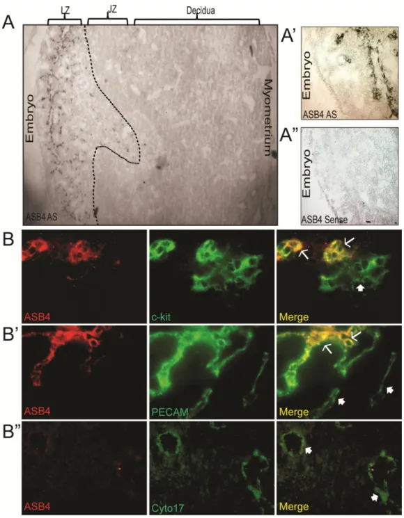
Asb4 is localized to areas of high vascular activity and is highly expressed in the developing placenta [19,87]. ID2 is also critical in the early development of the placenta, including the maturation of the placental vasculature [77]. Therefore, we hypothesized that ASB4 would be an important modulator of differentiation in the placental vasculature. As shown in Figure 2.1A (4x magnification) and 2.1A’ (20x magnification), Asb4 mRNA was only expressed in the labyrinth zone of E11.5 placentas. This zone exhibits high vascular activity [115] and contains the reservoir of TB cells that cross the junctional zone into the maternal decidua as they mature into functional endothelial-like cells [15,116]. This observation supports our hypothesis that ASB4 is involved in early vascular differentiation events in the placenta.
29
trophoblasts express c-kit, but terminally differentiated trophoblast giant cells and spongiotrohoplasts do not. Further, c-kit and its ligand SCF are implicated have been implicated as being required for trophoblast spreading and implantation, due to their role in trophoblast differentiation [119]. In addition, ASB4 also co-localized with a subset of PECAM-positive cells (Figure 2.1B’), suggesting that ASB4 is involved with cells that are differentiating into vascular lineages at this time point. Further supporting a specific role in early vascularization, ASB4 did not co-localize with cytokeratin 17, which is a marker of mature placental endothelial-like cells (Figure 2.1B”).
Because ASB4 co-localized with early markers of the endothelium, we hypothesized that Asb4 deletion would lead to functional consequences later in development. Specifically, if ASB4 promoted TB-to-endothelial cell differentiation, then placentas of Asb4-/- mice should have less mature endothelium than those of wild-type mice. Placentas from wild-type and Asb4-/- mice were examined at E17.5 and labeled for cytokeratin 17 (Cyto17) to visualize differentiated, mature endothelial-like cells. In wild-type mice, there abundant Cyto17
30
failure of the placenta to undergo integrin switching, indicating an immature and
undifferentiated placenta. Placental disc invasion, during which the fetal components of the placenta extend and expand into the maternal decidual layers, was more shallow in Asb4 -/-placentas compared with wild-type mice (Figure 2.2C) further confirming that placental development is compromised in Asb4-/- mice in a manner that is consistent with abnormal differentiation [108]. Additional observations indicate that the vasculature expressed markers of injury and dysfunction in near-term (E17.5) Asb4-/-placentas (Figure S2.2A) and were mislocalized within the junctional zone rather than the stage-appropriate outer deciduas (Figures S2.2B and S2.2C). These results indicate that differentiation defects in Asb4 -/-placentas may have deleterious effects that are observed into late gestation.
Because Asb4-/-placentas showed signs of early differentiation defects and impaired vascularization (Figures 2.2A, B), we examined ID2 expression due to its anti-differentiation role in the placenta [79]. We hypothesized that ID2 expression would be increased in the Asb4-/- placenta due to ID2’s anti-differentiation role in the placenta, and thus might underlie the observed differentiation defects in Asb4-/-placentas. In wild-type mice, ID2 expression is downregulated as TB cells differentiate. However, in whole placental cell lysates at E12.5, placentas from Asb4-/- mice have a ~2 fold increase in ID2 expression compared with placentas from wild-type mice (Figure 2.3A), and this finding is confirmed by
31
ASB4 negatively regulates ID2 expression through polyubiquitination and proteasome dependant degradation
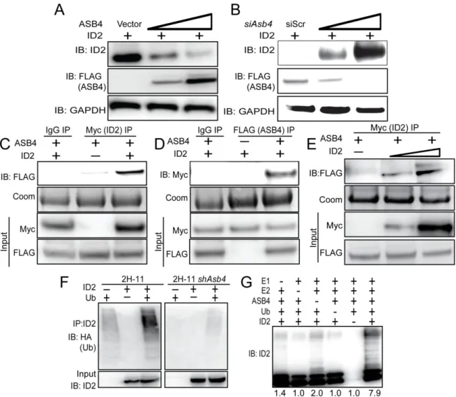
Given our observation that ID2 was significantly upregulated in Asb4-/-placentas, we hypothesized that ID2 may be a substrate of ASB4s ubiquitin ligase activity. To test this hypothesis, wild-type Id2 was co-transfected with Asb4 in JAR cells, and ID2 expression was examined. JAR cells are a desirable cell type because they do not express endogenous ID2 or ASB4, allowing us to modulate both proteins without endogenous protein interference. Because ID2 is rapidly turned over by myriad other proteins, resulting in a very short half-life (Figure S2.4B and [67]), we added a low dose of MG-132 to experiments in Figure 2.4A-E to slow proteasomal degradation events and visualize ID2 protein expression. ASB4 degraded ID2 in a dose-dependent manner in Asb4 and Id2 co-transfected JAR cells (Figure 2.4A). To test whether ID2 expression increased in the absence of ASB4, we transfected Id2 into 2H-11 endothelial cells that stably overexpressed Asb4. ID2 expression increased when co-transfected with increasing amounts of an siRNA duplex targeting Asb4 (siAsb4) (Figure 2.4B). To determine whether ASB4 binds directly to ID2, we performed
co-immunoprecipitation assays using co-transfected 3x-FLAG-tagged Asb4 and 6x-myc-tagged Id2 in JAR cells. As shown in Figure 2.4C, ASB4 was detected when ID2 was
32
Because ASB4 is an E3 ligase, we hypothesized that ASB4 regulates ID2 protein levels by polyubiquitinating ID2 and targeting it for proteasomal degradation. To test this hypothesis, we first co-transfected HA-tagged ubiquitin and myc-tagged Id2 in 2H-11 cells that either express endogenous Asb4 or have constitutively knocked down Asb4 expression to levels undetectable at either the transcript or protein level. We immunoprecipitated ID2 and blotted for HA, expecting a ubiquitin “smear” if ID2 was modified by polyubiquitination. As shown in Figure 2.4F, ID2 ubiquitination increased dramatically in cells that expressed Asb4, compared to cells that do not express Asb4. We then tested whether ASB4 could directly ubiquitinate ID2 by performing an in vitro ubiquitination assay. We combined recombinant ID2 with ASB4 and the minimal components required for ubiquitination and saw that ASB4 ubiquitinated ID2 four-fold more than the reaction absent of ASB4 (Figure 2.4G, lane 6 versus lane 3). Importantly, these reactions were performed in the absence of Roc1/Rbx1, the RING-finger protein that associates with ASB4, indicating that ASB4 does not require a RING-finger protein for ubiquitination. Previous reports into the mechanism of ID2 ubiquitination have demonstrated that ID2 is only susceptible to N-terminal ubiquitination [67]. To determine whether ID2’s N-terminus is sensitive to ASB4-mediated degradation, we co-expressed ASB4 with ID2 mutants that lack all lysine residues (LL-ID2) or have 6x-myc tags on either the N-terminus (degradation resistant, DR-ID2) or C-terminus (degradation sensitive, DS-ID2) in JAR cells. Only the N-terminally tagged ID2 (DR-ID2) expression level remained unchanged in the presence of ASB4 (Figure S2.3), indicating that ASB4 mediates ID2 degradation via N-terminal ubiquitination.
33
ectopically expressing ID2 and either ASB4 or vector control and compared ID2 expression to cells that were not treated with MG-132. In cells treated with DMSO, ASB4 expression led to reduced expression of ID2 as in Figure 2.4A. While a high dose of MG-132 increased total ID2 expression in the absence of ASB4, this increase was not diminished by the co-expression of ASB4 in the presence of MG-132 indicating that ID2 is degraded via the proteasome (Figure S2.4A). Further, when cells ectopically expressing ASB4 and ID2 were treated with cycloheximide to block protein translation, the half-life of ID2 decreased in the presence of ASB4 (Figure S2.4B). To ensure that the reduction in ID2 expression in the soluble fraction assayed above was not caused by ASB4 inducing ID2 translocation to an insoluble part of the cell, we performed a cell fractionation assay (Figure S2.4C). There was no observable accumulation of ID2 in any of the cell fractions when co-expressed with ASB4, suggesting that ID2 is not translocated to other insoluble fractions of the cell upon treatment with ASB4.
ASB4 mediates placental cell differentiation and function in vitro
In culture, TB cells can induce endothelial turnover [113] and increase the stability of endothelial cell networks [114], recapitulating the in vivo events that occur when TB cells are differentiating into endothelial-like cells. Because Asb4-/- placentas express markers of
34
cells that were only transfected with ASB4. Of note, DR-ID2 and ASB4 co-expression elevated apoptosis compared with vector control cells, but this was significantly less than cells that did express ASB4 alone (Figure 2.5A, B). These data demonstrate that ASB4 promotes a functional vascular phenotype that recapitulates in vivo endothelial replacement with differentiating TB cells and that ID2 represses this effect.
Previous reports demonstrated that endothelial cells induce TB migration in culture and that TB cells stabilize these endothelial vascular networks [114], representing a model of the in vivo events that occur during TB differentiation [15]. To examine whether ASB4 could promote TB cell stabilization of endothelial cell networks, we measured the ability of JAR cells transfected with Asb4 and Id2 to form stable vascular networks over time, using
35
ASB4 mediates these effects by degrading ID2, since DR-ID2 attenuates this ASB4-mediated effect in placental cells.
Because ASB4 mediates vascular differentiation in ES cells [19] and we have demonstrated that ASB4 negatively regulated the anti-differentiation protein ID2 (Figure 2.4A, B), we hypothesized that ASB4 would mediate placental cell differentiation through the regulation of ID2 and tested this hypothesis in vitro. First, TBSCs were isolated from the extraembryonic ectoderm of early post-implantation (E7.5) wild-type and Asb4-/- embryos and cultured on a feeder layer of mitotically inactivated MEFs, which promote the long-term maintenance and proliferation of undifferentiated stem cells [19]. Large-scale multipotent differentiation is expected for the first several passages, so cultures were grown 6-8 weeks prior to serum-withdrawal. Terminally differentiated cells were sub-cultured out, leaving only the undifferentiated embryoid bodies of TBSCs. Although the factors required for TBSC-to-endothelial transformation are not yet know, TBSCs readily differentiate into trophoblast giant cells (TGCs) [111]. Based on previous work from this laboratory [123], we used serum withdrawal to promote TBSC differentiation. Thus, we used the appearance of TGCs as an index of TBSC differentiation. After serum withdrawal for 72 hours, we visualized the isolated cells with bright-field microscopy. As shown in Figure 2.6A, wild-type TBSCs largely differentiated into large, multinucleated TGCs, which were
36
To determine whether ASB4’s influence on TB cell differentiation involves ID2, we examined human chorionic gonadotropin (hCG) secretion, a well-established marker of trophoblast differentiation [112], in JAR cells that ectopically express ASB4 and ID2. hCG secretion was stimulated via the addition of dbcAMP to the growth medium following the indicated transfection for 48 hours and was subsequently measured in the medium by ELISA. ASB4 stimulated hCG production approximately 2 fold compared with the vector control. Co-transfecting wild-type Id2 with ASB4 did not abolish hCG production, but
co-transfection of Asb4 and DR-Id2 prevented hCG stimulation (Figure 2.6B). Together with data in Figure 2.5, these results illustrate that ASB4 promotes placental cell differentiation and function in vitro, and that ID2 mutants resistant to ASB4-mediated degradation can inhibit the differentiation and function of TB cells in vitro.
Asb4-/- mice phenocopy human patients with pre-eclampsia
Because our data indicate that ASB4 mediates placental cell differentiation and function (Figures 2.4 and 2.5), and that Asb4 deletion has negative consequences in the placental vasculature throughout development (Figure 2.2 and Figure S2.2), we investigated whether the placental abnormalities found in Asb4-/- mouse placentas contributed to the placenta-specific disease pre-eclampsia, whose pathogenesis may stem from abnormal placental vascular development [7]. Asb4-/- female mice produced significantly smaller litter sizes compared with wild-type female mice (Figure 2.7B) due to spontaneous abortion mid-gestation (Figure 2.7A). Similarly, Asb4+/- breeding pairs produced non-Mendelian ratios of pups that were significantly skewed toward higher numbers of wild-type animals at the expense of Asb4-/- pups (Figure 2.7C). When investigating the source of lethality in the Asb4
-
37
subset of Asb4-/- embryos. These embryos lacked functioning placental vascularization (Figure 2.7A, and data not shown), which may contribute to the abortion and fetal reabsorption seen in Asb4-/- embryos [5].
Because ID2 expression is elevated in trophoblast cells placentas of women with pre-eclampsia [79] and Asb4-/- mouse placentas (Figure 2.2D, E), combined with the vascular defects observed in Asb4-/- placentas (Figure 2.2A, Figure S2.2), we investigated whether our Asb4-/- mice shared traits with human patients with pre-eclampsia, which is widely believed to be a disease of the placental vasculature [101]. Two hallmarks of pre-eclampsia are maternal hypertension and proteinuria during late-stage pregnancy. Pregnant Asb4-/- female mice had increased blood pressure during late gestation (E14-term), as compared to both gestationally age-matched wild-type mice and Asb4-/-mice during the first week of gestation (Figure 2.7D). Further, pregnant Asb4-/-female mice had higher ratios of albumin:creatinine protein in their urine during late stage pregnancy than wild-type mice (Figure 2.7E).
Together, these results suggest that Asb4-/-mice phenocopy human pre-eclampsia and may serve as a model for both early placental vascularization and human placental disease.
Discussion
Strict control over the vascular patterning of the placenta is critical for both maternal and fetal survival [124]. Aberrant differentiation events early in development negatively affect the later formation of the vasculature [15], but relatively little is known what drives early differentiation events. Although none of the limited data that identify putative substrates or functions of ASB4 support a central function for ASB4 in vivo
38
we utilized Asb4-/-mice, in conjunction with placenta-derived cells, to determine the function of ASB4 during placental vascular differentiation. Consistent with our previous work [19], we found that ASB4 is largely localized to the early endothelium in the placenta. We also found that Asb4 deletion induces the expression of markers of undifferentiation in the placenta, including the anti-differentiation protein ID2. Based on this data, along with the expression pattern of various markers of TB cells and endothelial differentiation in Asb4 -/-placentas, we determined that ASB4 is involved in the earlier stages of differentiation events, and the consequences of Asb4 deletion persist into later stages of gestation resulting in insufficient placental vascularization.
39
Placental remodeling requires three unique vascular events for proper function: TBSC differentiation, replacement of endothelium with trophoblast cells, and vascular stabilization to form high capacity vessels [15]. Possibly because the JAR cell line was isolated from CTB cells in choriocarcinomas, these cells can be induced to mimic in vivo cells under certain conditions. We adapted several methods to assess cell differentiation and function in culture, and whether ASB4 promoted these events through the inhibition of ID2. Although these methods do not completely recapitulate in vivo events, they collectively indicate that ASB4 has a pro-vascular differentiation function in placental cells. By ectopically expressing ASB4 in the JAR cells, we were able to determine that ASB4 promotes all three aspects of placental vascular remodeling. In addition, using isolated TBSCs, we were able to observe primary TB cell differentiation in culture. TBSCs that lack Asb4 remained in undifferentiated embryoid bodies, in contrast to wild-type TBSCs which differentiated into TGCs upon serum
withdrawal. Because exact markers of TBSC and endothelial differentiation are not well defined in the placenta, future studies will be needed to more precisely address these differentiation events.
40
of the placenta. Both third trimester hypertension and proteinuria, hallmarks of
41
Figure 2.1. ASB4 is expressed in the developing placental vasculature.
A) Asb4 mRNA is expressed only in the labyrinth zone of developing placentas. In situ hybridization was performed on E11.5 placental sections and imaged with bright field microscopy. Wide-field (4x, A) and higher magnification (20x, A’) anti-sense (AS)-probed sections illustrate Asb4 localized to the labyrinth zone. A sense probe was used as a negative control (A”).
42
43
Figure 2.2. Asb4-/-placentas express markers of undifferentiated vasculature and TB cells.
A) Placentas lacking Asb4 have reduced cytokeratin 17 expression in near-term placentas. E17.5 placental sections from wild-type and Asb4-/- mice were labeled with cytokeratin 17 (cyto17), a marker of terminally differentiated endothelial-like TB cells. Blood vessels (BVs) in Asb4-/- placentas display reduced cytokeratin 17 labeling compared with BVs in wild-type placentas.
B) Placentas from E15.5 wild-type and Asb4-/- placentas were labeled for integrin alpha V, a marker of mature, terminally differentiated TB cells and integrin beta 4, a marker of
immature, undifferentiated TB cells. Wild-type placentas express alpha V but not beta 4 integins. Cells in Asb4-/-placentas retain integrin beta 4 expression and fail to express integrin alpha V.
C) Placental disc invasion is reduced in Asb4-/- mothers at E17.5, indicating restricted
44
Figure 2.3. ID2 expression increases in placentas that lack Asb4.
A) Lysates from three E13.5 wild-type and Asb4-/- placentas were immunoblotted against ID2 (top panel, asterisk denotes nonspecific band) and quantified (bottom panel). p<0.01. JAR-WCL= whole cell lysates transfected with Id2 or vector and run as a positive immunoblotting control.
45
Figure 2.4. ASB4 negatively regulates ID2 expression through polyubiquitination and associates with ID2 in JAR cells.
A) ASB4 represses ID2 expression in a dose-dependent manner. Wild-type Id2 and vector, 0.5, or 2 µg of Asb4 were co-transfected in JAR cells. ID2 expression decreases as the ASB4 expression increases.
B) ID2 expression increases as ASB4 expression decreases. 2H-11 cells that constitutively express high levels of ectopic ASB4 were transfected with Id2 and either a scrambled
nucleotide siRNA duplex (siScr) or increasing doses (0.15 nM, 0.5 nM) of siAsb4 duplex. As ASB4 expression decreases, ID2 expression concurrently increases.
46
E) 2H-11 cells that stably express FLAG-tagged Asb4 were transfected with increasing amounts of myc-tagged Id2. Cells were lysed and pre-cleared as in C and D, then
immunoprecipitated with anti-myc conjugated agarose beads and then blotted for FLAG. FLAG expression increases in parallel with myc expression, indicating specific interaction between ID2 and ASB4.
F) ID2 ubiquitination increases in cells with ASB4 expression. Wild-type Id2 and HA-tagged ubiquitin were transfected into either 2H-11 cells that express endogenous Asb4 or 2H-11 cells that have Asb4 constitutively knocked down. ID2 was immunoprecipitated using anti-ID2 and then blotted against HA. Reactions were blotted on the same membrane. Input represents 2.5% of total lysate. Ubiquitination of ID2 increases in endothelial cells that express ASB4 compared with cells that do not.
G) ASB4 directly ubiquitinates ID2 in vitro. Recombinant ID2 was incubated with
47
Figure 2.5. ASB4 promotes JAR cell-mediated endothelial apoptosis and stabilization of endothelial cell networks.
A) JAR cells expressing ASB4 promote 2H-11 cell apoptosis. JAR cells were transfected with vector, Asb4, Asb4 and wild-type Id2, or Asb4 and DR-Id2 prior to being seeded on top of 2H-11 monolayers.
B) TUNEL-positive cells were counted and are presented as the percent of total endothelial cells within the field. Asb4-transfected cells increase apoptosis of the underlying endothelial cells, even when transfected with wild-type Id2. DR-Id2 co-transfected with Asb4 inhibits JAR-mediated 2H-11 apoptosis. * p<0.01 as compared to vector/vector. † p<0.01 compared to Asb4-only transfection.
C) JAR cells transfected with Asb4 promote endothelial tube stability. 2H-11 cells were placed on Matrigel and allowed to form tube-like networks. JAR cells transfected as in A were then plated on the networks, and total network area was measured at the times indicated.
D) JAR cells expressing DR-ID2 destabilize 2H-11 cell networks at 16 hours, while cells expressing ASB4 or ASB4 and wild-type ID2 maintained the size of these 2H-11 cell
48
Figure 2.6. ASB4 promotes TB cell differentiation in vitro.
A) TB stem cells (TBSCs) were isolated from wild-type and Asb4-/- extraembryonic ectoderm at E7.5. Cells isolated from each conceptus were cultured in isolation, and these data represent 4 unique populations of cells for each genotype. Serum withdrawal induces the formation of large, multinucleated trophoblast giant cells (TGCs, arrows) that differentiate from TBSCs (asterisks). As shown, wild-type TBSCs largely differentiate into TGCs (left panel) while Asb4-/- cells remain in undifferentiated embryoid bodies (right panel). MEF-feeder cells are indicated by filled arrows. Dashed outlines indicate the border of non-MEF cell clusters.
49
Figure 2.7. Pregnant Asb4-/- mice display symptoms of pre-eclampsia.
A) A subset of Asb4-/- embryos dies in utero. Asb4-/- littermates are shown at E12.5,
illustrating the lack of placental vasculature and dramatically reduced fetal growth in a subset of Asb4-/-embryos.
B) Quantification of the average litter size of wildtype mice compared to Asb4-/- mice, taken from more than 25 litters from each group.
C) Heterozygous breeding results in a lower than expected number of Asb4-/- pups (p < 0.01, Fisher’s exact test).
D) Pregnant Asb4-/- mice have significantly elevated mean blood pressure in the third trimester of pregnancy compared with both Asb4-/-mice in the first week of pregnancy and wild-type mice in the third week of pregnancy. * p<0.01.
50
Figure S2.1. Diminished mature vasculature in Asb4-/- placentas is not due to increased apoptosis or abnormal proliferation.
E15.5 placental sections from wild-type and Asb4-/- mice were evaluated for aberrant
51
Figure S2.2. Asb4 deletion induces vascular dysfunction and mislocalization of blood vessels in the placenta.
A) Near-term (E17.5) placental sections were harvested and labeled with von Willibrand factor to measure thrombus response and DBA to determine uterine natural killer cell response. Asb4-/- placentas display elevated thrombus/thrombosis response (left panel) compared with wild-type placentas, indicating damaged vasculature. Further, there is a dramatic increase in activated uterine natural killer cells (right panel) in Asb4-/- tissues, indicating elevated macrophage and immune response, compared to wild-type tissue.
B) E17.5 placental sections were stained with hematoxylin and eosin and examined for gross morphology. Blood vessels (arrows) were counted and classified based on their location in the labyrinth (LZ), junctional (JZ), or decidual (DE) zones. Blood vessels in wild-type placentas are seen at the edge of the deciduas in, whereas significantly more vessels in Asb4 -/-placentas are located in the junctional zone, at the expense of the decidual zone.
52
53
Figure S2.4. ASB4 degrades ID2 in a proteasome-dependant manner, and does not affect ID2 cellular location.
A) JAR cells co-transfected with Id2 and either vector or wild-type Asb4 were treated with DMSO or MG-132. While overall ID2 expression increases in the presence of MG-132, ID2 expression decreases only in the presence of ASB4 in DMSO-treated cells, suggesting that ID2 is sensitive to proteasomal degradation when co-expressed with ASB4.
54
55 CHAPTER 3
GENERAL DISCUSSION
Placental development is a critical, but poorly understood process. Previous work indicated that ASB4 mediated early vascular differentiation, and was localized to the placenta. Thus I sought to fully characterize the role of ASB4 in placental vascularization and trophoblast differentiation.
The experiments described herein attempt to fully characterize the role of ASB4 during placental vascular development by uncovering its expression, molecular function, biologic consequences, and molecular regulation. Data generated here reveal that 1) Asb4 is expressed in pluripotent trophoblast cells committed to the vasculature during a narrow time window during differentiation; 2) ASB4, as a ubiquitination complex, degrades the HLH protein ID2; and promotes differentiation of the vascular lineage through, in part, its
degradation of ID2; and 4) deletion of Asb4 in mice induces a placental vascular phenotype and recapitulates pre-eclampsia in Asb4-/- mothers (Chapter 2).
The role of ASB4 in vascular commitment and patterning
56
Earlier work from our laboratory on ASB4 function [19,36] led us to examine vascular development in the placenta. Ferguson, et al [19] described how Asb4 expression was restricted to a narrow spatiotemporal window in which the placenta was developing. This study also elucidated that ASB4 functioned as an oxygen sensor, further implicating its function in the placenta, where hemodynamic stresses and hypoxia/normoxia events are required for proper vascularization. Likewise, Bode, et al. [36] described how laminar shear stress repressed Asb4 expression. We therefore examined the gross morphological and physiological consequences of globally deleting Asb4 expression in mice, which was generated in [87].
Our initial investigation of Asb4-/- mice established that there was embryonic lethality in a subset of pups that was occurring at or about E9.5 (Figure 2.7A). This spontaneous abortion appeared to occur in the absence of any gross fetal defect or pathology, but the placentas of said pups resigned to intrauterine demise were dramatically less vascular or developed (Figure 2.7A, lower conceptus) compared with littermates that appeared to be morphologically “normal” (Figure 2.7A, upper conceptus). To begin understanding this pathology, a histological examination was undertaken and revealed that ASB4 expression only coincided with cells that were either pluripotent (Figure 2.1B) or cells that had committed to the vascular lineage (Figure 2.1B’). Cells that had fully differentiated into endothelium (Figure 2.1B”) and cells of different lineages (spongiotrophoblasts, trophoblast giant cells, syncytiotrophoblasts, etc.) did not express Asb4.
Though there have been recent advances in understanding mouse placental
57
cell types through the use of protein expression markers in the mouse placenta. To identify pluripotent/stem cells in the context of the mouse placenta, we relied on the receptor c-Kit (also known as CD117 or mast/stem cell growth factor receptor [SCFR]). c-Kit is a receptor tyrosine kinase that binds to stem cell factor ligand and mediates proliferation,
differentiation, and serves as a marker of progenitor cells of mature endothelium [128]. However, c-kit expression is also found in trophoblasts that invade, migrate, and differentiate into the TGC lineage [118,119], indicating that it may also serve as a marker undifferentiated CTBs and TGCs, though this expression pattern has not been empirically determined.
Similarly, the use platelet endothelial cell adhesion molecule (PECAM) (also known as CD31) and cytokeratin 17 for the determination of endothelial committed and mature endothelial-like cells, respectively, was based on studies investigating their roles in human placental tissue [129,130]. While we are confident that there are few or no discrepant functions of these broad endothelial specific molecules between mice and humans, we must acknowledge that there is a dearth of data that definitively describe these protein markers in differentiating mouse trophoblasts.
These experiments illuminate the role of ASB4 in placental vascular determination and differentiation. While we are limited by the scope of markers of endothelial
58 ASB4 as an E3 ligase
Like all E3 ubiquitin ligase proteins, the functional ramifications of ASB4 are dependent upon the function of the substrate protein(s) that it degrades. Multiple attempts to identify binding partners of ASB4 have not yielded the identification of ASB4 ubiquitination substrate proteins. Through the use of yeast 2-hybrid, the Patterson laboratory identified FIH as an interaction partner of ASB4, though no putative substrate proteins were identified [19]. This result not only highlights the limitations of this assay, but also that of identifying substrate proteins in general. The use of co-immunoprecipitation has also been successfully utilized to identify co-factors of ASB4 [19], but protein turn-over, lack of specificity, and difficulty in separating signal-to-noise all confound any results indicating a putative ASB4 target (data not shown and [87]).
Therefore, we took a candidate approach in our investigation into ASB4 substrates. We knew that any substrate of ASB4 must be involved in early differentiation of the
vasculature, be expressed in CTBs in vivo, and have overexpression models that mimic Asb4 deletion. Literature searches and coinciding data (Figure 2.3B) indicated that members of the ID family would fit these criteria. Specifically, studies of the retinoblastoma (Rb) protein in mice indicate that Rb-deficient mice die embryonically due to abnormal trophoblast
proliferation and placental dysplasia [82]. This effect, however, is ameliorated by the
59
Because ID2 mediates differentiation, invasion, and migration in CTBs [79], we hypothesized that ASB4 normally regulates ID2 via its role as an E3 ligase. The role of ID2 in human placental differentiation has served as the crux for our further investigation in our Asb4-/- mice. Specifically, in cells driven to overexpress ID2, invasion was abrogated, migration was abnormally enhanced, and these cells retained characteristics of
undifferentiated cells. Also of note, the abnormal maintenance of ID2 expression throughout placental development was associated with placentas from pre-eclamptic patients. Taken together, this report specifies the role of ID2 in the context of placental differentiation and pre-eclampsia [79]. Other studies have also illuminated the importance of ID2 in the context of proper placental development. Specifically, ID2 mediates transcriptional activity in the placenta [80]; ID2 inhibits extravillous trophoblast-to-TGC differentiation [131]; and Id2 is expressed and involved in trophoblast stem cell differentiation [132-134]. In conjunction with the known expression profile of Asb4, the hypothesized function of ASB4, the
consequence of Asb4 deletion in mice, and our data demonstrating that ID2 and ASB4 have reciprocal expression patterns (Figure 2.3B), we hypothesized that ID2 may be one substrate of ASB4.