Cu- and Ni-Doping Effect on Structure and Magnetic
Properties of Fe-Doped ZnO Nanoparticles
Jefferson A. Wibowo, Nadia F. Djaja, Rosari Saleh*
Departemen Fisika, FMIPA-Universitas Indonesia, Depok, Indonesia Email: *[email protected], *[email protected]
Received January 7,2013; revised February 8, 2013; accepted February 18, 2013
ABSTRACT
Cu- and Ni-codoped FeZnO particles with the wurzite structure were successfully synthesized at low temperature by a co-precipitation method. The samples were characterized using a vibrating sample magnetometer, X-ray diffraction, energy dispersive X-ray spectroscopy, UV-Vis spectrophotometry and electron spin resonance. The results demonstra- ted that room temperature ferromagnetic order was observed in both samples and the magnetization was higher than that of Fe-doped ZnO. The correlation between the structural and magnetic properties is discussed.
Keywords: Codoped ZnO Nanoparticles; Room-Temperature-Ferromagnetic; Co-Precipitation
1. Introduction
Dilute magnetic semiconductors (DMSs) in which some of the cations host lattice are replaced by a transition metal ions have attracted considerable attention due to their potential as spin-polarized carrier sources and their potential applications in spintronic devices [1-5]. The main challenge for practical application of DMSs is the attainment of Curie temperature above room tempera- ture [6]. Following the theoretical prediction of room temperature ferromagnetic by Dietl et al. [7], several studies involving magnetic ions doped II-VI semicon- ductors were performed by different researcher in tran- sition metal doped ZnO. It is known that ZnO has high solubility for transition metals and superior semicon- ductor properties [8]. Moreover, ZnO is a wideband gap semiconductor with a relative large exciton binding en- ergy. Among transition metal, ZnO doped with Fe ions without any modification of the structure has been the most considerable interest. Ferromagnetism with Curie temperature higher than room temperature has been ob- served in Fe-doped [9-12], Co-doped [13-15], Mn-doped [16-18], Ni-doped [19-21], Cu-doped [22] and V-doped [23] ZnO nanoparticles. Meanwhile, several codoped ZnO have also been reported with the expectation that codoping can lead to remarkable changes in the proper- ties of the materials [24-26]. Presence of two different kind of transition ions simultaneously in a host material produces magnetic property that can be different from the magnetic property due to single transition metal ions. For instance, Han et al. [27] reported that the Curie tem-
perature of bulk Zn0.94Fe0.05Cu0.01O was above room
temperature and the maximum saturation of magneti- zation was larger than that of the sample without Cu [27,28]. Shim et al. [29] also prepared FeCu co-doped ZnO sample and reported that the room temperature fer- romagnetic in the sample is due to the secondary phase ZnFe2O4 [27,29].
Despite the considerable amount of data a great deal of controversy remains, especially regarding the fundamen- tal issue of whether the system actually exhibits room temperature ferromagnetic at all; and in the case where it does, whether the effect is intrinsic to the material. Fur- ther studies suggested that the inconsistencies in the lit- erature regarding the ferromagnetic ordering of transition metal doped ZnO indicate that these materials are very sensitive to the fabrication and processing conditions. Therefore, this paper we attempt to study the effect of Cu- and Ni co-doping on the weakest ferromagnetic Fe- doped ZnO (1 at% of Fe). The co-precipitation method was chosen for the synthesis of these materials because it is cost effective, requires low temperature processing and offers a higher degree of solubility. The effects of Cu and Ni doping on the structural, optical and magnetic proper- ties of nanocrystalline Fe-doped ZnO particles was in- vestigated using X-ray diffraction (XRD), energy disper- sive X-ray (EDX), UV-Vis spectroscopy (UV-Vis), elec- tron spin resonance (ESR) and vibrating sample magne- tometer (VSM). It was found that the incorporation of Cu and Ni in Fe-doped ZnO nanoparticles not only enhances ferromagnetic properties to the host materials but also changes lattice constant and the optical properties.
2. Experimental
For the synthesis of Cu- and Ni-doping of Fe-doped ZnO nanoparticles in this study the following starting materials were used without further purification: zinc (II) sulfate (ZnSO4·7H2O, 99%, Merck), iron (II) sulfate (FeSO4·7H2O,
99%, Merck), cooper (II) sulfate (CuSO4·5H2O, 99%,
Merck) and nickel (II) nitrate (Ni(NO3)2·6H2O, 99%
Merck). FeSO4·7H2O and CuSO4·5H2O, FeSO4·7H2O
and Ni(NO3)2·6H2O, were added simultaneously to the
ZnSO4·7H2O, solution under continuous stirring to get
homogeneous solutions. These mixtures (solution A) were placed in an ultrasonic cleaner operating at 57 kHz for 2 h. Simultaneously, 44 mmol NaOH solution was prepared in 440 ml of deionized water (solution B). Then solution B was added drop wise to solution A with constant stirring for 2 h until a pH of 13 was reached. The mixed solution was allowed to stand at room tem- perature for 18 h. Subsequently, the solution was cen- trifuged and washed several times with ethanol and distilled water to remove residual and unwanted im- purities. The final product was dried in a vacuum oven at 200˚C for 1 h to yield Fe/Cu and Fe/Ni-codoped ZnO powders.
Elemental analyses of the samples were carried out using scanning electron microscope (SEM) with EDX attachment. To evaluate the phase purity of the samples, XRD measurements were performed using a Philips PW 1710 and monochromatic Cu-Kα (λ = 1.54060 Å) ra- diation operated at 40 kV and 20 mA in the range of 10˚
to 80˚. The instrumental broadening including the ins- trumental symmetry was calibrated using a Si powder standard sample. The X-ray diffraction patterns were analyzed by means of the MAUD program using the Rietveld whole profile fitting method to determine the crystal structure and lattice parameters.
To study the electronic interaction near the optical band gap resulting from the addition of dopant atoms, diffuse reflectance UV-Vis measurements were per- formed using a Shimadzu UV-Vis spectrophotometer with an integrating sphere and a spectral reflectance standard in the wavelength range of 200 - 800 nm. The diffuse reflectance, R, of the sample is related to the Kubelka-Munkfunction, F(R), according to the follow- ing equation: F(R)= (1 − R)2/2R [30]. The energy band
gap of the samples was calculated from the diffuse reflectance spectra by plotting the F(R)2 as a function of
energy and extrapolating to F(R)2 = 0.
Magnetic measurements were performed on Oxford Type 1.2 T vibrating sample magnetometer (VSM). These measurements were taken from 0 to ±1 Tesla field. To obtain information on electronic structure ESR was carried out using X-band JEOL JES-RE1X at room temperature. The shape and area of the ESR spectra
were analyzed using standard numerical methods.
3. Results
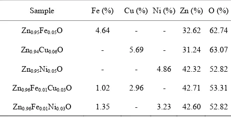
To confirm the presence of Fe, Cu and Ni ions in the synthesized nanocrystalline ZnO particles, EDX mea- surements were performed. Four different random areas in the sample were chosen and about the same Fe, Cu and Ni concentration was obtained for all of them. This result suggested that the distribution of doping is homo- geneously. The EDX data from concentrations of Fe, Cu and Ni are listed in Table 1. It is seen that the amounts of Fe, Cu and Ni incorporated in the samples are slightly lower than their nominal composition introduced in the synthesis.
The XRD patterns for Zn0.96Fe0.01Cu0.03O and Zn0.96
Fe0.01Ni0.03O samples are presented in Figure 1. Also
shown the XRD patterns of Zn0.95Fe0.05O [31], Zn0.94
Cu0.06O [32], Zn0.95Ni0.05O [33] and undoped ZnO [34]. It
has been observed that all of peaks of XRD pattern belong to the hexagonal lattice of ZnO with three most preferred orientations namely (100), (002) and (101). Most importantly, all of the XRD peaks were attributed to ZnO and no other undesired peaks were observed due to secondary phases or impurity phases within the de- tection limit of the X-ray diffractometer. From the 2Θ
values, the inter-planar spacing d of the peaks is cal- culated.
The values are listed in Table 2. A good agreement between the observed and the calculated d values is found to exist indicated a suitability of unit cell para- meters and the crystal structure.
[image:2.595.310.536.622.737.2]The lattice constants, calculated from Rietveld refine- ment using MAUD programs, unit cell volume, the values ratio (c/a) are summarized in Table 2. The results are compared with those of Fe-doped ZnO. The average crystallite size and strain were also obtained from Rietveld refinement of the X-ray diffraction patterns of the samples obtained by constructing Williamson-Hall plots [35] with different peaks for the same families. In the present study, (100), (002), (101), (102), (110), (103),
Table 1. EDX data of Fe-, Cu-, Ni-doped ZnO and Cu- and Ni-codoped FeZnO nanoparticle.
Sample Fe (%) Cu (%) Ni (%) Zn (%) O (%)
Zn0.95Fe0.05O 4.64 - - 32.62 62.74
Zn0.94Cu0.06O - 5.69 - 31.24 63.07
Zn0.95Ni0.05O - - 4.86 42.32 52.82
Zn0.96Fe0.01Cu0.03O 1.02 2.96 - 42.71 53.31
Figure 1. XRD patterns of ZnO, Zn0.95Fe0.05O, Zn0.94 Cu0.06O,
Zn0.95Ni0.05O, Zn0.96Fe0.01Cu0.03O and Zn0.96Fe0.01 Ni0.03O na-
noparticles.
and (112) peaks were used to construct the Williamson- Hall plot. From the linear fit to the data, the average crystallite size, <D>, was extracted from the y-intercept and the strain, ε, from the slope of the fit of:
cos K D 2 sin
β θ = λ + ε θ
In this calculation the strain was assumed to be uniform in all directions of the samples. The average crystallite size, <D> and the strain, ε, are shown in Table 2. These results indicate that the <D> in the Zn0.96Fe0.01
Cu0.03O and Zn0.96Fe0.01Ni0.03O samples have a similar
average crystallite size with Zn0.97Fe0.03O. These data
showed that the substitutional doping does not influence the crystal structure significantly.
To study the electronic interactions near the optical band gap region of Zn0.96Fe0.01Cu0.03O an Zn0.96Fe0.01
Ni0.03O samples diffuse-reflectance measurements were
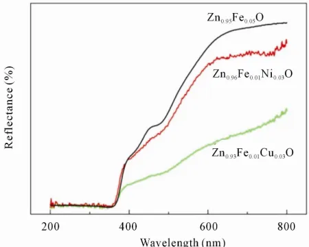
performed on the samples in the UV-Vis region at room temperature. All spectra were obtained in the range of 200 - 800 nm. Figure 2 shows the diffuse-reflectance spectra, R, as a function of wavelength. The band gap energy of the doped ZnO samples was calculated from the diffuse-reflectance spectra by plotting the square of the Kubelka-Munk function F(R)2 vs. the energy in elec-
tron volts. The linear part of the curve was extrapolated to F(R)2 = 0 to calculate the direct band gap energy. The
Table 2 also shows the band of Zn0.96Fe0.01Cu0.03O and
Zn0.96Fe0.01Ni0.03O samples. It is seen that the absorption
edge is slightly different with the addition of Cu and Ni in Fe-doped ZnO sample compare to Fe-doped ZnO sample itself.
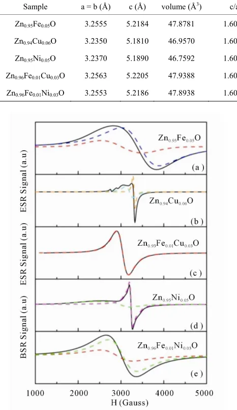
[image:3.595.311.536.86.266.2]To gain insight into the oxidation state of the dopant cations involved in the spin coupling and site occupancy of the dopant ion in the host material, ESR spectra were
Figure 2. The diffuse-reflectance spectra of Zn0.96Fe0.01
Cu0.03O and Zn0.96Fe0.01Ni0.03O compared with Zn0.95Fe0.05O
nanoparticles.
measured at room temperature. Typical ESR spectra of both the Zn0.96Fe0.01Cu0.03O and Zn0.96Fe0.01Ni0.03O par-
ticles are provided in Figure 3. For interpretation of Zn0.96Fe0.01Cu0.03O and Zn0.96Fe0.01Ni0.03Oresults a com-
parison with Fe-, Ni- and Cu-doped ZnO ESR spectra was also instructive. In Zn0.95Fe0.05O the ESR signal can
be considered as a superposition of two overlapping signals, a broad and intense signal attributed to Fe2+ and
another weak and narrow signal assigned to Fe3+ [31,33].
In the case of Zn0.95Ni0.05O, the ESR spectra had similar
features, which exhibited two overlapping resonance peaks. One peak corresponded to the broad resonance while the other peak located at higher magnetic field was much narrower. The linewidth and the g-values of the broad signal in our Zn0.95Ni0.05O was consistent with the
line shape and position of the previously reported Ni-doped ZnO samples [33,36] and have been attributed to a ferromagnetic resonance due to Ni2+ ions. A com-
parison of the g-values of the narrow ESR signal with the ESR signals of Ni in Li1−XNi1+XO2 [37,38], SnO2 [39]
and TiO2 [40], which have g-values in the range of 2.13 -
2.18, suggests that the narrow resonance in our Zn0.95
Ni0.05O samples is attributable to paramagnetic Ni3+ ion
centers.
The electronic configuration of Cu2+ ion is 3d9 and the
electronic ground state is 3S
1/2. The only natural isotope
is 63Cu, which has nuclear spin 3/2. The ESR spectrum
of Zn0.94Cu0.06O sample shown in Figure 3 revealed the
presence of broad signal, which is superimposed on poor-resolved quadruplet signals and a pronounce narrow resonance. The broad signal at g value of 2.05 is associated with Cu2+ interacting with nearby Cu2+ via
Table 2. The The lattice constants, unit cell volume, ratio of lattice parameters, interplanar spacing, average crystallite size, strain and band gap energy of Zn0.95Fe0.05O, Zn0.94Cu0.06O, Zn0.95Ni0.05O, Zn0.96Fe0.01Cu0.03O, andZn0.96Fe0.01Ni0.03O nanopar-
ticle.
Sample a = b (Å) c (Å) volume (Å3) c/a d
101 (Å) <D> nm ε
(
×10−4)
Eg (eV)Zn0.95Fe0.05O 3.2555 5.2184 47.8781 1.6029 2.4805 21 21.2 3.29
Zn0.94Cu0.06O 3.2350 5.1810 46.9570 1.6015 2.4644 18 47.0 3.32
Zn0.95Ni0.05O 3.2370 5.1890 46.7592 1.6030 2.4664 27 33.9 3.28
Zn0.96Fe0.01Cu0.03O 3.2563 5.2205 47.9388 1.6032 2.4812 19 9.16 3.33
Zn0.96Fe0.01Ni0.03O 3.2553 5.2186 47.8938 1.6031 2.4804 18 13.70 3.36
found that the line-width and the line intensity can be deconvoluted in Fe2+ and Ni2+ signals. Although, it was
reported that the g-value of Ni metallic species is centered at 2.2 [53], the presence of Ni metallic might be ruled out since the line width of this species is much broader than that of our ESR spectra of Zn0.96Fe0.01
Ni0.03O. The g-values, total number of spins associated
with each signals and the line width are quite variable as shown in Table 3.
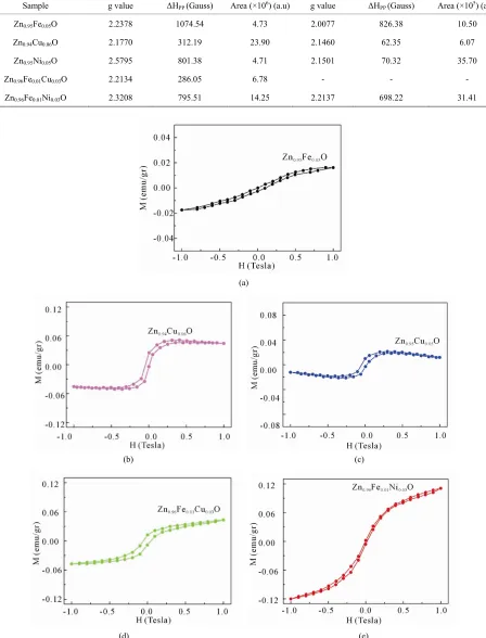
The room temperature ferromagnetic behavior of both the Zn0.96Fe0.01Cu0.03O and Zn0.96Fe0.01Ni0.03O particles in
the magnetic field range from 0 to ±1 T using VSM measurements have been shown in Figure 4. The mag- netization is plotted as a function of magnetic field for different dopant ions incorporated in Fe-doped ZnO par- ticles. The diamagnetic contribution from the sample holder has already been subtracted to estimate the actual ferromagnetic contribution of each sample. Also shown in Figure 4 the magnetization of Zn0.95Fe0.05O, Zn0.94
Cu0.06O and Zn0.95Ni0.05O. The comparative M(H) loops
showed that, the Zn0.96Fe0.01Ni0.03O exhibits higher mag-
netization than that of Zn0.97Fe0.03O as well as Zn0.95
Ni0.05O. The same result was also observed in Zn0.96Fe0.01
Cu0.03O. In the case of Zn0.96Fe0.01Cu0.03O sample a coer-
cive field (HC) and the remnant magnetization (MR) are
found to be 554 Oe and 0.012 emu/g, while for Zn0.96Fe0.01Ni0.03O smaller values are observed, namely
[image:4.595.57.290.127.530.2]120 Oe and 0.004 emu/g. However, the saturation mag- netization for Ni incorporation in Fe-doped ZnO is clearly higher than that of Cu co-doping.
Figure 3. The electron spin resonance spectra of Zn0.96Fe0.01
Cu0.03O and Zn0.96Fe0.01Ni0.03O compared with Zn0.95Fe0.05O,
Zn0.94Cu0.06O, and Zn0.95Ni0.05O nanoparticles.
ped on an oxygen vacancy site [42-52].
Comparing the ESR spectra of Zn0.95Fe0.05O and Zn0.94
Cu0.06O with that of Zn0.96Fe0.01Cu0.03O, the line width
and the g-value of Zn0.96Fe0.01Cu0.03O can be attributed to
Fe2+, since the g-value observed here does not agree with
the reported value for Cu2+. In addition, hyperfine
structure due to 63Cu and 63Cu nuclei necessary for iden-
tification of Cu-related center was not observed. So the peak observed here would not be attributed to the Cu ions themselves.
The mechanism responsible for the observed ferro-magnetism at room temperature in transition metal- doped ZnO is also not clear and has been debated over the years. Several explanations are discussed below. Nevertheless a few researchers have claimed to observe ferromagnetic behavior arising only from a secondary phase and not from the material itself. The results of the XRD and EDX measurements in our samples demon- strate that the dopant ion was incorporated into the wurtzite lattice at Zn sites forming a solid solution in- stead of precipitates. However, a secondary phase might It is apparent from Figure 3 that the two resonances of
Zn0.96Fe0.01Ni0.03O are too close to be separated with
Table 3. The g value, linewidth (ΔHpp), and peak area of Zn0.96Fe0.01Cu0.03O and Zn0.96Fe0.01Ni0.03O compared with Zn0.95Fe0.05
O, Zn0.94Cu0.06O, and Zn0.95Ni0.05O nanoparticle.
Sample g value ΔHPP (Gauss) Area (×106) (a.u) g value ΔHPP (Gauss) Area (×105) (a.u)
Zn0.95Fe0.05O 2.2378 1074.54 4.73 2.0077 826.38 10.50
Zn0.94Cu0.06O 2.1770 312.19 23.90 2.1460 62.35 6.07
Zn0.95Ni0.05O 2.5795 801.38 4.71 2.1501 70.32 35.70
Zn0.96Fe0.01Cu0.03O 2.2134 286.05 6.78 - - -
Zn0.96Fe0.01Ni0.03O 2.3208 795.51 14.25 2.2137 698.22 31.41
(a)
(b) (c)
[image:5.595.73.521.117.705.2](d) (e)
Figure 4. The room temperature ferromagnetic of Zn0.96Fe0.01Cu0.03O and Zn0.96Fe0.01Ni0.03O compared with Zn0.95Fe0.05O,
n0.94Cu0.06O, and Zn0.95Ni0.05O nanoparticles.
Z
exist in the sample even though it was not detected in our states, with the concentration of trivalent state increased XRD spectra. Thus, it is useful to consider all possible
ferromagnetic impurity phases that might be present in both samples. It is known that Cu-related oxides such as CuO, Cu2O or Cu clustering could not contribute to the
room temperature ferromagnetism, because none of them exhibit ferromagnetism above room temperature [54-56]. Therefore the ferromagnetism behavior observed in our Cu-doped samples studied here does not seem to be re- lated with the presence of any secondary phases or Cu clusters, while Cu clusters and its oxides are generally considered to be non-ferromagnetic and could not contri- bute to the room temperature ferromagnetic. In the case of Fe-doped samples nearly all possible Fe-based oxides, such as FeO and Fe2O3 are antiferromagnetic with TN
values of 198 and 963 K, respectively [57,58]. The ex- ception to this is Fe3O4, which is ferromagnetic with a Tc
of approximately 858 K [59]. Another secondary phase that can be found in Fe-doped ZnO samples is ZnFe2O4.
However, this phase is antiferromagnetic and can be ex- cluded as the origin of room temperature ferromagnetic in our samples. In the case of Ni co-doping, the forma- tion of secondary phase such as NiO is a unlikely source of ferromagnetism as NiO is antiferromagnetic in nature with TN values of 523 K [60] and 5 K [61] for bulk- and
nanocrystalline NiO, respectively. Accordingly, the fer- romagnetism behavior observed in our Zn0.96Fe0.01Cu0.03
O and Zn0.96Fe0.01Ni0.03O particles studied here does not
seem to be related with the presence of any secondary phases.
There is also an emerging consensus that ferromag- netic behavior in transition metal-doped ZnO is corre- lated with defects such as oxygen or zinc vacancies [62-64]. Karmakar et al. [11] investigated the origin of ferromagnetism in Fe-doped ZnO using local probe mea- surements such as ESR and Mössbauer spectroscopy. The results revealed that the Fe ions are present in both Fe2+ and Fe3+ valence states. The presence of uncoupled
Fe3+ ions is possibly due to hole doping in the system,
which was caused by cation (i.e., Zn) vacancies. By comparing the ESR measurements from our sample with the results obtained from Karmakar et al. [11] it is con- firmed that the ferromagnetism observed in our Zn0.97
Fe0.03O sample was due to the presence of Fe atoms in
the form of Fe2+ ions and Fe3+ ions.
According to Karmakar et al. [11] a cation vacancy near Fe can promote Fe2+ into Fe3+ and also mediate the
Fe2+-Fe2+ exchange interaction. Moreover, since the tran-
sition metal ion is slightly higher side towards cationic percolation threshold, Fe2+-Fe3+ exchange interaction
may also possible. Viswanatha et al. [65] investigated the origin of ferromagnetism in FeCu-codoped ZnO experi- mentally as well as theoretically. Their results revealed that the Fe ions are present in both Fe2+ and Fe3+ valence
with increasing Cu doping and redox-like pairs Fe2+ +
Cu2+
⇔ Fe3+ + Cu1+ can be occurred to stabilize the
ferromagnetism in codoped system. They believed that the ferromagnetism of this system is ascribed to a dou- ble-exchange interaction between the Fe atoms mediated by the Cu atom. It is obvious from ESR spectra of Zn0.96Fe0.01Cu0.03O show the presence of Fe2+ ion and the
absence of Cu2+ ion. However, our EDX result shows the
presence of Cu atom in our Zn0.96Fe0.01Cu0.03O. These
results suggested that the oxidation state of Cu is +1, since Cu in the +1 state has no unpaired spin. Usually, a Cu ion will contribute to the net ferromagnetic moment only if it is in the +2 state. Interestingly, the Zn0.96Fe0.01
Cu0.03O sample shows an evidence of ferromagnetic or-
der. Therefore we believed that a small amount of Fe3+
and Cu2+ ions would be found in our sample to neutralize the charge imbalance, although both ions (Fe3+ and Cu2+) were not detected in our ESR spectra. Comparing the XRD, EDX, VSM and ESR results for Zn0.96Fe0.01Cu0.03O
with Zn0.95Fe0.05O, Zn0.94Cu0.06O and the results obtained
in the literature, we conclude that Fe2+, Cu1+, Fe3+ and
Cu2+ are presence in our Zn
0.96Fe0.01Cu0.03O sample and
have played the important role in obtaining the room temperature ferromagnetism.
In the case of Zn0.96Fe0.01Ni0.03O, the bent nature of the
curve exhibits a shallow ferromagnetism in our sample. The ferromagnetism could arise due to possible reason: 1) secondary phase or clustering of metallic or 2) the pres- ence of charge carriers, or 3) the formation of defect structures such as oxygen- and zinc vacancies. It is al- ready discussed above that the formation of secondary phase is unlikely. Moreover, from the ESR spectra of Zn0.96Fe0.01Ni0.03O the presence of Ni metallic might be
ruled out. In addition the ESR measurement exhibits su- perposition of Fe2+ and Ni2+ signals. It is also known that
the presence of Ni in ZnO nanoparticle could enhance the magnetic d-d exchange interaction between the magnetic moment of Ni2+ contribute for the ferromagnetic state
[66]. Thus the observed ferromagnetism in the Zn0.96
Fe0.01Ni0.03O could be considered as a result of the ex-
change interaction between conductive electron with local spin polarized electron on the Ni2+ or Fe2+ ions. In
some reported transition metal doped ZnO systems, bound magnetic polaron (BMP) models are widely pro- posed mechanisms to explain the presence of room tem- perature ferromagnetism. The BMP model was used to explain room temperature ferromagnetism in semicon- ducting as well as insulating materials [67]. Other studies reported that defects and oxygen vacancies are common in Ni-doped ZnO nanostructures and are responsible for the formation of BMP [68]. However, the oxygen va- cancy signal was not observed in Zn0.96Fe0.01Ni0.03O ESR
spin polarized electron exchange interaction is the more probable mechanism in the present investigation.
4. Conclusion
In conclusion, the room temperature ferromagnetism of Zn0.96Fe0.01Cu0.03O and Zn0.96Fe0.01Ni0.03O nanoparticles
assumptions have been addres mperature ferromagnetism: the role of
im, T. Hwang, S. Lee, J. H. Park and S.-J. “Origin of Ferromagnetism in Fe- and Cu-Codoped Z
Applied Physics , 2005, Article ID:
082503. doi:10
were observed. Several to explain room te
sed
secondary phases, metallic clusters and defect-induced ferromagnetism. A detailed analysis of XRD, EDX, UV- Vis and ESR measurements revealed that the formation of secondary phases and metallic clusters in Zn0.96
Fe0.01Cu0.03O and Zn0.96Fe0.01Ni0.03O nanoparticles were
not responsible for the room temperature ferromagnetism. In Zn0.96Fe0.01Cu0.03O nanoparticles ESR and EDX analy-
sis revealed that Fe2+ ions and Cu1+ were present. How-
ever, to neutralize the charge imbalance we believed that a small amount of Fe3+ and Cu2+ ions would be found in
our sample and have played the important role in ob- taining the room temperature ferromagnetism. In Zn0.96
Fe0.01Ni0.03O the conductive electron with local spin po-
larized electron exchange interaction is the more prob- able mechanism for the origin of room temperature ferro- magnetism.
REFERENCES
[1] J. H. Sh Han,
nO,”
Letters, Vol. 86, No. 8
.1063/1.1868872
[2] S. A. Wolf, D. D. Awschalom, R. A. Buhrman, J. M. Daughton, S. Von Molnar, M. L. Roukes, A. Y. Chichel- kanova and D. M. Treger, “Spintronics: A Spin-Based Electronics Vision for the Future,” Science, Vol. 294, No. 5546, 2001, pp. 1488-1495. doi:10.1126/science.1065389 [3] S. Ghosh and K. Mandal, “Study of Zn1−xCoxO (0.02 < x
< 0.08) Dilute Magnetic Semiconductor Prepared by Me- chanosynthesis Route,” Journal of Magnetism and Mag-
netic Materials, Vol. 322, No. 14, 2010, pp. 1979-1984.
doi:10.1016/j.jmmm.2010.01.017
[4] H. Ohno, “Making Nonmagnetic Semiconductors Ferro- magnetic,” Science, Vol. 281, No. 5379, 1998, pp. 951- 956. doi:10.1126/science.281.5379.951
[5] G. A. Prinz, “Magnetoelectronics,” Science, Vol. 282, No. 5394, 1998, pp. 1660-1663.
doi:10.1126/science.282.5394.1660
[6] L. B. Duan, W. G. Chu, J. Yu, Y. C. Wang, L. N. Zhang,
x
Magnetic Materials,
G. Y. Liu, J. K. Liang and G. H. Rao, “Structural and Magnetic Properties of Zn1−xCo O (0 < x < 0.30) Nano-
particles,” Journal of Magnetism and
Vol. 320, No. 8, 2008, pp. 1573-1581. doi:10.1016/j.jmmm.2008.01.009
[7] T. Dietl,H. Ohno, F. Matsukura, J. Cibert and D. Ferrand, “Zener Model Description of Ferromagnetism in Zinc- Blende Magnetic Semiconductors,” Science, Vol. 287, No.
5455, 2000, pp. 1019-1022. doi:10.1126/science.287.5455.1019
mmm.2009.01.017
[8] X. Y. Xu and C. B. Cao, “Structure and Ferromagnetic Properties of Co-Doped ZnO Powders,” Journal of Mag-
netism and Magnetic Materials, Vol. 321, No. 14, 2009,
pp. 2216-2219. doi:10.1016/j.j
lov, “A Possibility to [9] J. M. Wesselinowa and A. T. Aposto
Obtain Room Temperature Ferromagnetism by Transition Metal Doping of ZnO Nanoparticles,” Journal of Applied
Physics, Vol. 107, No. 5, 2010, Article ID: 053917.
doi:10.1063/1.3329457
[10] P. K. Sharma, R. K. Dutta, A. C. Pandey, S. Layek and H. C. Verma, “Effect of Iron Doping Concentration on Mag- netic Properties of ZnO Nanoparticles,” Journal of Mag-
netism and Magnetic Materials, Vol. 321, No. 17,
pp. 2587-2591.
2009, 16/j.jmmm.2009.03.043
doi:10.10
[11] D. Karmakar, S. K. Mandal, R. M. Kadam, P. L. Paulose, A. K. Rajarajan, T. K. Nath, A. K. Das, I. Dasgupta and G. P. Das, “Ferromagnetism in Fe-Doped ZnO Nanocrystals: Experiment and Theory,” Physical Review B, Vol. 75, No. 14, 2007, Article ID: 144404.
doi:10.1103/PhysRevB.75.144404
[12] M. L. Dinesha, H. S. Jayanna, S. Ashoka and G. T. Chandrappa. J. Op-toel. Adv. Mater., Vol. 11, 2009, p. 964.
[13] S. K. Mandal, A. K. Das, T. K. Nath, D. Karmakar Satpati, “Microstructural and Magnetic Properties of ZnO: and B.
Article ID: 104315. doi:10.1063/1.2360387 TM (TM=Co, Mn) Diluted Magnetic Semiconducting Nano- particles,” Journal of Applied Physics, Vol. 100, No. 10, 2006,
[14] B. Martínez, F. Sandiumenge, L. Balcells, J. Arbiol, F. Sibieude and C. Monty, “Structure and Magnetic Pro- perties of Co-Doped ZnO Nanoparticles,” Physical Review B, Vol. 72, No. 16, 2005, Article ID: 165202.
doi:10.1103/PhysRevB.72.165202
[15] L. B. Duan, G. H. Rao, J. Yu and Y. C. Wang, “Ferro- magnetism of Lightly Co-Doped ZnO Nanoparticles,” Solid
State Communications, Vol. 145, No. 11, 2008, pp. 525-
528. doi:10.1016/j.ssc.2008.01.014
[16] G. J. Huang, J. B. Wang, X. L. Zhong, G. C. Zhou and H.
-4
L. Yan, “Synthesis, Structure, and Room-Temperature Ferromagnetism of Ni-Doped ZnO Nanoparticles,” Jour-
nal of Materials Science, Vol. 42, No. 15, 2007, pp. 6464-
6468. doi:10.1007/s10853-006-1256
[17] Z. X. Cheng, X. L. Wang, S. X. Dou, K. Otawa, H. Kimura and P. Munroe, “Synthesis, Structure, and Room-Tem- perature Ferromagnetism of Ni-Doped ZnO Nanopar- ticles,” Journal of Physics D, Vol. 40, No. 21, 2007, p. 6518. doi:10.1088/0022-3727/40/21/008
. 20, 2009, pp. [18] P. K. Sharma, K. Prashant, R. K. Dutta and K. Ranu, “Effect of Nickel Doping Concentration on Structural and Magnetic Properties of Ultrafine Diluted Magnetic Semi- conductor ZnO Nanoparticles,” Journal of Magnetism
and Magnetic Materials, Vol. 321, No
3457-3461. doi:10.1016/j.jmmm.2009.06.055
doi:10.1063/1.1873058
[20] J. B. Wang, G. J. Huang, X. L. Zhong, L. Z. Sun, Y. C. Zhou and E. H. Liu, “Raman Scattering and High Tem- perature Ferromagnetism of Mn-Doped ZnO Nanopar- ticles,” Appled Physics Letters, Vol. 88, No.
Article ID: 252502.
25, 2006
, pp. 358-363.
,
[21] O. D. Jayakumar, I. K. Gopalakrishnan, C. Sudakar, R. M. Kadam and S. K. Kulshreshtha, “Magnetization and Struc- tural Studies of Mn Doped ZnO Nanoparticles: Prepared by Reverse Micelle Method,” Journal of Crystal Growth, Vol. 300, No. 2, 2007
doi:10.1016/j.jcrysgro.2006.12.030
[22] H. L. Liu, J. H. Yang, Y. J. Zhang, Y. X. Wang, M. B. Wei, D. D. Wang, L. Y. Zhao, J. H. Lang and M. Gao, “Ferromagnetism in Cu-Doped ZnO Nanoparticles at Room Temperature,” Journal of Materials
Electronics, Vol. 20, No. 7, 2009, ppScience. 628-631. : Materials in doi:10.1007/s10854-008-9776-0
[23] N. Tahir, S. T. Hussain, M. Usman, S. K. Hasanain and A. Mumtaz, “Effect of Vanadium Doping on Structural, Mag- netic and Optical Properties of ZnO Nanoparticles,” Applied
Surface Science, Vol. 255, No. 20, 2009, pp. 8506-8510.
doi:10.1016/j.apsusc.2009.06.003
[24] L. B. Duan, G. H. Rao, Y. C. Wang, J. Yu and T. Wang, “Magnetization and Raman Scattering Studies of (Co, Mn) Codoped ZnO Nanoparticles,” Journal of Applied Physics, Vol. 104, No. 1, 2008, Article ID: 013909.
doi:10.1063/1.2952516
[25] O. D. Jayakumar, I. K. Gopalakrishnan and S. K. Kul- shreshtha, “The Structural and Magnetization Studies of Co-Doped ZnO Co-Doped with Cu: Synthesized by Co- Precipitation Method,” Journal of Material
Vol. 15, 2005, pp. 3514-3518. doi:10.1039/b507201hs Chemistry, [26] H. W. Zhang, Z. R. Wei, Z. Q. Li and G. Y. Dong,
“Room-Temperature Ferromagnetism in Fe-Doped, Fe- and Cu-Codoped ZnO Diluted Magnetic Semiconductor,”
Materials Letters, Vol. 61, No. 17, 2007, pp. 3605-3607.
doi:10.1016/j.matlet.2006.11.139
[27] H. Liu, J. Yang, Z. Hua, Y. Liu, L. Yang, Y. Zhang and J. Cao, “Cu-Doping Effect on Structure and Magnetic Pro- perties of Fe-Doped ZnO Powders,” Materials Chemistry
and Physics, Vol. 125, No. 3, 2011, pp. 656-659.
doi:10.1016/j.matchemphys.2010.10.002
[28] S. J. Han, J. W. Song, C. H. Yang, S. H. Park, J. H. Parket and Y. H. Jeong, “A Key to Room-Temperature Ferromagnetism in Fe Doped ZnO:Cu,” Applied Physics Letters, Vol. 81, No. 22, 2002, p. 4212.
doi:10.1063/1.1525885
[29] J. Shim, T. Hwang, J. Park, S. J. Han and Y. Jeong, “Origin of Ferromagnetism in Fe- and Cu-Codoped ZnO,” Applied
Physics Letters,Vol. 86, No. 8, 2005, Article ID: 082503.
doi:10.1063/1.1868872
[30] B. Hapke, “Theory of Reflectance and Emittance Spec- troscopy,” Cambridge University Press, Cambridge, 1993. doi:10.1017/CBO9780511524998
[31] R. Saleh, S. P. Prakoso and A. Fishli, “The Influence of Fe Doping on the Structural, Magnetic and Optical Pro-
.07.059
perties of Nanocrystalline ZnO Particles,” Journal of Mag-
netism and Magnetic Materials, Vol. 324, No. 5, 2012, pp.
665-670. doi:10.1016/j.jmmm.2011
[32] M. Mukhtar, L. Munisa and R. Saleh, “Co-Precipitation Synthesis and Characterization of Nanocrystalline Zinc Oxide Particles Doped with Cu2+ Ions,” Materials Sciences
and Applications, Vol. 3, No. 8, 2012, pp. 543-551.
doi:10.4236/msa.2012.38077
[33] R. Saleh, N. F. Djaja and S. P. Prakoso, “The Correlation between Magnetic and Structural Properties of Nano- crystalline Transition Metal-Doped ZnO Particles Pre- pared by the Co-Precipitation Method,” Journal of Al
and Compounds, Vol. 546, 2013, pp. 48-56. loys
doi:10.1016/j.jallcom.2012.08.056
[34] S. P. Prakoso and R. Saleh, “Synthesis and Spectroscopic Characterization of Undoped Nanocrytalline ZnO Par- ticles Prepared by Co-Precipitation,” Materials Sciences
and Applications, Vol. 3, No. 8, 2012, pp. 530-537.
doi:10.4236/msa.2012.38075
[35] G. K. Williamson and W. H. Hall, “X-Ray Line Broadening from Filled Aluminium and Wolfram,” Acta Metallurgica, Vol. 1, No. 1, 1953, pp. 22-31.
doi:10.1016/0001-6160(53)90006-6
[36] K. Srinivas, S. M. Rao and P. V. Reddy, “Preparation and
7.
Properties of Zn0.9Ni0.1O Diluted Magnetic Semicon-
ductor Nanoparticles,” Journal of Nanoparticle Research, Vol. 13, No. 2, 2011, pp. 817-83
doi:10.1007/s11051-010-0084-2
[37] C. B. Azzoni, A. Paleari, V. Massarotti and D. Capsoni, “Electron Paramagnetic Resonance Response and Mag- netic Interactions in Ordered Solid Solutions of Lithium Nickel Oxides,” Journal of Physic
Vol. 8, No. 39, 1996, p. 7339. s: Condensed Matter, doi:10.1088/0953-8984/8/39/010
[38] E. Zhecheva, R. Stoyanova, R. Alcántara, P. Lavela and J.-L. Tirado, “Cation Order/Disorder in Lithium Tran- sition-Metal Oxides as Insertion Electrodes for Lithium- Ion Batteries,” Pure and Applied
10, 2002, pp. 1885-1894.
Chemistry, Vol. 74, No.
doi:10.1351/pac200274101885
[39] L. He, X.-X. Wu, H.-G. Liu and W.-C. Zheng, “Theoretical Calculation of EPR g Factors for Ni3+ Ion at the Inter-
stitial Site of SnO2 Crystal,” Spectrochimica Acta Part A,
Vol. 68, No. 3, 2007, pp. 891-893. doi:10.1016/j.saa.2006.12.075
[40] S.-Y. Wu, X.-Y. Gao, J.-Z. Lin, Q. Fu and G.-D. Lu, “Studies on the Local Structures of the Substitutional and Interstitial Ni3+ Centers in Rutile,” Chemical Physics, Vol.
328, No. 1-3, 2006, pp. 26-32. doi:10.1016/j.chemphys.2006.06.004
Materials in Electronics,
[41] R. Elilarassi and G. Chandrasekaran,“Structural, Optical, and Magnetic Characterization of Cu-Doped ZnO Nano- particles Synthesized Using Solid State Reaction Method,”
Journalo of Materials Science:
Vol. 21, No. 11, 2010, pp. 1168-1173. doi:10.1007/s10854-009-0041-y
[42] X. L. Li, X. H. Xu, Z. Y. Quan, J. F. Guo, H. S. Wu and G. A. Gehring, “Role of Donor Defects in Enhacing Ferro- magnetism of Cu-Doped ZnO Films,” Journal of Applied
Physics,Vol. 105, No. 10, 2009, Article ID: 103914.
[43] A. J. Reddy, M. K. Kokila, H. Nagabhushana, R. P. S. Chakradhard, C. Shivakumara, J. L. Rao and B. M. Nagabhushana, “Structural, Optical and EPR Studies on ZnO:Cu Nanopowders Prepared via Low Temperat Solution Combustion S
ure ynthesis,” Journal of Alloys and
Compounds, Vol. 509, No. 17, 2011, pp. 5349-5355.
doi:10.1016/j.jallcom.2011.02.043
[44] N. Y. Garces, L. Wang, L. Bai, N. C. Giles, L. E. Halli- burton and G. Cantwell, “Role of Copper in the Green Luminescence from ZnO Crystals,” Applied Physics Let- ters, Vol. 81, No. 4, 2002, p. 622.
doi:10.1063/1.1494125
[45] D. M. Hoffmann, A. Hofstaetter, F. Leiter, H. Zhou, F. Henecker and B. K. Meyer, “Hydrogen: A Relevant Shal- low Donor in Zinc Oxide,” Physical Review Letters, Vol. 88, No. 4, 2002, Article ID: 045504.
t.88.045504 doi:10.1103/PhysRevLet
[46] K. M. Sancier, “ESR Investigation of Photodamage to Zinc Oxide Powders,” Surface Science, Vol. 21, No. 1, 1970, pp. 1-11. doi:10.1016/0039-6028(70)90059-2
[47] M. Schulz, “ESR Experiments on G
Crystals,” Physica Status Solidi, Vol. 27, No. 1, 1975, pp.a Donors in ZnO K5-K8. doi:10.1002/pssa.2210270140
[48] P. H. Kasai, “Electron Spin Resonance Studies of Donors and Acceptors in ZnO,” Physical Review, Vol. 130, No. 3, 1963, pp. 989-995. doi:10.1103/PhysRev.130.989 [49] A. Hausmann and B. Schallenberger, “Interstitial Oxygen
in Zinc Oxide Single Crystals,” Zeitschrift fur Physik, Vo
fects,” l. 31, No. 3, 1978, pp. 269-273.
[50] V. Ischenko, S. Polarz, D. Grote, V. Stavarache, K. Fink and M. Driess, “Zinc Oxide Nanoparticles with De
Advanced Functional Materials, Vol. 15, No. 12, 2003,
pp. 1945-1954. doi:10.1002/adfm.200500087
[51] N. G. Kakazev, T. V. Sreckovic and M. M. Ristic
21667 , “Electronic Paramagnetic Resonance Investigation of The Evolution of Defects in Zinc Oxide during Tribophysical Activation,” Journal of Materials Science, Vol. 32, No. 7, 2007, pp. 4619-4622. doi:10.1023/A:10186897
[52] L. S. Vlasenko, “Magnetic Resonance Studies of Intrinsic Defects in ZnO:Oxygen Vacancy,” Applied Magnetic Re-
sonance, Vol. 39, No. 1-2, 2010, pp. 103-111.
doi:10.1007/s00723-010-0140-1
[53] M. M. Selim and I. H. Abd El-Maksoud, “Spectroscopic
-278. .027
and Catalytic Characterization of Ni Nano-Size Catalyst for Edible Oil Hydrogenation,” Microporous and Meso-
porous Materials, Vol. 85, No. 3, 2005, pp. 273
doi:10.1016/j.micromeso.2005.06
tic lastic X-Ray Scat-
9547
[54] P. Thakur, V. Bisogni, J. C. Cezar, N. B. Brookes, G. Ghiringhelli, S. Gautam, K. H. Chae, M. Subramanian, R. Jayavel and K. Asokan, “Electronic Structure of Cu- Doped ZnO Thin Films by X-Ray Absorption, Magne Circular Dichroism, and Resonant Ine
tering,” Journal of Applied Physics, Vol. 107, No. 10, 2011, Article ID: 103915.
[55] M. Wei, N. Braddon, D. Zhi, P. A. Midgley, S. K. Chen, M. G. Blamire and J. L. MacManus-Driscoll, “Room Tem- perature Ferromagnetism in Bulk Mn-Doped Cu2O,”
Applied Physics Letters, Vol. 86, No. 7, 2005, Article ID:
072514. doi:10.1063/1.186
Materials, Vol. 322, No. 22,
[56] M. S. Seehra, P. Dutta, V. Singh, Y. Zhang and I. Wender, “Evidence for Room Temperature Ferromagnetism in CuxZn1−xO from Magnetic Studies in CuxZn1−xO/CuO
composite,” Journal of Applied Physics, Vol. 101, No. 9, 2007, Article ID: 09H107.
[57] D. Wang, Z. Q. Chen, D. D. Wang, J. Cong, C. Y. Cao, Z. Tang and L. R. Huang, “Effect of Thermal Annealing on the Structure and Magnetism of Fe-Doped ZnO Nano- crystals Synthesized by Solid State Reaction,” Journal of Magnetism and Magnetic
2010, pp. 3642-3647. doi:10.1016/j.jmmm.2010.07.014 [58] D. A. A. Santos and M. A. Macedo, “Study of the Mag-
netic and Structural Properties of Mn-, Fe-, and Co- Doped ZnO Powder,” Physica B, in Press.
[59] F. Lin, D. M. Jiang and X. M. Ma, “The Influence of Annealing on the Magnetism of Fe-Doped ZnO Prepared by Mechanical Alloying,” Physics B, Vol. 405, No. 6, 2010, pp. 1466-1469. doi:10.1016/j.physb.2009.12.010 [60] S. Yılmaz, E. McGlynn, E. Bacaksız, J. Cullen and R. K.
Chellappan, “Structural, Optical and Magnetic Properties of Ni-Doped ZnO Micro-Rods Grown by the Spray Pyrolysis Method,” Chemical Physics Letters, Vol. 525, 2012, pp. 72-76. doi:10.1016/j.cplett.2012.01.003 [61] M. Jagodic, Z. Jaglicic, A. Jelen, J. B. Lee, Y. M. Kim, H.
J. Kim and J. Dolinsek, “Surface Spin Magnetism of Antiferromagnetic NiO in Nanoparticle and Bulk Mor- pholgy,”Journal of Physics: Condensed Matter, Vol. 21, No. 21, 2009, Article ID: 215302.
doi:10.1088/0953-8984/21/21/215302
[62] C. J. Cong, J. H. Hong and K. L. Zhang, “Effect of Atmosphere on the Magnetic Properties of the Co-Doped ZnO Magnetic Semiconductors,” Materials Chemistry and
Physics, Vol. 113, No. 1, 2009, pp. 435-440.
2 doi:10.1016/j.matchemphys.2008.06.06
624589 [63] Z. Xiong, X.-C. Liu, S.-Y. Zhuo, J.-H. Yang, E.-W. Shi
and W.-S. Yan, “Oxygen Enhanced Ferromagnetism in Cr-Doped ZnO Films,” Applied Physics Letters,Vol. 99, No. 5, 2011, Article ID: 052513. doi:10.1063/1.3
rater, “Effect of [64] S. Ramachandran, J. Narayan and J. T. P
Oxygen Annealing on Mn Doped ZnO Diluted Magnetic Semiconductors,” Applied Physics Letters, Vol. 88, No. 24, 2006, Article ID: 242503. doi:10.1063/1.2213930 [65] R. Viswanatha, D. Naveh, J. R. Chelikowsky, L. Kronik
and D. D. Sarma, “Magnetic Properties of Fe/Cu Codoped ZnO Nanocrystals,” Journal of Physical Chemistry Let- ters, Vol. 3, No. 15, 2012, pp. 2009-2014.
doi:10.1021/jz300741z
[66] X. F. Liu and R. H. Yu, “Mediation of Room Temperature Ferromagnetism in Co-Doped SnO2 Nanocrystalline Films
by Structural Defects,” Journal of Applied Physics, Vol. 102, No. 8, 2007, Article ID: 083917.
doi:10.1063/1.2801375
[67] G. Venkataiah, M. R. S. Huang, H. L. Su, C. P. Liu and J. C. A. Huang, “Microstructure and Magnetic Properties of Ni:ZnO Nanorod/Zn:NiO Nanowall Composite Struc- tures,” Journal of Physics Chemistry C, Vol. 114
2010, pp. 16191-16196.
[68] M. El-Hilo, A. A. Dakhel and A. Y. Ali-Mohamed, “Room Temperature Ferromagnetism in Nanocrystalline Ni-Doped ZnO Synthesized Byco-Precipitation,” Journal of Mag-
netism and Magnetic Materials, Vol. 321, No. 24, 2009,