ISSN Print: 2160-8709
DOI: 10.4236/ojst.2018.810027 Oct. 24, 2018 287 Open Journal of Stomatology
Effects of Adjunctive Daily Blue Light
Toothbrushing on Dental Plaque and
Gingival Inflammation—A Randomized
Controlled Study
Nadja Bjurshammar
1, Sebastian Malmqvist
1, Gunnar Johannsen
1, Elisabeth Boström
1,
Jonas Fyrestam
2, Conny Östman
3, Annsofi Johannsen
11Department of Dental Medicine, Division of Oral Diseases, Karolinska Institutet, Huddinge, Sweden 2Department of Chemistry-BMC, Uppsala University, Uppsala, Sweden
3Department of Environmental Science & Analytical Chemistry, Division of Analytical Chemistry, Stockholm University,
Stockholm, Sweden
Abstract
Phototherapy with blue light emitting diodes (LEDs) is gaining interest be-cause of the efficient antimicrobial benefits reported in several studies in the last decade. The aim in this study was to investigate if a toothbrush with incor-porated blue light used in daily oral care can reduce dental plaque and gingival inflammation. An 8-week single-blinded randomized controlled clinical study including 48 subjects compared effects of toothbrushes with/without 450 nm blue LED light emission, on clinical parameters (plaque index, gingival index, bleeding on probing), and on inflammatory markers in saliva and gingival crevicular fluid. Significant reductions in dental plaque and gingival inflam-mation (p < 0.001), and in some inflammatory markers (p ≤ 0.05), matrix metalloproteinase (MMP)-8, tissue inhibitor metalloproteinase inhibitor (TIMP)-1, interleukin (IL)-1β and interleukin (IL)-8, were detected within both groups from baseline to follow-up. For all subjects dental plaque was reduced with 57%, and a reduction in gingival inflammation was demon-strated by a decrease in gingival index (GI) with 46% and in bleeding on probing (BOP) with a decrease of 15%. No significant differences were found between the groups at a level of p = 0.05. However, the amount of plaque was reduced by 62% in the blue light group and 51% in the control group, a dif-ference established at a level of p = 0.058. A toothbrush with a 450 nm LED did not show any statistical significant adjunctive effect of toothbrushing re-garding reduction in measurements of dental plaque and gingival inflamma-tion.
How to cite this paper: Bjurshammar, N., Malmqvist, S., Johannsen, G., Boström, E., Fyrestam, J., Östman, C. and Johannsen, A. (2018) Effects of Adjunctive Daily Blue Light Toothbrushing on Dental Plaque and Gingival Inflammation—A Rando-mized Controlled Study. Open Journal of Stomatology, 8, 287-303.
https://doi.org/10.4236/ojst.2018.810027
Received: August 12, 2018 Accepted: October 21, 2018 Published: October 24, 2018
Copyright © 2018 by authors and Scientific Research Publishing Inc. This work is licensed under the Creative Commons Attribution International License (CC BY 4.0).
DOI: 10.4236/ojst.2018.810027 288 Open Journal of Stomatology
Keywords
Phototherapy, Antimicrobial Blue Light, Dental Plaque, Gingival Inflammation, Toothbrushing
1. Introduction
One of the most common infectious diseases is periodontal disease, i.e. gingivitis and periodontitis, where the most prevalent are plaque-induced gingivitis and chronic periodontitis [1]. Epidemiological studies show that gingivitis is globally common in children and adolescents and periodontal destructive disease is found in 15% - 20% of middle-aged adults [2] [3]. Poor oral hygiene can trigger an inflammatory response [4], therefore daily oral care is crucial for reducing dental plaque and maintaining a healthy gingivae [5]. Studies have shown that persistent gingival inflammation is a risk factor for developing periodontitis [6] [7] [8]. Thus, it is of a great interest to prevent and treat gingival inflammation and develop effective therapies to control microbial biofilms, i.e. dental plaque. The increasing number of antibiotic resistant strains of microorganisms makes it even more important to develop antibiotic-free alternative treatments [9] [10] [11]. In a review, it is stated that antimicrobial phototherapy with short, daily exposures to blue light is a new research path for prophylaxis in oral hygiene that may have an impact on gingivitis and periodontitis [12]. A broad spectrum of light with wavelengths ranging from 380 - 520 nm could potentially give a threefold reduction of especially black pigmented bacteria such as Porphyromo-nas gingivalis and Prevotella spp. Antimicrobial phototherapy with blue light has two advantages: no chemicals are needed and blue light is regarded safe to use for short-term treatment [13] [14] [15], since exposing mammalian cells with a bactericidal dose (36 J/cm2) do not cause any damage [16]. Several bacteria that
DOI: 10.4236/ojst.2018.810027 289 Open Journal of Stomatology
disease was reduced [26]. Recently, toothbrushes with incorporated blue light have been introduced to the market. However, which wavelength is the most ef-fective has not been established. A pilot clinical study has reported that a blue light-emitting toothbrush with a central wavelength of 412 nm was significantly more effective compared to a standard toothbrush for the reduction of dental plaque and gingival inflammation [27]. The aim of the present study was to in-vestigate if toothbrushes with incorporated 450 nm LEDs can reduce dental plaque and gingival inflammation when used in daily oral care.
2. Materials and Methods
2.1. Study Design
This clinical study was designed as an 8-week single-blinded randomized con-trolled clinical study comparing toothbrushes with an incorporated 450 nm blue light LED and toothbrushes without light. The study population was divided in-to two intervention groups (Group A/C) and two control groups (Group B/D). The analysis followed a per-protocol-analysis concept that better reflected the effects of the treatment with a blue light toothbrush. This analysis was only re-stricted to the subjects who fulfilled the protocol in the terms of the eligibility, interventions, and outcome assessment.
2.2. Outcomes
Expected primary outcomes include: 1) reductions in dental plaque and 2) gingival inflammation from baseline to follow-up visit at week 8. In addition 3) reduction of gingival inflammation when using the two kinds of toothbrushes together with a peroxide compared to a non-peroxide toothpaste. The amount of dental plaque was measured using Plaque Index (PI) [28], while gingival inflammation was as-sessed using Gingival Index (GI) [29]. Bleeding on Probing Index (BOP), and analyzing inflammatory markers determined by bioanalytical methods were also used for the evaluation.
2.3. Study Population
DOI: 10.4236/ojst.2018.810027 290 Open Journal of Stomatology
Ethical approval was obtained from the Stockholm Regional Ethical Review Board (Dnr: 2013/200-31/3, 2014/1197-32, 2016/2348-31).
2.4. Randomization and Blinding
The non-blinded research leader (GJ) was responsible for the randomization process, as well as for distributing toothbrushes (with or without blue light) and toothpastes (with or without peroxide) to the subjects. Alphabetical lists of the eligible subjects were used by the research leader to assign the subjects with an odd ranking number on the alphabetical list to a control group, and subjects with an even number to an intervention group. The lists of the assignment of the participants were kept by the research leader until the end of the study. In 2014 both groups were given the same gel toothpaste and the intervention group used a 450 nm blue LED light toothbrush (Group A), while the control group used a toothbrush with the blue light disabled (Group B). In 2015 every second subject in each group used a toothpaste containing peroxide and the intervention group used a 450 nm blue LED light toothbrush (Group C), while the control group used a toothbrush with the blue light disabled (Group D). The subjects could for an obvious reason not be blinded as to which toothbrush they were using since one toothbrush was emitting blue light while the other was not. The clinical ex-amination was performed by three experienced clinicians of which one (AJ) was responsible for the sample collection of saliva and gingival crevicular fluid (GCF), while the other two (NB, SM) performed the clinical assessments and were both blinded regarding group assignment. Examiner NB registered both PI and GI at all visits on every subject. Examiner SM performed all measurements of BOP and probing pocket depth (PPD) at baseline as well as at the follow up visit after eight weeks.
2.5. Toothbrushes
The intervention groups were given a manual toothbrush incorporated with a light emitting diode (LED) emitting blue light with an emission maximum at 450 nm and with a power density of 13.5 mW/cm2 at 5.9 mm from the end of the
bristles1. This toothbrush had a built-in 2 minutes timer and the subjects in the
intervention groups were informed to brush their teeth until the blue light turned off. The toothbrush contains no harmful ultraviolet (UV) radiation and meets the standards of the Restriction of Hazardous Substances Directive 2002/95/EC [30]. Tests performed by Technical Research Institute of Sweden (SP) have shown that a 450 nm LED as the one used in this toothbrush is free from harmful UV radiation [31]. The control groups received the same kind of toothbrush with the light option disabled. The control groups were informed to brush 2 minutes twice a day, i.e. the same time as the intervention groups.
2.6. Toothpastes
As described above both the intervention groups (A/C) and the control groups
DOI: 10.4236/ojst.2018.810027 291 Open Journal of Stomatology
(B/D) used two kinds of commercially available toothpastes, a regular fluo-ride-containing gel toothpaste2 and a peroxide toothpaste containing fluoride3.
The latter has been accepted by the Council on Scientific Affairs, American Dental Association and is considered safe for home-use. According to the man-ufacturer of the toothbrushes, blue light in combination with peroxide can have an adjunctive antimicrobial effect. The ability of the blue light to pass through the toothpaste was tested. Each toothpaste was dissolved in deionized water and analyzed with an UV-Visible spectrophotometer4 scanning from 200 - 700 nm.
None of the toothpastes showed any absorbance peak in the blue light region (400 - 500 nm) and was therefore considered not to affect the intensity of the emitted light by absorption.
2.7. Sampling of Saliva and Gingival Crevicular Fluid
Collection of unstimulated saliva and gingival crevicular fluid (GCF) was per-formed at baseline and after 8 weeks. The subjects were requested not to eat or drink anything, except water, 1 hour before saliva collection minimizing possible food debris. All saliva produced during a 15 minutes period was transferred into a test tube. The samples were immediately centrifuged at 1500 rpm for 10 mi-nutes at 4˚C. After centrifugation, the supernatant was transferred to five Ep-pendorf tubes each containing 200 μL saliva. The tubes were sealed and stored at −80˚C pending analysis. GCF was collected from the same mesiobuccal gingival pockets of one maxillary first molar (14) and one mandibular first molar (19) in each subject. Sampling was performed using a paper strip5 which was inserted
into the gingival crevice and kept in place for 30 seconds. The paper strip was then retracted and put in a 1 mL Eppendorf tube containing 300 μL phosphate buffer solution. The tube was vortexed for 30 seconds, where after the strip was removed and the tube centrifuged for 5 minutes at 8000 rpm. The supernatant was transferred to a new Eppendorf tube which was stored at −80˚C until analy-sis of the content of selected immunological inflammatory markers was made.
2.8. Clinical Examination and Prophylaxis
A flow chart of the clinical study’s proceedings is shown in Table 1. Two weeks before baseline all subjects received a professional supra-gingival scaling and po-lishing performed by two examiners (NB, SM). At baseline, all subjects were subjected to a clinical periodontal examination including PI, GI PPD and BOP. These indices were measured at four sites on each tooth, while PPD and BOP were measured at six sites per tooth excluding third molars, using a manual pe-riodontal probe6. All subjects were then given oral prophylactic information and
instruction in oral hygiene procedures, i.e. to brush twice a day during two
2Apoteket AB, Solna, Sweden.
3Rembrandt, Deeply White, +Peroxide, Johnson & Johnson Healthcare Products Division of
McNeil-PPC, Inc, Skillman, NJ.
4EvolutionTM 60S, ThermoFisher, Waltham, MA, USA.
DOI: 10.4236/ojst.2018.810027 292 Open Journal of Stomatology
Table 1. Flow chart of the study design.
Week-2 - Scaling, polishing and flossing
Week 0 (Baseline)
- Clinical measurement of PI, GI and PPD/BOP. - Saliva and GCF sampling.
- Instructions in tooth brushing and flossing. - Handing out toothbrushes and toothpastes
Week 1 -- Clinical measurement of PI and GI. Re-instruction in tooth brushing.
Week 3 -- Clinical measurement of PI and GI. Re-instruction in tooth brushing.
Week 8
(Follow up) -- Clinical measurement of PI, GI and PPD/BOP. Saliva and GCF sampling.
minutes and instruction in flossing technique, as well as to refrain from using antiseptics. After both one and three weeks PI and GI scores were recorded and repeated oral hygiene instructions and a follow-up instruction in oral hygiene were given. After 8 weeks, the same clinical parameters and samples were col-lected in the same manner as at baseline.
2.9. Immunological Analysis
The levels of interleukin-1β (IL-1β), interleukin-6 (IL-6), interleukin-8 (IL-8), and tumor necrosis factor (TNF-α) were determined in GCF and saliva by a Lu-minex bead-based multiplex assay using a High Performance Assay kit7
accord-ing to the manufacturer instructions on a Bioplex Suspension Array System8.
The levels of matrix metalloproteinase (MMP-8) and metallopeptidase inhibitor (TIMP-1) in saliva were determined by the Enzyme Linked Immunosorbent As-say (ELISA)9. In brief, samples were thawed on ice, diluted, and added with
standard concentrations of MMP-8 and TIMP-1 to plates pre-coated with mo-noclonal antibodies specific for MMP-8 and TIMP-1. After 2 hours of incuba-tion, the plates were washed and antibodies conjugated with horseradish perox-idase were added and incubated for 2 hours. Following washing of the plates, substrate solution was added to generate a color reaction, which was stopped by adding stop solution. The color reactions on the plates were read at 450 nm us-ing a microplate spectrophotometer10.
2.10. Statistical Analysis
A power analysis was carried out prior to the study and showed that 48 subjects (24 intervention subjects and 24 control subjects) should be sufficient to detect a difference of 12.5% between the two groups with a power of 80% and a two tailed α of 5.0%, with BOP as primary outcome variable. For each patient the av-erage PI and GI indices were calculated as the sum of the individual index for each tooth surface divided with the total number of measured surfaces. The
av-7R & D Systems Inc; Minneapolis, MN, USA. 8Bio-Rad Laboratories, Hercules, CA, USA.
9R & D Systems Inc; Minneapolis, MN, USA
DOI: 10.4236/ojst.2018.810027 293 Open Journal of Stomatology
erage PPD was calculated as the sum of the measured PPD for each tooth with PPD ≥ 4 mm divided with the number of teeth having a PPD ≥ 4 mm. For BOP the sum of the number of surfaces showing bleeding upon probing was calcu-lated. The change in each index was calculated as:
Week8 Baseline
Absolute index change Index= −Index (1)
For some statistical analyses a normalized index change was used:
Week8 Baseline
Baseline
Index Index
Normalized index change 100
Index
−
= × (2)
Statistical data analysis was made by using Shapiro-Wilk test for normal dis-tribution, F-test for variance, and t-test for two sample means and paired t-test11
to determine the statistical significance in the changes of clinical indices (GI, PI and BOP) and in the concentrations of inflammatory markers (MMP-8, TIMP, IL-1β, IL-6, IL-8 and TNF-α).
3. Results
3.1. Subject Sample
Forty-eight subjects completed the entire clinical trial according to the protocol (twenty-five in the intervention groups, twenty-three in the control groups). There were thirteen drop-outs during the study and the reasons for that was that five subjects dropped out from the Dental Hygiene Program, one were pressed for time, one did not like the taste of the peroxide toothpaste, two experienced tooth hypersensitivity from the Pharmacy toothpaste, two felt the bristles of the toothbrush to hard and two of the drop-outs were of unknown causes.
At baseline, there were no significant differences observed regarding mean age and gender or in clinical indices PI, GI, BOP and PPD between subjects allocated to the control and intervention groups, Table 2. No adverse effects to hard or soft tissues was reported from the subjects or observed by the clinicians.
3.2. Clinical Assessments
According to the inclusion criteria, all subjects had a BOP-level above 20 %, thus all the subjects entered the study with gingival inflammation.
From baseline to the follow-up visit at week 8 a significant reduction in the average PI, GI and BOP indices was observed for all participants in both groups,
Table 3.
Figure 1 shows the average decrease the PI and GI indices for all subjects during the four visits. Both indices showed a statistical significant steady de-crease between each occasion during the study. A weak correlation was also found between the normalized decreases in these clinical parameters for all sub-jects from baseline to week 8, where GI and BOP showed a weak correlation (R2
= 0.31) which both measures gingival inflammation. For PPD as well as for sali-va flow no significant change in any of the groups was found during the study.
DOI: 10.4236/ojst.2018.810027 294 Open Journal of Stomatology
Table 2. Demographic and clinical data at baseline of subjects that completed the entire
study and followed the per-protocol analysis concept.
Parameter
Brushing groups Control
(Group B and D) (Group A and C) p-value Intervention
N 23 25
Age, mean (SD) 24.91 (±7.15) 23.68 (±5.09) 0.50
Gender (male/female) 4/19 2/23
Baseline data full mouth (excluding 3rd molars)
- PI 0 - 3, mean (SD) 0.49 (±0.17) 0.58 (±0.27) 0.19
- GI 0 - 3, mean (SD) 0.65 (±0.22) 0.67 (±0.23) 0.67
- BOP (%), mean (SD) 46.6 (±12.8) 52.1 (±14.7) 0.19
- number of sites with PD ≥ 4 mm 3.74 3.08
- number of sites with PD ≥ 6mm 0.22 0.04
Table 3. Mean values with standard deviation in parentheses for the clinical parameters
Plaque Index (PI), Gingival Index (GI), and Bleeding on Probing (BOP). All objects (n = 48), control groups (n = 23) and blue light intervention groups (n = 25). p-values are given for t-tests, see comments below.
Examination All p Control p Intervention p p
Plaque Index (PI)
Baseline 0.54 (0.23) - 0.49 (0.17) - 0.58 (0.27) - 0.19c Week 1 0.41 (0.27) <0.001a 0.41 (0.26) 0.052a 0.51 (0.26) 0.002a Week 3 0.30 (0.16) <0.001a 0.29 (0.17) 0.006 a 0.30 (0.15) 0.055a Week 8 0.23 (0.16) <0.001a 0.24 (0.19) 0.023a 0.22 (0.29) 0.002a PBaseline-Week 8 < 0.001b < 0.001b < 0.001b 0.058d
Gingival Index (GI)
Baseline 0.66 (0.21) - 0.65 (0.21) - 0.67 (0.23) - 0.67c Week 1 0.56 (0.17) <0.001a 0.55 (0.17) 0.028a 0.57 (0.18) 0.009a Week 3 0.46 (0.19) <0.001a 0.45 (0.19) 0.036a 0.48 (0.20) 0.012a Week 8 0.36 (0.18) <0.001a 0.37 (0.18) 0.055a 0.35 (0.18) <0.001a PBaseline-Week 8 <0.001b <0.001b <0.001b 0.39d
Bleeding on Probing Index (BOP)
Baseline 49.5 (14.1) 46.6 (12.8) 52.1 (14.7) 0.19c
Week 8 42.1 (14.9) 41.1 (14.7) 43.1 (15.1)
PBaseline-Week 8 <0.001b <0.001b <0.001b 0.29d a) p-value for paired t-test of the decrease in parameters between consecutive examinations; b) p-value for paired t-test of the decrease in parameters between baseline and week 8; c) p-value for t-test for difference between control and intervention groups at baseline; d) p-value for t-test for difference in decrease between control and intervention group from baseline to week 8.
[image:8.595.210.537.358.634.2]DOI: 10.4236/ojst.2018.810027 295 Open Journal of Stomatology
Figure 1. Change in the clinical parameters PI (dashed line) and GI (solid line) for all
subjects from Baseline (W0) to Week 8 (W8) plotted with the Standard Error of the Mean as the error bars. The decrease between each week of examinations were all significant with p < 0.001, n = 48.
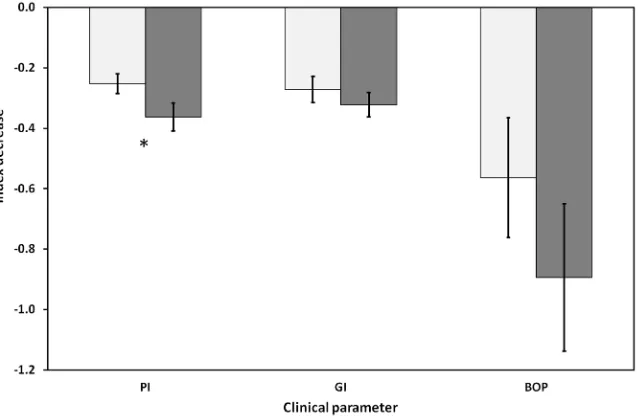
Figure 2. Decrease in the clinical parameters PI, GI and BOP for the control groups
(toothbrush without LED light, [light grey]) and the intervention groups (toothbrush with 450 nm blue LED light [dark grey]) with the standard error of the mean as the error bars. The bars for BOP have been multiplied with 0.1 in order to obtain a comparable scale in the diagram. * = difference between groups at p = 0.058.
[image:9.595.218.537.390.599.2]DOI: 10.4236/ojst.2018.810027 296 Open Journal of Stomatology
The difference in the decrease in PI from baseline to the last follow-up visit at week 8 is significant at a level of p = 0.058, close to accepted level p = 0.05. For GI and BOP there was no statistical significant difference with p = 0.39 and p = 0.29 respectively.
We also investigated if the effect of blue light on reducing plaque or gingival inflammation could be enhanced by the use of a peroxide containing toothpaste. The statistical analyses of the clinical parameters PI, GI and BOP indices as well as the inflammatory markers showed no differences between the toothpaste with or without peroxide (data not shown).
3.3. Immunological Assessments
The levels of MMP-8, TIMP-1, IL-1β, IL-6, IL-8 and TNF-α were determined in both saliva and GCF at baseline and at last follow-up visit at week 8, Table 4. For IL-6 and TNF-α in saliva and GCF as well as MMP-8 and TIMP-1 in GCF, the levels were close to, or below, limit of detection (LOD).
[image:10.595.210.536.400.689.2]These data were not subject to further statistical analysis. The concentrations of IL-1β and IL-8 in saliva were well above LOD, but showed dispersive results
Table 4. Mean values with standard deviation in parentheses for the inflammatory
mark-ers MMP1 and TIMP1 in saliva, and IL-1β and IL8 in gingival crevicular fluid (GCF) at Baseline and Week 8. p-values given for t-tests, see comments below. n = number of sub-jects. Concentrations given in picomolar.
Examination All n Control n Intervention n p
Matrix Metalloproteinase-8 (MMP8) in saliva Baseline 46.5 (40.2)
42 46.5 (37.7) 18 46.5 (44.0) 24 0.99 b
Week 8 27.5 (28.9) 26.3 (35.2) 30.8 (25.7)
PBaseline-Week 8 0.003a 0.042a 0.029a 0.54c
Metallopeptidase Inhibitor 1 (TIMP1) in saliva Baseline 263 (118)
47 270 (128) 22 237 (119) 25 0.72 b
Week 8 225 (111) 257 (110) 214 (104)
PBaseline-Week 8 0.002a 0.064a 0.008a 0.58c
Interleukin 1β (IL1β) in GCF Baseline 106 (143)
48 85.7 (83.4) 23 125 (182) 25 0.34 b
Week 8 46.9 (53.2) 56.2 (62.5) 38.3 (42.4)
PBaseline-Week 8 0.009a 0.13a 0.029a 0.44c
Interleukin 1β (IL1β) in GCF Baseline 325 (240)
46 310 (266) 22 338 (217) 24 0.72 b
Week 8 181 (142) 201 (152) 163 (133)
PBaseline-Week 8 0.002a 0.12a 0.002a 0.45c
DOI: 10.4236/ojst.2018.810027 297 Open Journal of Stomatology
making also these data not useful for further statistical analysis. Thus, the in-flammatory markers that were further analyzed were MMP-8 and TIMP-1 in sa-liva, and IL-1β and IL-8 in GCF.
When looking at all the subjects, salivary MMP-8 and TIMP-1 as well as IL-1β
and IL-8 in GCF decreased significantly (p < 0.01) from baseline to the final fol-low-up visit at week 8, Table 4. When comparing the intervention (A/C) and the control (B/D) groups, no significant difference (p ≥ 0.44) could be detected be-tween the groups. However, an interesting observation was that there were sig-nificant decreases in the salivary levels of both MMP-8 (p = 0.029) and TIMP-1 (p = 0.0073), as well as for IL-1β (p = 0.029) and IL-8 (p = 0.0017) in GCF in the blue light toothbrush intervention groups. For the control groups on the other hand, a significant decrease between baseline and week 8 could only be detected for MMP-8 in saliva (p = 0.042). As for the clinical indices, the statistical data analysis of the inflammatory parameters showed no difference when comparing peroxide or non-peroxide toothpastes, with or without blue light illumination (data not shown).
4. Discussion
The primary aim of this study was to investigate if a toothbrush with an incor-porated LED emitting blue light at 450 nm has any adjunctive effect on tooth-brushing regarding reduction of dental plaque and gingival inflammation.
4.1. Clinical Indices
For all subjects in the intervention as well as in the control groups, there were significant decreases in the clinical indices PI, GI and BOP from baseline to the final follow-up visit at week 8. The average PI for all subjects decreased with 57% (n = 48, p ≤ 0.0001), indicating that that the subjects in general had a substantial reduction of the amount of dental plaque. A significant reduction in gingival in-flammation was also demonstrated by reductions in both GI and BOP indices, 46% and 15% respectively (n = 48, p ≤ 0.0001). A meta-review has assessed 6-month-clinical controlled studies regarding self-performed toothbrushing in adults with gingivitis. Their conclusion was that a single hygiene instruction had a significant positive effect on the reduction of gingival inflammation even though it was small [32]. Improvements in the subjects’ oral health status shown in the present study verify these suggestions even though the observations only were conducted for 8 weeks. All subjects in both groups got repeated instruc-tions in toothbrushing at all visits except the final which can explain the signifi-cant decreases in all three clinical indices.
differ-DOI: 10.4236/ojst.2018.810027 298 Open Journal of Stomatology
ence observed in the GI and BOP indices (p > 0.29). In a recent study with a similar study design as in the present study, the effect of a blue light-emitting toothbrush with a central wavelength of 412 nm LED was investigated [27]. Af-ter four weeks significant improvements with up to 50% decrease in plaque and gingival inflammation indices were detected in the treatment group relative to the control group. Their results indicate that the wavelength chosen is of a great importance.
4.2. Inflammatory Markers
The levels of IL-1β and IL-8 in GCF, and MMP-8 and TIMP-1 in saliva demon-strated a significant decrease when looking at all subjects. The potential of in-flammatory markers in GCF and saliva to reflect gingival inflammation has been widely studied, where MMP-8 levels in GCF have been reported to differ be-tween periodontal healthy subjects and subjects with gingivitis and periodontitis
[33], putting forward that MMP-8 is a significant marker for determining the degree of inflammation. Some studies have detected elevated concentrations of IL-1β, IL-6, and IL-8 associated with experimental gingivitis [34] [35], while other studies have found no differences in the levels of IL-1β, as well as MMP-8, in saliva between gingivitis and healthy sites [36]. In a recent review it was con-cluded that there are sufficient evidence showing that IL-1β in saliva as well as in GCF can be used as markers of the degree of gingival inflammation [37]. In the study presented here, the decrease in the levels of IL-1β and IL-8 in GCF, and MMP-8 and TIMP-1 in saliva were in agreement with the decrease in the GI and BOP clinical indices in all studied objects. However, when comparing the con-trol and blue light intervention groups no differences could be found. Some in-teresting observations were made. In the blue light intervention groups there were statistical significant decreases in the inflammatory markers IL-1β and IL-8 in GCF, as well as in MMP-8 and TIMP-1 in saliva from baseline to the final week 8 in the intervention groups. In contrast, the control groups exhibited a statistical significant decrease in the concentration of MMP-8 in saliva. These results imply that there might be a possible adjunct effect of the blue light in combination with tooth brushing evidenced by potential subclinical changes in the levels of the investigated inflammatory markers. Subclinical changes in the IL-1B levels in GCF have previously been shown in response to plaque accumu-lation prior to clinical signs of inflammation [38]. In the present study, the IL-1β
concentration in saliva were well above LOD but showed very divergent results and because of that IL-1β could not be used as a reliable marker.
4.3. Porphyrins, Blue Light and the LED Toothbrush
The toothbrush used in this study was the commercially available UltraBlu
DOI: 10.4236/ojst.2018.810027 299 Open Journal of Stomatology
manufacturing company, but they were not able to finish it before the end of this project. This was a limitation in this study. A wavelength of 405 nm has been suggested to be more effective in a number of studies [22] [39] [40], as well as in our in vitro studies of bacteria killing efficiency and porphyrin content [17] [18] [41] [42]. In the latter studies we have shown a connection between the porphy-rin content in Escherichia coli and the killing efficiency of this bacterium by blue light at 405 nm. We also showed that at this wavelength, porphyrins are stimu-lated to produce singlet oxygen.
The proposed mechanism of phototherapy is that endogenous porphyrins, mainly protoporphyrin IX (PPIX), absorb the light and enter into an exited state
[23]. The excess energy in the molecule can then be transferred to yield reactive oxygen species (ROS) which are responsible for killing the bacteria. The porphy-rins present in oral bacteria are mainly intermediates in the endogenous heme synthesis. These porphyrins all have absorption maxima around 405 nm, while the absorption of light at a wavelength of 450 nm is much lower [24] [43] [44] [45]. Since the typical bandwidth at 50% emission level for the LED in the toothbrush used in this study was around 20 - 30 nm, only a small amount of the light energy can be adsorbed by e.g. PPIX. That 405 nm should be the optimal wavelength to include in a LED toothbrush is supported by several other studies. In a pilot study our research group has demonstrated efficient inhibition of the growth of A. actinomycetemcomitans cultivated on agar plates when illuminated with 405 nm LED light (unpublished data). In our study there are only weak in-dications that the 450 nm toothbrush improves the oral health status of the sub-jects. The discrepancy between our study and the results in studies by Genina
[27] and others [24] [43] [44] [45], is most likely explained by the differences in wavelengths of the LEDs used in the toothbrushes.
5. Conclusion
To include phototherapy treatment in a toothbrush represents a novel approach of interest to daily oral care, since it has the potential to be a promising and cost efficient adjunctive tool for treating gingivitis and preventing periodontitis. This study has demonstrated that a toothbrush with an incorporated blue light LED emitting with an emission maximum at 450 nm was not able to give a sufficient significant adjunctive effect to toothbrushing with regard to reduction in dental plaque and gingival inflammation as well as in immunological inflammatory markers. Still, our suggestion is that antimicrobial blue light incorporated in a toothbrush could be beneficial if a 405 nm LED is used to obtain optimal photo-therapeutic effect from a LED toothbrush. In future studies, the phototherapeu-tic of 405 nm LED toothbrushes should be investigated.
Acknowledgements
Universi-DOI: 10.4236/ojst.2018.810027 300 Open Journal of Stomatology
ty, Stockholm, Sweden supported the study. We thank Timothy Lawrence at TJL Enterprises, Long Beach, CA, USA and Michael Barns at TechLight Systems, Ocala, FL, USA for supplying the blue light toothbrushes used in this study. The authors report no conflicts of interest. We thank Daniela Bureik, Department of Dental Medicine, Division of Periodontology, Karolinska Institutet, Huddinge, Sweden, for technical laboratory assistance in the analysis of inflammatory markers.
Conflicts of Interest
The authors declare no conflicts of interest regarding the publication of this pa-per.
References
[1] Etiology and Pathogenesis of Periodontal Diseases.
https://www.ncbi.nlm.nih.gov/pubmed/15978238
[2] Albandar, J.M. (2005) Epidemiology and Risk Factors of Periodontal Diseases. Dental Clinics of North America, 49, 517-532.
https://doi.org/10.1016/j.cden.2005.03.003
[3] Jin, L.J., Armitage, G.C., Klinge, B., Lang, N.P., Tonetti, M., et al. (2011) Global Oral Health Inequalities: Task Group-Periodontal Disease. Advances in Dental Research, 23, 221-226. https://doi.org/10.1177/0022034511402080
[4] Marsh, P.D., Head, D.A. and Devine, D.A. (2015) Dental Plaque as a Biofilm and a Microbial Community-Implications for Treatment. Journal of Oral Biosciences, 57, 185-191. https://doi.org/10.1016/j.job.2015.08.002
[5] Chapple, I.L., Van der Weijden, F., Doerfer, C., Herrera, D., Shapira, L., et al. (2015) Primary Prevention of Periodontitis: Managing Gingivitis. Journal of Clinical Peri-odontolgy, 42, S71-S76. https://doi.org/10.1111/jcpe.12366
[6] Sheiham, A. (1997) Is the Chemical Prevention of Gingivitis Necessary to Prevent Severe Periodontitis? Periodontology, 15, 15-24.
https://doi.org/10.1111/j.1600-0757.1997.tb00100.x
[7] Albandar, J.M., Kingman, A., Brown, L.J. and Loe, H. (1998) Gingival Inflammation and Subgingival Calculus as Determinants of Disease Progression in Early-Onset Periodontitis. Journal of Clinical Periodontology, 25, 231-237.
https://doi.org/10.1111/j.1600-051X.1998.tb02433.x
[8] Lang, N.P., Schatzle, M.A. and Loe, H. (2009) Gingivitis as a Risk Factor in Peri-odontal Disease. Journal of Clinical Periodontology, 36, 3-8.
https://doi.org/10.1111/j.1600-051X.2009.01415.x
[9] Konig, K., Teschke, M., Sigusch, B., Glockmann, E., Eick, S., et al. (2000) Red Light Kills Bacteria via Photodynamic Action. Cellular and Molecular Biology (Noisy-le- Grand, France), 46, 1297-1303.
[10] Maisch, T. (2007) Anti-Microbial Photodynamic Therapy: Useful in the Future? Lasers in Medical Science, 22, 83-91. https://doi.org/10.1007/s10103-006-0409-7
[11] Yang, L., Liu, Y., Wu, H., Song, Z., Høiby, N., et al. (2012) Combating Biofilms. FEMS Immunology and Medical Microbiology, 65, 146-157.
https://doi.org/10.1111/j.1574-695X.2011.00858.x
DOI: 10.4236/ojst.2018.810027 301 Open Journal of Stomatology
Oral Biofilms. Periodontology, 55, 143-166.
https://doi.org/10.1111/j.1600-0757.2010.00346.x
[13] Kleinpenning, M.M., Smits, T., Frunt, M.H., van Erp, P.E., van de Kerkhof, P.C., et al. (2010) Clinical and Histological Effects of Blue Light on Normal Skin. Photo-dermatology, Photoimmunology and Photomedicine, 26, 16-21.
https://doi.org/10.1111/j.1600-0781.2009.00474.x
[14] Oplander, C., Hidding, S., Werners, F.B., Born, M., Pallua, N., et al. (2011) Effects of Blue Light Irradiation on Human Dermal Fibroblasts. Journal of Photochemistry and Photobiology B: Biology, 103, 118-125.
https://doi.org/10.1016/j.jphotobiol.2011.02.018
[15] Zhang, Y., Zhu, Y., Chen, J., Wang, Y., Sherwood, M.E., et al. (2016) Antimicrobial Blue Light Inactivation of Candida albicans: In Vitro and in Vivo Studies. Viru-lence, 7, 536-545. https://doi.org/10.1080/21505594.2016.1155015
[16] Ramakrishnan, P., Maclean, M., MacGregor, S.J., Anderson, J.G. and Grant, M.H. (2016) Cytotoxic Responses to 405 nm Light Exposure in Mammalian and Bacterial Cells: Involvement of Reactive Oxygen Species. Toxicology in Vitro, 33, 54-62.
https://doi.org/10.1016/j.tiv.2016.02.011
[17] Fyrestam, J., Bjurshammar, N., Paulsson, E., Johannsen, A. and Östman, C. (2015) Determination of Porphyrins in Oral Bacteria by Liquid Chromatography Electro-spray Ionization Tandem Mass Spectrometry. Analytical and Bioanalytical Chemi-stry, 407, 7013-7023.https://doi.org/10.1007/s00216-015-8864-2
[18] Fyrestam, J., Bjurshammar, N., Paulsson, E., Mansouri, N., Johannsen, A., et al. (2017) Influence of Culture Conditions on Porphyrin Production in Aggregatibac-ter actinomycetemcomitans and Porphyromonas Gingivalis. Photodiagnosis and Photodynamic Therapy, 17, 115-123.https://doi.org/10.1016/j.pdpdt.2016.11.001
[19] Soukos, N.S., Som, S., Abernethy, A.D., Ruggiero, K., Dunham, J., et al. (2005) Phototargeting Oral Black-Pigmented Bacteria. Antimicrobial Agents and Chemo-therapy, 49, 1391-1396.https://doi.org/10.1128/AAC.49.4.1391-1396.2005
[20] Cieplik, F., Spath, A., Leibl, C., Gollmer, A., Regensburger, J., et al. (2014) Blue Light Kills Aggregatibacter actinomycetemcomitans Due to Its Endogenous Photo-sensitizers. Clinical Oral Investigations, 18, 1763-1769.
https://doi.org/10.1007/s00784-013-1151-8
[21] Ashkenazi, H., Malik, Z., Harth, Y. and Nitzan, Y. (2003) Eradication of Propioni-bacterium acnes by Its Endogenic Porphyrins after Illumination with High Intensity Blue Light. FEMS Immunology and Medical Microbiology, 35, 17-24.
https://doi.org/10.1111/j.1574-695X.2003.tb00644.x
[22] Maclean, M., MacGregor, S.J., Anderson, J.G. and Woolsey, G. (2009) Inactivation of Bacterial Pathogens Following Exposure to Light from a 405-Nanometer Light- Emitting Diode Array. Applied Environmental Microbiology, 75, 1932-1937.
https://doi.org/10.1128/AEM.01892-08
[23] Dai, T., Gupta, A., Murray, C.K., Vrahas, M.S., Tegos, G.P., et al. (2012) Blue Light for Infectious Diseases: Propionibacterium acnes, Helicobacter pylori, and Beyond? Drug Resistance Updates, 15, 223-236.https://doi.org/10.1016/j.drup.2012.07.001
[24] Hope, C.K., Hindley, J.A., Khan, Z., de Jong Ede, J. and Higham, S.M. (2013) Lethal Photosensitization of Porphyromonas gingivalis by Their Endogenous Porphyrins under Anaerobic Conditions: An in Vitro Study. Photodiagnosis and Photodynamic Therapy, 10, 677-682.https://doi.org/10.1016/j.pdpdt.2013.08.006
DOI: 10.4236/ojst.2018.810027 302 Open Journal of Stomatology
Drug Discovery.
[26] Soukos, N.S., Stultz, J., Abernethy, A.D. and Goodson, J.M. (2015) Phototargeting Human Periodontal Pathogens in Vivo. Lasers in Medical Science, 30, 943-952.
https://doi.org/10.1007/s10103-013-1497-9
[27] Genina, E.A., Titorenko, V.A., Belikov, A.V., Bashkatov, A.N. and Tuchin, V.V. (2015) Adjunctive Dental Therapy via Tooth Plaque Reduction and Gingivitis Treatment by Blue Light-Emitting Diodes Tooth Brushing. Journal of Biomedical Optics, 20, Article ID: 128004.https://doi.org/10.1117/1.JBO.20.12.128004
[28] Silness, J. and Loe, H. (1964) Periodontal Disease in Pregnancy. II. Correlation be-tween Oral Hygiene and Periodontal Condition. Acta Odontologica Scandinavica, 22, 121-135.https://doi.org/10.3109/00016356408993968
[29] Loe, H. and Silness, J. (1963) Periodontal Disease in Pregnancy. I. Prevalence and Severity. Acta Odontologica Scandinavica, 21, 533-551.
https://doi.org/10.3109/00016356309011240
[30] Directive, E. (2002) 95/EC “Restriction of Hazardous Substances in Electrical and Electronic Equipment” (RoHS Directive 2002).
[31] SP Technical Research Institute of Sweden AB (2010) Measurement of Blue LEDs on Toothbrush. SP Report MTkPX05459, 4 p. (In Swedish)
[32] Van der Weijden, G.A. and Hioe, K.P. (2005) A Systematic Review of the Effective-ness of Self-Performed Mechanical Plaque Removal in Adults with Gingivitis Using a Manual Toothbrush. Journal of Clinical Periodontology, 32, 214-228.
https://doi.org/10.1111/j.1600-051X.2005.00795.x
[33] Sorsa, T., Mantyla, P., Tervahartiala, T., Pussinen, P.J., Gamonal, J., et al. (2011) MMP Activation in Diagnostics of Periodontitis and Systemic Inflammation. Jour-nal of Clinical Periodontology, 38, 817-819.
https://doi.org/10.1111/j.1600-051X.2011.01753.x
[34] Lee, A., Ghaname, C.B., Braun, T.M., Sugai, J.V., Teles, R.P., et al. (2012) Bacterial and Salivary Biomarkers Predict the Gingival Inflammatory Profile. Journal of Pe-riodontology, 83, 79-89.https://doi.org/10.1902/jop.2011.110060
[35] Zhou, M., Meng, H.X., Zhao, Y.B. and Chen, Z.B. (2012) Changes of Four Proin-flammatory Proteins in Whole Saliva during Experimental Gingivitis. The Chinese Journal of Dental Research: The Official Journal of the Scientific Section of the Chinese Stomatological Association (CSA), 15, 121-127.
[36] Syndergaard, B., Al-Sabbagh, M., Kryscio, R.J., Xi, J., Ding, X., et al. (2014) Salivary Biomarkers Associated with Gingivitis and Response to Therapy. Journal of Peri-odontology, 85, e295-e303.https://doi.org/10.1902/jop.2014.130696
[37] Boronat-Catala, M., Catala-Pizarro, M. and Bagan Sebastian, J.V. (2014) Salivary and Crevicular Fluid Interleukins in Gingivitis. Journal of Clinical and Experimen-tal Dentitry, 6, e175-e179.https://doi.org/10.4317/jced.51403
[38] Zhang, J., Kashket, S. and Lingström, P. (2002) Evidence for the Early Onset of Gingival Inflammation Following Short Term Plaque Accumulation. Journal of Clinical Periodontology, 29, 1082-1085.
https://doi.org/10.1034/j.1600-051X.2002.291206.x
[39] Murdoch, L.E., Maclean, M., Endarko, E., MacGregor, S.J. and Anderson, J.G. (2012) Bactericidal Effects of 405 nm Light Exposure Demonstrated by Inactivation of Escherichia, Salmonella, Shigella, Listeria, and Mycobacterium Species in Liquid Suspensions and on Exposed Surfaces. The Scientific World Journal, 2012, Article ID: 137805.https://doi.org/10.1100/2012/137805
DOI: 10.4236/ojst.2018.810027 303 Open Journal of Stomatology
(2014) 405 nm Light Technology for the Inactivation of Pathogens and Its Potential Role for Environmental Disinfection and Infection Control. The Journal of Hospital Infection, 88, 1-11.https://doi.org/10.1016/j.jhin.2014.06.004
[41] Fyrestam, J. and Ostman, C. (2017) Determination of Heme in Microorganisms Using HPLC-MS/MS and Cobalt (III) Protoporphyrin IX Inhibition of Heme Ac-quisition in Escherichia coli. Analytical and Bioanalytical Chemistry, 409, 6999-7010.
https://doi.org/10.1007/s00216-017-0610-5
[42] Fyrestam, J. (2017) Escherichia Coli Is Killed by 405 nm Blue Light Due to Its En-dogenous Porphyrins Induced by 5-Aminolevulinic Acid.
[43] Fukui, M., Yoshioka, M., Satomura, K., Nakanishi, H. and Nagayama, M. (2008) Specific-Wavelength Visible Light Irradiation Inhibits Bacterial Growth of Porphy-romonas Gingivalis. Journal of Periodontal Research, 43, 174-178.
https://doi.org/10.1111/j.1600-0765.2007.01009.x
[44] McDonald, R., Macgregor, S.J., Anderson, J.G., Maclean, M. and Grant, M.H. (2011) Effect of 405-nm High-Intensity Narrow-Spectrum Light on Fibrob-last-Populated Collagen Lattices: An in Vitro Model of Wound Healing. Journal of Biomedical Optics, 16, Article ID: 048003.https://doi.org/10.1117/1.3561903