A PROSPECTIVE RANDOMISED CONTROLLED STUDY TO COMPARE EFFICACY BETWEEN
ULTRASOUND GUIDED SINGLE SHOT RECTUS SHEATH BLOCK AND
POSTOPERATIVE ANALGESIA IN PATIENTS WITH
Dr. R. D. Patel, *Dr. Abhishek B. Rathod
Department of Anaesthesiology, Seth G. S. Medical College and KEM H
ARTICLE INFO ABSTRACT
Introduction:
study, analgesic efficacy between ultrasound guided single shot rectus sheath block was compared with epidural analgesia in patients who have undergone surgeries with midline anterior abdominal incisions. Material And Methods:
I and II were included in our study, which is a randomised
group A and group B. In group A, at the end of surgery, ultrasound guided single shot rectus sheath block was administered using 20 ml of 0.25% bupivacaine by supraumbilical approach on either side of midl above the posterior rectus sheath. In group B, before induction of general anaesthesia, epidural catheter is placed in sitting position by midline approach and received 4ml of 0.125% bupivacaine through it for postoperative pain relief. Postoperative
in immediate postoperative period. Analgesic efficacy was then evaluated by comparing VAS scores and need of rescue analgesia, among two techniques. All statistical calculations were don
programs Microsoft Excel 2007 (Microsoft Corporation, NY, USA) and SPSS (Statistical Package for the Social Science; SPSS Inc., Chicago, IL, USA) version 21.
Results:
3.20 in group A and 3.34 in group B), 30 to 60 minutes (mean VAS 2.72 in group A and 2.56 in group B), 60 to 90 minutes (mean VAS of 2.36 in group A and 2.74 in group B), 90 to 120 minutes (mean VAS of 2.02 in group A and 3.44 in gr
6 hours (mean VAS of 1.92 in group A and 4.10 in group B), 6 to 8 hours (mean VAS 2.40 of in group A and 4.38 in group B) as compared to group B. In group A, out of 50 patients 6
total group A population, received rescue analgesia, with 44 patients which is 88% of the total group A population did not receive any rescue analgesia. While in group B, out of 50 patients, 19 patients which is 38% of total gr
B population did not receive any rescue analgesia (P value <0.01). Statistically significant difference has also been observed in postoperative pulse rate among g
better pain relief among group A patients.With no complications noted in group A throughout study, while nausea and vomiting in two patients and hypotension among three patients of group B.
Conclusion:
immediate postoperative period at some timeintervals requiring less amount of rescue nonopioid analgesia without any complications.
Copyright © 2018, Patel et al. This is an open access distribution, and reproduction in any medium, provided
INTRODUCTION
Pain is one of the most common complaint of patients in immediate postoperative period; especially so after abdominal surgeries. This postoperative pain causes or rather further adds to patient’s anxiety, stress and dissatisfaction. Moreover, inadequately treated pain can have detrimental physiological, psychological, economic and social adverse effects
and Dolin, 2004). Hence effective pain relief forms an integral part of patient’s perioperative care and can modify postoperative surgical stress response and outcome.
ISSN: 0975-833X
Article History:
Received 04th November, 2017 Received in revised form 23rd December, 2017 Accepted 13th January, 2018 Published online 28th February, 2018
Citation: Dr. R. D. Patel, Dr. Abhishek B. Rathod
Between Ultrasound Guided Single Shot Rectus Sheath Block And Epidural For Postoperative Analgesia In Patients With Anterior Surgeries”, International Journal of Current Research
Key words:
Postoperative analgesia,
Ultrasound guided rectus sheath block, epidural,
VAS,
Rescue analgesia.
*Corresponding author: Dr. Abhishek B. Rathod
Department of Anaesthesiology, Seth G. S. Medical College and KEM Hopsital, Parel, Mumbai – 400012, India.
RESEARCH ARTICLE
A PROSPECTIVE RANDOMISED CONTROLLED STUDY TO COMPARE EFFICACY BETWEEN
ULTRASOUND GUIDED SINGLE SHOT RECTUS SHEATH BLOCK AND
POSTOPERATIVE ANALGESIA IN PATIENTS WITH ANTERIOR ABDOMINAL WALL SURGERIES
Dr. Abhishek B. Rathod and Dr. Nirav M.
Department of Anaesthesiology, Seth G. S. Medical College and KEM Hopsital, Parel, Mumbai
ABSTRACT
Introduction: Ultrasound allows precise placement of local anaesthetic
study, analgesic efficacy between ultrasound guided single shot rectus sheath block was compared with epidural analgesia in patients who have undergone surgeries with midline anterior abdominal incisions. Material And Methods: Hundred patients belonging to American society of anaesthesiologist (ASA) class I and II were included in our study, which is a randomised controlled study, and were divided in two groups, group A and group B. In group A, at the end of surgery, ultrasound guided single shot rectus sheath block was administered using 20 ml of 0.25% bupivacaine by supraumbilical approach on either side of midl above the posterior rectus sheath. In group B, before induction of general anaesthesia, epidural catheter is placed in sitting position by midline approach and received 4ml of 0.125% bupivacaine through it for postoperative pain relief. Postoperative pain assessed using visual analogue scale (VAS) for first eight hours in immediate postoperative period. Analgesic efficacy was then evaluated by comparing VAS scores and need of rescue analgesia, among two techniques. All statistical calculations were don
programs Microsoft Excel 2007 (Microsoft Corporation, NY, USA) and SPSS (Statistical Package for the Social Science; SPSS Inc., Chicago, IL, USA) version 21.
Results: Group A had better VAS scores in immediate postoperative period for fir
3.20 in group A and 3.34 in group B), 30 to 60 minutes (mean VAS 2.72 in group A and 2.56 in group B), 60 to 90 minutes (mean VAS of 2.36 in group A and 2.74 in group B), 90 to 120 minutes (mean VAS of 2.02 in group A and 3.44 in group B), 2 to 4 hours (mean VAS of 1.92 in group A and
6 hours (mean VAS of 1.92 in group A and 4.10 in group B), 6 to 8 hours (mean VAS 2.40 of in group A and 4.38 in group B) as compared to group B. In group A, out of 50 patients 6
total group A population, received rescue analgesia, with 44 patients which is 88% of the total group A population did not receive any rescue analgesia. While in group B, out of 50 patients, 19 patients which is 38% of total group B population, received rescue analgesia, with 21 patients which is 62% of the total group B population did not receive any rescue analgesia (P value <0.01). Statistically significant difference has also been observed in postoperative pulse rate among group A and group B with P value of 0.026, indicating better pain relief among group A patients.With no complications noted in group A throughout study, while nausea and vomiting in two patients and hypotension among three patients of group B.
Conclusion: Ultrasound guided rectus sheath block is a better analgesic modality than epidural analgesia in immediate postoperative period at some timeintervals requiring less amount of rescue nonopioid analgesia without any complications.
access article distributed under the Creative Commons Attribution License, the original work is properly cited.
Pain is one of the most common complaint of patients in immediate postoperative period; especially so after abdominal surgeries. This postoperative pain causes or rather further adds to patient’s anxiety, stress and dissatisfaction. Moreover, treated pain can have detrimental physiological,
psychological, economic and social adverse effects(Cashman
Hence effective pain relief forms an integral care and can modify postoperative surgical stress response and outcome. Surgeries
with incisions over anterior abdomen can lead to severe abdominal pain which causes shallow breathing as patients cannot breathe deep due to pain on deep inspiration, hen lowering lung volumes and capacities leading to basal pulmonary atelectasis, inadequate coughing with retention of secretions leading to various pulmonary complications
(Ferreyra et al., 2009). Various analgesic modalities including
pharmacotherapy and interventional therapy can be employed to relieve this pain for example NSAIDS (Non
inflammatory drugs), systemic opioids etc. NSAIDS may not be completely effective; also, has inherent nephrotoxic action,
International Journal of Current Research Vol. 10, Issue, 02, pp.65594-65599, February, 2018
Abhishek B. Rathod and Dr. Nirav M. Kotak, 2018. “A Prospective Randomised Controlled Study to Compare Efficacy Between Ultrasound Guided Single Shot Rectus Sheath Block And Epidural For Postoperative Analgesia In Patients With Anterior
International Journal of Current Research, 10, (02), 65594-65599.
A PROSPECTIVE RANDOMISED CONTROLLED STUDY TO COMPARE EFFICACY BETWEEN
ULTRASOUND GUIDED SINGLE SHOT RECTUS SHEATH BLOCK AND EPIDURAL FOR
ANTERIOR ABDOMINAL WALL SURGERIES
Dr. Nirav M. Kotak
opsital, Parel, Mumbai – 400012, India
Ultrasound allows precise placement of local anaesthetic agents to the desired site. In our study, analgesic efficacy between ultrasound guided single shot rectus sheath block was compared with epidural analgesia in patients who have undergone surgeries with midline anterior abdominal incisions.
Hundred patients belonging to American society of anaesthesiologist (ASA) class controlled study, and were divided in two groups, group A and group B. In group A, at the end of surgery, ultrasound guided single shot rectus sheath block was administered using 20 ml of 0.25% bupivacaine by supraumbilical approach on either side of midline, above the posterior rectus sheath. In group B, before induction of general anaesthesia, epidural catheter is placed in sitting position by midline approach and received 4ml of 0.125% bupivacaine through it for pain assessed using visual analogue scale (VAS) for first eight hours in immediate postoperative period. Analgesic efficacy was then evaluated by comparing VAS scores and need of rescue analgesia, among two techniques. All statistical calculations were done using computer programs Microsoft Excel 2007 (Microsoft Corporation, NY, USA) and SPSS (Statistical Package for the
Group A had better VAS scores in immediate postoperative period for first 30 minutes (mean VAS 3.20 in group A and 3.34 in group B), 30 to 60 minutes (mean VAS 2.72 in group A and 2.56 in group B), 60 to 90 minutes (mean VAS of 2.36 in group A and 2.74 in group B), 90 to 120 minutes (mean VAS of oup B), 2 to 4 hours (mean VAS of 1.92 in group A and 3.82 in group B), 4 to 6 hours (mean VAS of 1.92 in group A and 4.10 in group B), 6 to 8 hours (mean VAS 2.40 of in group A and 4.38 in group B) as compared to group B. In group A, out of 50 patients 6 patients which is 12% of the total group A population, received rescue analgesia, with 44 patients which is 88% of the total group A population did not receive any rescue analgesia. While in group B, out of 50 patients, 19 patients which is oup B population, received rescue analgesia, with 21 patients which is 62% of the total group B population did not receive any rescue analgesia (P value <0.01). Statistically significant difference has roup A and group B with P value of 0.026, indicating better pain relief among group A patients.With no complications noted in group A throughout study, while nausea and vomiting in two patients and hypotension among three patients of group B.
Ultrasound guided rectus sheath block is a better analgesic modality than epidural analgesia in immediate postoperative period at some timeintervals requiring less amount of rescue nonopioid analgesia
License, which permits unrestricted use,
with incisions over anterior abdomen can lead to severe abdominal pain which causes shallow breathing as patients cannot breathe deep due to pain on deep inspiration, hence lowering lung volumes and capacities leading to basal pulmonary atelectasis, inadequate coughing with retention of secretions leading to various pulmonary complications
Various analgesic modalities including interventional therapy can be employed to relieve this pain for example NSAIDS (Non-steroidal anti-inflammatory drugs), systemic opioids etc. NSAIDS may not be completely effective; also, has inherent nephrotoxic action,
INTERNATIONAL JOURNAL OF CURRENT RESEARCH
detrimental effect over platelets and systemic opioids are associated with side effects such as nausea, vomiting, sedation, pruritus, urinary retention, cardiovascular and respiratory
depression (Hazem et al., 2014). Local anesthetic agents when
given through ultrasonography (USG) guided truncal blocks for e.g. Rectus sheath block or through epidural route for analgesia, they provide excellent analgesia, reducing requirement of NSAIDS and systemic opioids. Rectus sheath block inhibits the sensory nerves that innervate the anterior abdominal wall taking care of superficial and deep somatic pain associated with incision of skin, subcutaneous tissue, rectus sheath and muscle. USG guides accurate placement of needle in rectus sheath compartment, allows visualization of local anesthetic solution spread in the appropriate plane, hence safe and precise. Epidural analgesia by means of local anesthetic agent introduced in epidural space by epidural catheter is also equally efficacious and provides good analgesia
(Susan et al.).
MATERIALS AND METHODS
Hundred patients belonging to American society of anaesthesiologist (ASA) class I and II were included in our study, which is a randomised controlled study, and were divided in two groups, group A and group B, drawing a card being the method of randomisation. After obtaining well informed written consent, standard monitors including pulse oximeter, cardioscope, noninvasive blood pressure monitor & capnograph will be attached. Preoperative readings will be noted. 18 G intravenous (IV) angiocatheter will be used to secure Iv line. Patient will be premedicated with Inj. Ondansetron 0.1mg/kg IV & Inj. Ranitidine 50mg IV. Intravenous ringers lactate will be started. Patients will receive intravenous sedation with Inj. Midazolam 0.05mg/kg IV and Inj. Fentanyl 2microgram/kg. Patients with group A will be given USG guided single shot rectus sheath block postoperatively, before reversing and extubating the patient of general anaesthesia and in patients with group B epidural catheter will be placed preoperatively, which will be activated postoperatively before reversing and extubating the patient of general anaesthesia. In patients of both groups, after preoxygenation, a standardised general anesthetic regime will be employed, consisting of Inj. Propofol 2mg/kg IV, Inj.
Vecuronium 0.1mg/kg, with intraoperative non-opioid
analgesia of paracetamol 15 – 20mg/kg and diclofenac 0.5mg/kg. For intraoperative maintenance of anaesthesia nitrous oxide and oxygen will be used in 2:1 ratio using closed circuit, with Isoflurane as a anaesthetic agent in a end tidal concentration of 0.5 to 1%. Polyvinyl chloride endotracheal tubes (ETT) will be used and sizes chosen will be 7.0 or 7.5 mm internal diameter (ID) for female patients and 8.0 or 8.5 mm ID for male patients. Atraumatic intubation will be performed with an oral ETT and its cuff will be inflated. In group A, following surgery, USG guided rectus sheath block will be given by supraumbilical approach. After proper painting and draping, under all aseptic precautions transducer is placed at midpoint between xiphoid process and umbilicus, then obtaining proper optimal image, stimuplex needle 100mm is insertedb3-6cms lateral to the edge of transducer and is then advanced in plane towards transducer in lateral to medial direction till it’s just lateral to lateral aspect of lineasemilunaris and lateral border of rectus abdominis muscle. Needle is further advanced until its deep to the potential space between posterior aspect of rectus abdominis muscle and posterior layer of rectus sheath. After that a small amount of 2ml of saline is
injected to confirm correct placement of needle tip, which is indicated by appearance of an anechoic fluid collection in this space. After confirming the correct placement of needle, 20 ml of Inj. Bupivacaine 0.25% plain is injected while observing for the expanding anechoic fluid collection, leading to clear separation (picture 3) of deep border of rectus abdominis muscle from posterior rectus sheath. Same procedure is repeated on contralateral side. Postoperative analgesic efficacy will be assessed by visual analogue scale. In patients of group B, before induction of general anaesthesia, in sitting position proper painting and draping done, under all aseptic precautions space is palpated and as per the extent of surgery, suitable space is selected. In this space, by midline approach, 18G/16G Tuohy’s needle is inserted and slowly advanced through the skin, subcutaneous tissue and interspinous ligament. Following removal of stylet from needle, low friction syringe filled with air is attached to the hub end of needle. Now with slow advancement of needle, loss of resistance is checked and with the loss of resistance, syringe is disconnected. For continuous type of epidural analgesia, epidural catheter is threaded for 3 to 5 cms into the epidural space. Now properly stabilising the catheter, epidural needle is slowly withdrawn. Analgesic dose of bupivacaine 4ml 0.125% will be given through this epidural catheter for postoperative pain relief. Following parameters will be evaluated every half hourly for first two hours in immediate postoperative period, then every hourly for next wo hours, then every 2 hourly for next 4 hours, for a total duration of eight hours of immediate postoperative period:
General condition of the patient, vital parameters like
pulse rate, blood pressure, temperature, respiration.
Postoperative abdominal pain assessed by visual
analogue scale (VAS) of 1 (no or minimal pain) to 10 (extremely unbearable pain).
Nausea, vomiting.
Motor blockade in bilateral lower limb / epidural band
of analgesia.
Any other complain or complication if any.
Inj. Paracetamol in a dose of 15mg/kg intravenously will be used for rescue analgesia.Following complications will be noted in our study.
Nausea and vomiting
Hypotension
Allergic reaction to drug
Injury to bowel
Local anaesthetic toxicity.
Statistical analysis
Demographic data of the patients will be presented using descriptive statistics. Data is of qualitative type and study type is prospective randomized controlled study. Hence, we
consider error up to 20%. So, N= 4PQ/L2, where P is a
happening event, Q is a non-happening event & L is an allowable error. Considering that 50% of patients get adequate postoperative analgesia after USG guided rectus sheath block by not requiring rescue analgesia; i.e. P=50%, hence Q= 100-P=50%. L= 20% OF 50 i.e. 20/100 X 50 = 10, so N= (4 X 50
X 50) / 102 = 100, So, sample size for this study is 10. 50 for
group A and 50 for group B. Parametric data will be analysed using Student’s t Test and will be represented as mean and
95% confidence interval if normally distributed.
Nonparametric data will be analysed using Fischer’s exact test if normally distributed. If both parametric and non-parametric
data are not normally distributed then it will be analysed using Mann-Whitney U test, which will be presented as median and minimum-maximum or interquartile range. A ‘’P’’ value of less than 0.05 will be considered significant.
consists of 100 patients. They will be divided into two groups “A” and “B”; and will randomly be allocated for giving USG guided single shot rectus sheath block in group A and epidural in group B, with drawing a card as a method of randomisation. Data were statistically described in terms of me
frequencies (number of cases) and percentages when appropriate. Data were tested first for normal distribution by Klomogorov– Smirnov test. Comparison of quantitative variables between the study groups was done using Student t test for independent samples if normally distributed. Mann Whitney U test was used for non-normally distributed quantitative data. For comparing categorical data, Chi square test was performed. Exact test was used instead when the expected frequency is less than 5. Pearson’s
coefficient was computed to evaluate the correlation between quantitative variables. A probability value (p value) less than 0.05 was considered statistically significant. All statistical calculations were done using computer programs Microsof Excel 2007 (Microsoft Corporation, NY, USA) and SPSS (Statistical Package for the Social Science; SPSS Inc., Chicago, IL, USA) version 21.
RESULTS
Group A had better VAS scores in immediate postoperative period for first 30 minutes (mean VAS 3.20 in gr
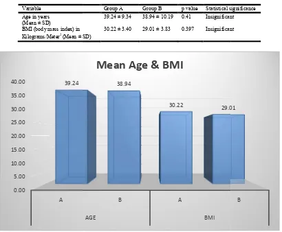
Variable Age in years (Mean ± SD)
BMI (body mass index) in Kilograms/Meter2 (Mean ± SD)
Graph 1. Patients age in years and BMI in kilograms /meter
0.00 5.00 10.00 15.00 20.00 25.00 30.00 35.00 40.00
A 39.24
distributed then it will be analysed using Whitney U test, which will be presented as median and maximum or interquartile range. A ‘’P’’ value of less than 0.05 will be considered significant. Our study divided into two groups “A” and “B”; and will randomly be allocated for giving USG guided single shot rectus sheath block in group A and epidural in group B, with drawing a card as a method of randomisation. Data were statistically described in terms of mean (±SD), frequencies (number of cases) and percentages when appropriate. Data were tested first for normal distribution by Smirnov test. Comparison of quantitative variables between the study groups was done using Student t t samples if normally distributed. Mann–
normally distributed quantitative data. For comparing categorical data, Chi square test was performed. Exact test was used instead when the expected frequency is less than 5. Pearson’s correlation coefficient was computed to evaluate the correlation between quantitative variables. A probability value (p value) less than 0.05 was considered statistically significant. All statistical calculations were done using computer programs Microsoft Excel 2007 (Microsoft Corporation, NY, USA) and SPSS (Statistical Package for the Social Science; SPSS Inc.,
Group A had better VAS scores in immediate postoperative period for first 30 minutes (mean VAS 3.20 in group A and
3.34 in group B), 30 to 60 minutes (mean VAS 2.72 in group A and 2.56 in group B), 60 to 90 minutes (mean VAS of 2.36 in group A and 2.74 in group B), 90 to 120 minutes (mean VAS of 2.02 in group A and 3.44 in group B), 2 to 4 hours (mean VAS of 1.92 in group A and
hours (mean VAS of 1.92 in group A and 4.10 in group B), 6 to 8 hours (mean VAS 2.40 of in group A
as compared to group B. In group A, out of 50 patients 6 patients which is 12% of the total group A population, received rescue analgesia in form of intravenous injection of paracetamol in dose of 15mg/kg as they had VAS score of more than 4 as stated in protocol, with 44 patients which is 88% of the total group A population did not receive any rescue analgesia. While in group B, out of 50 patients, 19 patients which is 38% of total group B population, received rescue analgesia, with 21 patients which is 62% of the total group B population did not receive any rescue analgesia (P value <0.01).
[image:3.595.95.505.431.769.2]Group B had received rescue analgesia more times than group A in immediate postoperative period indicating less efficient pain relief. Statistically significant difference has also been observed in postoperative pulse rate among group A and group B with P value of 0.026, indicating better pain relief among group A patients.
Table 1. Patient profile
Group A Group B p value Statistical significance 39.24 ± 9.34 38.94 ± 10.19 0.41 Insignificant
(Mean ± SD)
30.22 ± 3.40 29.01 ± 3.83 0.397 Insignificant
Graph 1. Patients age in years and BMI in kilograms /meter2
B A
AGE BMI
38.94
30.22 29.01
Mean Age & BMI
3.34 in group B), 30 to 60 minutes (mean VAS 2.72 in group A and 2.56 in group B), 60 to 90 minutes (mean VAS of 2.36
2.74 in group B), 90 to 120 minutes (mean VAS of 2.02 in group A and 3.44 in group B), 2 to 4 hours (mean VAS of 1.92 in group A and 3.82 in group B), 4 to 6 hours (mean VAS of 1.92 in group A and 4.10 in group B), 6 to 8 hours (mean VAS 2.40 of in group A and 4.38 in group B) In group A, out of 50 patients 6 patients which is 12% of the total group A population, received rescue analgesia in form of intravenous injection of paracetamol in dose of 15mg/kg as they had VAS score of than 4 as stated in protocol, with 44 patients which is 88% of the total group A population did not receive any rescue analgesia. While in group B, out of 50 patients, 19 patients which is 38% of total group B population, received rescue 1 patients which is 62% of the total group B population did not receive any rescue analgesia (P value
Group B had received rescue analgesia more times than group A in immediate postoperative period indicating less efficient ally significant difference has also been observed in postoperative pulse rate among group A and group B with P value of 0.026, indicating better pain relief among
Statistical significance Insignificant
Insignificant
DISCUSSION
Our study is a randomized controlled study comparing analgesic efficacy among two groups, group A receiving USG guided single shot rectus sheath block and group B receiving epidural analgesia, both the groups contain patients undergoing abdominal surgeries with midline abdominal incisions. Many studies have been done among various analgesic options available for patients undergoing abdominal surgeries. This was a prospective randomized interventional comparative study done after institutional ethics committee – I permission with two parallel groups. The study was conducted over a period of approximately one year. Total of one hundred patients were included in this study, having 53 male patients and 47 female patients of ASA I and II class of variable BMI
who underwent elective abdominal surgical procedures like exploratory laparotomy, open umbilical hernia repair involving vertical midline incisions over anterior abdominal wall, and were divided into two groups, namely group A and group B depending upon type of analgesic modality they receive, drawing a card being the method of randomisation, as follows: Group A receiving ultrasound guided bilateral rectus sheath block after surgical closure but prior to reversal and extubation of patient of general anaesthesia. Group B receiving epidural analgesia through a preoperatively placed epidural catheter prior to induction of general anaesthesia but activated after the surgical closure but before reversal and extubation of patients of general anaesthesia. Both the groups were similar and comparable with respect to age, sex, BMI and ASA status. The time of onset and duration of sensory and motor blockade was compared in these two groups. Preoperative and postoperative
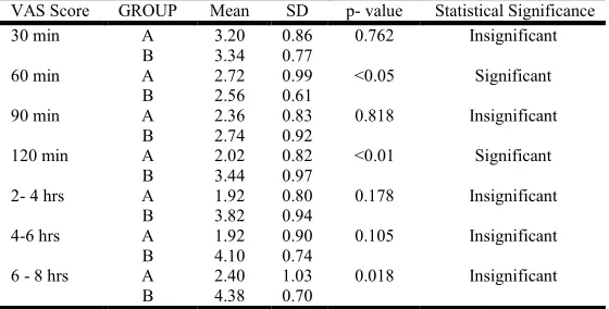
VAS score in postoperative periodtable 4 (vas score in postoperative period): Group A is patients receiving rectus sheath block and Group B is those receiving epidural analgesia
VAS Score GROUP Mean SD p- value Statistical Significance 30 min A 3.20 0.86 0.762 Insignificant
B 3.34 0.77
60 min A 2.72 0.99 <0.05 Significant
B 2.56 0.61
90 min A 2.36 0.83 0.818 Insignificant
B 2.74 0.92
120 min A 2.02 0.82 <0.01 Significant
B 3.44 0.97
2- 4 hrs A 1.92 0.80 0.178 Insignificant
B 3.82 0.94
4-6 hrs A 1.92 0.90 0.105 Insignificant
B 4.10 0.74
6 - 8 hrs A 2.40 1.03 0.018 Insignificant
[image:4.595.158.438.84.226.2]B 4.38 0.70
Table 2. Need for rescue analgesia
Need for Rescue Analgesia Group Group Total
Need for Rescue Analgesia A B
No 44 31 75
No 88.0% 62.0% 75.0%
Yes 6 19 25
Yes 12.0% 38.0% 25.0%
Total 50 50 100
100.0% 100.0% 100.0%
p- value <0.01 Statistically significant Statistically significant
Graph 2. VAS score in postoperative period
3.20
2.72
2.36
2.02 1.92 1.92 2.40
3.34
2.56 2.74
3.44 3.82
4.10 4.38
0.00 0.50 1.00 1.50 2.00 2.50 3.00 3.50 4.00 4.50 5.00
30 min 60 min 90 min 120 min 2- 4 hrs 4-6 hrs 6 - 8 hrs
VAS Comparison
A B
hemodynamic parameters and alterations were noted. Patients were followed up at regular intervals of 30 minutes for first two hours in immediate postoperative period, every two hours thereafter for first eight 8 hours in immediate postoperative period. Postoperative VAS score and need for any rescue analgesia, complications if any were noted. All these characteristics were compared statistically.
Group A had better VAS scores in immediate postoperative period for first 30 minutes (mean VAS 3.20 in group A and 3.34 in group B), 30 to 60 minutes (mean VAS 2.72 in group A and 2.56 in group B), 60 to 90 minutes (mean VAS of 2.36 in group A and 2.74 in group B), 90 to 120 minutes (mean VAS of 2.02 in group A and 3.44 in group B), 2 to 4 hours (mean VAS of 1.92 in group A and 3.82 in group B), 4 to 6 hours (mean VAS of 1.92 in group A and 4.10 in group B), 6 to 8 hours (mean VAS 2.40 of in group A and 4.38 in group B) as compared to group B. In group A, out of 50 patients 6 patients which is 12% of the total group A population, received rescue analgesia in form of intravenous injection of paracetamol in dose of 15mg/kg as they had VAS score of more than 4 as stated in protocol, with 44 patients which is 88% of the total group A population did not receive any rescue analgesia. While in group B, out of 50 patients, 19 patients which is 38% of total group B population, received rescue analgesia, with 21 patients which is 62% of the total group B population did not receive any rescue analgesia (P value <0.01)
Group B had received rescue analgesia more times than group A in immediate postoperative period indicating less efficient pain relief. Statistically significant difference has also been observed in postoperative pulse rate among group A and group B with P value of 0.026, indicating better pain relief among group A patients. With no complications noted in group A throughout study while nausea and vomiting in two patients, hypotension among three patients of group B.
Conclusion
From this study, we conclude that ultrasound guided rectus sheath block is a better analgesic modality than epidural analgesia in immediate postoperative period at some time intervals requiring less amount of rescue nonopioid analgesia without any complications.
REFERANCES
‘’Pain: current understanding of assessment, management and treatments’’. Joint Commission on Accreditation of healthcare Organizations and the National Pharmaceutical Council, Inc. December 2001. Retrieved January 2013.
Alsaeed et al, Ultrasound guided rectus sheath block in
children with umbilical hernia as a case series, Saudi Journal of Anaesthesia;oct-dec2013, Vol. 7/Issue 4, p432. Ayam A Kasem, 2015. Ashraf A AbdelkaderUltrasound
guided rectus sheath block versus local infiltration in management of pain after single-incision laparoscopic
cholecystectomy, Ains Shams Jurnal of Anaesthesiology,
8(1);100-106.
Cashman J. N., Dolin, S. J. 2004. Respiratory and haemodynamic effects of acute postoperative pain
management: evidence from published data, British
Journal of Anaesthesia, 93 (2):212-23.
Dilekozcengiz, Beyza Tekin Bayrak, Ersel Gulek, Murat Alkan, Yasemin Gunes, 2012. Rectus sheath block for postoperative pain relief in children undergoing major
abdominal surgery, Journal of Anaesthesiology and clinical
science, 1:5. http://dx.doi.org/10.7243/2049-9752-1-5.
Ferreyra G. et al. 2009. Respiratory complications after major
surgery, CurropinCrit Care, 15(4):342-8.
Godden AR, MJ Marshall, AS Grice, I.R. 2013. Daniels Ultrasonography guided rectus sheath catheters versus epidural analgesia for open colorectal cancer surgeries in a
single centre, Ann R Coll Surg Engl., 95:591-594.
Hazem El Sayed Moawad, Ehab M. Mokbel, 2014. Postoperative analgesia after major abdominal surgery: fentanyl-bupivacaine patient controlled epidural analgesia versus fentanyl patient controlled intravenous analgesia,
Egyptian Journal of Anaesthesia, Vol.30(4):393-397.
Kate M Wilkinson et al, 2014. Thoracic Epidural analgsia
versus Rectus sheath catheters for open midline incisions in major abdominal surgery within an enhanced recovery
programme (TERSC), Wilkinson et al. Trials., 15:400.
Khaled Abdelsalam, Ow MohamdinUltrasound guided rectus sheath block and transverse abdominis plane block for perioperative analgesia in patients with upper abdominal
surgery, Saudi journal of Anaesthesia, 2016;10(1):25-28
Khaled Elbahraw Y, Alaa El-Deeb, 2016. Rectus sheath block for postoperative analgesia in patients with mesenteric vascular occlusion undergoing laparotomy as a randomised
single-blinded study, Anaesthesia Essays and Researches,
10(3);516-520.
Marhofer P, Greher M, Kapral S. 2005. Ultrasound guidance in
regional anaesthesia. Br J Anaesth., 19;630-9.
Mattia c, Coluzzi F. 2009. What anaesthesiologist should know
about paracetamol (acetaminophen). Minerva Anestesiol.,
75:644-53.
Moore K, Dalley A. 2006. Clinically oriented anatomy. 5th ed.
Philadelphia: Lippincott Williams & Wilkins, 206.
Mukesh Kumar Shah, Sandeep S. Kulkarni, Wendy Fun, 2012. Analgesic efficacy of ultrasound guided modified rectus sheath block compared with wound infiltration in reduction of postoperative morphine consumption in women undergoing open hysterectomy or myomectomy: a
randomised controlled trial, Journal of Obstetric
Anaesthesia and Critical Care, Vol. 2/Issue 2.
Reips U. D. and F. Funke, 2008. “Interval level measurement with visual analogue scales in internet-based research: VAS Generator” doi: 10.3758/BRM.40.3.699.
RuchiSaxena et al. 2016. Comparative study of ultrasound
guided abdominal field blocks versus port infiltration in laparoscopic cholecystectomies for post-operative pain relief, Indian Journal of Anaesthesia, 60(8):578-583. Sandeman DJ, Dilley AV. 2008. Ultrasound guided rectus
sheath block and catheter placement. ANZ Journal of
Surgery, 78;621-3
Schleich CL. 1899. Schemerzloseoperationen. Berlin:
Springer, 240-1.
Scott Dingeman R et al. 2013. Ultrasonography-Guided
Bilateral Rectus Sheath Block vs Local Anesthetic Infiltration After Pediatric Umbilical Hernia -A Prospective
Randomised clinical Trial, JAMA Surg., 148(8)707-713.
Skinner AV, Lauder GR. Rectus sheath block: successful use in the chronic pain management.
Sotonye Fyneface-Ogan, Anatomy and clinical importance of the epidural space, www.intechopen.com
Anaesthetic Practice, 3rd edition. Wolters Kluwer; 2015: p 202, 203, 213.
Susan M Nimmo, Lorraine S Harrington, What is the role of epidural analgesia in abdominal surgery. http://www. medscape.com/viewearticle/837160_8.
Towards a mechanism-based classification of pain? Pain. 1998; 77(3)227-9.
What is this thing called pain? Journal of clinical investigation. 2010; 120(11):3742-4.
Yarwood, J, A Berrill, 2010. Nerve blocks of the anterior
abdominal wall, Contin Educ Anaesth Crit Care Pain,
doi:10. 1093/bjaceacp/mkq035.