VITAMIN D RECEPTOR GENE POLYMORPHISM IN OBESE AND NON OBESE INDIAN WOMEN
WITH POLYCYSTIC OVARY SYNDROME
1, *
Amar Nagesh Kumar,
2Uppala Satyanarayana,
Ramalingam,
1
Department of Biochemistry, Narayana Medical College and Hospital, Chinthareddypalem, Nellore,
2Department of Biochemistry, Dr Pinnamaneni Siddhartha Institute of Medical Sciences, Chinaoutapalli,
Gannavaram, Andhra Pradesh, India
3
Department of Obstetrics and Gynecology, Narayana Medical College and Hospital, Chinthareddypalem,
ARTICLE INFO ABSTRACT
Polycystic ovary syndrome (PCOS) is the most common endocrine disorder of the women in reproductive age group. Accumulating evidences from the recent studies indicate that vitamin D receptor (VDR) genetic variants may
the pathogenesis of polycystic ovary syndrome. The aim of the present study was to determine the VDR BsmI gene variant in intron 8 (A/G) (rs1544410) in normal controls, obese PCOS women and non obese PCOS women in India. A total
study. The subjects were divided into three groups as obese PCOS women, non obese PCOS women and healthy controls and each group consists of 75 participants. Genotypes of VDR gene in intron 8 (A/G)
statistical difference in genotype of AA, GA and GG between PCOS women and control women (p value >0.05). Our study suggests that there was no significant association of
both obese and non obese PCOS women.
Copyright © 2015 Amar Nagesh Kumar et al. This
unrestricted use, distribution, and reproduction in any medium, provided the original work is properly cited.
INTRODUCTION
Polycystic ovary syndrome (PCOS) affects 5
women in reproductive age group; however, the incidence might increase due to nutritional changes (Allahbadia and Merchant, 2008). An increase in the prevalence of PCOS among Indians is also a great concern. Observation of South Indian gynecologists from their experience reported 25
the women visiting them do suffer from PCOS (Muralidhara
al., 2015; Nidhi et al., 2011). Clinical manifestations of PCOS include oligomenorrhea, hirsutism, alopecia, obesity, and metabolic disturbances. Further, PCOS is associated with future complications such as cardiovascular abnormalities, type 2 diabetes mellitus, dyslipidemia, risk of malignancies and infertility (Kumar et al., 2014).
*Corresponding author: Amar Nagesh Kumar,
Department of Biochemistry, Narayana Medical College and Hospital, Chinthareddypalem, Nellore, Andhra Pradesh, India
ISSN: 0975-833X
Article History:
Received 07th May, 2015
Received in revised form 12th June, 2015
Accepted 27th July, 2015
Published online 31st August,2015
Key words:
Polycystic ovary syndrome, Insulin resistance, 25 – OH vitamin D, Vitamin D receptor gene, BsmI,
Infertility.
Citation: Amar Nagesh Kumar, Uppala Satyanarayana, Jupalle Nagaiah Naidu, Krishnan Ramalingam, Medabalmi Anitha
D receptor gene polymorphism in obese and non obese
Research, 7, (8), 19686-19691.
RESEARCH ARTICLE
VITAMIN D RECEPTOR GENE POLYMORPHISM IN OBESE AND NON OBESE INDIAN WOMEN
WITH POLYCYSTIC OVARY SYNDROME
Uppala Satyanarayana,
1Jupalle Nagaiah Naidu,
Ramalingam,
3Medabalmi Anitha
Department of Biochemistry, Narayana Medical College and Hospital, Chinthareddypalem, Nellore,
Andhra Pradesh, India
Pinnamaneni Siddhartha Institute of Medical Sciences, Chinaoutapalli,
Gannavaram, Andhra Pradesh, India
Department of Obstetrics and Gynecology, Narayana Medical College and Hospital, Chinthareddypalem,
Nellore, Andhra Pradesh, India
ABSTRACT
Polycystic ovary syndrome (PCOS) is the most common endocrine disorder of the women in reproductive age group. Accumulating evidences from the recent studies indicate that vitamin D receptor (VDR) genetic variants may influence the development of insulin r
the pathogenesis of polycystic ovary syndrome. The aim of the present study was to determine the VDR BsmI gene variant in intron 8 (A/G) (rs1544410) in normal controls, obese PCOS women and non obese PCOS women in India. A total of 225 women aged between 19
study. The subjects were divided into three groups as obese PCOS women, non obese PCOS women and healthy controls and each group consists of 75 participants. Genotypes of VDR gene in intron 8
with BsmI restriction enzyme were determined using the PCR
statistical difference in genotype of AA, GA and GG between PCOS women and control women (p value >0.05). Our study suggests that there was no significant association of
both obese and non obese PCOS women.
This is an open access article distributed under the Creative Commons Att use, distribution, and reproduction in any medium, provided the original work is properly cited.
Polycystic ovary syndrome (PCOS) affects 5-10% of the women in reproductive age group; however, the incidence might increase due to nutritional changes (Allahbadia and Merchant, 2008). An increase in the prevalence of PCOS among Indians is also a great concern. Observation of South heir experience reported 25-30% of the women visiting them do suffer from PCOS (Muralidhara et
Clinical manifestations of PCOS include oligomenorrhea, hirsutism, alopecia, obesity, and s associated with future complications such as cardiovascular abnormalities, type 2 diabetes mellitus, dyslipidemia, risk of malignancies
a Medical College and Hospital, Chinthareddypalem, Nellore, Andhra Pradesh, India.
The aetiopathology of PCOS reported the association of many environmental as well as genetic factors in different ethnic groups (Prapas et al., 2009; Fra
et al., 2006). A number of candidate genes involved in steroidogenesis (Gaaseenbeek
insulin signaling pathway (Jin et al.,
gonadotropin secretion (Li et al.,
to be associated with increased susceptibility to PCOS, but none is reported to have strong correlation with susceptibility to the disease. Increasing evidences suggests the contribution of vitamin D role in the pathogensis of polycystic ovary syndrome. Vitamin D deficiency might be a causal factor in the pathogenesis of insulin resistance and the metabolic syndrome in PCOS (Hahn et al
Hence vitamin D receptor (VDR) locus variations seem to have important impact on pathogenes
in PCOS women. Mahmoudi in 2009 reported that “bb” genotype (presence of restriction sites for ApaI and BsmI) has
Available online at http://www.journalcra.com
International Journal of Current Research
Vol. 7, Issue, 08, pp.19686-19691, August, 2015
INTERNATIONAL
Amar Nagesh Kumar, Uppala Satyanarayana, Jupalle Nagaiah Naidu, Krishnan Ramalingam, Medabalmi Anitha
m in obese and non obese Indian women with polycystic ovary syndrome”, International Journal of Current
z
VITAMIN D RECEPTOR GENE POLYMORPHISM IN OBESE AND NON OBESE INDIAN WOMEN
Jupalle Nagaiah Naidu,
1Krishnan
Department of Biochemistry, Narayana Medical College and Hospital, Chinthareddypalem, Nellore,
Pinnamaneni Siddhartha Institute of Medical Sciences, Chinaoutapalli,
Department of Obstetrics and Gynecology, Narayana Medical College and Hospital, Chinthareddypalem,
Polycystic ovary syndrome (PCOS) is the most common endocrine disorder of the women in reproductive age group. Accumulating evidences from the recent studies indicate that vitamin D the development of insulin resistance and so related to the pathogenesis of polycystic ovary syndrome. The aim of the present study was to determine the VDR BsmI gene variant in intron 8 (A/G) (rs1544410) in normal controls, obese PCOS women and of 225 women aged between 19-36 years participated in the study. The subjects were divided into three groups as obese PCOS women, non obese PCOS women and healthy controls and each group consists of 75 participants. Genotypes of VDR gene in intron 8 with BsmI restriction enzyme were determined using the PCR-RFLP method. There was no statistical difference in genotype of AA, GA and GG between PCOS women and control women (p value >0.05). Our study suggests that there was no significant association of BsmI genotypes with
is an open access article distributed under the Creative Commons Attribution License, which permits
The aetiopathology of PCOS reported the association of many environmental as well as genetic factors in different ethnic 2009; Fratantonio et al., 2005; Diamanti ). A number of candidate genes involved in steroidogenesis (Gaaseenbeek et al.,2004; Qin et al.,2006),
et al.,2006; Lee et al.,2008) and
et al.,2011) have been investigated to be associated with increased susceptibility to PCOS, but none is reported to have strong correlation with susceptibility to the disease. Increasing evidences suggests the contribution of vitamin D role in the pathogensis of polycystic ovary . Vitamin D deficiency might be a causal factor in the pathogenesis of insulin resistance and the metabolic
et al., 2006; Wehr et al., 2009). Hence vitamin D receptor (VDR) locus variations seem to have important impact on pathogenesis and insulin resistance in PCOS women. Mahmoudi in 2009 reported that “bb” genotype (presence of restriction sites for ApaI and BsmI) has INTERNATIONAL JOURNAL OF CURRENT RESEARCH
Amar Nagesh Kumar, Uppala Satyanarayana, Jupalle Nagaiah Naidu, Krishnan Ramalingam, Medabalmi Anitha, 2015. “Vitamin
been associated with higher levels of insulin and insulin resistance in comparison to ‘‘Ff/ff’’ and “BB and Bb” genotypes (Mahmoudi, 2009). Later in 2011, Ranjzad reported that there was significant association between VDR BsmI GG genotype and decreased levels of sex hormone binding globulin (SHBG) in PCOS women (Ranjzad 2011). Present investigation is the first to study the role of the VDR gene polymorphism (BsmI) in genetic susceptibility to PCOS in Indian women.
MATERIALS AND METHODS
The present study was conducted at Narayana Medical College and Hospital, Nellore, Andhra Pradesh, India during the period of October 2012 to December 2014. The study population included 75 obese and 75 non obese women with PCOS (cases) diagnosed based on Rotterdam criteria and 75 healthy women to serve as controls. All the control women had normal thyroid function, regular menstrual cycles, and no clinical signs of hyperandrogenism. All cases and controls were genetically unrelated. Diagnosis of PCOS was m
basis of the Rotterdam criteria (Rotterdam ESHRE/ASRM consensus, 2004). Two out of three of the following are required for diagnosis: oligo- and/or anovulation (defined by the presence of oligomenorrhea or amenorrhea); clinical and/or biochemical signs of hyperandrogenism [defined by presence of hirsutism (Ferriman–Gallwey score ≥6), acne or alopecia, and/or elevated androgen levels] and polycystic ovaries by gynecological ultrasound. Patients with congenital adrenal hyperplasia, Cushing’s syndrome, androgen
known hypothyroidism on treatment, other confounding factors as well as individuals who are already on treatment were excluded from the study. In addition, all subjects had polycystic ovaries by ultrasonography, but this was required inclusion criterion.
The study was approved by Institutional Ethics Committee, Narayana Medical College and Hospital and informed consent was obtained from all the subjects. Body mass index (BMI) was defined as the weight in kilograms divided by the square of the height in meters (kg/m2). Obesity was defined as a BMI ≥25.10 kg/m2 and non-obese as BMI < 25 kg/m
based on the consensus statement that the cut offs for overweight and obesity Asian Indians (Misra
About 5 ml of venous blood was obtained during a spontaneous bleeding episode of menstrual cycle after an overnight fast and collected in plain, fluoride and EDTA tubes from all the subjects. Specimens were immediately centrifuged and serum was separated and stored at -20oC until analysis. In both obese and non obese women with PCOS serum concentrations of luteinizing hormone (LH), follicle stimulating hormone (FSH), glucose, insulin, total calcium, and 25-OH vitamin D were measured. Serum glucose and calcium were assessed on Huma star-600 fully automated biochemistry analyzer by using commercial kits. Serum fasti insulin, luteinizing hormone (LH), follicle stimulating hormone (FSH) were measured with chemiluminiscence immunoassay (CLIA) method using Beckman Coulter Access – 2 fully automated analyzer. The hormone kits used in the Beckman Coulter Access analyzer 2(USA) were manufactured from Beckman Coulter, Ireland. Serum 25-OH vitamin D was been associated with higher levels of insulin and insulin resistance in comparison to ‘‘Ff/ff’’ and “BB and Bb” otypes (Mahmoudi, 2009). Later in 2011, Ranjzad et al., reported that there was significant association between VDR BsmI GG genotype and decreased levels of sex hormone binding globulin (SHBG) in PCOS women (Ranjzad et al.,
the first to study the role of the VDR gene polymorphism (BsmI) in genetic susceptibility to
The present study was conducted at Narayana Medical College and Hospital, Nellore, Andhra Pradesh, India during the period of October 2012 to December 2014. The study population included 75 obese and 75 non obese women with PCOS Rotterdam criteria and 75 healthy women to serve as controls. All the control women had normal thyroid function, regular menstrual cycles, and no clinical signs of hyperandrogenism. All cases and controls were Diagnosis of PCOS was made on the basis of the Rotterdam criteria (Rotterdam ESHRE/ASRM Two out of three of the following are and/or anovulation (defined by the presence of oligomenorrhea or amenorrhea); clinical and/or al signs of hyperandrogenism [defined by presence ≥6), acne or alopecia, and/or elevated androgen levels] and polycystic ovaries by gynecological ultrasound. Patients with congenital adrenal ome, androgen-secreting tumors, known hypothyroidism on treatment, other confounding factors as well as individuals who are already on treatment were excluded from the study. In addition, all subjects had polycystic ovaries by ultrasonography, but this was not a
The study was approved by Institutional Ethics Committee, Medical College and Hospital and informed consent was obtained from all the subjects. Body mass index (BMI) was defined as the weight in kilograms divided by the square ). Obesity was defined as a BMI se as BMI < 25 kg/m2. This was based on the consensus statement that the cut offs for overweight and obesity Asian Indians (Misra et al., 2009). About 5 ml of venous blood was obtained during a spontaneous bleeding episode of menstrual cycle after an rnight fast and collected in plain, fluoride and EDTA tubes from all the subjects. Specimens were immediately centrifuged C until analysis. In both obese and non obese women with PCOS serum izing hormone (LH), follicle stimulating hormone (FSH), glucose, insulin, total calcium, OH vitamin D were measured. Serum glucose and 600 fully automated biochemistry analyzer by using commercial kits. Serum fasting insulin, luteinizing hormone (LH), follicle stimulating hormone (FSH) were measured with chemiluminiscence immunoassay (CLIA) method using Beckman Coulter Access 2 fully automated analyzer. The hormone kits used in the 2(USA) were manufactured OH vitamin D was
estimated by high performance liquid chromatography (HPLC) with commercial column and reagents from RECIPE (Germany) and Younglin HPLC (Korea).
was estimated by formula for the homeostatic model assessment-insulin resistance (HOMA
2004). Whole blood collected in EDTA tubes was used for DNA isolation. Genomic DNA was isolated from peripheral blood (200µl) leukocytes with the commercial kit
[image:2.595.327.538.189.320.2]DNA extraction kit) according to manufacturer’s instructions. Concentration of DNA was estimated using nano drop (Thermo Fischer Scientific) in ng/µl.
[image:2.595.327.534.349.499.2]Figure 1. Genomic DNA extracted from different subjects
Figure 2. PCR amplification of human genome using primers targeted against VDR gene. Lane1, 100 bp DNA ladder; lane 2
amplified products from different genomic DNA samples
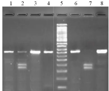
Figure 3. Restriction pattern of VDR gene fragment with BsmI. Lane 1, 3, 4, 6, 8 represents AA genotype, Lane 2
Lane 7 - GG genotype and Lane 5
estimated by high performance liquid chromatography (HPLC) with commercial column and reagents from RECIPE (Germany) and Younglin HPLC (Korea). Insulin resistance ed by formula for the homeostatic model insulin resistance (HOMA- IR) (Legro et al.,
[image:2.595.328.536.547.722.2]Whole blood collected in EDTA tubes was used for DNA isolation. Genomic DNA was isolated from peripheral blood (200µl) leukocytes with the commercial kit (Bioserve DNA extraction kit) according to manufacturer’s instructions. Concentration of DNA was estimated using nano drop (Thermo Fischer Scientific) in ng/µl.
Figure 1. Genomic DNA extracted from different subjects
PCR amplification of human genome using primers targeted against VDR gene. Lane1, 100 bp DNA ladder; lane 2-8,
different genomic DNA samples
The genotyping of VDR gene (rs1544410) loci in intron 8 (G/A) detected by restriction enzyme BsmI using restriction fragment length polymorphism - polymerase chain reaction (RFLP-PCR) method. Polymerase chain reaction is an in vitro
method for replicating the desired DNA fragment so that its amount increases exponentially. Figure 1 presents isolated genomic DNAs from different patients or control subjects on 1.5% agarose gel, which shows a characteristic band of genomic DNA near the well side. The genomic DNA from different subjects was amplified by polymerase chain reaction using primers targeted against the human VDR gene containing intron 8. PCR was performed using the following primers: forward: 5’ GGG AGA CGT AGC AAA AGG 3’ and reverse 5’ AGA GGT CAA GGG TCA CTG 3’. PCR amplification of the human genome using primers of the VDR gene, as mentioned above, yielded a fragment of 360bp. Figure 2shows a representative gel photograph of the amplified DNA product on 2% agarose gel.
Following DNA amplification, PCR products were digested with BsmI (CutSmart, New England BioLabs) at 65oC for 15 min (Table 1). The digested products were analyzed for the presence or absence of recognition sites by ethidium bromide staining of fragments separated through a 2% agarose gel and photographed. Three types of genotypes have been observed in the study subjects represented as AA, AG and GG. The genotype designated as AA shows absence of restriction site and yielded only one band in agarose gel electrophoresis at 360bp region which is of same size of PCR product. The genotype designated by GG shows presence of restriction site and yielded two bands of size 191 bp and 169 bp. The GA genotype presented with three bands in gel electrophoresis and size of fragments is 360 bp, 191bp, 169 bp (Figure 3).
Statisticalanalysis
Hardy Weinberg Equilibrium was tested to find out the gene polymorphism. The chi-square () test was performed to compare VDR BsmI (rs1544410) intron 8 (G/A) genotypic and allelic distributions between patients and healthy control group. The association between genotypes was examined by odds ratio with 95% Confidence Interval (CI). Chi-Square analysis using Open epi 6 software (open Epi version 2.3.1 from the department of epidemiology, Rollins School of Public Health, Emory University, Atlanta, GA 30322, USA). Allelic frequencies were calculated according to the number of different alleles observed and total number of alleles examined.
RESULTS
[image:3.595.75.519.454.508.2]The studied group consisted of 75 obese PCOS women, 75 non obese PCOS women and 75 healthy women as normal controls with age ranging from 19 to 36 years. The mean age of obese PCS women was, 25.96 ± 3.22 years, non obese PCOS women was 25.60 ± 4.08, and that of control women was 26.06 ± 3.58. Statistically significant difference between cases and controls was not found regarding age (p-value >0.05). But with respect to BMI (kg/m2) there is statistically significant difference between obese PCOS women (mean ± SD, 28.29 ± 3.20), non obese PCOS women (mean ± SD, 21.91 ± 1.30) and controls (mean ± SD, 24.86 ± 2.98) (p <0.001). PCOS women had a higher LH, FSH levels and low vitamin D and calcium levels when compared to controls. The LH/FSH ratio was significantly higher in obese PCOS women (1.02 ± 0.15) (p <0.05) than non obese PCOS women (0.95 ± 0.15) when compared to healthy controls (0.94 ± 0.13) (Table 2).
Table 1. Type of SNPs, site of SNPs, and PCR Conditions
Gene Location PCR details
VDR/BsmI (rs1544410)
Intron VIII (G/A)
PCR protocol: 94oC for 1 min, 54oC for 1 min, 72oC for 1 min for 30 cycles.
5’ GGG AGA CGT AGC AAA AGG 3’ 5’ AGA GGT CAA GGG TCA CTG 3’
[image:3.595.119.477.534.680.2]un-cut PCR product size: 360(bp) Restriction enzymes, Incubation temperature: Bsml, 65°C for 15 min
Table 2 Demographic and clinical characteristics of control and PCOS women
Parameter Control women Obese PCOS women Non obese PCOS women
Age (Years) 26.06 ± 3.58 25.96 ± 3.22 25.60 ± 4.08
BMI (kg/m2) 24.86±2.9 27.82 ± 3.14** 21.91±1.30**
WC (cm) 71.81±4.96 85.55±5.40** 74.53±5.31**
HC(cm) 86.29±5.87 87.83±4.22 83.20±11.16*
W/H ratio 0.83±0.07 0.96±0.05** 0.88±0.05**
Glucose (mg/dl) 86.69±7.60 83.48±5.48** 81.89±4.90**
Insulin (µIU/ml) 6.65±1.25 7.29±1.29** 5.93±1.12**
HOMA – IR 1.42± 0.36 1.49±0.31 1.20±0.27
LH (mIU/L) 6.00±1.76 8.05±1.24** 6.78±2.05**
FSH (mIU/L) 6.28±1.55 7.85±1.02** 7.09±1.89**
LH/FSH 0.94 ± 0.13 1.02 ± 0.15** 0.95 ± 0.15
Vitamin D (ng/ml) 31.91±8.28 30.36±8.33 30.99±6.64
Ca (mg/dl) 8.55±0.50 7.36±1.08** 6.93±1.17**
Phosphorous (mg/dl) 4.41±0.75 4.18±0.31* 4.36±0.33
*Significant at 0.05 level with control group; **Significant at 0.01 level with control group
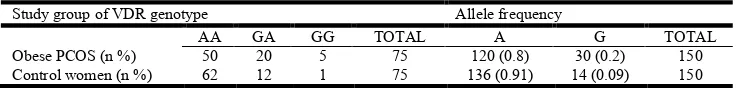
Table 3. Distribution of VDR gene intron 8 (G/A) genotypes and allelic frequencies of the obese POCS women and control women
Study group of VDR genotype Allele frequency
AA GA GG TOTAL A G TOTAL
Obese PCOS (n %) 50 20 5 75 120 (0.8) 30 (0.2) 150
Control women (n %) 62 12 1 75 136 (0.91) 14 (0.09) 150
For GG vs. AA; p = 0.07; odds ratio = 0.16 (95% CI: 0.018 – 1.42); for GG vs. GA + AA - = 2.7;
p = 0.09; odds ratio = 5.2 (95% CI: 0.6 – 46.37); G vs A = 6.79; p=0.009; odds ratio = 0.41 (95% CI: 0.2-0.81)
[image:3.595.114.482.714.758.2]The genotypic distribution of VDR BsmI (rs1544410) intron 8 (A/G) polymorphism and allelic frequency of A and G alleles in obese and non obese PCOS women and controls have been given in Table 2 and Table 3. In the case of VDR BsmI (rs1544410) intron 8 (A/G), obese PCOS women ( = 3.39 <3.84, p value with degree of freedom 2 = 0.07 >0.05) and non obese PCOS women ( = 2.6 <3.84, p value with degree of freedom 2 = 0.1 >0.05) were consistent with Hardy Weinberg equilibrium. VDR BsmI (rs1544410) intron 8 (A/G) AA, GA, GG, A and G genotypic/allelic frequencies were 50 (66.6%), 20 (26.7%), 5(6.7%), 120(80%), and 30(20%) in obese PCOS women, 47(62.7%), 24(32%), 4(5.3%), 118(78.6%), and 32(21.3%) in non obese PCOS women and 62(82.6%), 12(16%), 1(1.4%), 136(90.6%), and 14(9.4%) in controls, respectively. The frequencies of A and G alleles were 0.8% and 0.2% in obese PCOS women and that of non obese PCOS women were 0.78%, 0.22%. While the frequencies of A and G alleles were 0.91% and 0.09% in controls. Statistical analysis showed that the differences in genotypic frequencies between the cases and controls were not statistically significant regarding VDR BsmI (rs1544410) intron 8 (A>G) (p >0.05) (Table 3 and Table 4). Our study reported that there was no statistical difference in genotype of AA, GA and GG between obese PCOS/non obese PCOS women and control women (p value >0.05). But, we observed statistically significant difference in allelic frequencies of A and G in both obese and non obese PCOS women when compared to controls (p <0.05).
DISCUSSION
The aim of present study was to assess the role of VDR BsmI (rs1544410) (A/G) genetic variation in PCOS for the first time in Indian women. Current understanding of the molecular actions of vitamin D in the fertility of women and calcium homeostasis prompted the design of the present study. The goal of this investigation was to study whether the VDR BSmI gene variant in intron 8 (A/G) (rs1544410) is related to onset of PCOS for the first time in Indian women. PCOS is known as a syndrome and affects ovarian function. PCOS most commonly occur during adolescence and characterized by several different features including amenorrhoea, oligomenorrhoea, infertility as well as other metabolic problems in medical findings (Golbahar et al., 2012). It has been indicated that some females with syndrome will show polycystic ovary without clinical features of androgen excess. A patient with PCOS has an increased risk of obesity, bleeding disorders, hyperandrogenemia, endometrial carcinoma, breast cancer, chronic anovulation, infertility, insulin resistance, diabetes, hypertension, primary hyperparathyroidism, dyslipidemia and coronary artery disease (Pfeifer, 2005).
Research studies indicated that the reason of the ovarian overproduction of testosterone in PCOS women is due by inability of women to mediate insulin effectively (insulin resistance or hyperinsulinemia) (Mahmoudi, 2009; Wehr et al.,
2011). Hyperandrogenemia and insulin resistance are important indicators of PCOS. In this condition, level of insulin hormone within the blood is too high; therefore the ovaries produce higher level of testosterone (Wehr et al.,
2011). SHBG is a carrier protein which regulates the level of unbound steroids in peripheral blood (Golbahar et al., 2012). It has been demonstrated that VDR genetic variations have been associated with LH and SHBG levels in PCOS women (Ranjzad et al., 2011). It has been demonstrated that SHBG expression is reduced in the stromal compartment of endometria of women with polycystic ovary syndrome (Maliqueo et al., 2007). Increase in the levels of androgens bioavailability result in hyperandrogenemia by hyperinsulinemia in PCOS women with VDR BsmI GG genotypes via lower serum level of SHBG (Ranjzad et al.,
2011; Mahmoudi, 2009).
Insulin resistance is in association with reproductive abnormalities in PCOS women. Insulin resistance is correlated with vitamin D metabolism in PCOS (Pfeifer 2005). Biological responses and functions of vitamin D are mediated via the VDR within the vitamin D endocrine system in more than 30 target tissues (Chiu et al., 2001; Harris et al., 1997; Vigo Gago
et al., 2005; Holick 2007; Kinuta et al., 2000). In the body, vitamin D regulates calcium homeostasis; an important function in development of the skeletal system as well as in bone mineralization (Hassan et al., 2012). Since vitamin D is the main regulator for calcium and phosphate translocation.
[image:4.595.111.485.88.131.2]From the small intestine into the circulation, defects observed in the mutant VDR and calcium absorption lead to decreased level of mineral transport and hypocalcemia (Hassan et al., 2012; Ranjzad et al., 2011). Vitamin D and calcium repletion predict reproductive success following fertilization (Brannon and Picciano, 2011; Grundmann and Von Versen-Hoynck, 2011). Liang et al., indicated a dynamic role for Ca level in oocyte maturation and early embryonic development. Other studies are consistent with Liang et al., and imply that regulation of the ovum activation, follicular development and mammalian embryo development are calcium dependent (Liang et al., 2011). The findings of Oh and Barrett-Connor in 2002, suggest that VDR gene variant may be associated with glucose intolerance independent of defective insulin secretion and with insulin resistance (Oh and Barrett-Connor, 2002). Mahmoudi, indicated that VDR gene variant may affect PCOS development as well as insulin resistance in women with PCOS. Insulin resistance and increased levels of LH are usual signs of PCOS. Higher levels of LH, not only has an effect on oocyte maturity and human reproduction but also on lower
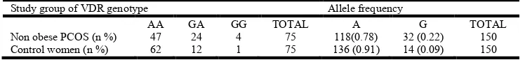
Table 4. Distribution of VDR gene intron 8 (G/A) genotypes and allelic frequencies of the non obese POCS women and control women
Study group of VDR genotype Allele frequency
AA GA GG TOTAL A G TOTAL
Non obese PCOS (n %) 47 24 4 75 118(0.78) 32 (0.22) 150
Control women (n %) 62 12 1 75 136 (0.91) 14 (0.09) 150
For GG vs. AA; p = 0.1; odds ratio = 0.19 (95% CI: 0.02 – 1.7); for GG vs. GA + AA - = 1.85; p = 0.17;
fertility and higher miscarriage prevalence. Still there were controversial findings about the action of LH on oocyte, embryo quality, fertility, implantation and miscarriage prevalence (Oh and Barrett-Connor, 2002; Gordon et al., 2001). In our study though PCOS women carrying the VDR BsmI “GG” and “GA” genotype showed low vitamin D, calcium, and phosphorous levels as compared with individuals in the “AA” genotype, but it is not statistically significant. The molecular mechanism through which this polymorphism influences LH levels is not known at present; however, previous studies have shown significant associations between VDR gene variants and insulin resistance on one side (Oh and Barrett-Connor, 2002), and insulin resistance and LH on the other. Furthermore, it has been suggested a modulating role of 1, 25 – OH vitamin D in the control of FSH secretion. Therefore, these findings are consistent with a recent report (Mahmoudi, 2009) that showed the VDR gene variants might have a role in pathogenesis of PCOS.
In the present study out of 75 obese POCS women, 50 women had AA genotype, 20 women had GA and 5 women showed GG genotype. In non obese PCOS women group 47 women presented AA genotype, 24 women had GA genotype, and 4 women presented with GG genotype. While in control 62 women were presented with AA genotype, 12 women had GA and only one women presented with GG genotype. In our study, statistical analysis showed that the differences in genotypic/allelic frequencies between the cases (obese and non obese PCOS women) and controls were not statistically significant with respect to VDR BsmI (rs1544410) intron 8 (A>G) (p >0.05). The findings of the present study were consistent with some reports (Wehr et al., 2011; Bagheri et al., 2012) and inconsistent with other (Mahmoudi, 2009; Ranjzad
et al., 2011; Jain et al., 2012) regarding the VDR BsmI gene variant. The exact aetiopathogenesis of PCOS are not known regarding vitamin D and insulin resistance. Several molecular mechanisms have been suggested to describe the relationship between the VDR locus variations and PCOS in different ethnic groups. We had some limitations regarding low sample size. Studies with a large sample size and more information such as other candidate gene variants, haplotypes and genetic linkage assessment are needed for further analysis (Tabor
et al., 2002; Cardon and Palmer, 2003; Colhoun et al., 2003).
Conclusion
Although our sample size is small, this study was well designed and focused on the role of vitamin D metabolism related gene polymorphism on metabolic and biochemical characteristics of Indian women with PCOS. It can be concluded that VDR BsmI (rs1544410) intron 8 (A/G) were not associated with PCOS susceptibility in our population. However, further studies with increased numbers of PCOS patients are required to validate these findings.
Acknowledgements
The first author (Mr Amar Nagesh Kumar) would like to thank Dr NTR University of Health Sciences, Vijayawada for sanctioning fellowship and helping him to carry out his research work. Authors would also like to thank all the subjects participated in the study.
Conflict of interest
There was no conflict of interest in this study.
Financial support
We did not receive any external funding to carry out this study.
REFERENCES
Allah badia, G. N. and Merchant, R. 2008. Polycystic ovary syndrome in the Indian Subcontinent. Semin Reprod Med., 26: 22-34.
Bagheri, M., Rad, I.A., Jazani, N. H. and Nanbakhsh, F. 2012. Lack of Association of Vitamin D Receptor FokI (rs10735810) (C/T) and BsmI (rs1544410) (A/G) Genetic Variations with Polycystic Ovary a Syndrome Risk: a Case control Study from Iranian Azeri Turkish Women. Maedica: J. Clin. Med., 7(4): 303 – 308.
Brannon, P. M. and Picciano, M. F. 2011. Vitamin D in pregnancy and lactation in humans. Annu Rev Nutr., 31: 89-115.
Cardon, L. R. and Palmer, L. J. 2003. Population stratification and spurious allelic association. Lancet, 361: 598-604. Chiu, K. C., Chuang, L. M. and Yoon, C. 2001. The vitamin D
receptor polymorphism in the translation initiation codon is a risk factor for insulin resistance in glucose tolerant Caucasians. BMC Med Genet., 2: 2.
Colhoun, H. M., McKeigue, P. M. and Davey, Smith, G. 2003. Problems of reporting genetic associations with complex outcomes. Lancet, 361:865-72.
Diamanti-Kandarakis, E., Kandarakis, H. and Legro, R. 2006. The role of genes and environment in the etiology of PCOS. Endocrine., 30:19-26.
Fratantonio E., Vicari E., Pafumi C., et al. 2005. Genetics of polycystic ovarian syndrome. Reprod Biomed Online., 10: 713-720.
Gaasenbeek, M., et al. 2004. Large scale analysis of the relationship between CYP11A promoter variation, polycystic ovarian syndrome, and serum testosterone. J Clin Endocrinol Metab., 89: 2408–2413.
Golbahar, J., Al-Ayadhi, M., Das, N.M., et al. 2012. Sensitive and specific markers for insulin resistance, hyperandrogenemia, and inappropriate gonadotrophin secretion in women with polycystic ovary syndrome: a case-control study from Bahrain. Int J Womens Health., 4: 201-206.
Gordon, U.D., Harrison, R.F., Fawzy, M., Hennelly, B. and Gordon, A.C. 2001. A randomized prospective assessor-blind evaluation of luteinizing hormone dosage and in vitro fertilization outcome. Fertil Steril., 75(2): 324-331.
Grundmann, M., and von, Versen-Hoynck, F. 2011. Vitamin D-roles in women’s reproductive health? Reprod Biol Endocrinol., 9: 146.
Hahn, S., Haselhorst, U., Tan, S., Quadbeck, B., Schmidt, M., Roesler, S., Kimmig, R., Mann, K. and Janssen, O.E. 2006. Low serum 25-hydroxyvitamin D concentrations are associated with insulin resistance and obesity in women with polycystic ovary syndrome. Exp Clin Endocrinol Diabetes., 114: 577–583.
Harris, S.S., Eccleshall, T.R., Gross, C., Dawson-Hughes, B. and Feldman, D. 1997. The vitamin D receptor start codon polymer phism (FokI) and bone mineral density in premenopausal American Black and White women. J Bone Miner Res., 12(7): 1043-1048.
Hassan, N.E., El-Orabi, H.A., Eid, Y.M. and Mohammed, N.R. 2012. Effect of 25-hydroxyvitamin D on metabolic parameters and insulin resistance in patients with polycystic ovarian syndrome. Mid East Fertil Society J.,
17: 76–180.
Holick, M.F. 2007. Vitamin D deficiency. N Engl J Med., 357(3): 266-281.
Jain, R., Hurst, P.R., Stonehouse, W., Love, D.R., Higgins, C.M. and Coad, J. 2012. Association of vitamin D receptor gene polymorphisms with insulin resistance and response to vitamin D. Metab Clin Exp., 61: 293 – 301.
Jin, L., Zhu, X.M., Luo, Q., Qian, Y., Jin, F. and Huang H.F. 2006. A novel SNP at exon 17 of INSR is associated with decreased insulin sensitivity in Chinese women with PCOS. Mol Hum Reprod., 12:151–155.
Kinuta, K., Tanaka, H., Moriwake, T., Aya, K., Kato, S. and Seino, Y. 2000. Vitamin D is an important factor in estrogen biosynthesis of both female and male gonads.
Endocrinol., 141(4): 1317-3124.
Kumar, A.N., Naidu, J.N., Stayanarayana, U. and Anitha, M. 2014. Past, present and future of insulin gene and its related genes in relation to polycystic ovary syndrome. J Mol Genet Med., 8: 107.
Lee, E.J., Oh, B., Lee, J.Y., Kimm, K., Lee. S.H. and Baek, K.H. 2008. A novel single nucleotide polymorphism of INSR gene for polycystic ovary syndrome. Fertil Steril., 89: 1213–1220.
Legro, R.S., Castracane, V.D. and Kauffman, R.P. 2004. Detecting insulin resistance in polycystic ovary syndrome: purposes and pitfalls. Obst Gynecol Survey., 59:141–154. Li Q, et al,. 2011. Common genetic variation in the
3'-untranslated region of gonadotropin-releasing hormone receptor regulates gene expression in cella and is associated with thyroid function, insulin secretion as well as insulin sensitivity in polycystic ovary syndrome patients. Hum Genet., 129: 553–561.
Liang, S.L., Zhao, Q.J., Li, X.C., Jin, Y.P., Wang, Y.P., Su, X.H., et al. 2011. Dynamic analysis of Ca²+ level during bovine oocytes maturation and early embryonic development. J Vet Sci., 12(2): 133-142.
Mahmoudi, T. 2009. Genetic variation in the vitamin D receptor and polycystic ovary syndrome risk. Fertil Steril., 92: 1381-1383.
Maliqueo, M., Bacallao, K., Quezada, S., et al. 2007. Sex hormone binding globulin expression in the endometria of women with polycystic ovary syndrome. Fertil Steril., 87: 321-328.
Misra, A., et al. 2009. Consensus statement for diagnosis of obesity, abdominal obesity and the metabolic syndrome for Asian Indians and recommendations for physical activity, medical and surgical management. J Assoc Physicians India., 57: 163-170.
Muralidhara, K.D., Adhikari, P.M. and Muralidhara, D.V. 2015. A Study on the Pattern of Genetic Inheritance of Polycystic Ovarian Syndrome. British J Med Medical Research., 10: 1230-1238.
Nidhi, R., Padmalatha, V., Nagarathna, R. and Ram, A. 2011. Prevalence of polycystic ovarian syndrome in Indian adolescents. J Pediatr Adolesc Gynecol., (4);24: 223-227. Oh, J.Y. and Barrett-Connor, E. 2002. Association between
vitamin D receptor polymorphism and type 2 diabetes or metabolic syndrome in community-dwelling older adults: the Rancho Bernardo Study. Metabolism., 51(3): 356-359. Pfeifer, S.M. 2005. Polycystic ovary syndrome in adolescent
girls. Semin Pediatr Surg., 14(2): 111-117.
Prapas, N., Karkanaki. A., Prapas I., et al. 2009. Genetics of polycystic ovary syndrome. Hippokratia., 13: 216-223. Qin, K., Ehrmann, D.A., Cox, N., Refetoff, S. and Rosenfield,
R.L. 2006. Identification of a functional polymorphism of the human type 5 17beta-hydroxysteroid dehydrogenase gene associated with polycystic ovary syndrome. J. Clin. Endocrinol. Metab., 91:270–276.
Ranjzad, F., Mahban, A., Shemirani, A.I., et al. 2011. Influence of gene variants related to calcium homeostasis on biochemical parameters of women with polycystic ovary syndrome. J Assist Reprod Genet., 28: 225-232.
Tabor, H.K., Risch, N.J. and Myers, R.M. 2002. Candidate-gene approaches for studying complex Candidate-genetic traits: practical considerations. Nat Rev Genet., 3: 391–397.
The Rotterdam ESHRE/ASRM-sponsored consensus
workshop group. 2004 Revised 2003 consensus on diagnostic criteria and long term health risks related to polycystic ovary syndrome (PCOS). Human Reproduction., 19:41–47.
Vigo, Gago, E., Cadarso-Suarez, C., Perez-Fernandez, R., Romero, Burgos, R., Devesa, Mugica. J. and Segura, Iglesias, C. 2005. Association between vitamin D receptor FokI polymorphism and serum parathyroid hormone level in patients with chronic renal failure. J Endocrinol Invest., 28(2): 117-121.
Wehr, E., Pilz, S., Schweighofer, N., Giuliani, A., Kopera, D., Pieber T. and Obermayer-Pietsch B. 2009. Association of hypovitaminosis D with metabolic disturbances in polycystic ovary syndrome. European Journal of Endocrinology., 161: 575–582.
Wehr, E., Trummer, O., Giuliani, A., Gruber, H.J., Pieber, T.R. and Pietsch, B.O. 2011. Vitamin D-associated polymorphisms are related to insulin resistance and vitamin D deficiency in polycystic ovary syndrome. European Journal of Endocrinology., 164(5):741–749.