POSTEXTRACTION SOCKET GRAFTING USING COMBINATION OF DEMINERALIZED
FREEZED-DRIED BONE ALLOGRAFT AND PLATELET
RICH FIBRIN
*
Dr. Shobhit Arora, Dr
Department of Periodontology, Santosh Dental College, Pratap Vihar, Ghaziabad, Uttar Pradesh, India
ARTICLE INFO ABSTRACT
Alveolar ridge bone resorption is a biologic phenomenon that occurs following tooth extraction and cannot be prevented.
improve the est
periodontal tissue engineering. Phosphate (β
with periodontally compromised tooth due to trauma with no labial cortical bone present which was confirmed by pre operative CBCT and was treated by extractions along with augmentation of the alve
blood was centrifuged to obtain PRF. Allograft bone graft was mixed with PRF particles and applied to fill the defect. After 6
use of PRF as co
months the site appeared precociously healed and the bone volume increased. This new approach represents a predictable method of augmenting deficient alveolar bone.
Copyright © 2015 Dr. Shobhit Aroraet al. This is an open access article distributed under the Creative Commons Att use, distribution, and reproduction in any medium, provided the original work is properly cited.
INTRODUCTION
The key processes of tissue modeling and remodeling after tooth extraction have been well documented in both animals and humans (Horowitz, 2012). Tooth extraction whether due to caries, trauma or advanced periodontal disease is a traumatic procedure often resulting in immediate destruction and loss of alveolar bone and surrounding soft tissues (Caplanis, 2005 general, the alveolar bone remodeling that occurs after tooth loss yields diminished alveolar ridge dimensions in both the vertical and horizontal planes up to 40% to 60% bone loss height and width as early as 3 months. On an average, grafted extraction sites have reported a loss of width < 2mm and a loss of height < 0.5mm as compared to non-grafted extraction sites which have reported losses of ridge width from 2
ridge height of 1 mm with great variations (Brownfield, 2012 To prevent this clinical situation, different authors have described several surgical procedures, ranging from regenerative techniques for socket preservation to immediate implant placement. Regenerative techniques have been widely tested in controlled and uncontrolled stud
materials and clinical approaches: bone grafting alone,
*Corresponding author: Dr. Shobhit Arora,
Department of Periodontology, Santosh Dental College, Pratap Vihar, Ghaziabad, Uttar Pradesh, India.
ISSN: 0975-833X
Vol.
Article History:
Received 25th September, 2015 Received in revised form 21st October, 2015
Accepted 27th November, 2015 Published online 30th December,2015
Key words:
Alveolar ridge augmentation, β- Tri Calcium Phosphate, Platelet Rich Fibrin.
Citation: Dr. Shobhit Arora, Dr. Shweta Bali and Dr. Priyanka Thukral demineralized freezed-dried bone allograft and platelet rich fibrin (PRF) 24225-24230.
RESEARCH ARTICLE
POSTEXTRACTION SOCKET GRAFTING USING COMBINATION OF DEMINERALIZED
DRIED BONE ALLOGRAFT AND PLATELET
RICH FIBRIN (PRF) –A CASE SERIES
Shobhit Arora, Dr. Shweta Bali and Dr. Priyanka Thukral
of Periodontology, Santosh Dental College, Pratap Vihar, Ghaziabad, Uttar Pradesh, India
ABSTRACT
Alveolar ridge bone resorption is a biologic phenomenon that occurs following tooth extraction and cannot be prevented. Newer techniques are evolving in restorative dentistry and periodontics to treat these defects to improve the esthetics, form and functions of the dentition. Alveolar bone augmentation remains the holy grail of periodontal tissue engineering.21The purpose of this paper was to evaluate the efficacy of
Phosphate (β-TCP) and Platelet Rich Fibrin (PRF) in post extraction socket preservation.
with periodontally compromised tooth due to trauma with no labial cortical bone present which was confirmed by pre operative CBCT and was treated by extractions along with augmentation of the alve
blood was centrifuged to obtain PRF. Allograft bone graft was mixed with PRF particles and applied to fill the defect. After 6 months a Cone-Beam Computed Tomography was performed to evaluate bone regeneration. The use of PRF as cover membrane permitted a rapid epithelisation and represented an effective barrier. After 6 months the site appeared precociously healed and the bone volume increased. This new approach represents a predictable method of augmenting deficient alveolar bone.
is an open access article distributed under the Creative Commons Attribution License, which use, distribution, and reproduction in any medium, provided the original work is properly cited.
processes of tissue modeling and remodeling after tooth extraction have been well documented in both animals Tooth extraction whether due to caries, trauma or advanced periodontal disease is a traumatic g in immediate destruction and loss of Caplanis, 2005). In general, the alveolar bone remodeling that occurs after tooth loss yields diminished alveolar ridge dimensions in both the vertical and horizontal planes up to 40% to 60% bone loss height and width as early as 3 months. On an average, grafted n sites have reported a loss of width < 2mm and a loss grafted extraction sites which have reported losses of ridge width from 2-6 mm and Brownfield, 2012). inical situation, different authors have described several surgical procedures, ranging from regenerative techniques for socket preservation to immediate implant placement. Regenerative techniques have been widely tested in controlled and uncontrolled studies with various materials and clinical approaches: bone grafting alone,
Department of Periodontology, Santosh Dental College, Pratap
including autografts, allografts,
alone or in combination with absorbable or nonabsorbable membrane (Mazor, 2009).
introduced Platelet Rich Fibrin (PRF), a second platelet concentrate that improves healing of the both hard soft tissues (Choukroun et al
concentrations of the collected platelets, which allow slow release of growth factors (GFs) (Kang
include Vascular Endothelium Growth Factor (VEGF), Platelet-Derived Growth Factor (PDGF), Fibroblast Growth Factor (FGF), Epidermal Growth Factor (EGF), Hepatocyte Growth Factor (HGF),
Insulin-Transforming Growth Factor-Β (TGF role in replacing lost tissue, resurfacing of restoring vascular integrity, (
Compared to other platelet concentrates, PRF releases these factors at a sustained rate over a longer period, thereby optimizing wound healing (Reddy
also been shown to stimulate the growth of osteoblasts and periodontal ligament cells, both of which are significant for the regeneration of periodontal defects
2011). Here, we present a case where PRF was used in combination with Demineralized Freeze
(DFDBA) for ridge augmentation procedure.
International Journal of Current Research
Vol. 7, Issue, 12, pp.24225-24230, December, 2015
Dr. Shobhit Arora, Dr. Shweta Bali and Dr. Priyanka Thukral,2015. “Postextraction socket grafting using combination of dried bone allograft and platelet rich fibrin (PRF) –A Case Series”, International Journal of Current Research,
POSTEXTRACTION SOCKET GRAFTING USING COMBINATION OF DEMINERALIZED
DRIED BONE ALLOGRAFT AND PLATELET
Priyanka Thukral
of Periodontology, Santosh Dental College, Pratap Vihar, Ghaziabad, Uttar Pradesh, India
Alveolar ridge bone resorption is a biologic phenomenon that occurs following tooth extraction and cannot be Newer techniques are evolving in restorative dentistry and periodontics to treat these defects to hetics, form and functions of the dentition. Alveolar bone augmentation remains the holy grail of The purpose of this paper was to evaluate the efficacy of β- Tri Calcium post extraction socket preservation. The patients presented with periodontally compromised tooth due to trauma with no labial cortical bone present which was confirmed by pre operative CBCT and was treated by extractions along with augmentation of the alveolar bone. The patient's blood was centrifuged to obtain PRF. Allograft bone graft was mixed with PRF particles and applied to fill the Beam Computed Tomography was performed to evaluate bone regeneration. The ver membrane permitted a rapid epithelisation and represented an effective barrier. After 6 months the site appeared precociously healed and the bone volume increased. This new approach represents a
ribution License, which permits unrestricted
including autografts, allografts, xenografts, and alloplasts alone or in combination with absorbable or nonabsorbable Recently, Choukroun et al. introduced Platelet Rich Fibrin (PRF), a second-generation platelet concentrate that improves healing of the both hard and
et al., 2006). It consists of high
concentrations of the collected platelets, which allow slow release of growth factors (GFs) (Kang et al., 2011). These GFs include Vascular Endothelium Growth Factor (VEGF), owth Factor (PDGF), Fibroblast Growth Factor (FGF), Epidermal Growth Factor (EGF), Hepatocyte -Like Growth Factor (IGF), and Β (TGF-β). All of these play a role in replacing lost tissue, resurfacing of the wound, and , (D M.G.Ara´ ujo, 2009). Compared to other platelet concentrates, PRF releases these factors at a sustained rate over a longer period, thereby Reddy, 2014). Recently, PRF has shown to stimulate the growth of osteoblasts and periodontal ligament cells, both of which are significant for the regeneration of periodontal defects (Shah, 2012 and Sharma, Here, we present a case where PRF was used in ed Freeze-Dried Bone Allograft (DFDBA) for ridge augmentation procedure.
OF CURRENT RESEARCH
Postextraction socket grafting using combination of
Case report I
[image:2.595.315.553.166.323.2]A 18 years old patient reported to the Department of Periodontics and Oral Implantology, Santosh Dental College, Ghaziabad with a chief complaint of root stump in lower front region of the jaw. On clinical examination, it was found that right mandibular central incisor was periodontally compromised with no labial bone as a result of trauma (Fig.1). Radiographic investigation (IOPA, CBCT) of the mandibular anterior region showed labial bone dehiscence (Fig.2). The patient was in good health and had no contraindications to surgical therapy with absence of local inflammation and absence of mucosal disease. A localized ridge augmentation was necessary to obtain an esthetic prosthetic reconstruction and thus it was decided to augment the site using combination of Demineralized Freezed-Dried Bone Allograft (β- TCP) and Platelet Rich Fibrin (PRF).
[image:2.595.36.274.260.416.2]Fig. 1. Pre-operative view of #41 showing periodontally compromise tooth with no labial bone
Fig. 2. Pre operative CBCT showing inadequate bone
Case report II
A 22 years old patient reported to the Department of Periodontics and Oral Implantology, Santosh Dental College, Ghaziabad with a chief complaint of root stump in upper front
[image:2.595.328.538.353.570.2]region of the jaw (Fig.3). Radiographic investigation (IOPA, CBCT) of the maxillary anterior region showed labial bone fenestration (Fig.4). A localized ridge augmentation was necessary to obtain an esthetic prosthetic reconstruction and thus it was decided to augment the site using combination of Demineralized Freezed-Dried Bone Allograft (β- TCP) and Platelet Rich Fibrin (PRF). The patient was in good health and had no contraindications to surgical therapy with absence of local inflammation and absence of mucosal disease.
Fig. 3. Pre operative view of tooth #12 showing root stump
Fig. 4. Pre operative CBCT showing inadequate bone
A written consent from the patients was taken after the whole procedure was explained to them.
Presurgical Procedures
Each individual was subjected to a full diagnostic workup including:
1. A detailed case history record.
2. Study cast and complete clinical photographs. 3. Routine lab investigations.
4. Radiographic evaluation using CBCT. 5. Oral Prophylaxis.
[image:2.595.65.261.463.666.2]Preparation of Platelet Rich Fibrin (Shah, 2012 and Sharma, 2011)
The PRF preparation for the test group was done before surgery.
Nine millilitres of whole blood was drawn by venipuncture of the antecubital vein and was collected into two blood collection tubes without anticoagulant for PRF preparation. The blood collection was performed quickly (Fig.5).
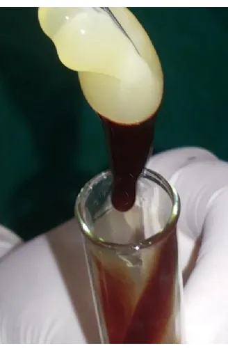
[image:3.595.374.496.52.284.2] The tubes were immediately centrifuged at 400g for 10min at the rate of 3000 revolutions per minute with a specific table centrifuge at room temperature (Fig.6).
Blood centrifugation immediately after collection allows the composition of a structured fibrin clot in the middle of the tube, just between the red corpuscles at the bottom and the acellular platelet poor plasma at the top (Fig.7).
[image:3.595.55.272.300.482.2] PRF was easily separated from the red corpuscles base (preserving a small RBC layer) using a sterile tweezer and scissors just after removal of PPP (Platelet Poor Plasma) and then transferred onto a dapen dish (Fig.8).
Fig. 5. Drawing blood (9 ml of blood as per requirement)
Fig. 6. The Centrifuge
Surgical Treatment
A full thickness mucoperiosteal flap was elevated to expose both the labial and palatal/lingual aspects of the alveolar ridge (Fig.9 and Fig.10).
[image:3.595.352.516.312.562.2]Fig.7. Separated fractions
Fig. 8. Activated gel-like PRF easily carried with forceps
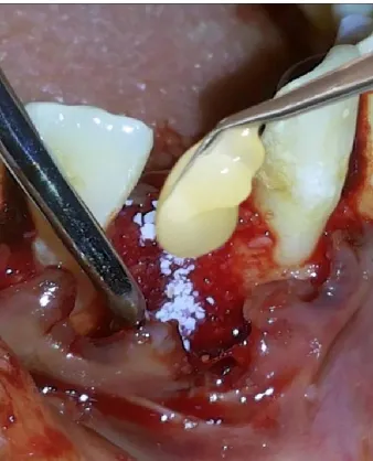
Atraumatic extraction using periotome and extraction forceps was performed. The extraction socket was carefully curetted to remove all the soft tissue. After flap reflection, β- TCP bone graft plug along with PRF was inserted up to the level of the crest of the socket (Fig.11 and Fig.12). The labial and palatal/lingual flaps were approximated back into position by giving vertical mattress sutures using a 3-0 mersilk suture in order to prevent any exfoliation of the graft.
Postoperative Care and Evaluation
[image:3.595.54.272.513.679.2]Fig. 9. Conventional flap procedure showing dehiscence of the labial bone #41
Fig. 10. Conventional flap procedure showing fenestration on the labial bone #12
Fig.11. PRF along with DFDBA was placed in the defect #41
The patients were asked to abstain from brushing on the surgical area for at least 1 week and they were recalled 1 week postoperatively during which sutures were removed and the
[image:4.595.54.271.285.444.2]operated area was evaluated for healing, infection and any signs of ulceration and necrosis. Patients were reevaluated at the end of 6th month (Fig.13 and Fig. 14).
Fig. 12. PRF along with DFDBA was placed in the defect #12
Fig. 13. Post operative width of the alveolar ridge at 6 months # 41
Fig. 14. Post operative width of the alveolar ridge at 6 months #12
RESULTS
During the course of the study, wound healing was uneventful. There were no post operative complication and patient was satisfied with the results. The increase in the amount of tissue present was adequate to permit placing an esthetic fixed restoration. Six months postsurgically, there was 1.39 mm gain in buccolingual width and 1.0 mm gain in vertical dimension
[image:4.595.324.542.306.439.2] [image:4.595.327.538.479.633.2] [image:4.595.79.248.483.692.2]in case I (Fig.15) and 0.94 mm gain in buccolingual width and 6.58 mm gain in vertical dimension in case II (Fig.16).
Fig.15 Post oprative CBCT sgowing bone regeneration at 6 months
Fig. 16. Post oprative CBCT sgowing bone regeneration at 6 months
DISCUSSION
The importance of healing of an extraction socket cannot be undermined as the internal and external changes that occur, ultimately affect the shape of the alveolar ridge, (Darby, 2009). Majority of the studies indicate that bone is not able to regenerate to the level of bone crest or even to the level of the neighboring teeth, during healing, therefore 100% socket fill does not occur, (Ara´ ujo and Lindhe, 2009). Using an animal model, Araujo and Lindhe showed that in the first 8 weeks following extraction, there is marked osteoclastic activity, resulting in the resorption of the facial and lingual bone walls, especially in the crestal region. They also noted that bone resorption was greater on the facial wall and that any loss of ridge height was accompanied by a horizontal loss on both facial and lingual walls of the extraction site (Araujo and Lindhe, 2009). In 1993 Misch and Dietsh suggested different
graft materials and techniques for socket grafts based on the number of bony walls that remained after the tooth is removed. A recent publication showed that the combination of β-TCP and type I collagen used for simple preservation of a maxillary extraction socket without a barrier membrane resulted in new bone formation 9 months after the procedure with 62.6% of mineralized bone and 21.1% of bone marrow. β-TCP resorption occurs concurrently with new bone formation. Horch et al. reported that 65% resorption of β-TCP occurred 1 year after being used as a bone substitute in large mandibular cystic defects, alveolar clefts and for maxillary sinus floor augmentations. Simunek et al. reported that the mean graft area occupied by β-TCP was 39%, 9 months after sinus augmentation procedures. During the last decade several different ridge augmentation techniques have been developed, most of which include the use of a graft material. This increases the treatment cost as well as increases the risk of disease transmission. Studies also indicate that in many cases, the graft material is not totally incorporated into the newly formed bone and when compared to sites without graft material, they show less vital bone formation.
In some cases it requires the use of collagen membranes. In these cases a 25% membrane exposure rate has been reported, and this directly affects the amount of bone fill that takes place within the socket (Darby, 2009).PRF was first described by Choukroun et al., 2006. It is considered a second-generation platelet concentrate and has been used in various surgical procedures in an attempt to enhance wound healing. It is autogenous and is not associated with any issues related to immune reactions or infections. Besides, its gelatinous consistency enhances clot and graft stability. Platelets release cytokines, such as Vascular Endothelium Growth Factor (VEGF), Platelet-Derived Growth Factor (PDGF), Fibroblast Growth Factor (FGF), Epidermal Growth Factor (EGF), Hepatocyte Growth Factor (HGF), Insulin-Like Growth Factor (IGF), and Transforming Growth Factor-Β (TGF-Β). Choukroun PRF is known to release growth factors for at least 7 days.
[image:5.595.74.252.331.516.2]least 95% of the platelets of the collected blood into a fibrin mesh (Ehrenfest, 2010). The fibrin mesh can then be easily manipulated into a membrane that allows it to be transferred to any surgical site. Recently, PRF has also been shown to stimulate the growth of osteoblasts and periodontal ligament cells, both of which are significant for the regeneration of periodontal defects (Reddy, 2014; Shah and Gujjari, 2013; Shah, 2012; Sharma, 2011). Currently, PRF has been successfully tested in a number of procedures including maxillofacial surgery, periodontal surgery, and implantology. According to a study carried out by Mazor et al. PRF was successfully used as the only grafting material in a series of sinus augmentation procedures. They also demonstrated that PRF could stimulate new bone formation in areas that were previously deficient of the amount of bone that is required for implant placement (Shah, 2012).
Conclusion
In the above case series, the successful use of Demineralized Freezed-Dried Bone Allograft combines with Platelet Rich Fibrin (PRF) in a postextraction ridge preservation procedure. The biomaterial acts by releasing high-concentration growth factors to the wound site, thereby stimulating healing and new bone formation. Unlike other procedures, the use of PRF is a simple method that requires minimal cost and reduces the need for specialized grafting material. Because it is a completely autologous product, the risk of disease transmission and graft rejection is negated.
REFERENCE
Araujo, M. and Lindhe, J., 2009. Ridge alterations following tooth extraction with and without flap elevation: an experimental study in the dog. Clinical Oral Implants
Research 20 545-549.
Blair, P. and Flaumenhaft, R., 2009. Platelet alpha - granules: basic biology and clinical correlates. Blood Reviews 23 177-189.
Bozidar, M.B. et al., 2012. Beta-tricalcium phosphate/type I collagen cones with or without a barrier membrane in human extraction socket healing: clinical, histologic, histomorphometric, and immunohistochemical evaluation.
Clin Oral Invest; 16:581-590.
Brownfield, L.A. and Weltman, R.L. 2012. Ridge preservation with or without an osteoinductive allograft: a clinical, radiographic, micro-computed tomography, and histologic study evaluating dimensional changes and new bone formation of the alveolar ridge. J Periodontol; 83: 581-583. Caplanis, N. et al., 2005. Extraction Defect Assessment,
Classification and Management.CDA Journal; 33:853. Corso, M. et al. 2009. Clinical evaluation of a modified
coronally advanced flap alone or in combination with a platelet-rich fibrin membrane for the treatment of adjacent multiple gingival recessions: a 6-month study. Journal of
Periodontol., 80 1694-1697
D M.G.Ara´ ujo and Lindhe J. Ridge alterations following tooth extraction with and without flap elevation: an experimental study in the dog. Clinical Oral Implants
Research 2009; 20:545-549.
Darby, S.T., Chen, and Buser, D. 2009. Ridge preservation techniques for implant therapy. The International Journal
of Oral & Maxillofacial Implants; 24:260-27.
Ehrenfest, D. et al., 2010. Three-dimensional architecture and cell composition of a Choukroun’s platelet-rich fibrin clot and membrane. Journal of Periodontology 81 546-555. Horowitz, R. et al., 2012. A review on alveolar ridge
preservation following tooth extraction. J Evid Based Dent
Pract. 2012 Sep;12(3 Suppl):149-60.
Hussain, et al., 2015. Soft tissue ridge augmentation of maxillary anterior region using combination of allograft and GTR membrane. International Journal of Current
Research, vol 7, issue 06 pp.17448-17451.
Kang, Y et al., 2011. Platelet-rich fibrin (PRF) is a bioscaffold and reservoir of growth factors for tissue regeneration. Tissue Engineering 17 349-359.
Kang, Y et al., 2011. Platelet-rich fibrin is a Bioscaffold and reservoir of growth factors for tissue regeneration. Tissue
Engineering 17 349–359.
Mazor, Z et al., 2009. Sinus floor augmentation with simultaneous implant placement using Choukroun’s platelet-rich fibrin as the sole grafting material: a radiologic and histologic study at 6 months. Journal of Periodontol 80 2056-2064.
Orgeas, G.V. et al., Surgical techniques for alveolar socket preservation: A systematic review. International Journal of
oral and maxillofacial implant 2013; 28:1049-1061.
Ramakrishnan, T. et al., 2009. Umasudhakar, Platelet-rich-fibrin: a novel root coverage approach. Journal of Indian
Society of Periodontology 13 50- 54.
Reddy, et al., 2014. Extraction socket preservation using β tricalcium phosphate bone graft plug and platelet rich fibrin membrane – A case series. International Journal of
Applied Dental Sciences 2014; 1(1): 36-40
Shah, M. and Gujjari, S. K., 2013. Alveolar ridge augmentation utilizing platelet rich fibrin in combination with demineralized freeze-dried bone allograft – a case report. International Journal of Basic and Applied Medical
Sciences ISSN: 2277-2103. Vol. 3 (1) January-April,
pp.18-23
Shah, M. et al., 2012. Double papilla flap with platelet rich fibrin in isolated gingival recession – A case report.
Journal of Contemporary Dental Sciences 2(1) 36-40.
Sharma, A. and Pradeep, A., 2011. Autologous platelet rich fibrin in the treatment of mandibular degree II furcation defects: a randomized clinical trial. Journal of Periodontol 82 1396-1403.
Sharma, A. and Pradeep, A., 2011. Treatment of 3-wall intrabony defects in chronic periodontitis subjects with autologous platelet rich fibrin-a randomized controlled clinical trial. Journal of Periodontol 82 1705-1712. Sheikh, et al., 2015. Bone Replacement Materials and
Techniques Used for Achieving Vertical Alveolar Bone Augmentation. Vol 8, 2953-2993.
Simonpieri, A et al., Simultaneous sinuslift and implantation using microthreaded implants and leukocyte and platelet-rich fibrin as sole grafting material: a six-year experience. Implant Dentistry 20 2-12.