Original citation:
Eslamian, Ladan, Borzabadi-Farahani, Ali, Tavakol, Pegah, Tavakol, Ali, Amini, Nazila
and Lynch, Edward. (2015) Effect of multiple debonding sequences on shear bond
strength of new stainless steel brackets. Journal of Orthodontic Science, Volume 4
(Number 2). pp. 37-41. ISSN 2278-0203
Permanent WRAP url:
http://wrap.warwick.ac.uk/67504
Copyright and reuse:
The Warwick Research Archive Portal (WRAP) makes this work of researchers of the
University of Warwick available open access under the following conditions.
This article is made available under the Creative Commons
Attribution-NonCommercial-ShareAlike 3.0 (CC BY-NC-SA 3.0) license and may be reused according to the
conditions of the license. For more details see:
http://creativecommons.org/licenses/by-nc-sa/3.0/
A note on versions:
The version presented in WRAP is the published version, or, version of record, and may
be cited as it appears here.
ABSTRACT
Objectives: This in‑vitro study aimed at evaluating the effect of three debonding sequences on the shear bond strength (SBS) of new stainless steel (SS) brackets.
Materials and Methods: Stainless steel twin brackets (0.022‑inch, American Orthodontics, Sheboygan, WI, USA) were bonded with light cure adhesive (Transbond XT, 3M Unitek, St. Paul, MN, USA) to 80 newly extracted human premolars after acid etching with 37% phosphoric acid (30 s). Brackets were debonded with a universal testing machine, and new brackets were bonded to teeth using the same adhesive and same manner. This process was repeated twice, and brackets were debonded within 24 h after bonding. The longitudinal changes of average SBS were assessed with the repeated measures ANOVA. Post‑hoc tests using the Bonferroni correction were also used to compare the average SBS at three debonding sequences.
Result: The mean SBS decreased significantly after each debonding sequence (P < 0.01). The
corresponding mean values (standard deviation, 95% CI) after the first, second, and third debonding
sequences were 22.88 MPa (4.08, 21.97–22.79), 19.36 MPa (4.54, 18.62–20.64), and 16.67
MPa (4.27, 15.72–17.62), respectively. There was no significant difference among the adhesive
remnant index (ARI) scores of three debonding sequences (χ2 = 5.067, df = 6, P = 0.53).
Conclusion: Average SBS after three debonding sequences was significantly decreased, but was
above the recommended 5.9–7.8 MPa. In‑vivo studies are required to validate the finding of this study.
Key words: Adhesive remnant index, enamel, multiple debonding, orthodontic brackets, shear bond strength
Effect of multiple debonding sequences on shear bond
strength of new stainless steel brackets
Ladan Eslamian
1, Ali Borzabadi‑Farahani
2,3, Pegah Tavakol
4, Ali Tavakol
4, Nazila Amini
4and Edward Lynch
2INTRODUCTION
Orthodontic brackets are the main means of tooth movement for orthodontist. The elimination of the remaining adhesive material following failure of brackets or debonding procedures removes about 50 µm of enamel[1,2] and the processes of rebonding
may led to a significantly different shear bond strength (SBS)
between the bracket and tooth surface. Clinicians may use new brackets or recycled stainless steel (SS) brackets, a process that is associated with the structural changes of brackets. The common methods for bracket recycling are; heat application to burn the bonding agent that follows by electrolytic polishing for oxide removal; as well as the combined use of high‑frequency
vibrations, electrochemical polishing, and chemicals to dissolve the bonding agent.[3‑5] These methods can be associated
with reduction in bond strength, particularly after thermal recycling;[6,7] although it has been claimed that recycled brackets
offer a similar bond failure profile to new brackets.[8] Some
clinicians may also reuse debonded brackets with in‑office
reconditioning of the debonded bracket using sandblasting[9‑13]
or laser reconditioning,[14] as a method of rebonding of the same
bracket, perhaps to address the drawbacks associated with commercial recycling.
In order to address the issues associated with recycling brackets, clinicians can use new brackets. Previous studies on the effect of multiple bondings on the SBS, using new brackets, are limited;[15‑21] these studies often used small sample
Address for correspondence: Dr. Ali Borzabadi‑Farahani, Warwick Dentistry, Warwick Medical School, University of Warwick, Coventry, UK.
E‑mail: [email protected]
1Department of Orthodontics and Dentofacial Deformities
Research Center, School of Dentistry, Shahid Beheshti University of Medical Sciences, Tehran, Iran, 2Warwick
Dentistry, Warwick Medical School, University of Warwick, Coventry; 3NHS England (Locum Orthodontic Consultant),
UK, 4Private Practice of Orthodontics, Tehran, Iran
Access this article online Quick Response Code:
Website:
www.jorthodsci.org
DOI:
Eslamian, et al.: Shear bond strength after three sequences of debonding of SS brackets
sizes[15‑17,20,21] or used bovine teeth.[16] The aim of the present
in‑vitro study was to examine the effect of three debonding sequences on the SBS of new SS brackets bonded to human teeth.
Null Hypothesis
The null hypothesis for this study was “There is no difference among the SBSs of new SS brackets bonded to human teeth after three sequences of debonding.”
MATERIALS AND METHODS
This study was performed using 80 noncarious freshly extracted premolars after ethical approval granted by the ethics committee of the Shahid Beheshti University of Medical Sciences. The age of the patients whose extracted teeth we used varied between 11 and 16 years. All teeth were caries‑free
and did not have any cracks, fractures, hypercalcification on the buccal surface, which could influence the bonding process.
The teeth were cleaned, lightly pumiced, and stored in distilled water at room temperature before use.
Assessment of Shear Bond Strength
Teeth were polished with fluoride‑free pumice paste (Dentatus,
Tehran, Iran), using rubber cap for 15 s, then washed with tap water for 15 s and air‑dried. One operator performed the bonding process, after etching the specimens with 37% phosphoric acid (3M Unitek, St. Paul, MN, USA) for 30 s. Each bracket (0.022‑inch twin brackets, American Orthodontics, Sheboygan, WI, USA) was bonded with a Transbond XT adhesive (3M Unitek, St. Paul, MN, USA) and light‑cured (Bonart‑Art‑L2 Light Curing Unit, Bonart Co. Ltd., Taipei, Taiwan) according to the instruction provided by the adhesive’s manufacturer. The bracket base size was approximately 11.85 mm². No bond enhancer was used for bonding procedures. Overall, 240 SS brackets were used and bonded with 4 mm distance from the occlusal surface. The excess adhesives were removed using a dental explorer.
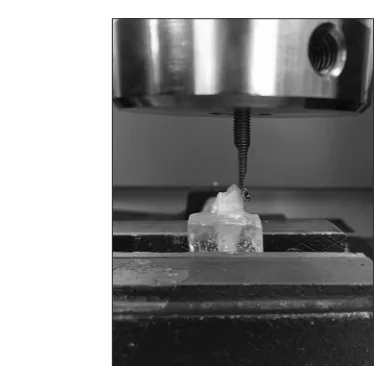
In order to ensure all brackets were bonded in the same plane a mounting jig appliance consisting of a stand containing a 0.021 × 0.025 inch SS wire was used; this was placed in the bracket’s slot when teeth were put in molds containing self‑cure acrylic resin [Figure 1]. The specimens were not exposed to thermocycling and brackets were debonded after 24 h. During force application, each tooth with its own acrylic base was put in one jaw of the universal testing machine (Zwick/Roell Zo20, Ulm, Germany) and a specimen holder was used to ensure constant load parallel to the tooth surface [Figure 2]. The other part of the machine exerted an occlusal‑gingival load to the upper surface of the bracket between the upper wings and bracket base using a blade, producing a shear force at the bracket tooth interface. The blade, which was perpendicular to the bracket’s slot, exerted a force at a crosshead speed of 1 mm/min until rupture of the bracket‑tooth union.[22] The required forces
for debonding (failure load) were recorded as Newtons, and subsequently, the SBSs (MPa) calculated and used as control for future comparisons. The day after debonding, residual adhesives on tooth surfaces were removed using a low‑speed TC tungsten carbide bur (Mani, Tokyo, Japan) until the enamel surface reached its glaze. In order to make sure the entire adhesive remnant has been removed, a new bur was used for every 2 specimens and the etched surface was
also evaluated by the operator under magnification to ensure
all of the adhesive remnants had been removed. Teeth were subsequently cleaned and etched as mentioned earlier. After each debonding sequence, new SS brackets were bonded to the teeth with the same orthodontic adhesive. This process was repeated twice, and the SBS values were calculated.
Adhesive Remnant Index
The buccal surfaces and bracket bases were evaluated using Stereomicroscope (Siemens, Munich, Germany) and one operator (PT) used the adhesive remnant index (ARI), as described by Årtun and Bergland,[23] and scored the adhesive
remaining on the teeth:
• 0, no adhesive left on the tooth
• 1, less than half of the adhesive left on the tooth • 2, more than half of the adhesive left on the tooth • 3, all the adhesive left on the tooth with the mesh pattern
visible.
Statistical Analysis
[image:3.612.363.511.531.705.2]The statistical analysis was performed using SPSS software, Ver. 17 (Statistical Package for Social Sciences, SPSS Inc., Chicago, IL, USA). Descriptive statistics such as means, standard derivations (SD), range, and 95% confidence intervals (CI) were calculated for average SBS at three debonding sequences. For assessing the longitudinal changes of average SBS in three debonding sequences, the repeated measures ANOVA were used. Post‑hoc tests using the Bonferroni correction were also used to compare the average SBS at three debonding sequences. The Chi‑square test was
also used to evaluate differences in the ARI scores among the
three sequences of debonding. The level of significance was
set at P < 0.05.
RESULTS
The descriptive statistics for three debonding sequences is
shown in Table 1. The analysis of variance indicated significant
differences among the SBS values for all three debonding sequences (P < 0.01). The mean SBS decreased after each debonding sequence, which was statistically significant (P < 0.01) [Table 2]. The mean SBS (SD, 95% CI) after the
first, second, and third debonding sequences were 22.88
MPa (4.08, 21.97–22.79), 19.36 MPa (4.54, 18.62–20.64), and 16.67 MPa (4.27, 15.72–17.62), respectively.
The amount of residual adhesive on the buccal tooth surface as evaluated by the ARI scores is shown in Table 3. The
Chi‑square test did not reveal significant differences among
the ARI scores of three sequences of debonding (χ2 = 5.067,
df = 6, P = 0.53). Therefore, the null hypothesis of the present study was fully rejected.
DISCUSSION
Bracket failure can be common in practice and its effect on the subsequent bond strengths is important. In a busy
orthodontic practice, a significant number of teeth may need to
be rebonded[17] and clinicians may choose to rebond a new or
a recycled bracket. Reports on the effect of multiple debonding and bonding procedures on SBS, using new/recycled brackets, are sometimes contradictory. The present study had a sample size of 80 and was adequately powered to detect the differences in SBS following multiple debonding of new brackets.
A significant decline in the SBS after each debonding
sequence was observed in the present study; the first stage
had the highest SBS (mean SBS = 22.88 MPa) followed by
the second and third stages (Mean SBS of 19.63 and 16.67
MPa, respectively). Comparison of the present findings and
the previous studies would be challenging. This is due to the different retaining devices that were used (human/bovine teeth or plastic cylinders), or different study sample sizes, bracket types (different brands, new, recycled), bracket base sizes, recycling methods (thermal, chemical, or sandblasting), or methods of bond strength assessment (shear or tensile) that were used. The observed reduction in the SBS is probably due to the partial destruction of the etching pattern[18,19] and the
weaker retentive enamel morphology.[17,24] Nonetheless, our
findings contrast with the reports of no significant change in the
SBS[15‑17,20] or increase in the SBS following debonding with the
new brackets.[18,19,21] The different findings in those studies could
be due to the small sample sizes[15‑17,20,21] that were not able to
detect the differences between groups, dissimilar specimen types (molars[16,17] vs. premolars[21]). Alternatively, different
etching times (30 s,[16‑18] or 60 s[15]) or curing times, differences
in the time gap between bonding and debonding (½‑h after bonding[16,17] vs. 24 h[21]), and dissimilar used bonding materials
could explain the contradictory findings.
[image:4.612.54.241.52.235.2]After bond failure, clinicians may use new or recycled brackets. One of the earliest studies that evaluated the effect of recycling on bond strength concluded that it was dependent on the bracket type and the recycling method.[4] The present study
[image:4.612.315.558.82.141.2]Figure 2: Profile view of a specimen before shear bond strength testing
Table 1: Mean, SD and 95% Cl of SBS in MPa for 3 debonding sequences
Debonding
sequence N Mean SBS (SD) of mean95% CI Range
First 80 22.88 (4.08) 21.97-22.79 10.92-31.76
Second 80 19.36 (4.54) 18.62-20.64 10.70-28.62
Third 80 16.67 (4.27) 15.72-17.62 10.06-18.37
[image:4.612.315.557.198.257.2]SD – Standard deviation; SBS – Shear bond strength; CI – Confidence interval
Table 2: The post‑hoc tests using the Bonferroni correction show the longitudinal changes in the average SBS at each debonding sequence which was highly significant (P<0.01)
Debonding
sequence Debonding sequence Mean difference in SBS (MPa) Significance
First Second 3.52 0.00
First Third 6.21 0.00
Second Third 2.69 0.00
SBS – Shear bond strength
Table 3: The ARI scores on buccal surfaces of the teeth after 3 debonding sequences
ARI
scores* First Debonding sequencesSecond Third
0 0 2 0
1 40 38 35
2 36 37 40
3 4 3 5
[image:4.612.317.559.304.376.2]Eslamian, et al.: Shear bond strength after three sequences of debonding of SS brackets
assessed the SBS but looking into other studies that assessed the bond strength, including the tensile bond strength, we found similar patterns, similar to investigations that used recycled brackets[6,7,13,25‑28] such as the study by Wright and Powers.[25]
They assessed the tensile bond strength using small sample sizes of 5 brackets in each recycled bracket group and used plastic cylinders as retaining devices.[25] Another study with
a small sample size also reported a decline in tensile bond strength, which was dependent on the method of recycling.[29]
Reddy etal.[27] investigated the effect of thermal recycling and
similarly reported a reduction in bond strength (shear and tensile) as a result of recycling. The reduction in SBS following
recycling is not a common finding,[28] and depending on the
bracket type and the method of recycling, conflicting results
have been reported. For instance, when self‑ligation brackets were bonded to bovine enamel, the reconditioning process lowered the SBS of Smart‑Clip and Damon3 MX brackets,
but significantly increased the bond strength values for Quick
brackets.[28]
A limitation of the study was the lack of information on the mechanism for the reduced adhesion as no scanning electron microscopy (SEM) of the enamel or bracket surfaces was performed. The present study used a common method (ARI)[20‑24,30] to assess the amount of adhesive left on the enamel
surface. An analysis of the failure sites demonstrated that ARI scores were found to be similar after all three debonding sequences. However, the similar pattern of ARI did not explain the changes in SBS, which needs further investigation using the SEM technique. Although we did not use recycled bracket it
was interesting that the present findings were in agreement with
some studies that evaluated the ARI scores of rebonded new or reconditioned/recycled brackets.[20,28,29] The average SBS after
two debonding processes was still above the recommended 5.9–7.8 MPa by Reynolds.[31] The minimum effective etching
time with 37% phosphoric acid was reported to be 30 s,[32]
which was employed in the present investigation. It seems that resurfacing the enamel using a tungsten carbide bur, acid‑etching the enamel for 30 s,[32] and use of a new bracket
offer reasonable bond strength.
The limitations of the in‑vitro studies should be considered in
interpreting the present findings. Most reported in vivo bond
strengths might not be as high as those measured using the in‑vitro models. The average reported in vivo bond strengths were approximately 40% less than the in‑vitro studies.[33] The
gradual decrease in bond strength of composites due to aging and storage of material in saliva is another factor that should be considered before making clinical recommendations.[34]
The storage medium for the present study was distilled water. According to a recent systemic review,[35] many studies used
distilled water; however, it has been suggested that teeth must be ideally stored in thymol solutions and not water as this may
reduce bond strength significantly. The purpose of this study
was to compare the SBS after bonding procedures using new
brackets and as the storage medium was the same for all
specimens, the effect on the findings of comparisons should
be minimal. Bearing in mind the discussed limitations, the
present findings suggest the average SBS of new SS brackets
is decreased after two debonding procedures, but is still above the recommended required bond strength.
CONCLUSION
The SBS of new SS brackets after two debonding procedures
significantly decreased, but was still above the recommended
required bond strength (5.9–7.8 MPa). In‑vivo studies are
further required to validate the finding of the presen study.
REFERENCES
1. Ryf S, Flury S, Palaniappan S, Lussi A, van Meerbeek B, Zimmerli B. Enamel loss and adhesive remnants following bracket removal and various clean-up procedures in vitro. Eur J Orthod 2012;34:25-32.
2. Faust JB, Grego GN, Fan PL, Powers JM. Penetration coefficient, tensile strength, and bond strength of thirteen direct bonding orthodontic
cements. Am J Orthod 1978;73:512-25.
3. Buchman DJ. Effects of recycling on metallic direct‑bond orthodontic
brackets. Am J Orthod 1980;77:654-68.
4. Mascia VE, Chen SR. Shearing strengths of recycled direct-bonding brackets. Am J Orthod 1982;82:211-6.
5. Buchwald A. A three-cycle in vivo evaluation of reconditioned direct-bonding brackets. Am J Orthod Dentofacial Orthop 1989;95:352-4.
6. Regan D, van Noort R, O’Keeffe C. The effects of recycling on the tensile bond strength of new and clinically used stainless steel orthodontic
brackets: An in vitro study. Br J Orthod 1990;17:137-45.
7. Wheeler JJ, Ackerman RJ Jr. Bond strength of thermally recycled metal brackets. Am J Orthod 1983;83:181-6.
8. Cacciafesta V, Sfondrini MF, Melsen B, Scribante A. A 12 month clinical study of bond failures of recycled versus new stainless steel
orthodontic brackets. Eur J Orthod 2004;26:449‑54.
9. Millett D, McCabe JF, Gordon PH. The role of sandblasting on the retention of metallic brackets applied with glass ionomer cement. Br
J Orthod 1993;20:117-22.
10. Sonis AL. Air abrasion of failed bonded metal brackets: A study of shear
bond strength and surface characteristics as determined by scanning
electron microscopy. Am J Orthod Dentofacial Orthop 1996;110:96-8.
11. Basudan AM, Al‑Emran SE. The effects of in‑office reconditioning on
the morphology of slots and bases of stainless steel brackets and on the shear/peel bond strength. J Orthod 2001;28:231-6.
12. Quick AN, Harris AM, Joseph VP. Office reconditioning of stainless steel orthodontic attachments. Eur J Orthod 2005;27:231‑6. 13. Wendl B, Muchitsch P, Pichelmayer M, Droschl H, Kern W. Comparative
bond strength of new and reconditioned brackets and assessment
of residual adhesive by light and electron microscopy. Eur J Orthod 2011;33:288-92.
14. Ahrari F, Basafa M, Fekrazad R, Mokarram M, Akbari M. The efficacy of Er, Cr: YSGG laser in reconditioning of metallic orthodontic brackets.
Photomed Laser Surg 2012;30:41-6.
15. Mui B, Rossouw PE, Kulkarni GV. Optimization of a procedure for rebonding dislodged orthodontic brackets. Angle Orthod 1999;69:276-81.
16. Bishara SE, VonWald L, Laffoon JF, Warren JJ. The effect of repeated bonding on the shear bond strength of a composite resin orthodontic
adhesive. Angle Orthod 2000;70:435-41.
17. Bishara SE, Laffoon JF, Vonwald L, Warren JJ. The effect of repeated
bonding on the shear bond strength of different orthodontic adhesives. Am J Orthod Dentofacial Orthop 2002;121:521-5.
multiple bracket bonding with or without repeated etching. Eur J
Orthod 2011;33:521-7.
19. Fischer‑Brandies H, Monsees M. Enamel damage from multiple
debonding in the bracket adhesion technic. Fortschr Kieferorthop 1993;54:143-7.
20. Endo T, Ozoe R, Shinkai K, Aoyagi M, Kurokawa H, Katoh Y, et al.
Shear bond strength of brackets rebonded with a fluoride‑releasing
and-recharging adhesive system. Angle Orthod 2009;79:564-70.
21. Montasser MA, Drummond JL, Evans CA. Rebonding of orthodontic
brackets. Part I, a laboratory and clinical study. Angle Orthod 2008;78:531-6.
22. Eslamian L, Borzabadi-Farahani A, Mousavi N, Ghasemi A.
A comparative study of shear bond strength between metal and ceramic brackets and artificially aged composite restorations using different surface treatments. Eur J Orthod 2012;34:610‑7.
23. Artun J, Bergland S. Clinical trials with crystal growth conditioning as an alternative to acid‑etch enamel pretreatment. Am J Orthod
1984;85:333-40.
24. Regan D, LeMasney B, van Noort R. The tensile bond strength of new and rebonded stainless steel orthodontic brackets. Eur J Orthod
1993;15:125-35.
25. Wright WL, Powers JM. In vitro tensile bond strength of reconditioned
brackets. Am J Orthod 1985;87:247-52.
26. Liu JK, Tsai MY, Huang PH. Tensile bond strength of reused orthodontic metal brackets. Zhonghua Ya Yi Xue Hui Za Zhi 1991;10:30‑5. 27. Reddy YN, Varma DP, Kumar AG, Kumar KV, Shetty SV. Effect of thermal
recycling of metal brackets on shear and tensile bond strength. J Contemp Dent Pract 2011;12:287-94.
28. Sfondrini MF, Xheka E, Scribante A, Gandini P, Sfondrini G.
Reconditioning of self‑ligating brackets. Angle Orthod 2012;82:158‑64. 29. Faltermeier A, Behr M. Effect of bracket base conditioning. Am J
Orthod Dentofacial Orthop 2009;135:12.e1-5.
30. Eslamian L, Borzabadi‑Farahani A, Mousavi N, Ghasemi A. The effects
of various surface treatments on the shear bond strengths of stainless
steel brackets to artificially‑aged composite restorations. Aust Orthod
J 2011;27:28-32.
31. Reynolds IR. A review of direct orthodontic bonding. Br J Orthod
1975;2:171-8.
32. Gardner A, Hobson R. Variations in acid‑etch patterns with different acids and etch times. Am J Orthod Dentofacial Orthop 2001;120:64‑7. 33. Hajrassie MK, Khier SE. In‑vivo and in‑vitro comparison of bond
strengths of orthodontic brackets bonded to enamel and debonded at various times. Am J Orthod Dentofacial Orthop 2007;131:384‑90. 34. Hariri I, Shimada Y, Sadr A, Ichinose S, Tagami J. The effects of aging on
shear bond strength and nanoleakage expression of an etch-and-rinse
adhesive on human enamel and dentin. J Adhes Dent 2012;14:235‑43.
35. Finnema KJ, Ozcan M, Post WJ, Ren Y, Dijkstra PU. In‑vitro orthodontic bond strength testing: A systematic review and meta‑analysis. Am J
Orthod Dentofacial Orthop 2010;137:615-22.e3.
How to cite this article: Eslamian L, Borzabadi‑Farahani A, Tavakol P, Tavakol A, Lynch N, Lynch E. Effect of multiple debonding sequences on shear bond strength of new stainless steel brackets. J Orthodont Sci 2015;4:37‑41.
Source of Support: Nil, Conflict of Interest: None declared.
Author Help: Online submission of the manuscripts
Articles can be submitted online from http://www.journalonweb.com. For online submission, the articles should be prepared in two files (first page file and article file). Images should be submitted separately.
1) First Page File:
Prepare the title page, covering letter, acknowledgement etc. using a word processor program. All information related to your identity should be included here. Use text/rtf/doc/pdf files. Do not zip the files.
2) Article File:
The main text of the article, beginning with the Abstract to References (including tables) should be in this file. Do not include any informa-tion (such as acknowledgement, your names in page headers etc.) in this file. Use text/rtf/doc/pdf files. Do not zip the files. Limit the file size to 1 MB. Do not incorporate images in the file. If file size is large, graphs can be submitted separately as images, without their being incorporated in the article file. This will reduce the size of the file.
3) Images:
Submit good quality color images. Each image should be less than 4096 kb (4 MB) in size. The size of the image can be reduced by decreas-ing the actual height and width of the images (keep up to about 6 inches and up to about 1800 x 1200 pixels). JPEG is the most suitable file format. The image quality should be good enough to judge the scientific value of the image. For the purpose of printing, always retain a good quality, high resolution image. This high resolution image should be sent to the editorial office at the time of sending a revised article. 4) Legends: