warwick.ac.uk/lib-publications
A Thesis Submitted for the Degree of PhD at the University of Warwick
Permanent WRAP URL:
http://wrap.warwick.ac.uk/93414
Copyright and reuse:
This thesis is made available online and is protected by original copyright. Please scroll down to view the document itself.
Please refer to the repository record for this item for information to help you to cite it. Our policy information is available from the repository home page.
Assay Development Towards the
Characterisation of the Bifunctional Activity
of P. aeruginosa and E. coli Class A PBPs
Amy Mae O’Reilly
Thesis submitted in partial fulfillment of the requirements for
the degree of Doctor of Philosophy in Mathematical Biology and
Biophysical Chemistry
The University of Warwick
Department of Chemistry
Thesis Contents
Table of Contents………...……….………..….II Acknowledgements……….…...XIV Declaration……….………...XVI Abstract……….………XVII Abbreviations……….….…XVIII List of Tables………..XXI List of Figures………...XXII
Chapter 1 Introduction
1.1 Gram-Positive vs Gram-Negative Bacterial Cell Envelopes……….1
1.1.1 The Gram-Positive Bacterial Cell Wall………..1
1.1.2 The Gram-Negative Bacterial Cell Wall………2
1.2 Targets for Antibiotics………..3
1.2.1 Protein Synthesis as an Antibiotic Target………..5
1.2.2 DNA and RNA Synthesis………...5
1.2.3 Cell Wall Biosynthesis………...5
1.3 Mechanisms of Antibiotic Resistance in Bacterial Pathogens………...7
1.3.1 Reduced Antimicrobial Uptake and Active Export by Efflux Pumps………....7
1.3.2 Modification of the Antibiotic………7
1.3.3 Modification of the Antibiotic Target………8
1.3.4 The Emergence of Antimicrobial Resistant (AMR) Superbugs……….8
1.4 Peptidoglycan as the Molecular Scaffold………9
1.4.1 Cytoplasmic Steps of Peptidoglycan Synthesis………10
1.4.2 Lipid-Linked Steps of Peptidoglycan Synthesis………...10
1.4.3 The Mechanism of Transglycosylation………11
1.4.4 Known Inhibitors of Transglycosylation………..12
1.4.4.1 Moenomycin: The ‘blueprint’ Transglycosylase Inhibitor and a Structural Tool………..13
1.4.4.3 Moenomycin Analogues as Transglycosylase Inhibitors……….15
1.4.4.4 Lipid II Analogues as Transglycosylase Inhibitors………..19
1.4.4.5 Prospects for New Transglycosylase Inhibitors………...20
1.4.5 The Mechanism of Transpeptidation………21
1.4.5.1 PBP Acylation………..24
1.4.5.2 PBP Deacylation………...24
1.4.6 Peptidoglycan Recycling and Maintenance……….25
1.4.6.1 The DD-Carboxypeptidase and DD-Endopeptidase Function of PBPs…26 1.4.7 Antibiotics Targeting Transpeptidation: The Structural Similarity of β-lactams to D-Ala-D-Ala………27
1.5 The Penicillin Binding Proteins……….28
1.5.1 The Classification and Topology of PBPs………29
1.5.2 Membrane Proteins as Drug Targets………31
1.5.3 Using Detergents to Study Membrane Proteins………...31
1.5.4 E. coli PBP1A………..31
1.5.5 E. coli PBP1B………...32
1.5.6 P. aeruginosa as a Clinically Important Pathogen………...36
1.5.7 P. aeruginosa PBP1A………...…37
1.5.8 P. aeruginosa PBP1B………...38
1.5.9 P. aeruginosa Resistance to β-Lactams………38
1.5.10 PBP-Associated Proteins………...……….38
1.5.10.1 The LpoA and LpoB Lipoproteins………...39
1.6 Underexploited Targets for Antimicrobials……….40
1.7 Strategies for Novel Antibiotic Discovery………41
1.7.1 Recent Developments in Compounds Against MDR P. aeruginosa………….42
1.7.2 Recently Approved Antimicrobials………43
1.7.3 Teixobactin - The Latest Discovery………...43
1.8 Antimicrobial Resistance (AMR) Public Policy………...44
1.9 Thesis Aims and Outlines………..45
2.2 Growth and Maintenance of Bacteria………..47
2.2.1 Bacterial Strains………47
2.2.2 Preparing Competent Cells………...47
2.2.3 Bacterial Transformation………...48
2.2.4 Bacterial Electroporation………..48
2.2.5 Bacterial Growth Media………...48
2.2.6 Preparation of Glycerol Stocks……….49
2.3 Molecular Biology Techniques………..49
2.3.1 Polymerase Chain Reaction………..49
2.3.2 DNA Quantification……….51
2.3.3 Agarose DNA Electrophoresis……….51
2.3.4 DNA Cloning………52
2.3.4.1 Screening for Positive Clones………..53
2.3.5 DNA sequencing………..54
2.3.6 Site-Directed Mutagenesis………54
2.4 Recombinant Protein Expression in E. coli………..54
2.4.1 Over-expression of P. aeruginosa PBP1A and E. coli PBP1A………54
2.4.2 Over-expression of P. aeruginosa PBP1B and E. coli PBP1B………54
2.4.3 EcPBP1B and PaPBP1B Expression Cell Line Trials……….55
2.4.4 Large-Scale Bacterial Cell Lysis for the Preparation of Crude Cell Lysates...55
2.4.5 Harvesting the Membrane Fraction………..55
2.4.6 Membrane Protein Solubilisation……….56
2.5 Protein Purification………56
2.5.1 Purification of Class A PBPs………56
2.5.2 Immobilised-Metal-Affinity Chromatography (IMAC)………...56
2.5.3 Ion Exchange Chromatography: Source S Column………..57
2.6 Protein Analysis and Detection……….57
2.6.1 SDS-PAGE………...57
2.6.2 Western Blotting………...58
2.6.3 Protein Concentration Determination………...59
2.6.3.1 BioRad Assay………...59
2.6.3.2 BCA Assay………...60
2.7 Compound Synthesis………..60
2.7.2 UDP-MurNAc Pentapeptide Purification……….61
2.7.3 Pentapeptide Quantification……….61
2.7.4 Labelling UDP-MurNAc-L-Ala-D-Glu-mesoDAP-D-Ala-D-Ala with Fluorescamine………...62
2.7.5 Pentapeptide Dansylation: UDP-N-actylmuramyl-L-Ala-γ-D-Glu-(Nε- dansyl)-mesoDAP-D-Ala-D-Ala (Dansyl-labelled UDP-MurNAc- pentapeptide)………62
2.7.6 Removal of the UDP Group from UDP-Pentapeptides………63
2.7.7 The Synthesis of Lipid II………..63
2.7.8 The Purification of Lipid II………..63
2.7.9 Thin Layer Chromatography………64
2.7.10 LII Quantification………...65
2.7.11 Preparing Samples for Mass Spectrometry………65
2.8 Enzyme Activity Assays……….65
2.8.1 Tris-Tricine Polyacrylamide Gel System……….65
2.8.2 BOCILLIN Assay……….66
2.8.3 A Continuous Fluorescence Assay for Transglycosylase Activity…………..66
2.8.4 The Spectrophotometric Phosphate Release Assay for Transglycosylase Activity……….67
2.8.5 Continuous Spectrophotometric Assay for D-Alanine Release………68
2.8.6 The Continuous Transpeptidase Spectrophotometric Assay………69
2.8.7 MurF Assay for dipeptide incorporation………..69
2.8.8 D-Ala-D-X Ligase (VanA) Assay……….69
2.9 Techniques for Protein Structure Analysis………..69
2.9.1 Circular Dichroism Spectroscopy………69
2.9.2 Protein Crystallisation………..70
Chapter 3
The Cloning, Expression and Purification of
PBPs and their Associated Proteins
3.1 Introduction………713.3 The Cloning, Expression and Purification of PBPs……….71
3.3.1 The Cloning and Purification of E. coli PBP1A………...74
3.3.2 The Purification of E. coli PBP1B………...75
3.3.3 The Cloning and Purification of Full-length P. aeruginosa PBP1A…………76
3.3.4 Cloning P. aeruginosa PBP1B……….77
3.3.5 Expression Trials with E. coli PBP1B and P. aeruginosa PBP1B…………...78
3.3.6 P. aeruginosa PBP1B Detergent Screen………..81
3.3.7 The Purification of PaPBP1B………..81
3.4 Initial Structural and Functional Characterisation of Purified PaPBP1A and PaPBP1B………..82
3.4.1 Labelling PaPBP1B with Fluorescent-Penicillin, BOCILLIN FLTM………...82
3.4.2 Circular Dichroism of PaPBP1A and PaPBP1B……….83
3.5 Further Investigation in the quest for active PaPBP1B………..84
3.5.1 The Cloning and Expression of Shorter, ‘Soluble’ PBP1B from E. coli and P. aeruginosa………84
3.5.2 Cloning P. aeruginosa PBP1B in to a P. putida Expression Construct and Homologous Protein Expression………..86
3.6 Purification of PBP Catalytic Active Site Mutants……….88
3.6.1 Site-Directed Mutagenesis and Purification of E. coli PBP1A and PBP1B Transglycosylase Active Site Mutants E86Q and E233Q Respectively……..88
3.6.2 The Effect of Over-Expression of E. coli PBP Transglycosylase Catalytic Active Site Mutants on Cell Culture Growth………...89
3.6.3 Site-Directed Mutagenesis and Purification of E. coli PBP1A and E. coli PBP1B Transpeptidase Active-Site Mutants S473A and S510A, respectively………..90
3.6.4 Site-Directed Mutagenesis and Purification of P. aeruginosa PBP1A Transglycosylase and Transpeptidase Active Site Mutants E86Q and S461A, respectively………...92
3.7 The Cloning and Purification of PBP-Associated Proteins………93
3.7.1 Cloning and Purification of P. aeruginosa and E. coli PBP3………..93
3.7.2 E. coli CpoB and P. aeruginosa YbgF ………94
3.7.3 The Expresion and Purification of Enterococcus faecium VanA………95
3.7.4 The ExprEssion and Purification of E. coli LpoA and LpoB ……….96
3.7.6 The Expression and Purification of E. coli FtsN………..97
3.8 PBP X-Ray Crystallography………97
3.8.1 Initial X-Ray Crystallography Screens of E. coli PBP1A, P. aeruginosa PBP1A and P. aeruginosa PBP1B………...97
3.9 Discussion………..101
3.9.1 The Lack of Activity of PaPBP1B……….101
3.9.2 Over-Expression of the EcPBP1B Transglycosylase Mutant………101
3.9.3 PBP Crystallography Trials………102
3.10 Conclusions……….102
3.11 Future Work………...102
Chapter 4
The Kinetic Analysis of the Transglycosylation Activity
of E. coli PBP1A and P. aeruginosa PBP1A using a Continuous
Fluorescence Assay
4.1. Current Transglycosylase Assays in the Literature……….1044.1.1 Thin Layer Chromatography Assays………..104
4.1.2 SDS-PAGE Analysis of Transglycosylation Products………...105
4.1.3 HPLC Analysis of Transglycosylation Products……….105
4.1.4 High-Through Put (HTP) Screening Assays Based on Moenomycin Displacement………..106
4.1.5 Continuous Fluorescence Assays………...106
4.2 Experimental Aims………...107
4.3 E. coli PBP1B as a Control Enzyme for the Study of Transglycosylation of Class A Transglycosylase Enzymes……….107
4.3.1 A Continuous Fluorescence Assay for Transglycosylase Activity…………107
4.3.2 Assay Scale-Up: From Fluorimeter to Plate Reader………..110
4.3.3 Dependence of E. coli PBP1B Transglycosylase Activity on Enzyme Concentration……….112
4.3.4 Michaelis-Menten Kinetic Constants……….113
4.4 Buffer Optimisation Screening with E. coli PBP1A and P. aeruginosa PBP1A and PBP1B………115 4.4.1 Optimal Detergent(s) for Transglycosylase Activity of E. coli and P.
aeruginosa PBP1A………115 4.4.2 Testing Optimal Detergent(s) for P. aeruginosa PBP1B Activity………….116 4.4.3 The Optimisation of pH for Transglycosylase Activity……….117 4.4.4 The Optimisation of NaCl Concentration for Transglycosylase Activity…..118 4.5 The Kinetic Characterisation of Transglycosylation of E. coli PBP1A and P. aeruginosa PBP1A and PBP1B………119 4.5.1 Dependence of E. coli PBP1A and P. aeruginosa PBP1A Transglycosylase Activity on Enzyme Concentration………119 4.5.2 The Kinetic Profile of E. coli PBP1A and P. aeruginosa PBP1A………….121 4.5.2.1 The Kinetic Profile of E. coli PBP1A and P. aeruginosa PBP1A with dansDAP-Lipid II C55 and C35………121 4.5.2.2 The Dual-Substrate Kinetic Equation and the Hill Equation………….123 4.5.2.3 The Kinetic Profile of E. coli PBP1A and P. aeruginosa PBP1A with dansLys-Lipid II C55………...125 4.6 The Activity of Transglycosylase Active Site Mutants………..128 4.7 The Effect of PBP-Associated Proteins on Transglycosylase Activity……….129
4.7.1 The Effect of E. coli LpoA and LpoB on the Transglycosylase Activity of PBP1A………...130 4.7.2 The Effect of E. coli PBP2 and FtsN on the Transglycosylase Activity of Class A PBPs………..131 4.8 Discussion………..133 4.8.1 Plate Reader versus Fluorimeter for Transglycosylase Activity
Measurement………..133 4.8.2 Different Substrates Exhibit Different Levels of Fluorescence in the
Continuous Assay………...134 4.8.3 Defining the Optimum pH of the Transglycosylase Activity of PBPs……...134 4.8.4 The Effect of Detergent on Substrate Fluorescence in the
4.10.1 Optimising the Detergent for Transglycosylation Activity………..137 4.10.2 Further Investigation of PaPBP1B Activity……….137 4.10.3 The Lack of an LpoB Protein in Pseudomonas aeruginosa……….138
Chapter 5
A Discontinuous Spectrophotometric Assay for
The Detection of Transglycosylase-Dependent
Undecaprenyl Pyrophosphate Production
5.1 Transglycosylation Generates the By-Product Undecaprenyl Pyrophosphate (C55-PP) ……….139 5.2 Undecaprenyl Pyrophosphate Phosphatases Dephosphorylate C55-PP……...140 5.3 Phosphate Release as a Means of Quantifying Lipid II Turnover…………...140 5.3.1 Advantages of this Discontinuous Assay for Transglycosylation
Detection……….143 5.3.2 Disadvantages of this Discontinuous Assay for Transglycosylation
Detection……..………...143 5.4 Experimental Aims………...143 5.5 The Expression and Purification of E. coli PgpB………..144 5.6 The Development of a Spectrophotometric Assay for the Detection of
Transglycosylase Activity………145 5.6.1 Phosphate Dose-Response Curve with the MESG-PNP Coupling System…145 5.6.2 Initial Rate Dependence on E. coli PgpB Concentration………...146 5.6.3 Comparison Between Substrates of E. coli PgpB………..146 5.6.3.1 The Kinetics of Dependence of E. coli PgpB on Undecaprenyl
Pyrophosphate (C55-PP) and Diacylglycerol
5.6.8.1 The Activity of E. coli PgpB when purified in TritonX-100, CHAPS and
DDM………...155
5.6.8.2 E.coli PgpB Activity in Ethylene Glycol Alkyl Ether Detergents…….156
5.6.9 E. coli PgpB Activity ± MgCl2, in a Variety of Non-ionic Detergents……..158
5.7 Discussion………..159
5.7.1 Comparing the Continuous Fluorescence Assay with the Phosphate Release Spectrophotometric Assay………159
5.7.2 Ceasing Transglycosylation Polymerisation………160
5.7.3 E. coli PgpB Activity in Different Detergents……….160
5.8 Conclusions………...160
5.9 Future Work……….161
5.9.1 The PgpB Assay has the Potential to Monitor Transglycosylation Continuously………...161
5.9.2 The Stability and Activity of PgpB in Different Detergents………..162
5.9.3 Scrutinise the Kinetic Capability of E. coli PgpB in a Wider Range of Detergents………...162
5.9.4 Investigate Alternative Undecaprenyl Pyrophosphate Phosphatases to PgpB………...162
5.9.5 The PgpB Assay and Bacitracin……….162
Chapter 6
The Study of Transpeptidation in Class A Penicillin
Binding Proteins from E. coli and P. aeruginosa
6.1 Defining Transpeptidase Donors and Acceptors………...1636.2 Transpeptidase Assays in the Literature………165
6.2.1 The Exchange Reaction………..166
6.3 Experimental Aims………...167
6.4 The Development of a FRET Assay Allowing the Kinetic Characterisation of Class A PBPs……….168
6.4.1 Definition of FRET……….…168
6.4.2 The Förster Radius (R0)………..169
6.5 Designing the Transpeptidase Donor and Acceptor Compounds
for FRET………..169 6.5.1 Criteria for a Successful FRET System………..170 6.5.2 Selecting the Optimum Fluorophores for Transpeptidase Donors and
Acceptors………171 6.5.2.1 Potential FRET Donor Fluorophores………..171 6.5.2.2 Potential FRET Acceptor Fluorophores………..172 6.5.2.3 Considering R0: The Förster distance between two FRET pairs………174 6.5.2.4 The Selected Fluorescent Compounds to be Synthesised………..174 6.5.3 The Fluorescent Properties of the Selected Fluorophores………..176 6.6 Synthesis of the Transpeptidase Acceptor Compounds………177 6.6.1 Synthesis of MurNAc-L-Ala-D-Glu-mesoDAP-D-AzaTrp-D-Ala…………...177 6.6.2 Synthesis of MurNAc-L-Ala-D-Glu-mesoDAP-D-Trp-D-Ala………..182 6.7 Synthesis of the Transpeptidase Donor Compounds………187 6.7.1 Synthesis of Dansylated (mesoDAP) Lipid II………187 6.7.2 Synthesis of Fluorescamine-labelled Lipid II (C55-UPP-MurNAcGlcNAc-L- Ala-D-Glu-(Nε-Fluorescamine)-mesoDAP-D-Ala-D-Ala……….188 6.7.2.1 Fluorescamine as a Primary Amine-Labelling Fluorophore…………..188 6.7.2.2 Enzymatic Characterisation of Fluorescamine DAP-LII as a substrate for E. coli PBP1B transglycosylation……….193 6.8 Analysis of the Activity of the Synthesised Compounds, as Functional
Transpeptidase Acceptors and Donors………...194 6.8.1 Activity of the Synthesised Transpeptidase Acceptors, Using Lys-LII as the Transpeptidase Donor………..195 6.8.2 Activity of the Synthesised Transpeptidase Acceptors, using dansDAP-LII as the Transpeptidase Donor………...198 6.8.3 Activity of the Synthesised Transpeptidase Acceptors, using Fluorescamine- labelled DAP-LII as the Transpeptidase Donor………200 6.9 Enzymology of Transpeptidation: Kinetic Characterisation of the
Transpeptidase Activity of E. coli PBP1A and P. aeruginosa PBP1A………203 6.9.1 The Transpeptidase Activity of E. coli PBP1B in the presence and
6.9.2.1 Comparing the Transpeptidase Activity of E. coli PBP1A with and
without a Hexa-His Tag……….206
6.9.2.2 Investigating Cryo-Preservation and Subsequent Transpeptidase Activity of E.coli PBP1A……….207
6.9.2.3 Determining the Dependency of a Transpeptidase Rate Upon the Presence of E. coli PBP1A and E. coli LpoA………208
6.9.2.4 The Sensitivity of E. coli PBP1A Transpeptidase Activity to Inhibition by Moenomycin A and Ampicillin………209
6.9.2.5 The Ability of E. coli PBP1A to Utilise Structurally Diverse Transpeptidase Acceptor Compounds………...210
6.9.2.6 Testing E. coli PBP1A Catalytic Active Site Transglycosylase and Transpeptidase Mutants in the Continuous Transpeptidase Assay……212
6.9.3 The Transpeptidase Activity of P. aeruginosa PBP1A in the Presence and Absence of P. aeruginosa YbgF………212
6.9.3.1 Testing P. aeruginosa PBP1A Catalytic Transglycosylase and Transpeptidase Active-Site Mutants in the Continuous Transpeptidase Assay……….213
6.10 Discussion………215
6.11 Conclusions……….215
6.12 Future Work………...216
!
Chapter 7
Discussion, Conclusions and Future Work
7.1 The Continuous Fluorescence Assay for Transglycosylase Activity…………2187.1.1 Buffer Conditions for the Continuous Fluorescence Assay………...218
7.1.2 Initial Lag-Phase Observed with Class A PBPs……….218
7.1.3 The Pros and Cons of DMSO upon Transglycosylase Activity Assays and Future Work……….…219
7.1.4 The Kinetic Constants Derived for Class A PBPs, from Transglycosylase Activity………..220
7.1.5 PBP-Associated Proteins and their Impact on Transglycosylase Activity...222
7.2 The Oligomeric Status of PBP1A and PBP1B in vitro………..222
7.3.1 Kinetic Measurements of Transglycosylation using E. coli PgpB…………224
7.3.2 E. coli PgpB Detergent Screening………..224
7.3.3 Limitations of the Current PgpB Assay Protocol………...225
7.3.3.1 The Development of a Continuous Assay………..225
7.4 No Activity Observed of P. aeruginosa PBP1B ……….225
7.4.1 The Lack of LpoB in P. aeruginosa………...226
7.5 The FRET Transpeptidase Assay………...226
7.6 The Continuous Transpeptidation D-Ala Release Assay………..227
7.7 Thesis Summary………...228
Appendix 1 : A table of oligos for gene synthesis or site-directed mutagenesis………...231
Appendix 2 : The Percentage and concentrations of each detergent typically used in the extraction and purification of membrane proteins………...232
Appendix 3 : Derivation of the dual substrate Equation……..………..233
Acknowledgements
I would like to thank my supervisors, Prof. Tim Bugg and Dr. David Roper. Thanks to Tim who is always really positive about my research and results and I appreciate his efforts in critiquing this thesis before submission. A huge thank you goes to Dr. Adrian Lloyd for his patience, his willingness to share results and collaborate on multiple areas of this project. Adrian went out of his way to help in all aspects of the bringing together of this thesis.
Thanks to Anita, Julie and Smita for help with compound synthesis. I appreciate the numerous flasks of media supplied by Cerith and Mark, totalling over 1000 Litres of media over 3.5 years. I would like to thank fellow lab members: Claire, Christine, Ailidh and Mussa for being fantastic bench partners. Kat, Steve, Conor, Greg and Mike for pub Fridays, DannyMc for consecutive secret santa exchanges and Kyle for
sophisticated dinner parties. Thanks to Chris for all the films, cooking sessions and Family Guy episodes we shared. Thank you to Soph and Adrian for being so accommodating and flexible.
Thank you to my advisory panel for keeping my project on the straight and narrow. A particular mention to Dr. Ann Dixon for her support and interest in my progression. A huge heart-felt thank you to Prof. Alison Rodger who awarded me the EPSRC funding to conduct this research. I appreciate everything that comes with the Doctoral Training Centre family, from life-long friendships and team-building to weekly seminars and multi-disciplinary problem solving. Thank you to Naomi for organising the greatest annual conferences.
Finally, a big love to Rich who has always been there for support no matter what and has been extremely patient during the write up of this thesis.
There may be times when we are powerless to prevent injustice, but there must never be a time when we fail to protest.
This thesis is dedicated to Dr. Adrian Lloyd. Without his overwhelming support and trust,
this thesis would not have been written.
"Strong people stand up for themselves; stronger people stand up for others” Steve Oedekerk, 2006.
Declaration
Abstract
Pseudomonas aeruginosa is a Gram-negative species of bacteria that is of high clinical importance and is on the ESKAPE list of pathogens, causing the most serious nosocomial infections World-wide. Current antibiotics in clinical use against P. aeruginosa include carbenicillin, cefepime, ceftazidime, and the fluoroquinoline ciprofloxacin. The multi-drug resistant (MDR) phenotype exhibits widespread resistance to β-lactams and antibiotics from multiple classes often need to be administered in conjunction with β-lactamase inhibitors.
Penicillin binding proteins (PBPs) are essential for cell viability and are important antibiotic targets, with the Class A bifunctional PBPs synthesising the principle component of the bacterial cell wall, peptidoglycan. The focus of this project was to investigate the kinetics of transglycosylation and transpeptidation mechanisms with a view to improving our
understanding of bacterial cell wall synthesis, maintenance and regeneration.
In this project, the function of Class A PBPs 1A and 1B from Escherichia coli and P. aeruginosa, with particular focus on PBP1A has been investigated. Transglycosylation and transpeptidation have been probed from multiple angles using orthogonal assays and fluorescently-labelled and native lipid substrates, as well as Lipid II structural variants. P. aeruginosa PBP1A transglycosylase and transpeptidase activities are demonstrated
continuously, and compared to the model organism equivalent, E. coli PBP1A. The continuous monitoring of activity gave insights into the possibility of two catalytic sites for
transglycosylation: a donor and an acceptor site. Data was fitted to different kinetic models to elucidate the best fit for extracting meaningful kinetic parameters.
The data acquired over the course of this project suggests that P. aeruginosa PBP1A and E. coli PBP1A exhibit similar functional behaviour to each other, with E. coli PBP1B showing distinct activity from PBP1A. PaPBP1B activity was not detected at a level required to kinetically characterise the enzyme and it is possible that this PBP has an as yet undetermined stimulatory cofactor. The assays developed and optimised in this thesis will pave the way for further in-depth studies of P. aeruginosa PBP1A, which could be used to screen for putative inhibitor compounds and potentially identify certain characteristics required for antimicrobial efficacy.
Abbreviations
2YT 2 x yeast trypone media
Axxx Absorbance
ATP Adenosine 5’-triphosphate
AU Absorbance Unit
BCA Bicinchoninic acid
BODIPY Boron dipyrromethene dye
BSA Bovine Serine Albumin
C55-PP Undecaprenyl pyrophosphate
CD Circular dichroism
CHAPS 3-[(3-Cholamidopropyl) dimethylammonio]-1-propanesulfonate CMC Critical Micellar Concentration
Da Dalton
DAAO D-amino acid oxidase
Dans dansylated
DAP D-aminopimelic Acid
DDM n-Dodecyl-β-D-maltopyranoside
DEAE Diethyl-aminoethyl
DMSO Dimethyl Sulphoxide
DNAaseI Deoxyribonuclease I enzyme
dNTPs deoxy
Ec Escherichia coli
ECL Electrochemiluminescence
EGTA ethylene glycol-bis(β-aminoethyl ether)-N,N,N',N'-tetraacetic acid EDTA Ethylenediaminetetraacetic acid
FRET Fluorescence resonance energy transfer GlcNAc N-acetylglucosamine
HEPES 4-(2-hydroxyethyl)-1-piperazineethanesulfonic acid
HMW High molecular weight
HPLC High-Performance Liquid Chromatography HRP Horseradish peroxidase
IMAC Immobilised Metal-Affinity Chromatography IPTG Isopropyl-β-D-thiogalactopyranoside
Kcat Michaelis-Menton Constant
KD Dissociation constant
Km Michaelis-Menton Constant
kpsi kilopound per square inch
LB Luria Broth
LDAO Lauryldimethylamine oxide
Lipid I Undecaprenyl pyrophosphoryl-N-acetylmuramyl-L-Alanyl-D-Glutamyl-L-Lysyl-D-Alanyl-D-Alanine
LII Undecaprenyl pyrophosphoryl-N-acetylmurmayl (Nacetyl-glucosaminyl)- L-Alanyl-D-Glutamyl-X-D-Alanyl DAlanine mAU Milli-absorbance unit
MESG 7-methyl-6-guanosine MurNAc N-acetylmuramic acid m/z Mass to charge ratio
NADH Nicotinamide adenine dinucleotide
Pa Pseudomonas aeruginosa
PAGE Polyacrylamide electrophoresis PBP Penicillin Binding Protein PCR Polymerase chain reaction
PEP Phosphoenol pyruvate
PgpB phosphatidylglycerophospate phosphatase B
pI Isoelectric point
Pi Inorganic phosphate
PMSF Phenyl methyl sulphonyl fluoride PNP Purine Nucleoside Phosphorylase RFU Relative Fluorescence Units SDM Site-Directed Mutagenesis SDS Sodium dodecyl sulfate
TAE Tris acetate ethylenediaminetetraacetic acid TEV Tobacco etch virus protease
TM Transmembrane domain
TP Transpeptidase
Tris Tris(hydroxymethyl)aminomethane
TX-100 Triton X-100
UDP Uridine diphosphate
List of Tables
Chapter 1 Introduction
Table 1.1 A List of Antibiotics and their Mode of Action Against Metabolic
Processes in Bacterial Cells………4 Table 1.2 A Summary of Antimicrobial Agents that Target the Bacterial Cell Wall…….6 Table 1.3 A Summary of Analogues and Small Molecule inhibitors of Moenomycin
and Lipid II.………..…17
Chapter 2 Materials and Methods
Table 2.1 Bacterial Strains used for Cloning and Protein Over-Expression………47 Table 2.2 PCR Reaction Mixtures for HF Phusion Polymerase………...…50 Table 2.3 The PCR program used for Phusion DNA Polymerase………....50 Table 2.4 Typical Vector : Insert Ligation Reactions………...53
Chapter 3 The Cloning, Expression and Purification of PBPs and their Associated Proteins
Table 3.1 A Comprehensive Library of all PBP and PBP-associated DNA
Constructs used Throughout this Project………..71
Chapter 4 The Kinetic Analysis of the Transglycosylation Activity of E. coli PBP1A and P. aeruginosa PBP1A using a Continuous Fluorescence Assay
Table 4.1 A Summary of the Vmax and Km data derived from the Michaelis-
Menten equation for the two substrates C55-PP and DGPP………115 Table 4.2 The Kinetic Parameters for E. coli PBP1A Transglycosylase Activity
Fitted to the Dual-Substrate Equation………127 Table 4.3 The Kinetic Parameters for E. coli PBP1A Transglycosylase Activity
Fitted to the Hill Cooperativity Equation………..127 Table 4.4 The Kinetic Parameters for P. aeruginosa PBP1A Transglycosylase
Table 4.5 The Kinetic Parameters for P. aeruginosa PBP1A Transglycosylase
Activity Fitted to the Hill Cooperativity Equation………127
Chapter 5 A Discontinuous Spectrophotometric Assay for The Detection of Transglycosylase-Dependent Undecaprenyl Pyrophosphate Production
Table 5.1 Table 5.1 A Summary of the Vmax and Km data derived from the
Michaelis-Menten equation for the two substrates C55-PP and DGPP……...149 Table 5.2 Table 5.2 The Kinetic Constants for each PBP, with data fitted to the
Michaelis-Menten equation………153 Table 5.3 Table 5.3 The Kinetic Constants for each PBP, with data fitted to the
Hill equation………...153 Table 5.4 A Summary of the Ethylene Glycol Ether Detergents Tested for
Suitability for EcPgpB Activity………..156
Chapter 6 The Study of Transpeptidation in Class A Penicillin Binding Proteins from E. coli and P. aeruginosa
Table 6.1 Potential Fluorescent Amino Acids that can be Incorporated in to the
Pentapeptide Stem of the Transpeptidase Acceptor Molecule………...171 Table 6.2 A Comparison Between Assay Buffers for Transglycosylation and
Transpeptidation for EcPBP1A and PaPBP1A………
List of Figures
Chapter 1 Introduction
Figure 1.1 The Structural Architecture of Gram-Positive and Gram-Negative
Figure 1.8 The Structure of Penicillin G (6-aminopenicillanic acid) and the
D-alanyl-D-alanine end of the Peptidoglycan Strand………...28
Figure 1.9 The Classification of Gram-negative PBPs (using E. coli as the model) and a schematic representation of the two Class A bifunctional PBPs:
E. coli PBP1A and PBP1B………...30 Figure 1.10 The Conserved Motifs in the TG51 Structural Family, with this E. coli
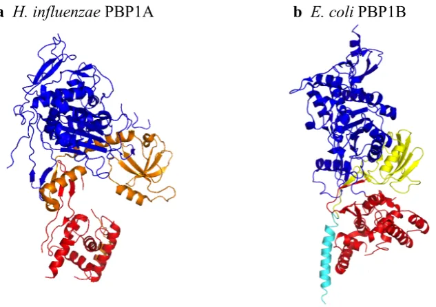
PBP1B Domain Bound to Moenomycin A, Residing in the Catalytic Cleft....34 Figure 1.11 The Crystal Structures of the two Gram-Negative Class A Bifunctional
PBPs (PBP1A and 1B). H. influenzae PBP1A and E. coli PBP1B…………..36 Figure 1.12 The ‘Divisome’ machinery and ‘Elongasome’ Complexes involved in
both Cell Elongation and Cell Division………...39
Chapter 3 The Cloning, Expression and Purification of PBPs and their Associated Proteins
Figure 3.1 PCR Amplification of Full-length E. coli PBP1A………74 Figure 3.2 The Immobilised Metal Affinity Chromatography and Cation
Exchange Purification of Full-length E. coli PBP1A………...74 Figure 3.3 SDS-PAGE Analysis of the Ni2+ IMAC Purification of E. coli PBP1B
M46-N844………....75
Figure 3.4 The Amplification of Full-length P. aeruginosa PBP1A by PCR……….…..76 Figure 3.5 The Purification of pET28(b)::PaPBP1A1-822 by Ni2+ IMAC and
SourceS Cation Exchange………77 Figure 3.6 Amplification of PaPBP1B W23-N774 by PCR……….78 Figure 3.7 A Bacterial Cell-Line Expression Trial of PaPBP1B and EcPBP1B……...…79 Figure 3.8 Expression Temperature Trials for EcPBP1B and PaPBP1B………..80 Figure 3.9 A Detergent Screen of P. aeruginosa PBP1B in a Variety of Ionic,
Anionic and Non-Ionic Detergents………..81 Figure 3.10 The Purification of pET28(b)::PaPBP1B W23-N774 in four detergents: CHAPS,
TritonX-100, LDAO and DDM………82 Figure 3.11 BOCILLINTM-FL Labelling of PaPBP1B, EcPBP1B and PaMurF………….83 Figure 3.12 Circular Dichroism Analysis of P. aeruginosa PBP1A and 1B…………...…84 Figure 3.13 The PCR Amplification of EcPBP1B Y107-N844 and PaPBP1B
Figure 3.15 The Cloning of PaPBP1B23-774 in to the P.Putida Expression
Vector pIZ1016……….87
Figure 3.16 A Summary of the Cloning and Purification of all Four Class A
PBPs: EcPBP1A, EcPBP1B, PaPBP1A and PaPBP1B………...88 Figure 3.17 The Purification by IMAC of E. coli PBP1A and 1B
Transglycosylase Mutants………89 Figure 3.18 The Growth Curves of the Expression of the Transglycosylase
Active Site Mutants EcPBP1A and EcPBP1B, independently……….90 Figure 3.19 The Purification of E. coli PBP1A Active Site Mutant S473A and
its Ability to Bind BOCILLINTM………..91 Figure 3.20 The Purification of E. coli PBP1B Transpeptidase Active-Site
Mutant S510A and its Ability to Bind BOCILLINTM………..92 Figure 3.21 The Purification P. aeruginosa PBP1A Transglycosylase E86Q
Mutant and Transpeptidase S461A Mutant………..93 Figure 3.22 The PCR Amplification and Purification of E. coli PBP3 and
P. aeruginosa PBP3………..94 Figure 3.23 The Expression and Purification of E. coli CpoB (formally EcYbgF)……….94 Figure 3.24 The Expression and Purification of full-length P. aeruginosa YbgF………...95 Figure 3.25 The Purification of E. faeccium VanA……….…95 Figure 3.26 The Purification of E.coli LpoA and LpoB………..…96 Figure 3.27 The PCR Amplification of the PaLpoA Gene and Purification
of PaLpoA Protein………...…….96 Figure 3.28 The Purification of E. coli FtsN………....97 Figure 3.29 Crystallisation Screen of E. coli PBP1A, in its apo-enzyme form
and co-crystallised with its ligand Moenomycin………...98 Figure 3.30 Crystallisation Screen of P. aeruginosa PBP1A, in its apo-enzyme
form and co-crystallised with its ligand Moenomycin……….…99 Figure 3.31 Crystallisation Screen of P. aeruginosa PBP1B, in its apo-enzyme
form and co-crystallised with its ligand Moenomycin………...100
Chapter 4 The Kinetic Analysis of the Transglycosylation Activity of E. coli PBP1A and P. aeruginosa PBP1A using a Continuous Fluorescence Assay
Figure 4.1 The Continuous Fluorometric Assay for the Detection of Glycan
Figure 4.2 The Continuous Fluorometric Transglycosylation Assay in a
Fluorimeter Compared to a Plate-Reader………..110 Figure 4.3 The Continuous Fluorometric Transglycosylase Assay - Proof of
Principle assay with Positive and Negative Controls……….111 Figure 4.4 Dependence of E. coli PBP1B Transglycosylase Activity on
Enzyme Concentration………...113 Figure 4.5 The Initial Rate of Transglycosylation in EcPBP1B with
Increasing dans-DAP-LII Substrate Concentration………114 Figure 4.6 The Transglycosylase Activity of PaPPBP1A and EcPBP1A in
LDAO, DDM, CHAPS and TritonX-100………...116 Figure 4.7 The Transglycosylase Activity of P. aeruginosa PBP1B after Extraction and
Purification in to CHAPS, DDM, TritonX-100 and LDAO………...117 Figure 4.8 The Dependence of E.coli and P. aeruginosa PBP1A
Transglycosylase Activity on pH………...118 Figure 4.9 The Transglycosylase Activity of E. coli and P. aeruginosa
PBP1A at Increasing Ionic (NaCl) Concentration………..119 Figure 4.10 The Dependence of Initial Rate of Transglycosylation on E. coli
PBP1A Concentration at 3 and 10 µM dans-DAP-LII………...120 Figure 4.11 The Dependence of Initial Rate of Transglycosylation on P. aeruginosa PBP1A
Concentration at 3 and 10 µM dans-DAP-LII………...121 Figure 4.12 The Dependency of E. coli and P. aeruginosa PBP1A upon LII
Substrate Concentration (dans-DAP-LII C55 and dans-DAP-LII C35)………122 Figure 4.13 The Dependency of E. coli and P. aeruginosa PBP1A upon
dansLys-LII C55 Substrate Concentration……….…..125 Figure 4.14 The Transglycosylase Activity of PBP Transglycosylase Active-
Site Mutants………....129 Figure 4.15 The Effect of PaLpoA upon the Transglycosylase Activity of PaPBP1A….130 Figure 4.16 A Dose-Response of EcPBP1B Transglycosylase Activity to
EcLpoB and EcPBP1A Transglycosylase Activity to EcLpoA….………….131 Figure 4.17 The Effect of E. coli FtsN and PBP2 on PBP1A and PBP1B……….…132
Chapter 5 A Discontinuous Spectrophotometric Assay for The Detection of Transglycosylase-Dependent Undecaprenyl Pyrophosphate Production
Characterisation by Circular Dichroism (CD)………144 Figure 5.3 Correlating phosphate addition to the total change in absorbance of
MESG at 355 nm……….145 Figure 5.4 The Dependence of the Initial rate of Reaction upon the EcPgpB
Enzyme Concentration………...146 Figure 5.5 A Direct Comparison of Substrate Preference of E. coli PgpB, between the
native substrate undecaprenyl pyrophosphate (C55-PP/UPP) and the non- native substrate diacyl glycerol pyrophosphate (DGPP)………147 Figure 5.6 Kinetics of Dependence of E. coli PgpB Phosphatase Activity with its
natural substrate undecaprenyl pyrophosphate C55-PP and the non-native substrate DGPP………...148 Figure 5.7 Time-dependence of Undecaprenyl Pyrophosphate Generation in
the Transglycosylation Polymerisation Reaction………...150 Figure 5.8 Comparing the Substrate Preference of EcPBP1A, EcPBP1B and PaPBP1A
with both Lys- and DAP-LII………..151 Figure 5.9 The Kinetic Profile of E. coli PgpB with DAP-LII generated
undecaprenyl pyrophosphate (C55-PP) from 3 different Class A PBPs……..152 Figure 5.10 The activity of both transglycosylase and transpeptidase wildtype
and active site mutants were tested using EcPgpB……….154 Figure 5.11 The activity of E. coli PgpB when extracted and purified in DDM,
TritonX-100 and CHAPS………...155 Figure 5.12 E. coli PgpB activity tested in the presence of the non-ionic
ethylene glycol alkyl ether detergents E4C12, E5C10, E6C12 and E8C10……....157 Figure 5.13 The Activity of E. coli PgpB with and without Magnesium Chloride
(MgCl2) in a Range of Detergents: DDM, TritonX-100, E3C10, E4C12,
E5C10,E6C12,E3C10………158
Chapter 6 The Study of Transpeptidation in Class A Penicillin Binding Proteins from E. coli and P. aeruginosa
Figure 6.1 Transpeptidase Donor and Acceptor Compounds………...164 Figure 6.2 The D-Amino Acid Exchange Reaction……….167 Figure 6.3 Potential FRET Fluorophore Pairs and their Fluorometric Properties……...173 Figure 6.4 A Schematic of the Concept Behind the Proposed FRET Assay
to Function as FRET Donors and Acceptors as well as
Transpeptidase Acceptors and Donors………...175 Figure 6.6 The Emission Maxima of the Four Fluorophores Dansyl, Fluorescamine,
D-Tryptophan and D-Azatryptophan………...176 Figure 6.7 The Ligation of D-Tryptophan and D-Azatryptophan on to D-Ala by the D
-Ala-D-Lac ligase E. coli VanA………...178 Figure 6.8 The Incorporation of Non-Canonical Dipeptides (D-Azatryp-D-Ala) at the
C-Terminus of the Pentapeptide Stem, using MurF………...179 Figure 6.9 Purification of the potential transpeptidase acceptor UDP-MurNAc-L-Ala-
D-Glu-mesoDAP-D-Azatrp-D-Ala………..180 Figure 6.10 Determining the presence of pentapeptide by release of the terminal D-Ala
residue from UDP-MurNAc-L-Ala-D-Glu-mesoDAP-D-Azatrp-D-Ala...181 Figure 6.11 MonoQ analysis of UDP-MurNAc-L-Ala-D-Glu-mesoDAP-D-Azatrp-
D-Ala………..……….181
Figure 6.12 The mass spectrometry results of UDP-MurNAc-L-Ala-D-Glu-mesoDAP-D
-Azatrp-D-Ala………..182
Figure 6.13 The Incorporation of the Dipeptide D-Trp-D-Ala into the pentapeptide stem, using PaMurF……….183 Figure 6.14 The purification of the reaction products of the enzymatic synthesis of
UDP-MurNAc-L-Ala-D-Glu-mesoDAP-D-Trp-D-Ala……….184 Figure 6.15 Determining the presence or absence of UDP-MurNAc-L-Ala-D
-Glu-mesoDAP-D-Trp-D-Ala in peaks from its elution profile from Source 30Q Purification……….184 Figure 6.16 Analysis of the purity of UDP-MurNAc-L-Ala-D-Glu-mesoDAP-D-Trp-
D-Ala………...185
Figure 6.17 Negative Ion Nanospray mass spectroscopy analysis of UDP-MurNAc-L -Ala-D-Glu-mesoDAP-D-Trp-D-Ala...186 Figure 6.18 The Purification of dansDAP-LII (Source 30Q)………187 Figure 6.19 A Schematic of Fluorescamine as a Fluorophore………...189 Figure 6.20 The Purification of Fluorescamine-labelled DAP-Pentapeptide and enzymatic
analysis of the elution products………..190 Figure 6.21 Mass Spectral Analysis of Purified Fluorescamine-Labelled UDP-
MurNAc-L-Ala-D-Glu-mesoDAP-D-Ala-D-Ala……….…191 Figure 6.22 Thin Layer Chromatography of eluted fractions of purified
Figure 6.24 The polymerisation of Fluorescamine-labelled DAP-LII by E. coli PBP1B..194 Figure 6.25 Analysing the Capability of Lys-LII to act as a Transpeptidase Donor with
MurNAc-L-Ala-D-Glu-mesoDAP-D-Ala-D-Ala as the Transpeptidase
Acceptor………...196 Figure 6.26 The Capability of the Three Synthesised Compounds as Transpeptidase
Acceptors, all with Lys-LII as the Donor………....197 Figure 6.27 The Ability of dansDAP-LII to Function as a Transpeptidase Donor, using
MurNAc-L-Ala-D-Glu-mesoDAP-D-Ala-D-Ala as the Transpeptidase
Acceptor………..198 Figure 6.28 Evaluation of the Three Synthesised Compounds as Transpeptidase Acceptors,
with dansDAP-LII as the Donor………..…199 Figure 6.29 The Capability of Fluorescamine-DAP-LII to Function as a Transpeptidase
Donor with MurNAc-L-Ala-D-Glu-mesoDAP-D-Ala-D-Ala as Acceptor…...200 Figure 6.30 The Ability of the Three Synthesised Compounds as Transpeptidase
Acceptors, all with Fluorescamine-DAP-LII as the Donor…..………...……201 Figure 6.31 Schematic of the Proposed FRET Assay………...…..202 Figure 6.32 A Schematic of the Continuous Transpeptidase coupled Assay……….204 Figure 6.33 The Continuous Spectrophotometric Detection of E. coli PBP1B
Transpeptidase Activity………...…205 Figure 6.34 The Continuous Spectrophotometric Detection of E. coli PBP1A
Transpeptidase Activity………...206 Figure 6.35 The Durability and Stability of E. coli PBP1A to withstand one freeze-thaw
cycle at -80 or -20 °C………...207 Figure 6.36 The Dependency of Transpeptidase Activity upon the Presence of E. coli
PBP1A or E. coli LpoA………...208 Figure 6.37 Sensitivity of the Transpeptidase assay to Ampicillin or
Moenomycin A………209 Figure 6.38 The Ability of E. coli PBP1A to Utilise Non-Canonical Transpeptidase
Acceptor Compounds………..210 Figure 6.39 The ability of D-lactate to Function as a Transpeptidase Acceptor, and D,L
-DAP-LII to Function as both a Transpeptidase Donor and Acceptor……….211 Figure 6.40 E. coli PBP1A Transglycosylase and Transpeptidase Catalytic Active Site
Mutant Proteins Tested for Transpeptidase Activity………...212 Figure 6.41 The Transpeptidase Activity of Pseudomonas aeruginosa PBP1A, with and
Site Mutant Proteins Tested for Transpeptidase Activity………642
Chapter 7 Discussion, Conclusions and Future Work
Chapter 1 Introduction
1.1 Gram-Positive vs Gram-Negative Bacterial Cell Envelopes
The exclusivity of the cell wall to bacteria has resulted in its exploitation as a valid target for antimicrobials, particularly β-lactams such as penicillin and cephalosporins. Antimicrobial inhibition of cell wall synthesis can be targeted to several steps in the pathway, by antibiotics including Fosfomycin, Cycloserine, Bacitracin, Vancomycin glycolipids and β-lactams. Increasing prevalence of antibiotic resistance has fuelled research into novel targets for antibacterial drugs.
1.1.1 The Gram-Positive Bacterial Cell Wall
Gram-positive cells contain an inner cell membrane called the plasma membrane, which follows the most widely accepted model for membrane structure: the fluid mosaic model. The structure is dynamic and flexible, containing peripheral and integral membrane proteins. This membrane comprises of a phospholipid bilayer with
hydrophilic surfaces and hydrophobic interior, with most bacterial membranes lacking sterols. It provides a selectively permeable barrier that keeps the cytoplasm separate from the external environment. The plasma membrane is the site for metabolic processes such as lipid synthesis, cell wall synthesis and harbours chemo-response receptor molecules (Silhavy et al., 2010).
Surrounding the plasma membrane is a thick layer of the organic polymer
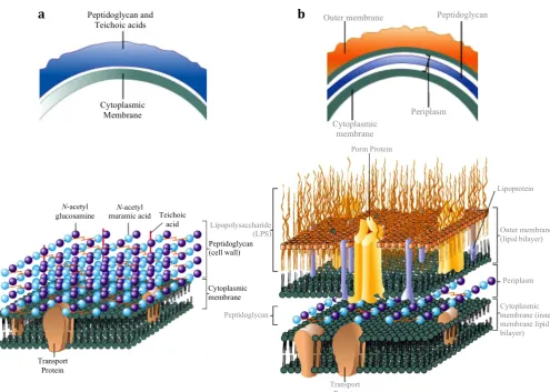
Figure 1.1 The Structural Architecture of (a) Gram-Positive and (b) Gram-Negative Bacterial Cell Envelopes (Adapted from Prescott, Harley & Klein’s Microbiology 7th Ed, 2008).
1.1.2 The Gram-Negative Bacterial Cell Wall
This thesis is focused on the Gram-negative bacteria Escherichia coli and Pseudomonas aeruginosa. The Gram-negative bacterial cell envelope is thinner, less compact and more chemically complex than in positive cells. The cell envelope of Gram-negatives is comprised of an outer membrane, a cell wall, and an inner (cytoplasmic) membrane (Figure 1.1 b). The outer membrane is a distinguishing feature of Gram-negative bacteria, where the outer leaflet is composed of the glycolipid
lipopolysaccharide and the inner leaflet contains phospholipids. The outer membrane serves as a semi-permeable molecular sieve preventing lysis-promoting molecules such as Penicillin G and lysozyme from entering the cell (Silhavy, et al., 2010). It comprises of a plasma membrane, a layer of peptidoglycan surrounding the plasma membrane and an outer cell wall. The peptidoglycan layer in Gram-negatives is extremely thin (~ one
N-acetyl
glucosamine muramic acid Teichoic N-acetyl acid Peptidoglycan (cell wall) Cytoplasmic membrane Transport Protein Lipopolysaccharide (LPS) Peptidoglycan Transport Protein Lipoprotein Outer membrane (lipid bilayer) Periplasm Cytoplasmic membrane (inner membrane lipid bilayer) Porin Protein Peptidoglycan and Teichoic acids Cytoplasmic Membrane
Outer membrane Peptidoglycan
Cytoplasmic membrane
Periplasm
[image:32.595.76.572.82.435.2]layer thick) being only 5-10% of the cell wall, making them much more challenging for antibiotics to penetrate. Cross-linking in Gram-negative peptidoglycan is mostly by direct 3-4 cross-linking between adjacent peptide stems, whereas in Gram-positive peptidoglycan, peptides can be cross-linked by additional amino acids due to potential peptide stem side-branching (amidation). Modification of the peptide stem alters the efficacy of antibiotics, as well as transpeptidase specificity (Münch et al., 2015). The Gram-negative cell envelope has to have more advanced mechanisms to evade
destruction by β-lactams as they have a thinner peptidoglycan layer compared to Gram-positive bacteria. The β-lactam antibiotics penicillin and cephalosporins interfere with the cross-linking between peptides in peptidoglycan, compromising cell wall integrity. Gram-negative bacteria are known to have developed a higher resistance to antibiotics compared to Gram-positive bacteria, Pseudomonas spp. in particular (Miller, 2016).
An outer membrane composed of lipids, lipoproteins and lipopolysaccharides (LPS) surround the thin layer of peptidoglycan, with no teichoic acids in Gram-negative cell walls. This outer membrane is more permeable than the plasma membrane as it is composed of large LPS molecules and porins, which form channels through which small molecules (600-700 Da) can pass (van den Berg, et al., 2015). LPS can be harmful and are classified as endotoxins, however, they are necessary as they prevent antibiotics from entering the periplasm in Gram-negative bacteria (Delcour, 2009).
1.2 Targets for Antibiotics
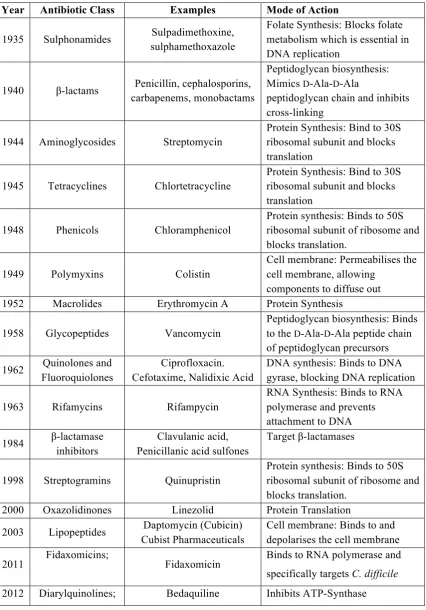
Table 1.1 A List of Antibiotics and their Mode of Action Against Metabolic Processes in Bacterial Cells. The first year of the commercial availability of each antibiotic class is shown and examples from each class are given, with the mode of action (not an exhausted list). Adapted from (Lewis, 2013).
Year Antibiotic Class Examples Mode of Action
1935 Sulphonamides Sulpadimethoxine, sulphamethoxazole
Folate Synthesis: Blocks folate metabolism which is essential in DNA replication
1940 β-lactams Penicillin, cephalosporins, carbapenems, monobactams
Peptidoglycan biosynthesis: Mimics D-Ala-D-Ala
peptidoglycan chain and inhibits cross-linking
1944 Aminoglycosides Streptomycin
Protein Synthesis: Bind to 30S ribosomal subunit and blocks translation
1945 Tetracyclines Chlortetracycline
Protein Synthesis: Bind to 30S ribosomal subunit and blocks translation
1948 Phenicols Chloramphenicol
Protein synthesis: Binds to 50S ribosomal subunit of ribosome and blocks translation.
1949 Polymyxins Colistin
Cell membrane: Permeabilises the cell membrane, allowing
components to diffuse out 1952 Macrolides Erythromycin A Protein Synthesis
1958 Glycopeptides Vancomycin
Peptidoglycan biosynthesis: Binds to the D-Ala-D-Ala peptide chain of peptidoglycan precursors
1962 Quinolones and Fluoroquiolones
Ciprofloxacin. Cefotaxime, Nalidixic Acid
DNA synthesis: Binds to DNA gyrase, blocking DNA replication
1963 Rifamycins Rifampycin
RNA Synthesis: Binds to RNA polymerase and prevents attachment to DNA
1984 β-lactamase inhibitors
Clavulanic acid, Penicillanic acid sulfones
Target β-lactamases
1998 Streptogramins Quinupristin
Protein synthesis: Binds to 50S ribosomal subunit of ribosome and blocks translation.
2000 Oxazolidinones Linezolid Protein Translation
2003 Lipopeptides Daptomycin (Cubicin) Cubist Pharmaceuticals
Cell membrane: Binds to and depolarises the cell membrane
1.2.1 Protein Synthesis as an Antibiotic Target
Protein synthesis in bacteria is unique from that in Eukaryotes, and is a complex and viable target for antibiotics. The ribosome is central to protein synthesis, made up of two subunits: a 30S and 50S that associate to form a 70S ribosome upon protein synthesis. Initiation, elongation and termination are all targeted by streptogramins, macrolides and phenicols, which target the 50S subunit. Aminoglycosides (E.g.
Kanamycin A) target the 30S subunit and cause misreading of the mRNA, incorporating the incorrect amino acids. Tetracyclines bind to part of the rRNA in the 30S subunit and compete with tRNA for the acceptor binding site. Chloramphenicol and
tetrahydropyrimidinones block translation, while Oxazolidinones (e.g. Linezolid) inhibit the ribosomal peptidyl transferase activity (Arenz & Wilson 2016).
1.2.2 DNA and RNA Synthesis
The RNA polymerase initiation complex that precedes transcription can be disrupted by oxazolidinone drugs, and rifampicin also binds to the RNA polymerase initiation
complex, inhibiting RNA synthesis. DNA gyrase is unique to bacteria/prokaryotes and is inhibited by the quinolone family. The fluoroquinolone ciprofloxacin A targets and inhibits the DNA synthesis enzymes Topoisomerase IV and DNA gyrase (Fournier et al., 2000).
1.2.3 Cell Wall Biosynthesis
Table 1.2 A Summary of Antimicrobial Agents that Target the Bacterial Cell Wall
Antimicrobial
Agent Cell Wall Target
Inhibitors of Muramyl-Pentapeptide Synthesis
Fosfomycin The N-acetylmuramic acid component of the bacterial cell wall is derived from N-acetyl glucosamine by the addition of a lactic acid substituent derived from phospheenolpyruvate. The pyruvyl transferase enzyme involved is MurA and is inhibited by fosfomycin.
D-Cycloserine The first three amino acids of the pentapeptide chain are added
individually, but the terminal two residues D-alanyl-D-alanine are added as dipeptide. This dipeptide is produced by racemization of L-Ala to D-Ala and subsequent ligation by a D-ala-D-ala ligase (DDlb). Both of these reactions can be inhibited by D-cycloserine as it is a structural analogue of D-alanine.
Inhibitors of reactions occurring at or in the membrane
Ramoplanin Ramoplanin is a lipoglycodepsipeptide active against Gram-positive bacteria. It inhibits the uptake of N-acetylglucosamine by growing cells with a resultant accumulation of UDP-MurNAc-pentapeptide. Inhibition is of N-acetyl-glucosaminyl-transferase (MurG) that adds N-acetyl
glucosamine to the undecaprenyl-muramyl-pentapeptide.
Bacitracin The lipid carrier involved in transporting the cell wall UDP-MurNAc pentapeptides across the membrane are C55 isoprenyl phosphates. In the transport process the lipid acquires an extra phosphate moiety and to continue in another cycle of transport it must be dephosphorylated.
Bacitracin is a cyclic peptide that binds to the isoprenyl pyrophosphate and prevents dephosphorylation.
Inhibitors of chain cross-linking and formation of the cell wall structure β-Lactams β-Lactams are based around a thiazolidine ring structure and mimic the
D-Ala-D-Ala stem peptide of peptidoglycan. To prevent the final cross-linking reactions in peptidoglycan biosynthesis that gives the
peptidoglycan sacculus its characteristic rigidity. β-Lactams specifically inhibit the transpeptidase domain of penicillin-binding proteins (PBPs) by acting as suicide substrates, which form inactive acyl-enzyme complexes. This results in the accumulation of a pool of inactivated PBPs incapable of catalysing transpeptidation with resulting cell lysis.
1.3 Mechanisms of Antibiotic Resistance in Bacterial Pathogens
Resistance mechanisms include the breakdown of β-lactams by β-lactamases, which are present in the periplasm of the Gram-negative cell envelope. Permeability of the outer membrane can be altered due to altered porins in the outer membrane, preventing access of β-lactams to the cell. Also, PBPs are showing increasing signs of reduced affinity to β-lactams in particular (Moya et al., 2009).
1.3.1 Reduced Antimicrobial Uptake and Active Export by Efflux Pumps
For inhibition of DNA topoisomerases, RNA polymerase, protein synthesis and the cytoplasmic stage of peptidoglycan biosynthesis, navigation through the plasma
membrane is required (two in the case of Gram-negatives) and then accumulation in the cytoplasm. The outer membrane in Gram-negatives provides a selective barrier to antimicrobials, excluding most hydrophobic molecules and macromolecules but the presence of hydrophilic porins allows slow absorption through the membrane (Kumar & Schweizer, 2005). Efflux pumps are found in all bacteria but most cannot respond to an antibiotic without specific mutations. Active efflux pumps and selective uptake work in synergy to reduce the ability of antimicrobials penetrating the cell wall. Gram-negative cells can reduce the permeability of their outer membrane and can also lower the antibiotic concentration near the targets using efflux proteins. P. aeruginosa spp does not contain the porin OprD, and also contains the active β-lactamase AmpC, which confers resistance to the β-lactam Imipenem (Ochs et al., 1999). P. aeruginosa
absorption efficiency of drug molecules through outer membrane porins is 10-100 fold less than those in E. coli (Hancock and Speert, 2000).
1.3.2 Modification of the Antibiotic
Two examples of antibiotic modification are enzymatic modification of
adenyltranserases, respectively (Shaw, et al., 1993). Modification reduces their affinity to the ribosomal RNA.
Amidase enzymes have evolved to cleave the β-lactam ring of penicillins and
cephalosporins. Also, β-lactamases hydrolytically cleave the β-lactam ring using either an active site serine nucleophile or a nucleophilic water molecule (activated via a Zn2+ centre). The β-lactam ring mimics the terminal D-Ala-D-Ala of the peptide stem, acting as a pseudosubstrate. β-lactamases have a similar active site structure to the
transpeptidases and they also hydrolyse the β-lactam ring, so the β-lactam can no longer act as a suicide substrate for the inhibition of PBPs (Philippon et al., 1989). The spread of β-lactamase genes has been increased by their integration in to mobile genetic elements – plasmids and transposons. Multi-drug resistance cassettes can confer
resistance not only to β-lactams, but to aminoglycosides, macrolides, sulphonimides and chloramphenicol.
1.3.3 Modification of the Antibiotic Target
Interaction of the β-lactam with a PBP involves a rapid transformation of a non-covalent complex, blocking PBP function. Many PBPs have evolved with decreased antibiotic affinity including the transpeptidase S. aureus PBP2a, which confers resistance to Methicillin as its rate constant for transpeptidation is reduced 1000-fold (Lim & Strynadka, 2002). The glycopeptide antibiotic vancomycin has a concave binding pocket that is a heptapeptide core, forming five Hydrogen bonds with the D -Ala-D-Ala terminus of the pentapeptide, stalling peptidoglycan synthesis. Resistance to vancomycin occurs in some strains including Vancomycin-Resistant Enterococci (VRE) due to the vanHAX and vanRE genes structurally altering the D-Ala-D-Ala target to D -Ala-D-Lac (Walsh et al., 1996). Vancomycin binds to D-Ala-D-Lac with only four Hydrogen bonds, reducing affinity by 1000-fold and causing resistance.
1.3.4 The Emergence of Antimicrobial Resistant (AMR) Superbugs
for 80,000 infections, and 11,285 deaths in the USA (Centre for Disease Control, USA 2014). The ESKAPE pathogens (Enterococcus faecium, Staphylococcus aureus, Klebsiella pneumoniae, Acinetobacter baumannii, Pseudomonas aeruginosa, and Enterobacter species) are the leading cause of nosocomial (hospital-acquired) infections throughout the world. Most of them are multidrug resistant isolates, which is one of the greatest challenges in clinical practice (Santajit & Indrawattana, 2016). Roughly 400 deaths per year are attributed to these infections (Data from Centre of Disease Control, 2013, USA). Some strains of MDR P. aeruginosa have been found to be resistant to nearly all antibiotics, including aminoglycosides, cephalosporins, fluoroquinolones, and carbapenems.
1.4 Peptidoglycan as the Molecular Scaffold
The bacterial cell is encompassed inside a cell wall that is essential for its viability. This sacculus is composed of multiple layers of a carbohydrate polymer peptidoglycan. Peptidoglycan (murein) is the principal constituent of the bacterial cell wall and is responsible for rigidity of the cell against internal osmotic pressure. The biosynthetic pathway of peptidoglycan is intricate with multiple complexes involved (Figure 1.2). Peptidoglycan a regular mesh-like structure of numerous glycan strands cross-linked for extra tensile strength (Barreteau, et al., 2008). During bacterial growth, the murein layer is continually being replenished at a very high rate.
1.4.1 Cytoplasmic Steps of Peptidoglycan Synthesis
The biosynthesis of peptidoglycan is a multistep pathway of 11 steps, divided into 3 main phases: (1) Formation of UDP-N-acetyl muramic acid (UDP MurNAc) (2) a pentapeptide chain is added sequentially to UDP MurNAc by four ATP-dependent amide bond ligases (Figure 1.3) (3) transport through the cytoplasmic membrane and incorporation into the growing peptidoglycan layer.
Figure 1.3 The Cytoplasmic Steps of Peptidoglycan Biosynthesis. The four Mur ligases: Mur C, D E and F catalyse the sequential addition of the pentapeptide side chain to the D-lactyl group of UDP-MurNAc.
Peptidoglycan is formed by polymerisation and cross-linking of a lipid II monomer into a mesh structure. Lipid II (undecaprenyl-P-P-MurNAc-[pentapeptide]-GlcNAc) is formed in the cytoplasmic phase and is ‘flipped’ to the other cytoplasmic surface via a ‘flippase’ mechanism performed by either FtsW (Mohammadi et al. 2011), or MurJ (Sham et al., 2014), or a synergistic effort by both. The extracellular processing of the monomer Lipid II occurs, with the membrane-associated transglycosylation reaction, followed by transpeptidation.
1.4.2 Lipid-Linked Steps of Peptidoglycan Synthesis
Peptidoglycan is comprised of alternating acetylglucosamine (NAG) and N-acetylmuramic acid (NAM) residues linked by β1-β4 Carbon-bonds and serves as a protective layer unique to bacteria (Lovering, et al., 2007). Polymerisation of each glycan strand is catalysed by transglycosylation and cross-linking between glycan chains is catalysed by transpeptidation. In the case of bifunctional peptidoglycan synthases (more commonly referred to as Penicillin-Binding Proteins or PBPs), these two functions can be performed by one bifunctional PBP. N-acetylmuramic acid has a pentapeptide stem attached to its D-lactyl group that helps form the bridge between two glycan chains. The structure of the pentapeptide in Gram-negative bacteria is commonly
MurD MurE MurF
UDP-MurNAc !
!
UDP-MurNAc-L-Ala UDP-MurN
Ac-Pentapeptide MurC
L-Ala D-Glu
! ! !
L-Ala-D-Glu-mesoDAP-D-Ala-D-Ala. The cross-linking bridge is formed between L -alanyl-γ-D-glutamyl-meso-2,6-diaminopimelic acid of one glycan and the forth D -alanine on the new glycan. The most common form of cross-linking extends between the residue at position 3 of one peptide stem (the acyl acceptor) to the D-Ala at the forth position of another peptide stem (the acyl donor), known as 3-4 cross-linkage (Lovering et al., 2012). mesoDAP contains both an L- and a D-stereocentre. In peptidoglycan, the L-stereocentre is incorporated into the peptide backbone, whereas the cross-linking side-chain contains the D-stereocentre of mesoDAP.
1.4.3 The Mechanism of Transglycosylation
Transglycosylation is the polymerisation of the monomer unit LII in to linear glycan strands, catalysed by the transglycosylase domain of Penicillin Binding Proteins (PBPs). The initial reaction in transglycosylation is the linking of two LII molecules together to form Lipid IV. After which, the successive addition of a single LII unit on to the growing end of the glycan chain forms an extended glycan chain (Figure 1.4). These two reactions are similar, differing only in the length of substrate used. The terms transglycosylation donor and acceptor differentiate between the growing chain (donor) and the incoming LII unit (acceptor). The transglycosylases are processive enzymes anchored to the membrane by a TM alpha-helix, which have a donor site where the growing glycan chain resides and an adjacent acceptor site for the incoming lipid II monomer. As a result the enzyme active site is comparatively long and extended and must accommodate at least four sugar binding sites. Similar active site architecture is seen in lysozyme which also binds alternating N-acetyl-glucosamine and N
Figure 1.4 Transglycosylation: The Mechanism. The transglycosylase domain of Penicillin-Binding Proteins (PBPs) catalyse the polymerisation of Lipid II in to elongated linear peptidoglycan polymers. The growing glycan chain attached to undecaprenyl pyrophosphate is the glycosyl donor substrate and is transferred to the 4-OH group of the GlcNAc unit of the incoming LII, which is the glycosyl acceptor substrate. β-1-4 C-linked N-acetylmuramic acid-N-acetylglucosamine [(NAG-NAM)n] polymers are formed by transglycosylation.
1.4.4 Known Inhibitors of Peptidoglycan Transglycosylation
[image:42.595.115.537.59.360.2]1.4.4.1 Moenomycin: The ‘blueprint’ Transglycosylase Inhibitor and a Structural Tool
The moenomycins are a family of glycolipid antibiotics naturally produced as a complex of related compounds by Streptomyces ghananensis with moenomycin A representing the major component with antimicrobial activity (Welzel 2005, Ostash & Walker 2010). The structure of moenomycin consists of a pentasaccharide of units B, C, D, E and F with a chromophore (unit A) and a C25 lipid chain connected to the F
[image:43.595.109.553.258.621.2]saccharide via a phosphoglycerate linker (Figure 1.5).
Figure 1.5 The Chemical Structure of Moenomycin A. The only known potent inhibitor for bacterial transglycosylases. The region highlighted in blue is the minimal inhibitory pharmacophore, which is often used as a scaffold for the design of new potential inhibitors.
The C25 chain is required for antimicrobial action and the moenomycin structure resembles that of the Lipid IV product formed within the transglycosylase active site. Moenomycin has amphiphilic properties due to the hydrophilic nature of the A to F carbohydrate units, the phosphate group of the phosphoglycerate linker and the folded
O O O O O HO OH HO O C N H A O OH B O OH HO NHAc O HO NHAc O D HO HO HO OH O OH O H2N
O F O CONH2 P O -O O OH O O E C
Losing the D ring does not impact on antibacterial activity.
The C25 chain is similar to the C55 chain in Lipid II.
Both the C25 chain and the phosphoglycerate play a role in binding to the transglycosylase – the phosphoryl group forms electrostatic interactions with conserved residues in the transglycosylase active site.
The phosphoric acid diester group is similar in structure to pyrophosphate in Lipid II and could therefore mimic it. The function of the phosphoric acid could be to direct the C25 lipid chain towards the membrane.
!
Sugar units C,E and F are the minimal units of the pentasaccharide sugars required for antibacterial activity. Sugar units E and F still inhibit
transglycosylase function, but lack antibacterial activity.
Rings C and E bind in the same way as a NAM-NAG disaccharide of the Lipid II substrate.