Review Article
Simple ligation versus stump invagination for the
appendix stump: a systematic review and meta-analysis
Youquan Shi1,2, Dong Tang2, Xiaoli Tang2,3, Daorong Wang2
1Department of General Surgery, Fengxian People’s Hospital of Jiangsu Province, Fengxian, Jiangsu Province,
P. R. China; 2Department of Gastrointestinal Surgery, Subei People’s Hospital of Jiangsu Province, Yangzhou,
Jiangsu Province, P. R. China; 3Department of Gastrointestinal Surgery, The Second Xiangya Hospital of Central
South University, Changsha, Hunan Province, P. R. China
Received October 20, 2017; Accepted October 13, 2018; Epub November 15, 2018; Published November 30, 2018
Abstract: Aim: To evaluate the clinical efficacy and safety of simple ligation (SL) and stump invagination (SI) to treat
the appendix stump. Methods: The Cochrane Library, Embase, Pubmed, Web of science, VIP, and Wanfang data
-bases were searched systematically to identify relevant randomized controlled trials (RCTs) and quasi-RCTs. The study quality was assessed and the relevant data was extracted. Inter-study heterogeneity was assessed using the
Cochran Q test, the I2 test,and the Galbraith figure. The source of heterogeneity was determined using subgroup and
sensitivity analyses. Publication bias was tested using funnel plots. Results: Twenty RCTs including 3677 patients were included in this meta-analysis. In open surgery group, there were no differences in adhesive intestinal obstruc
-tion, wound infec-tion, and post-operative fever between the SL and SI groups. The patients in the SL group had shorter hospitalization and operating time, a lower rate of paralytic ileus, and shorter temperature recovery time after surgery. In the laparoscopy group, the SL group had a higher rate of adhesive intestinal obstruction, longer hos
-pital stay of hos-pital, and shorter surgery time compared with those in the SI group. Conclusions: Our meta-analysis revealed that SL might be a superior method when applying in open surgery. By contrast, SI seemed to be a better solution in laparoscopic surgery.
Keywords: Simple ligation, stump invagination, appendix stump, meta-analysis
Introduction
In general surgery, appendicitis is a common disease, comprising inflammatory changes in the appendix caused by a variety of factors. To treat appendicitis, surgery is usually perform-ed, including open and laparoscopic methods. Open appendectomy (OA) has been the classi -cal treatment for acute appendicitis in adults for decades. In 1983, the German gynecologist Semm successfully performed a laparoscopic appendectomy (LA) for the first time [1]. LA has been recognized by surgeons and physicians to have advantages in terms of accurate diag-nosis, less trauma, mild postoperative pain, rapid recovery, and fewer complications [2]. The treatment of the appendix stump is the key to the success of the operation, and is mainly achieved by simple ligation (SL) or stump invag -ination (SI); however, there is controversy con -cerning the selection of these two methods. In SL, the root of the appendix is ligated using silk thread. In SI, the appendix stump is taken into
the cecum wall and subjected to a pouch suture. In the present study, we carried out a meta-analysis to compare the clinical efficacy and safety of SL and SI to treat the appendix stump.
Materials and methods
Search strategy
Inclusion criteria and exclusion criteria
The inclusion criteria were: (1) A clear diagnosis of acute or chronic appendicitis in the clinic; (2) A quantitative comparison was made between simple ligation and stump invagination; (3) The study monitored the patients using objective and relevant indicators; (4) There were no age, gender, race, language, or publication status restrictions; (5) The study was an RCT or quasi-RCT. The exclusion criteria were: (1) Ambiguous diagnosis; (2) Duplicate studies; and (3) Ob- servational studies or other non-RCT studies.
Data extraction
Studies were retrieved, screened, and extract-ed independently by two researchers, and selected according to the inclusion and exc- lusion criteria. If there were inconsistent opin-ions between them, a third researcher decided whether to include the study or not. The main data extracted were as follows: (1) General
study heterogeneity. If the heterogeneity was small (P ≥ 0.1, I2< 50%), a fixed effects model
was used; otherwise, a random effects model was selected. If I2 was greater than 50%, we
analyzed the sources of heterogeneity using subgroup and sensitivity analyses. If the het -erogeneity was too large, descriptive analysis was used. When the number of studies was greater than nine, we conducted a funnel plot to test the publication bias.
Results
Study selection
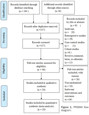
[image:2.612.90.378.71.442.2]A total of 192 articles were initially retrieved, and 137 articles were included after screening for duplicates. After excluding irrelevant stud-ies, non-RCT, irrelevant interventions and out-comes, and not full-text studies, twenty studies [3-22] were eventually included. Figure 1 shows the flow diagram of study identification and selection.
Figure 1. PRISMA flow
diagram.
information (title, author, da- te of publication); (2) Studies characteristics (country, oper -ation method, sex, age, sam-ple size); (3) Outcome mea -sures (the rate of adhesive intestinal obstruction, hospi-talization time, wound infec -tion, operating time, post-operative fever, paralytic ileus (24 to 48 hours after surgery) and temperature recovery time after surgery).
Quality assessment
We used the Cochrane risk of bias tool (version 5.0) for quality evaluation, comprising analysis of the generation of random sequences, alloca-tion concealment, the blind-ing method for patients and testers, the blinding method for the outcome evaluator, and Selective reporting.
Data synthesis and analysis
inter-Table 1. Study characteristics
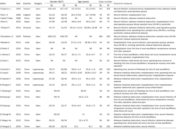
Author Year Country Surgery Gender (M/F) Age (years) Case number Outcome measure
SL SI SL SI SL SI
3 Engstrom, L 1985 Sweden Open 196/165 187/187 29 (15-91) 29 (14-85) 361 374 Wound infection; incisional hernia; hospitalization time; adhesive intesti
-nal obstruction; post-operative pyrexia
4 Dick J A 1984 England Open 21/23 29/30 18.1 (5-43) 21.7 (6-46) 44 59 Wound infection; hospitalization time
5 Habel P Dass 1989 Oman Open 48/16 48/18 NA NA 64 66 Wound infection; adhesive intestinal obstruction
6 Khan S 2010 Nepal Open 27/58 22/48 30.8 ± 9.8 30.0 ± 8.8 80 70 Wound infection; adhesive intestinal obstruction; hospitalization time; post-operative pyrexia; fistula; paralytic ileus (24-48 h); peritonitis 7 Chalya P L 2012 Tanzania Open 20/23 21/23 24.12 ± 12.14 26.28 ± 14.58 43 44 Wound infection; adhesive intestinal obstruction; post-operative pyrexia;
hospitalization time; operating time; paralytic ileus (24-48 h); vomiting;
peritonitis; residual abdominal abscess
8 Chaudhary I A 2005 Pakistan Open 169/213 138/157 NA NA 382 295 Wound infection; adhesive intestinal obstruction; paralytic ileus (24-48 h);
peritonitis; residual abdominal abscess
9 Mukesh S 2013 India Open 24/30 23/33 27.11 ± 4.9 28.36 ± 15.5 54 56 Hospitalization time; wound infection; post-operative pyrexia; paralytic
ileus (24-48 h); vomiting; peritonitis; residual abdominal abscess
10 Peizhi Z 2014 China Open NA NA NA NA 99 99 Hospitalization time; the time of anal exsufflation; temperature recovery
time after operation
11 Haihua Z 2015 China Open 22/15 20/17 32.2 ± 7.1 31.6 ± 6.7 37 37 The time of anal exsufflation; white blood cell count; temperature recovery time after operation; hospitalization time
12 Xuhui L 2013 China Open NA NA NA NA 62 62 Wound infection; white blood cell count; operating time; amount of
bleeding; the time of anal exsufflation; temperature recovery time after
operation
13 Guozhi X 2013 China Laparoscopy 78/72 82/68 50.6 ± 1.4 50.2 ± 1.5 150 150 Operating time; amount of bleeding; the time of anal exsufflation 14 Long H 2016 China Laparoscopy 19/11 18/22 30.50 ± 6.50 29.50 ± 6.17 30 30 Adhesive intestinal obstruction; hospitalization time; operating time; ap
-pendix stump inflammation; abdominal pain; hospitalization expense
15 Xiaotian Y 2016 China Laparoscopy 27/29 26/30 45.0 ± 1.5 45.2 ± 8.5 56 56 Adhesive intestinal obstruction; hospitalization time; hospitalization
expense
16 Hongxia C 2015 China Laparoscopy 31/14 30/15 35.2 ± 2.3 34.6 ± 3.1 45 45 Adhesive intestinal obstruction; hospitalization time; hospitalization expense; abdominal pain; appendix stump inflammation
17 Zhiqiang Z 2015 China Open 40/20 38.23 30 30 Operating time; amount of bleeding; the time of anal exsufflation; tem
-perature recovery time after operation
18 Zhiyong Y 2013 China Open 94/74 32.5 ± 3.4 84 84 Adhesive intestinal obstruction; hospitalization time; wound infection; the time of anal exsufflation; white blood cell count; temperature recovery
time after operation; abdominal pain
19 Caiyang L 2014 China Open 20/19 22/17 42.9 ± 6.6 41.8 ± 7.2 39 39 Adhesive intestinal obstruction; hospitalization time; wound infection; temperature recovery time after operation; the time of anal exsufflation;
white blood cell count
20 Zhongli W 2013 China Open 65/35 32 50 50 Adhesive intestinal obstruction; hospitalization time; wound infection; abdominal abscess; the time of anal exsufflation
21 Mugou Hu 2012 China Open 29/31 26/34 32 ± 4.75 60 60 Adhesive intestinal obstruction; wound infection; abdominal abscess;
operating time; white blood cell count; the time of anal exsufflation 22 Hongcai Z 2004 China Open 60/36 62/43 37.2 41.7 96 105 Adhesive intestinal obstruction; wound infection
[image:3.792.95.692.86.523.2]Study characteristics
This meta-analysis included twenty RCTs [3-22] with a total of 3677 patients. The SL group contained 1866 patients, while the SI group contained 1811 patients. Thirteen [10-22] of the RCTs were conducted in China, and the others were conducted in Sweden [3], Engl-and [4], Oman [5], Nepal [6], Tanzania [7], Pakistan [8], and India [9]. The publication dates ranged from 1984 to 2016. Sixteen RCTs [3-12, 17-22] adopted open surgery, and the remainder [13-16] performed laparoscopic surgery. There was no significant difference between the two groups regarding the age and sex of the patients. Table 1 shows the specific characteristics of the studies.
Quality assessment
All the studies mentioned were randomized. Four studies [7, 11, 12, 20] used the random number table method, one study used a lottery method [9], and the rest of the studies did not
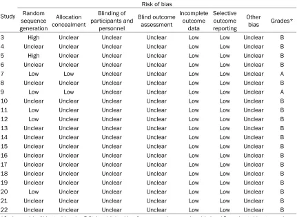
[image:4.612.92.523.86.401.2]mention the randomization and allocation con -cealment methods. With regard to random sequence generation, five studies [7, 9, 11, 12, 20] were low risk and two studies [3, 5] were high risk; in the other studies, the risk was unclear. With regard to allocation concealment, two studies [7, 9] were low risk, and in the other studies, the risk was unclear. All studies are unclear regarding the blinding of partici-pants and personnel, blinding the outcome assessment, and other potential sources of bias. All studies were low risk in terms of incom-plete outcome data and selective outcome reporting. We graded them according to the Cochrane risk of bias tool (version 5.0), Level A: low bias, four or more items are low risk. Level B: moderate bias, two or three items were low risk. Level C: High bias, less than or equal to one items were of low risk. After quality ev-aluation, we found that two [7, 9] studies were type A, and the others were type B. Table 2 shows the quality assessment of bias in the included studies.
Table 2. Quality assessment of bias in the included studies
Study
Risk of bias Random
sequence generation
Allocation concealment
Blinding of
participants and personnel
Blind outcome
assessment
Incomplete outcome
data
Selective outcome reporting
Other
bias Grades*
3 High Unclear Unclear Unclear Low Low Unclear B
4 Unclear Unclear Unclear Unclear Low Low Unclear B
5 High Unclear Unclear Unclear Low Low Unclear B
6 Unclear Unclear Unclear Unclear Low Low Unclear B
7 Low Low Unclear Unclear Low Low Unclear A
8 Unclear Unclear Unclear Unclear Low Low Unclear B
9 Low Low Unclear Unclear Low Low Unclear A
10 Unclear Unclear Unclear Unclear Low Low Unclear B
11 Low Unclear Unclear Unclear Low Low Unclear B
12 Low Unclear Unclear Unclear Low Low Unclear B
13 Unclear Unclear Unclear Unclear Low Low Unclear B
14 Unclear Unclear Unclear Unclear Low Low Unclear B
15 Unclear Unclear Unclear Unclear Low Low Unclear B
16 Unclear Unclear Unclear Unclear Low Low Unclear B
17 Unclear Unclear Unclear Unclear Low Low Unclear B
18 Unclear Unclear Unclear Unclear Low Low Unclear B
19 Unclear Unclear Unclear Unclear Low Low Unclear B
20 Low Unclear Unclear Unclear Low Low Unclear B
21 Unclear Unclear Unclear Unclear Low Low Unclear B
22 Unclear Unclear Unclear Unclear Low Low Unclear B
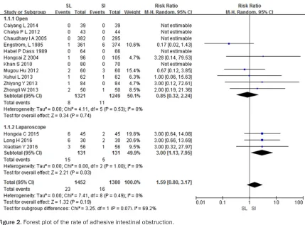
Figure 2. Forest plot of the rate of adhesive intestinal obstruction.
Figure 3. Funnel plot. Adhesive
Meta analysis
The frequency of adhesive intestinal obstruc-tion: Fourteen studies (2832 patients) [3, 5-8, 12, 14-16, 18-22] investigated the frequency of adhesive intestinal obstruction. There was statistical heterogeneity among the studies (P
= 0.07, I2 = 69.2%); therefore, the random ef-
fects model was chosen. The overall analysis showed that there was no statistically signifi -cant difference between the SL and SI groups (RR = 1.59, 95% CI: [0.80, 3.17]). We per -formed subgroup analysis based on different
SL group than in the SI group (RR = 3.00, 95% CI: 1.13-7.95) (Figure 2). A funnel plot to test the publication bias showed that publication bias existed (Figure 3A).
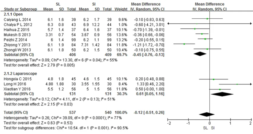
Hospitalization time: Ten studies (1077 pa- tients) [7, 9-11, 14-16, 18-20] reported the hos -pitalization time for the two different surgical procedures. The overall analysis showed that statistical heterogeneity existed among these studies (P < 0.001, I2 = 77%); therefore, the
[image:6.612.91.517.70.296.2]ran-dom effects model was chosen. We found no statistically significant difference in hospitaliza -Figure 4. Forest plot of hospitalization time.
Figure 5. Sensitivity analysis of hospitalization time.
operation methods (open or laparoscopy). The meta-analy -sis results showed that the heterogeneity decreased sig -nificantly when analyzing the open surgery group [3, 5-8, 12, 18-22] (P = 0.53, I2= 0%)
and the laparoscopy group [14-16] (P = 0.49, I2 = 0%)). In
[image:6.612.92.518.78.557.2]tion time between the SL and SI groups (MD = -0.12, 95% CI: -0.51-0.26). We then performed subgroup analysis between the open and lapa -roscopic surgery subgroups. The meta-analysis results showed that the heterogeneity among the studies decreased for the analysis of the open subgroup [7, 9-11, 18-20] (P = 0.04, I2=
55%) and the laparoscopy subgroup [14-16] (P
= 0.13, I2 = 51%). In the open subgroup, the
hospitalization time of the SL group was short -er than that of the SI group. Howev-er, in the laparoscopy group, the hospitalization time was longer in the SL group than in the SI group (Figure 4). We conducted a funnel plot to test the publication bias (Figure 3B). The plot was basically symmetrical, indicating little publica -tion bias. We also conducted sensitivity analy -sis (Figure 5) and found no significant source of sensitivity.
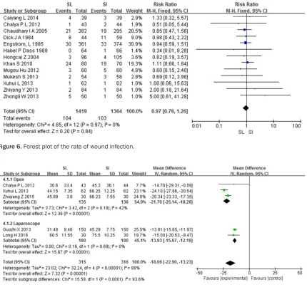
The frequency of wound infection: Thirteen studies (2783 patients) [3-9, 12, 18-22] report -ed the frequency of wound infection. No hetero -geneity was found among these studies (P = 0.97, I2 = 0%); therefore, the fixed effects model
[image:7.612.93.523.71.474.2]was chosen. We found no statistically signifi -cant difference between the SL and SI groups in terms of the frequency of wound infection (RR = 0.97, 95% CI: 0.76-1.26). Both groups had similar frequencies of wound infection (Figure 6). A funnel plot to test showed that publication bias existed among these studies (Figure 3C).
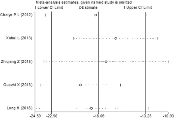
Surgery time: Five studies (631 patients) [7, 12-14, 17] reported the surgery time, including three open surgery studies [7, 12, 17] and two laparoscopic surgery studies [13, 14]. The over -all analysis showed that the there was signifi -Figure 6. Forest plot of the rate of wound infection.
cant heterogeneity (P < 0.00001, I2 = 88%)
[image:8.612.92.378.73.268.2]among these studies; therefore, the random effects model was chosen. The surgery time of the SL group was statistically significantly shorter than that of the SI group (MD = -18.06, 95% CI: -22.90 to -13.23). We then perform- ed a subgroup analysis. The meta-analysis re-sults showed that the heterogeneity decreas-ed in the analyses of the open surgery group [7, 12, 17] (P = 0.18, I2= 42%) and the
laparos-copy group [13, 14] (P = 0.69, I2 = 0%). In the
open surgery group, the surgery time of the SL group was shorter than that of the SI group (MD = -21.70, 95% CI: -25.14 to -18.26), and in the laparoscopy group, the surgery time of the SL group was longer than that the SI group (MD = -13.93, 95% CI: -15.67 to -12.19) (Figure 7). Sensitivity analysis showed no significant source of sensitivity (Figure 8).
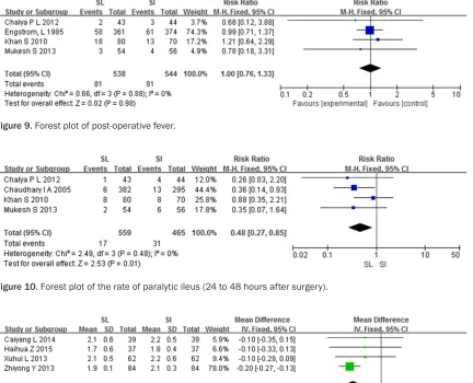
Post-operative fever: Four studies (1082 pa- tients) [3, 6, 7, 9] mentioned post-operative fever. There was no statistical heterogeneity among the studies (P = 0.88, I2 = 0%);
there-fore, the fixed effects model was chosen. There was no statistically significant difference in the frequency of post-operative fever between the two groups (RR = 1.00, 95% CI: 0.76-1.33) (Figure 9).
The frequency of paralytic ileus (24 to 48 hours after surgery): Four studies (1021 patients) [6-9] mentioned paralytic ileus (at 24 to 48 hours after surgery). There was no statistical heterogeneity among the studies (P = 0.48, I2 =
significantly shorter than that of SI group (MD = -0.18, 95% CI: -0.24 to -0.12) (Figure 11). Discussion
Twenty studies comprising a total of 3677 patients were included in this meta-analysis. This study mainly concentrated on the frequen -cy of adhesive intestinal obstruction, hospital -ization time, wound infection, surgery time, post-operative fever, paralytic ileus (at 24 to 48 hours after surgery), and temperature recovery time after operation.
For adhesive intestinal obstructions, the meta-analysis found that there was no statistically significant between the studies in the open sur -gery subgroup; however, in the laparoscopy subgroup, the SL group showed more frequent adhesive intestinal obstructions than in the SI group. The wound surface of the stump invagi-nation is smooth, which can reduce the possi-bility of the residual stump entering the abdom -inal cavity, thus preventing postoperative intestinal adhesion [23], which is consistent with the results of in the laparoscopy subgroup. Watters et al, [24] showed that there was no significant difference in terms of the ileus between the SL and SI groups, which was con -sistent with the results of the open surgery sub -group reported here. The open surgery and laparoscopy group showed opposite results. The reasons may be as follows: firstly, in laparo -scopic surgery, the view of the abdomen is clear; damage to abdominal tissues and organs Figure 8. Sensitivity analysis of operating time.
0%); therefore, the fixed ef-fects model was chosen. The frequency of paralytic ileus in the SL group was significantly lower than that in the SI group (RR = 0.48, 95% CI: 0.27-0.85) (Figure 10).
Temperature recovery time after operation: Four studies (444 patients) [11, 12, 18, 19] reported the temperature recovery time after surgery. There was no statistical het-erogeneity among the studies (P = 0.60, I2 = 0%); therefore,
is reduced and excessive extrusion of the intes-tinal wall are avoided. As a result, the incidence of adhesive intestinal obstruction is reduced compared with laparotomy. Secondly, because of the limited number of samples in laparo-scopic group, there is a possibility of bias.
SI is a more complicated procedure than SL; therefore the surgery time of the SI group was longer than that of the SL group. For wound infection, our meta-analysis found that there was no statistically significant difference be-tween SL and SI groups. SI can avoid the expo -sure of the appendix stump to the abdominal cavity, reducing the infection rate. For inflam -matory appendicitis with severe edema, SI might separate the bowel wall, increasing the likelihood of infection. Therefore, the results of the meta-analysis in terms of wound infection
are understandable. Postoperative fever is clo- sely related to infection; however, there was no statistically significant difference between the SL group and SI group in terms of postop -erative infection.
[image:9.612.94.526.75.425.2] [image:9.612.95.518.76.177.2]In the SI group, the incidence of paralytic ileus was higher, which could have occurred for sev-eral reasons. First, the appendix is required to be buried in the serosa, which could result in deformation and ischemia in the distal cecum during surgery. Secondly, the tightness of the pouch suture also affects ischemia of the cecum, and surgeons, particularly younger or inexperienced surgeons, find it difficult to com -plete a modest string suture. Both of these fac -tors might increase the incidence of paralytic ileus [25]. In addition, the absorption time of the inflammation of the appendiceal stump was Figure 9. Forest plot of post-operative fever.
Figure 10. Forest plot of the rate of paralytic ileus (24 to 48 hours after surgery).
[image:9.612.94.528.362.456.2]shortened significantly because of the lack of a string suture in the SL group. The temperature recovery time after surgery is closely related to inflammation, and as a result, the temperature recovery time in the SL group was shorter.
For hospitalization time, our meta-analysis showed that in the open surgery subgroup, the hospitalization time for the SL group was short -er than that of the SI group, but long-er in la- paroscopy surgery. Street [26] showed that compared with the SL group, the SI group had little difference in postoperative complications, length of hospital stay, or may even be better in some respects. This conflicts with the results of our meta-analysis. During laparoscopic surgery, there are fewer invasions into the abdomen, and the surface of the cecum wall is smoother than that after in SI, which leads to a faster recovery and shorter hospital stay.
Our study has some limitations. First, the com -mon types of appendicitis are acute simple appendicitis, acute purulent appendicitis, and acute perforative appendicitis, and this meta-analysis has not carried out based on these classifications. Second, there were fewer RCTs concerning laparoscopic appendectomy; we only identified four RCTs. Third, although we identified 20 RCTs, we found that high-quality studies were still lacking after quality evalua -tion. Finally, twelve of the 20 studies were from China, thus there is a possibility of regional bias.
Simple ligation and stump invagination are the most common surgical methods to treat the appendix stump, and are chosen by many gen -eral surgeons when performing an appendec-tomy. There are few meta-analyses in this field currently. We believe that this meta-analysis provides feasible options for physicians when treating a patient with appendicitis.
Acknowledgements
We thank all our colleagues at the Department of Gastrointestinal Surgery, Subei People’s Hospital of Jiangsu Province for their assitance. We also thank the native English speaking sci-entists of Elixigen Company (Huntington Beach, California) for editing our manuscript.
Disclosure of conflict of interest
None.
Address correspondence to: Daorong Wang, Depart-
ment of Gastrointestinal Surgery, Subei People’s
Hospital of Jiangsu Province, 98 Nantong West,
Yangzhou 225001, Jiangsu Province, P. R. China.
Tel: 8618051062590; E-mail: wdaorong666@sina. com
References
[1] Semm K. Endoscopic appendectomy. Endos
-copy 1983; 15: 59-64.
[2] Heikkinen TJ, Haukipuro K and Hulkko A.
Cost-effective appendectomy. Open or laparoscop
-ic? A prospective randomized study. Surg En -dosc 1998; 12: 1204-8.
[3] Engström L and Fenyö G. Appendicectomy: as -sessment of stump invagination versus simple
ligation: a prospective, randomized trial. Br J
Surg 1985; 72: 971-972.
[4] Watters DA, Walker MA and Abernethy BC. The
appendix stump: should it be invaginated? Ann R Coll Surg Engl 1984; 66: 92-3.
[5] Dass HP, Wilson SJ, Khan S, Khan A, Parlade S
and Uy A. Appendicectomy stumps: ‘to bury or not to bury’. Trop Doct 1989; 19: 108-9. [6] Khan S. Assessment of stump invagination
versus simple ligation in open
appendicecto-my. Tohoku J Exp Med 2010; 32: 57-74. [7] Chalya PL and Mchembe M. Is invagination of
appendicular stump in appendicectomy neces
-sary? A prospective randomized clinical study. East and Central African Journal of Surgery
2012; 17: 85-89.
[8] Chaudhary IA, Samiullah, Mallhi AA, Afridi A and Bano A. Is it necessary to invaginate the stump after appendicectomy? Pak J Med Sci
2005; 21: 35-38.
[9] Suvera MS, Kharadi AH, Asari US, Damor PB, Shah MT and Patel MB. Open appendicectomy
stump: invaginate or not to invaginate? Inter-national Journal of Research in Medical Sci-ences 2013; 1: 248-251.
[10] Zhang PZ. The effects of two different methods
of ligation of the appendecal stump on
postop-erative recovery of patients. Chinese Journal of Trauma and Disability Medicine 2015; 22:
82-83.
[11] Zhang HH. The effects of two kinds of appen
-dectomy on postoperative recovery of patients
with appendicitis. Chinese Journal of Trauma
and Disability Medicine 2015; 23: 63-64. [12] Lu XH. The comparison of clinical efficacy of
different appendiceal stump ligation methods. Modern Practical Medicine 2013; 25: 913-914.
[13] Xu GZ, Peng Y and Li DS. Clinical observation
of simple ligation of appendix stump in
laparo-scopic appendectomy. Journal of Bethune Mili
[14] Hua L, Wang CY, Zhang JJ and Chen JT. Effect
of purse string suture on appendiceal stump
during laparoscopic appendectomy. Chin J
Mod Drug Appl 2016; 10: 107-108.
[15] Yu XT. Comparative analysis of laparoscopic
pouch suture embedding and simple ligation
in appendectomy. China Continuing Medical
Education 2016; 8: 113-115.
[16] Cui HX. Comparison of the effect of
laparo-scopic appendectomy on whether the appendi -ceal stump is embedded. Henan Medical Re-search 2016; 24: 108-109.
[17] Zhang ZQ. Analysis of the effect of string su -ture and simple ligation on the stump of
ap-pendectomy. Medical Information 2015; 28:
117.
[18] Yang ZY. Clinical effect analysis of purse-string
suture and simple ligation on appendiceal
stumps in appendectomy. China Modern Medi -cine 2013; 20: 33-34.
[19] Liu CY. Comparison between the purse-string
suture and simple ligation on appendiceal
stumps in appendectomy. Medical Aesthetics and Cosmetology 2014; 589-590.
[20] Wang ZL. Evaluation of the clinical effect of ap
-pendectomy with purse-string suture and sim -ple ligation. Mod Diagn Treat 2013; 24: 3956-3957.
[21] Hu MG. Discussion on the treatment of stump
in appendectomy. Seek Medical and Ask The
Medicine 2012; 10: 558-559.
[22] Zhong HC, Chi DZ, Huang WK, Chen KJ and Lu K. Comparison and analysis of clinical effects
of three different methods on handling
appen-diceal stump. Military Medical Journal of South
China 2004; 18: 40-42.
[23] Güller U, Rosella L, Mccall J, Brügger LE and Candinas D. Negative appendicectomy and
perforation rates in patients undergoing
lapa-roscopic surgery for suspected appendicitis. Br
J Surg 2011; 589-595.
[24] Watters DA, Walker MA and Abernethy BC. The
appendix stump: should it be invaginated? Ann R Coll Surg Engl 1984; 66: 92-3.
[25] Qian D, He Z, Hua J and Song Z. Stump invagi -nation versus simple ligation in open
appendi-cectomy: a systematic review and meta-analy -sis. Int Surg 2015; 100: 1199.
[26] Street D, Bodai BI, Owens LJ, Moore DB, Wal
-ton CB and Holcroft JW. Simple ligation vs stump inversion in appendectomy. Arch Surg