Original Article
SUMO-1 expression modulates non-small
cell lung cancer progression
Changkang Ke1*, Kai Zhu2*, Ying Sun1, Zhipei Zhang1, Yunfeng Ni1, Xiaofei Li1
1Department of Thoracic Surgery, Tangdu Hospital, Fourth Military Medical University, Xi’an, China; 2National
Local Joint Engineering Research Center of Biodiagnostics and Biotherapy, The Second Affiliated Hospital of Xi’an Jiaotong University, Xi’an, China. *Equal contributors.
Received July 21, 2017; Accepted April 5, 2018; Epub June 15, 2018; Published June 30, 2018
Abstract: Previous studies indicate that the role of small ubiquitin-related modifier-1 (SUMO1) is responsible for mul-tiple cancer progression including non-small-cell lung cancer (NSCLC). This study aims to explore the expression and significance of SUMO1 in NSCLC. 179 NSCLC patients with resected lung tissue were used for immunofluorescence to detect the expression level of SUMO1 in tumor cells. We also evaluated the association of SUMO1 expression with clinicopathological parameters, including the Ki-67 labeling index (LI). We used Kaplan-Meier survival analysis and Cox proportional hazards models to estimate the effect of SUMO1 expression on survival. We found that SUMO1 ex-pression was significantly elevated in NSCLC tissues when compared with adjacent lung tissues (p<0.01). Moreover, SUMO1 expression was significantly associated with histological type (p<0.01), lymphatic metastasis (P<0.001), and pTNM stage (p<0.05). Kaplan-Meier survival analysis showed that overall survival of patients with high expres-sion of SUMO1 was significantly shorter (n = 163, p<0.05). In concluexpres-sion, SUMO1 is closely related to the progres-sion in NSCLC and could be served as a potential prognostic biomarker and therapeutic target for NSCLC.
Keywords: SUMO1, NSCLC, survival, oncogene
Introduction
Lung cancer is the leading cause of cancer-related death worldwide [1]. Most cases of lung cancer are non-small cell lung cancer (NSCLC), however, the long-term survival rate of NSCLC patients remains disappointing. A majority of NSCLC patients die from distant metastases and recurrent disease even after undergoing curative surgical resection [2-4]. There is an urgent need to identify new prognostic markers that can facilitate a better assessment of the survival probabilities and optimized therapies for individual patients.
Small ubiquitin-related modifier-1 (SUMO-1) is
the best-characterized member of a rapidly growing family of ubiquitin-like proteins that are
involved in post-translational modifications [5-7]. SUMO-1 is reversibly attached to other proteins in a ubiquitination-like manner.
SUMO-1 congestion (sumoylation) affects the subcel-lular localization of substrates and stability as
well as transcriptional activities [8-11]. Many transcriptional regulatory proteins have been
identified as sumoylation substrates, and many
more sumoylation substrates are expected to exist [12, 13]. A couple of oncoproteins, for example c-Myb [14], PML [15] and c-Jun [16], have been corroborated to be substrates of
sumoylation. The significance of sumoylation in
the function of the oncogenes varies among them. Given the substrates involved, protein sumoylation would be expected to play impor-tant role in the course of tumorigenesis and alter in different human cancer [17]. However, there is still no explicit evidence to support this theory.
To study the clinicopathologic features and
prognostic implications of SUMO1 expression
in patients with NSCLC, we investigated the
expression of SUMO1 in NSCLC by immunohis -tochemical staining and analyzed the
Material and methods
Subjects
Study population and tissue samples collection: Paraffin-embedded tissue specimens from 163 patients with confirmed NSCLC were ob -tained from the thoracic oncology tissue re-pository in the Department of Thoracic Sur-gery of Tangdu Hospital, between March 2006 to September 2009. Patients who received preoperative chemotherapy, radiotherapy or
EGFR-targeted therapy were excluded from this study. Detailed information was obtained
serial slides. The slides were then dewaxed in xylene and rehydrated through a graded series of ethanol solution. Endogenous per-oxidase activity was neutralized by immers-ing the slides in a solution of 3% hydrogen peroxide in methanol for 15 min at room tem-perature. Combined sodium citrate (pH 6.0) and incubation in a pressure cooker (3 min, 125°C) was used for antigen retrieval. To
re-duce nonspecific binding, slides were
block-ed with goat serum for 30 min. Then, the slides
[image:2.612.90.383.95.519.2]were incubated in a humidified chamber at 4°C overnight with primary anti-SUMO1 (diluted 1:400, Abcam, USA) or anti-Ki-67 (diluted 1:50,
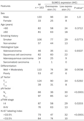
Table 1. Correlation between SUMO1 expression and clinicopatho -logical features in 163 patients with NSCLC.
Features n = 163All
SUMO1 expresion (IHC)
Overexpres-sion (%) Low expres-sion (%) P-value* Sex
Male 130 96 34 1.0
Female 33 25 8
Age (years)
<60 82 58 24 0.3712
≥60 81 63 18
Smoking history
Smoker 106 77 29 0.5773
Non-smoker 57 44 13
Histological type
Adenocarcinoma 46 35 11 0.9337
Squamous cell carcinoma 82 60 22
Adenosquamous carcinoma 34 25 9
Sarcomatoid carcinoma 1 1 0
Differentiation
Well + Moderately 110 74 36 0.0038
Poorly 53 47 6
pT factor
T1-2 124 90 34 0.5292
T3-4 39 31 8
pN factor
N0 66 36 30 <0.0001
N1-2 97 85 12
pTNM stage
I-II 87 58 29 0.0203
III-IV 76 63 13
Ki-67 labeling index
>33.5% 79 47 32 <0.0001
<33.5% 84 74 10
*P-value of χ2-test are shown.
from the medical records of the enrolled patients in a computerized registry da-tabase including age, gen-der, smoking history, clini- cal manifestation, tumor st- atus, histological differen-tiation, nodal status and follow-up information. Fol-low-up lasted through De- cember 1, 2013, with a me-dian follow-up period of 52 months for living patients (range, 45-80 months). The day of surgery was con- sidered as the starting day for estimating postopera- tive survival time.
Histologi-cal classification of tumors
was reviewed by
patholo-gists and based on the Wo-rld Health Organization cri -teria. All tumors were stag- ed according to the pa- thological tumor/node/me-
tastasis (pTNM) classifica -tion (7th edi-tion) of the
In-ternational Union against
Cancer [18]. The study pro-tocol was approved by the Regional Ethics Committee for Clinical Research of the
Fourth Military Medical Uni -versity. Informed consent was taken from all subjects.
Immunohistochemistry
Paraffin-embedded tissue
Thermo, USA) antibodies. A two-step
polymer-HRP method (Dako, Carpinteria, CA) was used for detection. No staining was observed for negative controls, which included incubation of lung tissue with a non-immune primary an-tibody.
Evaluation of immunohistochemical staining
Five random fields from each slide were
view-ed under a light microscope (Leica DM4000B, Germany) at ×400 magnification. The expres
-sion of SUMO1 was scored by multiplication of
the percentage of positive tumor cells and the staining intensity. Initially, the percentage of positive cells was scored as 0 (0%), 1 (1-10%), 2 (11-50%) and 3 (51-100%). Thereafter, inten-sity of staining was scored as follows: 0 (nega-tive), 1 (weakly posi(nega-tive), 2 (moderately
posi-Meier method, and the significance of the dif -ference was evaluated using the log-rank test. Correlation analyses of the survival time and various clinicopathological variables were per-formed by univariate and multivariate analyses
using the Cox regression model. P<0.05 was considered to be statistically significant. All
analyses were performed with Prism 5.01 soft-ware (GraphPad Softsoft-ware, Inc.) and SPSS 18
(Inc., Chicago IL, USA).
Results
General characteristics of the subjects
[image:3.612.89.377.72.397.2]The clinicopathologic characteristics of the patients are summarized in Table 1. There were 33 female and 130 male patients with a medi-an age of 59 years (rmedi-ange, 30-81 years). The
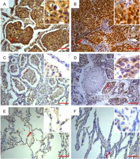
Figure 1. Expression of SUMO1 in NSCLC. A. Optical overexpression SUMO1 staining in ADC. B. Overexpression SUMO1 staining in SCC. C. Low expres-sion SUMO1 staining in ADC. D. Low expresexpres-sion SUMO1 staining in SCC. E. Low expression SUMO1 staining in corresponding adjacent non-cancerous tissue. F. Absence of SUMO1 in corresponding adjacent non-cancerous tis-sue staining in corresponding adjacent non-cancerous tistis-sue. Scale bar = 100 µm.
tive) and 3 (strongly positive).
By ROC analyses, the case with a final scores ≥5 was classified as overexpression (Sensitivity 81.9, Specificity 95.1), the case with a final
scores between 3-5 was
clas-sified as low expression. For
Ki-67, the expression of Ki-67 was assessed based on the labeling index (LI) determined by counting 500-1000 tumor cells randomly selected in a
high-power field. The median
value of positive tumor cells was 33.5% in the current
se-ries, therefore, we defined tumors with ≥33.5% of Ki-67
as high Ki-67. All slides were assessed by 3 independent investigators who were blind-ed to the clinical features and
outcomes. The final immuno -histochemical staining score reported is the average of the scores from the three investi-gators.
Statistical analysis
Kaplan-patients were diagnosed with squamous cell
carcinoma (SCC; n = 82, 50.3%), adenocarci
-noma (ADC; n = 46, 28.2%), adenosquamous carcinoma (ASC; n = 34, 20.9%) and sareoma
-todes carcinoma (SC; n = 1, 0.61%).
Histop-athologic diagnosis included: 29 well differen-tiation (17.8%), 81 moderately differendifferen-tiation (49.7%), and 53 poorly differentiation tumors (33.1%). Postoperative staging evaluation dem-onstrated stage I disease in 24 patients, stage II disease in 64 patients, stage III disease in 71 patients, and stage IV disease in 4 patients.
Expression pattern of SUMO1 in NSCLC and correlation with clinicopathological character-istics
Overall, 74.23% (121/163) tumor sections were classified as SUMO1 overexpression
(Figure 1A, 1B), the rest of 42 (25.774%) tumor
sections were classified as low expression
(Figure 1C, 1D), while 5.52% (9/163) homolo-gous adjacent non-cancerous tissue sections
were classified as SUMO1 low expression
(Figure 1E) and the rest of adjacent
non-can-cerous tissues were classified as negative
(Figure 1F). Positive staining was mainly locat-ed in the cytoplasm. In order to evaluate the
role of SUMO1 in NSCLC, we analyzed wheth-er SUMO1 expression was associated with any
of the clinicopathological features (Table 1).
Statistical results showed that SUMO1 over-expression was significantly associated with differentiation (P = 0.0038), pN factor (P< 0.0001), pTNM stage (P = 0.0203) and Ki-67 labeling index (P<0.0001). No significant rela
-tionship was noted between SUMO1 expres
-sion and sex (P = 1.0), age (P = 0.3712), smok
-ing history (P = 0.5773), histological type (P = 0.9337), and pT factor (P = 0.5292).
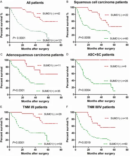
Relationship between SUMO1 expression and survival in NSCLC patients
To investigate the relationship between SUMO1
expression level and the clinical outcome of NSCLC patients, we compared the correlation
between patient survival and SUMO1 expres
-sion status. Patients with overexpres-sion SU-MO1 had an obviously worse prognosis than those with low level of SUMO1 expression (P<0.0001, Figure 2A). NSCLC patients with
SUMO1-overexpression (n = 121) had a
19-month median survival time and 23.17-19-month
mean survival time (95% CI = 19.91-26.42
months). The mean survival time of patients
with low level of SUMO1 expression (n = 42) was 46.76 months (95% CI = 42.41-51.11
months), whereas the median survival time had not reached. Compare with poorly
differen-tiated patients (n = 53, median survival time =
19 months), patients with well or moderately
differentiated tumors (n = 110, median survi-val time = 50) had a longer survisurvi-val time (P<
0.001). The median survival time of SCC
(Squamous cell carcinoma) patients with SU-MO1 overexpression (n = 60) was 12 months (95% CI = 5-19 months), while the median sur
-vival time of those with SUMO1 low-expression SCC (n = 22) was 51 months (95% CI was not reached). SCC patients with SUMO1 overex -pression had a worse prognosis than those
with SUMO1 low expression (P = 0.0056, Figure 2B). For the ADC (Adenocarcinoma) patients, the median and mean survival time of patients
with SUMO1-overexpression (n = 35) was 25
months, whereas the mean survival time of
those with SUMO1 low expression ADC (n = 11)
had not yet been reached and the mean sur-vival time was 45.45 months. ADC patients
with SUMO1 overexpression expression had a worse prognosis than those with SUMO1 low expression (P = 0.0301, Figure 2C). For the
ASC + SC (Adenosquamous carcinoma and
Sarcomatoid carcinoma) patients, the median
and mean survival time of patients with SUMO1-overexpression (n = 26) was 15 months, where
-as the mean survival time of those with SUMO1 low expression (n = 9) had not yet been reached
and the mean survival time was 54.11 months.
ASC + SC patients with SUMO1 overexpression
expression had a dramatically worse prognosis
than those with SUMO1 low expression (P =
0.0004, Figure 2D). The survival of patients with NSCLC was related with the grade of tumor differentiation and pTNM stage. Patients with
pTNM I/II tumors (n = 87) had a median survival
time of 49 months. The median survival time of
NSCLC patients with TNM III/IV tumors (n = 76) was 20 months (P = 0.0019, Figure S1).
We divided the enrolled NSCLC patients into
pTNM I/II and pTNM III/IV groups to investigate
the relationship between SUMO1 expression
and the clinical outcome. Among the I/II-stage group, the median survival time of patients
with SUMO1-overexpression (n = 58) was 24
months, whereas the mean survival time of
52.17 months and the median survival time
had not been reached. There were significant differences in the survival rate with SUMO1
expression level (P = 0.0001, Figure 2E). Am- ong the III/IV-stage group, the median survival
[image:5.612.92.524.73.593.2]time of SUMO1 overexpression patients (n =
60) was 15 months, whereas the median
sur-vival time of SUMO1 low expression patients (n = 16) had not yet been reached and the mean
survival time was 38.5 months. There also existed momentous difference in the survival
rate with SUMO1 expression in this group (P =
0.0019, Figure 2F). To further assess whether
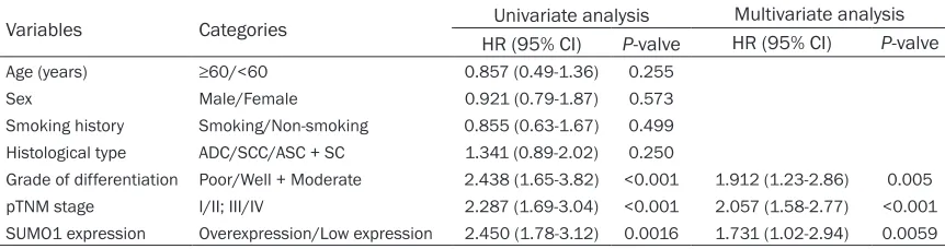
SUMO1 expression represents a prognostic cri -terion in patients with NSCLC, we carried out regression analysis using the Cox’s proportion-al hazards model. The covariate parameters included several clinicopathological factors in
addition to SUMO1 were shown in Table 2. In
univariate analysis, SUMO1 expression, grade
of differentiation and pTNM stage showed a
sig-nificantly higher hazard ratio for a poor progno -sis. Moreover, multivariate analysis was
per-formed using the significant factors observed
in univariate analysis. The results showed that, along with the TNM stage and grade of
differen-tiation, SUMO1 expression level was an autono
-mous prognostic factor (P = 0.0059, Table 2). These results adequately pointed out that the
SUMO1 expression in NSCLC patients was firm -ly related to a poor prognosis.
Discussion
Protein sumoylation can be expected to be important in the course of tumorigenesis [11, 19-21]. However, there is no evidence to sup-port this notion in lung cancer. In this study, we performed immunohistological analysis to
detect the expression of SUMO-1 in 163
patients with NSCLC. The result showed that 74.23% (121/163) of tumor sections were
over-expression for SUMO-1 over-expression, while none
of corresponding adjacent non-cancerous
tis-sue was overexpression. SUMO1 was highly
expression in NSCLC tissues and almost unde-tectable in normal lung tissue. This indicated
that SUMO-1 may be a new biomarker or diag
-nostic tool for lung cancer. We also
compar-ed the correlation between patient survival
and SUMO1 expression status and found that SUMO1 over expression in NSCLC patients
was notably associated with some clinical ch- aracteristic, such as pN factor and pTNM st-
age. These finding indicated that SUMO1 may
play an important role in the metastasis of NSCLC.
To the best of our knowledge, this is the first
time to indicate an association between
SUMO1-positivity in NSCLC patients and poor prognosis and to suggest that SUMO1 overex -pression may be an independent prognostic factor of NSCLC patients. Furthermore, the
prognostic significance of SUMO1 overexpres -sion was apparent in all kinds of NSCLC
patients. We also investigated whether SUMO1
overexpression was associated with prognosis in early stage NSCLC patients. The results
showed apparent prognostic significance of SUMO1 overexpression in early stage patients
and in late stage patients. This result indicated
that SUMO1 overexpression may also take part in the development of NSCLC. We hypothesize that SUMO1 overexpression may be related to
cancer cell proliferation and metastasis.
Some shortcomings present existed in this study. Subject to the limitations of sample size and follow-up, the median survival time in many groups was absent. To respond the patient’s survival, we listed the mean survival time, which was failed to accurately respond the patient’s survival as the median survival time. In addition, this study did not investigate the relationship between the expression level of
SUMO1 gene and the pathogenesis of lung can
-cer, which was very significant for a biomarker
[image:6.612.90.521.85.199.2]validation. To remedy these limitations, further multicenter clinical studies will be carried out Table 2. Cox proportional hazards model analysis of variables affecting survival in NSCLC patients
Variables Categories Univariate analysis Multivariate analysis
HR (95% CI) P-valve HR (95% CI) P-valve
Age (years) ≥60/<60 0.857 (0.49-1.36) 0.255
Sex Male/Female 0.921 (0.79-1.87) 0.573
Smoking history Smoking/Non-smoking 0.855 (0.63-1.67) 0.499
Histological type ADC/SCC/ASC + SC 1.341 (0.89-2.02) 0.250
Grade of differentiation Poor/Well + Moderate 2.438 (1.65-3.82) <0.001 1.912 (1.23-2.86) 0.005
pTNM stage I/II; III/IV 2.287 (1.69-3.04) <0.001 2.057 (1.58-2.77) <0.001
and an expanding sample size will enrich the means of detection.
Taken together with the results in this study,
we indicate that SUMO1 may be a potential
drug therapy target for NSCLC patients. How- ever, the mechanism underlying the promo-
tion of NSCLC by SUMO1 needs to be further
examined. Conclusions
We showed that a high proportion of NSCLCs express SUMO1 and that SUMO1 overexpres
-sion is significantly associated with grade of
tumor differentiation, pTNM stage, and lym-phatic metastasis. Moreover, our study is the
first to indicate a role for SUMO1 as an
inde-pendent factor predictive of poor prognosis in NSCLCs.
Acknowledgements
The work was supported by the grants from the National Natural Science Foundation of China (30973379 to Xiao-Fei Li).
Disclosure of conflict of interest
None.
Abbreviations
NSCLC, Non-small cell lung cancer; SUMO1, small ubiquitin-related modifier-1; SCC,
Squa-mous cell carcinoma; ADC, Adenocarcinoma; SC, sareomatodes carcinoma; ASC,
adeno-squamous carcinoma; CI, Confidence interval;
HR, Hazard Ratio.
Address correspondence to: Xiaofei Li, Depart- ment of Thoracic Surgery, Tangdu Hospital, Fourth Military Medical University, Xi’an, China. Tel: +86-029-84777928; E-mail: [email protected]; Yunfeng Ni, Department of Thoracic Surgery, Tangdu Hospital, Fourth Military Medical University, Xi’an, China. Tel: +86-029-84777936; E-mail: niyunfng@ fmmu.edu.cn; Zhipei Zhang, Department of Thoracic Surgery, Tangdu Hospital, Fourth Military Medical University, Xi’an, China. Tel: +86-029-84777934; E-mail: [email protected]
Reference
[1] Siegel RL, Miller KD and Jemal A. Cancer sta-tistics, 2016. CA Cancer J Clin 2016; 66: 7-30.
[2] Reungwetwattana T, Weroha SJ and Molina JR. Oncogenic pathways, molecularly targeted therapies, and highlighted clinical trials in non-small-cell lung cancer (NSCLC). Clin Lung Can-cer 2012; 13: 252-266.
[3] Pfannschmidt J, Muley T, Bulzebruck H, Hoff-mann H and DieneHoff-mann H. Prognostic assess-ment after surgical resection for non-small cell lung cancer: experiences in 2083 patients. Lung Cancer 2007; 55: 371-377.
[4] Ye X, Ji C, Huang Q, Cheng C, Tang R, Xu J, Zeng L, Dai J, Wu Q, Gu S, Xie Y and Mao Y. Isolation and characterization of a human putative re-ceptor protein kinase cDNA STYK1. Mol Biol Rep 2003; 30: 91-96.
[5] Hochstrasser M. Evolution and function of ubiquitin-like protein-conjugation systems. Nat Cell Biol 2000; 2: E153-157.
[6] Yeh ET, Gong L and Kamitani T. Ubiquitin-like proteins: new wines in new bottles. Gene 2000; 248: 1-14.
[7] Melchior F. SUMO--nonclassical ubiquitin. Annu Rev Cell Dev Biol 2000; 16: 591-626. [8] Muller S, Hoege C, Pyrowolakis G and Jentsch
S. SUMO, ubiquitin’s mysterious cousin. Nat Rev Mol Cell Biol 2001; 2: 202-210.
[9] Hay RT. SUMO: a history of modification. Mol Cell 2005; 18: 1-12.
[10] Muller S, Ledl A and Schmidt D. SUMO: a regu-lator of gene expression and genome integrity. Oncogene 2004; 23: 1998-2008.
[11] Gill G. Something about SUMO inhibits tran-scription. Curr Opin Genet Dev 2005; 15: 536-541.
[12] Mendes AV, Grou CP, Azevedo JE and Pinto MP. Evaluation of the activity and substrate speci-ficity of the human SENP family of SUMO prote-ases. Biochim Biophys Acta 2016; 1863: 139-147.
[13] Lin CH, Liu SY and Lee EH. SUMO modification of Akt regulates global SUMOylation and sub-strate SUMOylation specificity through Akt phosphorylation of Ubc9 and SUMO1. Onco-gene 2016; 35: 595-607.
[14] Bies J, Markus J and Wolff L. Covalent attach-ment of the SUMO-1 protein to the negative regulatory domain of the c-Myb transcription factor modifies its stability and transactivation capacity. J Biol Chem 2002; 277: 8999-9009. [15] Sternsdorf T, Jensen K and Will H. Evidence for
covalent modification of the nuclear dot-asso-ciated proteins PML and Sp100 by PIC1/ SUMO-1. J Cell Biol 1997; 139: 1621-1634. [16] Muller S, Berger M, Lehembre F, Seeler JS,
Haupt Y and Dejean A. c-Jun and p53 activity is modulated by SUMO-1 modification. J Biol Chem 2000; 275: 13321-13329.
[18] Goldstraw P, Crowley J, Chansky K, Giroux DJ, Groome PA, Rami-Porta R, Postmus PE, Rusch V, Sobin L; International Association for the Study of Lung Cancer International Staging Committee; Participating Institutions. The IASLC lung cancer staging project: proposals for the revision of the TNM stage groupings in the forthcoming (seventh) edition of the TNM classification of malignant tumours. J Thorac Oncol 2007; 2: 706-714.
[19] Zhang H, Kuai X, Ji Z, Li Z and Shi R. Over-ex-pression of small ubiquitin-related modifier-1 and sumoylated p53 in colon cancer. Cell Bio-chem Biophys 2013; 67: 1081-1087.
[20] Zhang J, Huang FF, Wu DS, Li WJ, Zhan HE, Peng MY, Fang P, Cao PF, Zhang MM, Zeng H and Chen FP. SUMOylation of insulin-like growth factor 1 receptor, promotes prolifera-tion in acute myeloid leukemia. Cancer Lett 2015; 357: 297-306.