ORIGINAL ARTICLE
In
fl
ammatory lung disease a potential
risk factor for onset of idiopathic
in
fl
ammatory myopathies: results from
a pilot study
Sevim Barbasso Helmers,1Xia Jiang,1David Pettersson,1Anna-Lis Wikman,2 Pia Axelman,1Åsa Lundberg,2Ingrid E Lundberg,2Lars Alfredsson1,3
To cite:Helmers SB, Jiang X,
Pettersson D,et al. Inflammatory lung disease a potential risk factor for onset of idiopathic inflammatory myopathies: results from a pilot study.RMD Open 2016;2:e000342.
doi:10.1136/rmdopen-2016-000342
▸ Prepublication history and additional material is available. To view please visit the journal (http://dx.doi.org/ 10.1136/rmdopen-2016-000342).
SB, XJ, IEL, LA authors contributed equally.
Received 20 July 2016 Revised 22 September 2016 Accepted 10 October 2016
For numbered affiliations see end of article.
Correspondence to Professor Ingrid Lundberg; Ingrid.Lundberg@ki.se
ABSTRACT
Objectives:To assess the association
between inflammatory lung disease and the risk of developing idiopathic inflammatory
myopathies.
Methods:A population-based case–control study was conducted. Adult myositis cases, identified from the Swedish inpatient registry (diagnosed between 1995 and 1997), and randomly selected controls matched to cases on the date of birth, gender and residency, were asked to fill out a questionnaire with questions on lifestyle, environmental exposures and health. Eventually, 100 cases and 402 controls responded to the questionnaire and were included in the analyses. Exposure was defined as self-reported preceding inflammatory lung diseases ( pneumonia, tuberculosis or sarcoidosis). The association between the exposure and risk of developing myositis was evaluated by calculating OR together with 95% CIs in logistic regressions.
Results:42 (42%) cases and 112 (28%) controls reported preceding inflammatory lung disease. Median duration between inflammatory lung disease and first symptom of myositis was 30 years. We observed a significant association between self-reported history of lung disease at study inclusion and diagnosis of myositis (crude OR=1.8 (1.1 to 2.9); smoking adjusted OR=1.9 (1.2 to 3.1)). We further identified a modestly increased, yet non-significant, association between preceding
inflammatory lung disease ( prior to index year) and diagnosis of myositis (smoking adjusted OR=1.6 (0.9 to 2.8)). The association was more
pronounced among the cases of myositis with concurrent interstitial lung disease (OR=3.8 (1.0 to 14.5)).
Conclusions:Patients with preceding inflammatory lung disease tend to have an increased risk of developing myositis compared to those without. The effect was more pronounced among patients with myositis with concurrent interstitial lung disease. Thus inflammatory lung disease may constitute a risk factor for myositis.
INTRODUCTION
Idiopathic inflammatory myopathies (IIM) col-lectively named myositis, are rare autoimmune disorders characterised by symmetrical muscle weakness and inflammation in skeletal muscle tissues. Based on clinical and histopathological features, IIMs can be subdivided into dermato-myositis (DM), polydermato-myositis (PM) and inclu-sion body myositis (IBM). More recently a subgroup named autoimmune necrotising myopathy has been identified. Although the
Key messages
What is already know about this subject? ▸ Myositis occurs as a combined effect of genetic
factors and environmental triggers. Lung symp-toms have been long recognised as a common complication of myositis which may even precede the muscle symptoms.
What does this study add?
▸ We observed a significant association between self-reported history of inflammatory lung disease at study inclusion and risk of myositis (crude OR=1.8 (1.1 to 2.9); smoking adjusted OR=1.9 (1.2 to 3.1)). We further identified a modestly increased yet non-significant associ-ation between preceding (before study inclusion) inflammatory lung disease and myositis (smoking adjusted OR=1.6 (0.9 to 2.8)), which, became more pronounced among the cases of myositis with concurrent interstitial lung disease (OR=3.8 (1.0 to 14.5)).
How might this impact on clinical practice? ▸ Our study suggests a potential link between
infection and certain autoimmune diseases such as myositis. This piece of evidence, together with other information such as family history, would hopefully aid early detection and early treatment of autoimmune diseases.
on May 7, 2020 by guest. Protected by copyright.
http://rmdopen.bmj.com/
aetiology remains unclear, several lines of evidence have indicated that myositis occurs as a combined effect of genetic and environmental factors. HLA-DRB1*0301 has been identified as the strongest genetic risk factor,1with a most pronounced effect in anti-Jo-1 positive myositis.2 Ultraviolet light exposure and deficient vitamin D levels are established environmental risk factors.3 4 Smoking increases risk of anti-Jo-1 positive myositis and seems to interact with DRB1*0301 genotype in anti-Jo-1 positive myositis.2Studies have suggested preceding infections, in particular infections in the lung, as an additional risk factor but with no convincing results.5 6 Lung symptoms have commonly been observed among patients with myo-sitis and may precede the muscle symptoms.7 Interstitial lung disease (ILD) has been found in up to 78% of patients with PM and DM but less frequently in IBM.8 Pneumonia, tuberculosis and sarcoidosis have been reported to be over-represented in patients with rheumatic disorders including myositis.9
Based on thesefindings, we hypothesised that a pre-ceding lung disease could confer a risk to develop myositis. We aimed to investigate the association between preceding inflammatory lung diseases (including pneumonia, tuberculosis and sarcoidosis) and risk to develop myositis using a nationwide population-based case–control study involving patients with adult onset myositis and randomly selected con-trols in Sweden. As many patients from the investi-gated patient population were deceased we also estimated the mortality rate.
MATERIAL AND METHODS Design and study population
This is a nationwide population-based case–control study with patients with myositis diagnosed between 1995 and 1997 in Sweden. The patients were identified through the Swedish National Inpatient Register (IPR), which covers information on discharges from inpatient care in Sweden since 1987. This register includes data on primary and secondary discharge diagnosis, based on codes according to the International Classification of Diseases (ICD) V.9 (valid from 1987 to 1996) and V.10 (valid from 1997). Cases were defined by having had ICD-9 code 710E (PM) or 710D (DM) or having had ICD-10 code M331 (DM) or M332 (PM) as primary or secondary diagnosis. The myositis diagnosis from the IPR was further verified by scrutinising the patient records.10ILD was identified from patient records based on chest X-ray and pulmonary function tests.
The controls were identified from the National popu-lation register in Sweden which contains information on Swedish residents’ dates of birth, death and residency. For each case six controls were randomly selected matched by date of birth, gender and residential area of the case. Each participant was invited to answer a ques-tionnaire. The Ethics Committee of Karolinska University Hospital approved the study.
Information on inflammatory lung disease and other exposures
Information on lifestyle, environmental exposures and disease history was collected between 1999 and 2002 through a questionnaire sent to the cases and controls and returned by regular mail. Unanswered or incom-pletely answered questionnaires were completed by mail or by telephone with assistance of trained personnel not connected to the individual clinics. The questionnaire was based on questions that have been previously used in a large epidemiological study (the EIRA case–control study11), contained a wide spectrum of questions cover-ing extensive areas such as demographic factors, symp-toms at onset of myositis, family history of diseases, own medical history, smoking habits and snuff habits, vacci-nations, pets, sun exposure, reproductive variables, occu-pational history, etc (original questionnaire in Swedish were attached as appendix 1a and 1b).
For each case the year when the first symptoms of myositis were reported was defined as the index year, and the same index year was used for the correspond-ing controls. There were three cases with no informa-tion reported on the time when first symptoms occurred, therefore the year at diagnosis was used as the index year. In this report we focused on the follow-ing questions regardfollow-ing disease history: “Do you have, or have you had the following diseases?” The answer alternatives were; “yes”, “no”; and “if yes, which year?” The diseases included in the questionnaire were: pneu-monia, tuberculosis, sarcoidosis, hepatitis, gastric ulcer, measles, mumps, rubella, inflammatory bowel disease, diabetes and psoriasis. Previous inflammatory lung disease was defined by reported occurrence of pneumo-nia, tuberculosis or sarcoidosis at least 1 year before the index year.
Statistical analysis
We calculated ORs for the association between reported inflammatory lung disease and myositis, with 95% CIs by means of conditional logistic regressions. The analyses were further adjusted for ever-smoking. Sensitivity ana-lysis excluding patients with IBM and sarcoidosis expos-ure was performed. ORs were also calculated for the association between ILD at myositis diagnosis and reported history of inflammatory lung disease. For valid-ation of myositis diagnosis, the positive predictive value (PPV) was assessed by comparing registered diagnoses in the IPR with information in patient medical records. For assessment of age and gender standardised mortality ratio (SMR), information on mortality among the general population aged 15–89 years by 5-year intervals and gender was gathered from Statistics Sweden. The SMR was obtained by dividing the observed number of deaths among the patients with myositis with the expected number of deaths if they would have the same mortality as the general population. The SAS software package V.9.2 (SAS Institute, Cary, North Carolina, USA) was used for all analyses.
on May 7, 2020 by guest. Protected by copyright.
http://rmdopen.bmj.com/
RESULTS
Validation of the IPR
Based on the ICD codes we identified a total of 214 patients with myositis from the IPR (see online supplementary figure 1). To verify the diagnosis, we managed to retrieve medical records from 158 (74%) of the cases. Twenty-four cases were found to have wrong diagnosis, resulting in a PPV of 85%.
Case–control study Cases
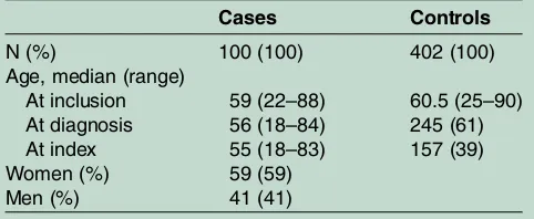
Of the overall 214 cases, 24 cases were excluded due to wrong diagnosis, 11 cases were excluded due to an estab-lished diagnosis before the study period; of the remain-ing 179 cases fulfilling the inclusion criteria, 49 (27%) cases were deceased at the time of data collection (33 women and 16 men, median age at diagnosis 72, range 25–86 years) (see online supplementary figure 1). We were not able to contact two cases (both PM, one male and one female). The remaining 128 cases (76 women and 52 men) received the questionnaire, and 100 cases returned the questionnaire (59 women and 41 men) (table 1), giving a response proportion of 78%.
Controls
For each of the 128 cases, 6 controls were randomly sampled. For 17 cases additional controls were sampled due to insufficient control response. Of the total 808 selected controls (467 women and 341 men), 17 were deceased and 503 (316 women and 187 men) returned the questionnaire, giving a response proportion of 64% (see online supplementaryfigure 1).
This report comprises data from the 100 cases and their correspondingly matched 402 controls who responded to the questionnaire (table 1). All 100 cases had minimum one matched control; 99 had at least two, 93 had three, 72 had four, 32 had five and 8 cases had six matched controls.
Verification of diagnosis
Diagnosis of myositis was verified by review of medical records, which were retrieved from 77 (77%) of the 100 cases that were included in the analysis. For some cases information was incomplete concerning the results of diagnostic procedures, for example, the
report from the muscle biopsy or EMG could be missing. Despite this missing data, 28 patients fulfilled diagnostic criteria for definitive, 25 for probable and 11 for possible PM or DM (10). Five patients had been diagnosed with IBM. In the remaining eight cases, information was incomplete for diagnostic verification according to the criteria (10).
Association between inflammatory lung disease and myositis
Inflammatory lung disease at study inclusion
The matched analysis suggested a significant association between reported history of lung disease and diagnosis of myositis (OR=1.8, 95% CI 1.1 to 2.9). After additional adjustment for smoking, the OR increased to 1.9 (95% CI 1.2 to 3.1) ( p=0.006).
Inflammatory lung disease before index year
[image:3.595.309.551.474.683.2]The median duration from index year to diagnosis was 1 year, and to inclusion in the study 4 years. The number of cases and controls that reported infl amma-tory lung disease was 42 (42%) and 112 (28%), respect-ively (table 2). The incident year of inflammatory lung disease was reported by 30 (71%) cases and 82 (73%) controls. Of these, 22 (73%) cases and 69 (84%) con-trols had been exposed to lung disease before the index year. The median duration between exposure and index year was 30 and 24.5 years, respectively, for cases and controls. A suggestive yet non-significant association between lung disease before index year and myositis risk was observed (OR=1.5, 95% CI 0.9 to 2.6). After
Table 1 Demographic data on cases and controls included in the matched analysis
Cases Controls
N (%) 100 (100) 402 (100)
Age, median (range)
At inclusion 59 (22–88) 60.5 (25–90) At diagnosis 56 (18–84) 245 (61) At index 55 (18–83) 157 (39) Women (%) 59 (59)
[image:3.595.46.287.637.736.2]Men (%) 41 (41)
Table 2 History of inflammatory lung disease among cases and controls
Cases Controls
Total, n (%) 100 (100) 402 (100) No reported inflammatory lung
disease, n (%)
58 (58) 290 (72)
Reported inflammatory lung disease at study inclusion, n (%)
42 (42) 112 (28)
Pneumonia* 35 (35) 106 (26)
Tuberculosis* 5 (5) 12 (3)
Sarcoidosis 3 (3) 1 (0)
Reported inflammatory lung disease before index year, n (%)†
22 (22) 69 (17)
Pneumonia‡ 18 (18) 62 (15)
Tuberculosis‡ 3 (3) 10 (2)
Sarcoidosis 1 (1) 1 (0)
Reported inflammatory lung disease after or at index year, n (%)†
8 (8) 13 (3)
*One case and six controls reported both pneumonia and tuberculosis.
†Numbers does not add up since 12 cases (29%) and 30 controls (27%) that had reported inflammatory lung disease did not report incidence year.
‡One case and six controls reported both pneumonia and tuberculosis.
on May 7, 2020 by guest. Protected by copyright.
http://rmdopen.bmj.com/
adjustment for ever-smoking, the OR increased slightly to 1.6 (95% CI 0.9 to 2.8) ( p=0.09). Excluding patients with IBM and sarcoidosis did not noticeably alter the OR (1.6 (0.9 to 2.8), p=0.12 and 1.5 (0.9 to 2.7), p=0.13 respectively).
No significant associations were found between myo-sitis and exposure to hepatitis, gastric ulcer, measles, mumps, rubella, inflammatory bowel disease, diabetes or psoriasis before index year.
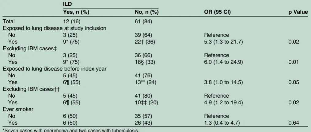
Interstitial lung disease
We further investigated the frequency of ILD at the time of myositis diagnosis among the cases. Medical records were available for 77 cases, in which four cases had no information on lung involvement. The ILD status of cases (n=73) and their corresponding reports for occur-rence of lung disease reported at study inclusion and before index year is presented in table 3. Overall recorded signs of ILD were found in 16% of the cases. In most cases we had only access to results from chest radiography instead of high-resolution computerised scans of the lungs or pulmonary function tests, why we might miss milder cases with ILD. A strong association was observed between myositis with concurrent ILD and reported lung disease both at study inclusion (OR=5.32, 95% CI 1.3 to 21.7) and before index year (OR=3.8, 95% CI 1.0 to 14.5) (table 3). Analysis regarding ILD and ever smoker status showed no significant association (OR=1.35, 95% CI 0.4 to 4.7).
Mortality
The overall mortality among the myositis cases from year 1995 up to year 1999 was 27% (49/179). For 44 (90%) of the deceased cases, the age at time of diagnosis ranged between 50 and 89 years, while the remaining 5 (10%) cases were diagnosed between the age 25 and 49 years. The myositis cases had an overall 7.1-fold (95% CI 5.1 to 9.0) age and gender-standardised risk of death, as compared with the general population. When dissect-ing the mortality ratio for different age categories, we observed that the SMR was 28 for cases diagnosed between 15 and 49 years, while it was 6.5 for cases diag-nosed between 50 and 89 years.
DISCUSSION
We conducted a pilot study comprised of 100 cases of myositis and 402 controls, and identified a link between inflammatory lung disease and myositis risk. Our results suggest that individuals with previous inflammatory lung disease had 60% higher risk of developing myositis com-pared to those without. In particular patients with myo-sitis and a concurrent ILD had a higher reported history of a preceding lung disease.
Several limitations need to be acknowledged. The myositis population in our study was identified from the inpatient register, thus it is likely that milder cases of myositis who have not been hospitalised during their
[image:4.595.44.552.446.661.2]first 3 years after diagnosis could have been missed. The validity of our patient population was supported by high
Table 3 ILD status at diagnosis of the cases with available information on lung disease in patient chart reviews (n=73) and associations with reported occurrence of inflammatory lung disease and smoking
ILD
Yes, n (%) No, n (%) OR (95 CI) p Value
Total 12 (16) 61 (84)
Exposed to lung disease at study inclusion
No 3 (25) 39 (64) Reference
Yes 9* (75) 22†(36) 5.3 (1.3 to 21.7) 0.02
Excluding IBM cases‡
No 3 (25) 36 (66) Reference
Yes 9* (75) 18§ (33) 6.0 (1.4 to 24.9) 0.01
Exposed to lung disease before index year
No 5 (45) 41 (76)
Yes 6¶ (55) 13** (24) 3.8 (1.0 to 14.5) 0.05
Excluding IBM cases††
No 5 (45) 41 (80) Reference
Yes 6¶ (55) 10‡‡(20) 4.9 (1.2 to 19.4) 0.02
Ever smoker
No 6 (50) 35 (57) Reference
Yes 6 (50) 26 (43) 1.3 (0.4 to 4.7) 0.64
*Seven cases with pneumonia and two cases with tuberculosis.
†Twenty cases with pneumonia (of which one exposed also to tuberculosis) and two cases with sarcoidosis.
‡Five verified (two with pneumonia) and two suspected (both with pneumonia) IBM cases; all negative for ILD. §Sixteen patients with pneumonia (including one case exposed also to tuberculosis) and two with sarcoidosis. ¶Four cases with pneumonia and two cases with tuberculosis.
**Eleven patients with pneumonia (including one case exposed also to tuberculosis) and two with sarcoidosis.
††Two verified (with pneumonia) and one suspected (with pneumonia) IBM cases; all negative for ILD.
‡‡Eight patients with pneumonia (including one case exposed also to tuberculosis) and two with sarcoidosis. IBM, inclusion body myositis; ILD, Interstitial lung disease.
on May 7, 2020 by guest. Protected by copyright.
http://rmdopen.bmj.com/
PPV which is in line with the corresponding values of other autoimmune diseases in the Swedish IPR. The lack of specific ICD code for IBM, and the inconsistent use of ICD codes for IBM in clinical practice, could explain the identification of five IBM cases in our study when we scrutinised the patient records. However, we consider this problem to be minor because no apparent changes of results were observed after excluding the IBM cases. None of the available biopsy reports sug-gested a necrotising myopathy. Our case–control study design with retrospective collection of exposures could introduce differential misclassification due to recall bias. However, the risk was reduced to the largest extent by including only recently-diagnosed cases; and informa-tion on the incident year of inflammatory lung disease was reported by comparable proportions of cases and controls. Another limitation is that we could not confirm the diagnosis of the preceding lung disease as we did not have access to relevant patient records before myo-sitis diagnosis. Moreover, we also lack information on autoantibody profiles for most patients as the data were collected before many of the myositis specific autobodies were known (eg, Jo-1, synthetase anti-bodies) and no biological samples were collected for autoantibody profiling.
Owing to the observation of a large dropout of myo-sitis cases because of death, a post hoc analysis of mortal-ity among cases was performed. The overall case mortality (27%) in this report is in line with previous studies on myositis cases followed for a similar time period.12 The myositis patients had a sevenfold age and gender standardised mortality, even higher than the only hitherto population-based study of mortality among polymyositis and dermatomyositis conducted in Finland.13 Various factors such as clinical characteristics of the cases, the length of follow-up period, type of recruitment centre, genetic background and even auto-antibody profile may influence the mortality, thus com-plicating comparisons. Respiratory failure, opportunistic infections in lungs and the digestive tract, as well as smoking were reported among common causes of death already within the first years after diagnosis of myositis. It is possible that a significant proportion of the deceased cases in this report had lung disease or were smokers, which might explain the relatively low rate of ILD diagnosis among the final cases as well as the low proportion of smokers (17% cases vs 21% controls).
Pneumonia and tuberculosis have infectious aetiology, whereas the origin of sarcoidosis is unclear. Interestingly, these lung diseases have been reported in combination with rheumatic manifestations which might suggest a common trigger.8 9 There are multiple mechanisms by which host infection by a pathogen can lead to auto-immunity. The pathogen may carry elements that are similar enough in amino acid sequence or structure to self-antigen that the pathogen acts as a self-‘mimic’. T or B cells that are activated in response to the pathogen are also cross-reactive to self and further activate of other
arms of the immune system. The pathogen may also lead to disease via epitope spreading. Patients with myositis with lung disease and anti-Jo-1 positivity were reported to be associated with type I interferon (IFN) activity that may lead to the induction of autoimmune disease.14 15A role for type I IFN in the lung during viral infection and in tuberculosis has been demonstrated. An upregulation of the type I IFN system and modification of host-antigen (s) in genetically predisposed individuals could result in the induction of autoimmune diseases including myo-sitis.15 The long duration between reported exposures and diagnosis is intriguing, but little is known on the time lapse between triggering of the immune system and the development of an autoimmune diseases. It may well be that the lung disease initiates a process that, together with other factors, after many years, manifests in a diag-nosis of myositis. In the case of rheumatoid arthritis con-vincing data suggest that autoantibodies can be present more than 10 years before disease onset, but little is known when the exposure that leads to production of autoantibodies occur. There are also data to support that conditions during pregnancy may affect risks to develop autoimmune diseases later in life.
In summary, patients with preceding inflammatory lung disease seem to have a modestly increased risk to develop myositis. This may be even more prominent in patients with myositis with an associated ILD. Inflammatory lung disease may thus potentially be a potential risk factor for the onset of inflammatory myop-athies, myositis, but a larger study is needed to confirm the data in this pilot study. In such a study we recommend to assess previous lung disease not only by self-reports but also by means of patient records and further to collect biological samples for autoantibody profiling.
Author affiliations
1Unit of Cardiovascular Epidemiology, Karolinska Institutet, Institute of
Environmental Medicine, Stockholm, Sweden
2Department of Medicine, Rheumatology Unit, Karolinska Institutet, Karolinska
University Hospital, Stockholm, Sweden
3Centre for Occupational and Environmental Medicine, Stockholm County
Council, Stockholm, Sweden
Acknowledgements The authors would like to thank Marie-Louise Serra for
selection of the controls.
Contributors SBH, XJ and DP analysed the data, interpreted the results and
drafted the manuscript; A-LW, PA and A-JL. interpreted the results and modified the manuscript. IEL and LA are the primary investigators of the EIRA study and provided important supports to carry out the study. All of the authors contributed significantly to the study design, data analysis, interpretation of results and manuscript writing.
Funding This study was supported by grants from the Swedish Research
Council K2014-52X-14045-14-3, Sweden, Swedish Rheumatism Association, COMBINE, King Gustaf V 80-year Foundation, Institutet of Environmental Medicine, Theme Inflammation, and“The regional agreement on medical training and clinical research (ALF)”at Karolinska Institutet/Stockholm County Council, and Centre for Research and Development, Uppsala University/ County Council of Gävleborg.
Competing interests None declared.
Patient consent Obtained.
on May 7, 2020 by guest. Protected by copyright.
http://rmdopen.bmj.com/
Ethics approval The Ethics Committee of Karolinska University Hospital approved the study.
Provenance and peer review Not commissioned; externally peer reviewed.
Data sharing statement No additional data are available.
Open Access This is an Open Access article distributed in accordance with
the Creative Commons Attribution Non Commercial (CC BY-NC 4.0) license, which permits others to distribute, remix, adapt, build upon this work non-commercially, and license their derivative works on different terms, provided the original work is properly cited and the use is non-commercial. See: http:// creativecommons.org/licenses/by-nc/4.0/
REFERENCES
1. O’Hanlon TP, Carrick DM, Targoff IN,et al. Immunogenetic risk and protective factors for the idiopathic inflammatory myopathies: distinct HLA-A, -B, -Cw, -DRB1, and -DQA1 allelic profiles distinguish European American patients with different myositis autoantibodies. Medicine (Baltimore)2006;85:111–27.
2. Chinoy H, Adimulam S, Marriage F,et al. Interaction of HLA-DRB1*03 and smoking for the development of anti-Jo-1 antibodies in adult idiopathic inflammatory myopathies: a European-wide case study.Ann Rheum Dis2012;71:961–5. 3. Hengstman GJ, van Venrooij WJ, Vencovsky J,et al. The relative
prevalence of dermatomyositis and polymyositis in Europe exhibits a latitudinal gradient.Ann Rheum Dis2000;59:141–2.
4. Azali P, Barbasso Helmers S, Kockum I,et al. Low serum levels of vitamin D in idiopathic inflammatory myopathies.Ann Rheum Dis 2013;72:512–6.
5. Zampieri S, Ghirardello A, Iaccarino L,et al. Polymyositis-dermatomyositis and infections.Autoimmunity2006;39:191–6. 6. Rider LG, Wu L, Mamyrova G,et al. . Environmental factors
preceding illness onset differ in phenotypes of the juvenile idiopathic inflammatory myopathies.Rheumatology (Oxford).2010;49:2381–90. 7. Love LA, Leff RL, Fraser DD,et al. A new approach to the
classification of idiopathic inflammatory myopathy: myositis-specific autoantibodies define useful homogeneous patient groups.Medicine (Baltimore)1991;70:360–74.
8. Fathi M, Vikgren J, Boijsen M,et al. Interstitial lung disease in polymyositis and dermatomyositis: longitudinal evaluation by pulmonary function and radiology.Arthritis Rheum2008;59:677–85. 9. Chatham W. Rheumatic manifestations of systemic disease:
sarcoidosis.Curr Opin Rheumatol2010;22:85–90.
10. Bohan A, Peter JB. Polymyositis and dermatomyositis (second of two parts).N Engl J Med1975;292:403–7.
11. Stolt P, Bengtsson C, Nordmark B,et al. Quantification of the influence of cigarette smoking on rheumatoid arthritis: results from a population based case–control study, using incident cases.Ann Rheum Dis2003;62:835–41.
12. Bronner Intramuscular, van der Meulen MF, de Visser M,et al. Long-term outcome in polymyositis and dermatomyositis.Ann Rheum Dis2006;65:1456–61.
13. Airio A, Kautiainen H, Hakala M. Prognosis and mortality of polymyositis and dermatomyositis patients.Clin Rheumatol 2006;25:234–9.
14. Eloranta ML, Barbasso Helmers S, Ulfgren AK,et al. A possible mechanism for endogenous activation of the type I interferon system in myositis patients with anti-Jo-1 or anti-Ro 52/anti-Ro 60
autoantibodies.Arthritis Rheum2007;56:3112–24.
15. Lundberg IE, Helmers SB. The type I interferon system in idiopathic inflammatory myopathies.Autoimmunity2010;43:239–43.
on May 7, 2020 by guest. Protected by copyright.
http://rmdopen.bmj.com/