Functionalisation of Ti6Al4V components fabricated using selective laser
melting with a bioactive compound
Jayasheelan Vaithilingam
a, Samuel Kilsby
b, Ruth D. Goodridge
a,⁎
, Steven D.R. Christie
b,
Steve Edmondson
c, Richard J.M. Hague
aa
Additive Manufacturing and 3D Printing Research Group, School of Engineering, The University of Nottingham, Nottingham NG7 2RD, UK b
Department of Chemistry, Loughborough University, Loughborough LE11 3TU, UK c
School of Materials, The University of Manchester, Manchester M13 9PL, UK
a b s t r a c t
a r t i c l e i n f o
Article history: Received 17 June 2014
Received in revised form 12 August 2014 Accepted 2 October 2014
Available online 5 October 2014
Keywords:
Selective laser melting (SLM) Self-assembled monolayers (SAMs) Additive manufacturing Functionalisation
X-ray photoelectron spectroscopy (XPS)
Surface modification of an implant with a biomolecule is used to improve its biocompatibility and to reduce post-implant complications. In this study, a novel approach has been used to functionalise phosphonic acid mono-layers with a drug. Ti6Al4V components fabricated using selective laser melting (SLM) were functionalised with Paracetamol (a pharmaceutically relevant biomolecule) using phosphonic acid based self-assembled mono-layers (SAMs). The attachment, stability of the monomono-layers on the SLM fabricated surface and functionalisation of SAMs with Paracetamol were studied using X-ray photoelectron spectroscopy (XPS) and surface wettability measurements. The obtained results confirmed that SAMs were stable on the Ti6Al4V surface for over four weeks and then began to desorb from the surface. The reaction used to functionalise the phosphonic acid mono-layers with Paracetamol was noted to be successful. Thus, the proposed method has the potential to immobilise drugs/proteins to SAM coated surfaces and improve their biocompatibility and reduce post-implant complications.
© 2015 The Authors. Published by Elsevier B.V. All rights reserved. This is an open access article under the CC BY license (http://creativecommons.org/licenses/by/4.0/).
1. Introduction
Selective laser melting (SLM) is a metal-based additive manufactur-ing (AM) technique, which is gainmanufactur-ing considerable interest for biomed-ical applications due to its ability to fabricate parts with customised and complex geometries directly from 3D model data[1–5]. However, limi-tations are present including the materials currently available and the surface quality of fabricated parts. Some of the biomaterials that can be processed by SLM include titanium and its alloys (such as Ti6Al4V and Ti6Al7Nb), cobalt–chromium alloy and 316L stainless steel. Control over the surface chemistry of an implant is essential since it governs the surface chemical reactions. It is not always possible to achieve the de-sired surface chemistry directly from the manufactured part and hence surface modifications are performed to obtain the desired surface chemistry.
In recent years, surface modification has been performed not just to improve the surfacefinish of the implant but also to make the implants biocompatible and a drug carrier[6–12]. For example, to reduce the re-stenosis rate in drug eluting stents (DES), the stents are coated with polymers containing drugs such as Paclitaxel. In addition, to improve
biocompatibility, the implants are coated with biocompatible proteins, ceramics and other biomaterials. Hence by changing the physicochem-ical characteristics, material and biologphysicochem-ical responses are altered[13].
Most implants are coated or surface modified with biocompatible materials using techniques such as dip-, spray- or spin-coating or solvent casting[14,15]. Implants coated with these conventional coating tech-niques have drawbacks including surface heterogeneity in the type and distribution of functional groups, hydrophilic or hydrophobic domains and surface roughness[16]. Limitations of using polymeric carriers for biomedical applications includefissures, cracks and waviness of the coat-ing (leadcoat-ing to uneven drug distribution), inflammatory and hypersensi-tivity reactions[17]. Thus a coating technique that offers unprecedented control over the location and orientation of chemical groups/biomole-cules on the surface is essential to cater for current needs.
Self-assembled monolayers (SAMs) are amphiphilic molecules with head and tail groups. Typically the head group is attached to the surface that needs modification and the tail group is used to introduce function-ality[18]. SAMs are formed on a substrate by the adsorption of mole-cules from a solution and are usually nanosized adding only a few nanometres of thickness (depending on the alkyl chain) to the implant surface[19]. SAMs can be designed at the molecular level to be biologi-cally inert and can be expanded uniformly (to cover the whole implant surface) with the implant surface maintaining their assembly, structural integrity and chemical composition. SAMs are inexpensive and versatile ⁎ Corresponding author at: Additive Manufacturing and 3D Printing Research Group,
School of Engineering, University Park, University of Nottingham, Nottingham, NG7 2RD, UK.
E-mail address:[email protected](R.D. Goodridge).
http://dx.doi.org/10.1016/j.msec.2014.10.015
0928-4931/© 2015 The Authors. Published by Elsevier B.V. All rights reserved. This is an open access article under the CC BY license (http://creativecommons.org/licenses/by/4.0/).
Contents lists available atScienceDirect
Materials Science and Engineering C
Various biomolecules including proteins, peptides, carbohydrates, antibodies and therapeutics have been attached to SAMs for biomedical applications[22]. Since SAMs can serve as a localised drug delivery sys-tem, the use of SAMs to attach drugs to biomedical implants is gaining considerable interest. There are several literature examples of the func-tionalisation of monolayers with drugs[11,17,23,24]. However, these drugs are either physisorbed or both physisorbed and covalently bound. A method was proposed by Mani et al.[17]to successfully bind Pac-litaxel to SAM coated surfaces; however, it has a number of limitations. In their procedure, a microdrop method was used to drop the drug solu-tion on to the cobalt–chromium surface. It was reported that from the 25μg of drug placed on the surface (1 cm2), only 25% of the drug was adsorbed to the surface and the rest was washed away by solvent cleaning. Although a certain amount of the drug can be recovered by recycling, this method can be time consuming and expensive. Also it should be noted that scaling up of this method will be difficult since coating drugs on implants with complex geometries using microdrop deposition would be challenging. Furthermore, the drug adsorbed on the surface was observed to be crystallised. Crystallisation of drugs on an implant surface can potentially increase the surface roughness of the implant and may cause post-implant complications. Also due to crystallisation, controlling drug release from the implant surface will be problematic.
In this study, one-to-one binding of a drug directly to monolayers adsorbed on a SLM surface was studied. The surface of a SLM-manufactured part was used due to the increased interest in the use of SLM to fabricate biomedical implants[3,25–27]. By allowing the mono-layers to react with drugs under optimal reaction conditions, drugs can be covalently bound to the monolayers. Such binding can avoid crystallisation of drugs on the implant surface and reduce the loss of drugs whilst coating. In this study, the stability of phosphonic acid monolayers on a Ti6Al4V surface was investigated. Following the stabil-ity of the monolayers, a novel approach was used to bind Paracetamol covalently to monolayers adsorbed on the SLM fabricated Ti6Al4V surface.
2. Materials and methods
2.1. Materials
Gas atomised grade 5 titanium alloy, Ti6Al4V powder, with an aver-age particle size distribution (volume weighted) of 33.35μm was pur-chased from LPW Technology Ltd., UK. Silicon carbide grits, polishing cloth, diamond paste and extender solutions were supplied by Buehler (Buehler-met® II). 16-Phosphanohexadecanoic acid (16-PhDA) (97%) and Paracetamol (analytical grade) used for the surface modification were purchased from Sigma-Aldrich, UK. Thionyl chloride (97%), tetra-hydrofuran (THF), hydrogen peroxide (30%) and sulphuric acid (98%) used for the cleaning and surface modification steps were also pur-chased from Sigma-Aldrich, UK. THF used for binding Paracetamol to monolayers was dried using benzophenone/sodium under argon atmo-sphere. This is referred in this study as“dry THF”.
2.2. Methods
2.2.1. Design and fabrication
Cuboidal Ti6Al4V parts with dimensions 10 × 10 × 3 mm were fab-ricated in a SLM 250 laser melting machine (Renishaw, UK) using a laser power of 200 W, hatch spacing of 100μm, exposure time of 200μs and a layer thickness of 50μm. A Meander scan strategy was used to build the part. The theory behind the SLM process and its working principles can be found in literature[25]. After fabrication, the parts were removed from the build plate and sonicated in deionised water.
1200μm diameter, for 5–7 min. These surfaces were then polished for 3 min with a polishing cloth using 6μm and 1μm diamond pastes. The mechanically polished surfaces were rinsed with deionised water and then immersed in a mixture of sulphuric acid, hydrogen peroxide and water in the ratio of 1:1:5 respectively for 15 min to remove surface contaminants. Finally the surfaces were sonicated in deionised water for 30 min (twice) to remove any traces of residual acids.
2.2.3. Self-assembly of monolayers
A solution immersion deposition method was used to coat 16-PhDA phosphonic acid SAMs on the mechanically polished Ti6Al4V plates. SAMs were deposited on the sample surfaces using a procedure previ-ously reported in the literature[17]. Briefly, 1 mM solution of 16-PhDA in 150 ml THF was prepared. After 24 h, the samples were removed from the THF and any residual solution was allowed to evapo-rate in air. Without rinsing, the samples were immediately transferred to an oven maintained at 120 °C. After 24 h, the SAM-coated specimens were removed from the oven and allowed to cool to room temperature before ultrasonicating in THF and deionised water for 1 min each.
2.2.4. Stability studies
The stability of the phosphonic acid monolayers assembled on the SLM fabricated Ti6Al4V surfaces was studied by immersing the SAM-coated samples in 20 ml of 10 mM Tris–HCl buffer solution (TBS) with a pH of 7.4. The samples were incubated at 36.5 °C for 6 weeks. The samples were removed from the TBS solution at 1, 2, 4 and 6 week time intervals and ultra-sonicated with deionised water for 1 min and dried with compressed air before characterisation.
2.2.5. Drug attachment
The SAM coated samples were transferred to a round bottomedflask (RBF) with the SAM coated surface facing upwards. The apparatus consisted of a RBFfitted to a reflux condenser. The entire reaction was carried out in a nitrogen atmosphere. The apparatus was dried using a flame/heat gun (an apparatus that producesflame) before use to re-move any moisture. 20 ml of anhydrous THF was added to the RBF con-taining the samples through a side tube followed by the addition of 1 mM of thionyl chloride (SOCl2). Care was taken to ensure that the mixture of THF and SOCl2added to the RBF covered the sample surface. The RBF together with the samples and SOCl2mixture were sonicated for an hour before syringing out the mixture from the RBF. Now the SOCl2reacted surfaces were sonicated thrice (for 2 min each time) using excess anhydrous THF without exposing the surfaces to the atmo-sphere. If exposed to the atmosphere, the chlorinated SAMs would have reacted with atmospheric air moisture and the acid chloride would have hydrolysed back to the carboxylic acid. After rinsing the samples, 5 mM of Paracetamol in 20 ml of anhydrous THF was added to the RBF and sonicated for an hour. Finally, the surfaces in the RBF were sonicated with an excess of anhydrous THF and deionised water thrice (each for 2 min) and characterised using XPS.Fig. 1shows a schematic of the chemical reaction that took place at each step to bind the drug to the monolayers.
2.2.6. Surface characterisation
2.2.6.1. Surface morphology.The surface morphology of the SLM fabricat-ed samples was imagfabricat-ed using a LEO 440 scanning electron microscope (SEM). SEM was operated at an extra-high tension (EHT) power of 10 kV and micrographs of the surfaces were obtained at magnifications including 50× and 500×.
samples were obtained using an Alicona InfiniteFocus® optical 3D measurement device. Surface roughness patterns were obtained from a 175μm × 175μm square area of the sample. Surface roughness
[image:3.595.99.493.60.521.2]was measured forfive samples and averaged. The data acquired was processed using the in-built software within the Alicona InfiniteFocus®.
Fig. 1.General reaction scheme to functionalise 16-PhDA SAMs with Paracetamol. Label description: 1. Hydroxylated titanium surface; 2. 16-Phosphanohexadecanoic acid; 3. SAMs adsorbed on a Ti6Al4V surface; 4. Chlorinated surface; 5. Ti6Al4V surface functionalised with Paracetamol.
[image:3.595.65.522.612.726.2]2.2.6.3. Surface wettability.Surface wettability of the samples before and after surface modification (SAM attachment and drug attachment) was determined by static water contact angle measurement using a contact angle goniometer (OCA-20). A drop volume of 2μl was placed on the surface and allowed to settle for approximately 5 s. The contact angle formed by the water drop on the sample surface was measured using the inbuilt software SCA 20. Contact angles were measured infive dis-tinct spots within the sample surface and averaged.
2.2.6.4. Surface chemistry.X-ray photoelectron spectroscopy (XPS) mea-surements were performed using a Thermo Scientific K-Alpha ESCA with a probing spot size of 400 μm. Using aluminium (Al) Kα monochromated radiation at 1486.6 eV, photoelectrons were collected using a take-off angle of 90°. In a constant analyser energy mode, survey spectra were collected at a pass energy of 100 eV and high resolution spectra were obtained at a pass energy of 20 eV. Peak deconvolution was performed using Gaussian–Lorentzian curves to investigate the dif-ferent chemical states of the detected elements. A built-in Thermo Sci-entific Avantage data system was used for data acquisition and processing. Previously reported works and the National Institute of Standards and Technology (NIST) database were used to identify the spectral lines.
3. Results and discussion
3.1. Surface morphology
The surface morphology of a typical SLM-fabricated Ti6Al4V sample is shown inFig. 2. Partially sintered particles observed in the micro-graph show that the SLM fabricated sample was rough. The average sur-face roughness (Ra) obtained using a sursur-face profilometer was 17.6 ± 3.7μm. The reason for such sintering of particles to the surface could be due to multiple factors including as a natural consequence of building parts from a powder bed, equipment based process parameters such as laser power, and laser beam spot size[28–30].
Some biomedical applications require a smooth surface (such as stents) to avoid cellular damage and some require a rougher, porous surface to promote cell adhesion and tissue integration and regenera-tion[13]. Although the SLM as-fabricated parts cannot be used for appli-cations that require a smooth surfacefinish, they could be used where a
porous surface is required. However, the SLM-produced surfaces are po-rous due to partially and loosely sintered particles. These particles could detach from the surface and can cause acute and chronic effects.
In addition, if particles detach after monolayer assembly, areas will be left without drug attachment since there will be no underlying monolayer to functionalise. XPS and contact angle measurements are sensitive to rough surface and hence, the use of SLM as-fabricated sur-face will affect the sursur-face characterisation results. Hence in this study, the SLM fabricated surfaces were mechanically polished to remove the partially-sintered particles and to attain a smooth surface before surface modification and functionalisation with a drug. The average surface roughness (Ra) of the mechanically polished surfaces obtained using a surface profilometer was 0.437 ± 0.045μm.
3.2. Monolayer attachment
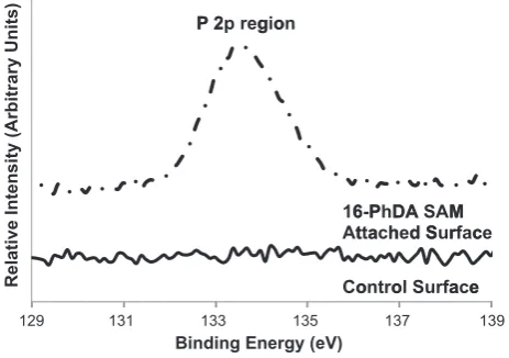
[image:4.595.320.552.60.223.2]Fig. 3shows the survey spectra obtained from the mechanically polished Ti6Al4V surface before and after surface modification using 16-PhDA SAMs. The spectra clearly indicate the introduction of a metal phosphonate peak at 133.3 eV showing that the surface has been modified with 16-PhDA monolayers. This metal phosphonate peak is due to the formation of Ti\O\P bonds after SAM attachment [31,32]. This has further been justified by an increase in the intensity of carbon and a reduction in the intensity of oxygen, titanium and alu-minium after surface modification (due to the limited penetration depth of XPS measurements, less of the underlying metal and oxide is detected).
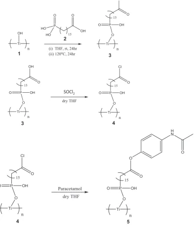
Table 1shows the relative atomic composition obtained for the Ti6Al4V surface before and after surface modification. For a surface modified using 16-PhDA, the expected ratio of C:P is 16:1 and in the cur-rent study, it is observed to be nearly the same (i.e. 15.6:1). The small variation is likely due to errors in the integration of the small phospho-rous peak. High resolution spectra obtained for the phosphophospho-rous 2p re-gion showed the formation of metal–phosphonate bond at 133.6 eV (Fig. 4) confirming the modification of the surface with monolayers [31].
Relave Intensity (Arbitrary Units) 0
TTi 3ssAAl 2
100
P P 2P, P
200 2S
Binding Energy (eV)
300 400 N 1S 2p
500 600
b
a
[image:4.595.45.289.60.175.2]Fig. 3.Survey spectra showing the change in surface chemistry of a Ti6Al4V surface after SAM attachment. (a) Control surface without SAM coating and (b) after 16-PhDA SAM coating.
Table 1
Relative atomic percentage of the elements detected in XPS.
Sample Relative atomic percentage
C O N Ti Al V P Cl S
Control 21.8 ± 1.7 54.5 ± 2.1 0.2 ± 0.8 18.2 ± 0.9 4.7 ± 0.7 0.6 ± 0.2 0 0 0
16-PhDA SAM 51.1 ± 2.3 32.9 ± 1.4 2.0 ± 0.5 8.7 ± 1.1 1.8 ± 0.7 0.3 ± 0.1 3.3 ± 0.5 0 0
129
Relative Intensity (Arbitrary Units)
131
Binding Energy (eV)
133 135
16 At
Co 6-P
tta
on
137
PhD ach
tro DA hed
ol S A S
d S
u AM Sur
rfa M rfac
ace
139
ce
e
[image:4.595.43.568.696.744.2]The average static contact angles obtained from the surface before and after surface modification with SAMs were 23.7° ± 3.4° and 35.1° ± 7.1° respectively. The contact angles can be observed to increase after SAM attachment showing a change in the surface wettability. However, since 16-PhDA SAMs are hydrophilic, displaying a carboxylic acid (COOH) terminal group, the surface remained highly wettable even after surface modification.
3.3. Stability studies
Stability of the 16-PhDA monolayers on the SLM fabricated Ti6Al4V surface was studied by immersing the SAM coated samples into Tris–
HCl buffer solution.Fig. 5shows the high resolution XPS spectra for the phosphorous 2p region of the samples soaked in the buffer solution for different time intervals. It can be witnessed from the spectra that the metal phosphonate peak at 133.3 ± 0.6 eV retained its position at the same binding energy for the whole duration of immersion in the buffer solution. However, its intensity and relative atomic composition de-creased over the course of the experiment showing desorption of mono-layers from the Ti6Al4V surface. Although the desorption of monomono-layers was noted to occur after four weeks, a small amount of monolayers was observed at the end of six weeks. The phosphorous 2p peak showed a small peak shift upon immersing the substrate in Tris–HCl buffer solu-tion. This may be due to the charging effect of the substrate during XPS characterisation[33].
A gradual increase in the contact angle was noted after immersing the samples in Tris–HCl buffer solution (although this change has low significance compared to the standard deviations of individual mea-surements). This may be attributed to a small amount of desorption (Fig. 6) leading to the change in the assembly pattern of the monolayers. Thus, both the XPS characterisation and static water contact angle mea-surements confirm that 16-PhDA monolayers are relatively stable on the Ti6Al4V surface for more than the 28 day time period when soaked in Tris–HCl buffer solution. A previous study showed that 16-PhDA SAMs are stable on both SLM as-fabricated (SLM-AF) surface and a me-chanically polished SLM surface (SLM-MP) for over 28 days[34]. This study also revealed that the surface roughness did not significantly af-fect the stability of the 16-PhDA monolayers formed on these rough (SLM-AF) and smooth (SLM-MP) surfaces.
3.4. Functionalisation of 16-PhDA monolayers with Paracetamol
After studying the stability of monolayers on the Ti6Al4V surface, a sample drug (Paracetamol) was used to functionalise the SAMs. The reac-tive carboxylic acid group at the tail end of 16-PhDA SAMs was reacted with thionyl chloride (SOCl2) to form an intermediate acid chloride com-pound with\COCl as the terminal group. This group was then allowed to react with the hydroxyl (\OH) group of Paracetamol to bind the drug co-valently to the monolayers. The whole reaction step was performed in an inert atmosphere. Anhydrous THF was used to prevent the SOCl2reacting with moisture in the atmosphere to form HCl that could corrode the metal surface and also hydrolyse the\COCl to inactive\COOH.
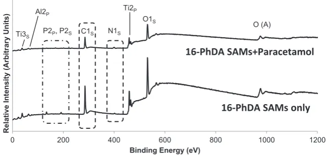
Fig. 7shows the survey spectra for the Ti6Al4V sample before and after drug attachment. The corresponding atomic percentages are
128 130 132 134 136 138
Relative Intensity (Arbitrary Units)
Binding Energy (eV)
Week 1
Week 4 Week 2 Week 0 P 2p region
[image:5.595.45.275.53.228.2]Week 6
Fig. 5.XPS spectra for the phosphorous 2p region showing the stability of monolayers on the SLM fabricated Ti6Al4V surface at various time intervals.
0 20 40 60 80 100
[image:5.595.38.282.287.427.2]Contact Angle (°)
Fig. 6.Static water contact angles formed on the Ti6Al4V surface before and after SAM coating.
0 200 400 600 800 1000 1200
Relative Intensity (Arbitrary Units)
Binding Energy (eV) P2P, P2S C1S
O1S
Ti2P
Ti3S
Al2P
O (A)
16-PhDA SAMs only
16-PhDA SAMs+Paracetamol
N1S [image:5.595.130.453.563.718.2]given inTable 2. It can be clearly noted that there is a significant change in the surface chemistry of the SAM-coated Ti6Al4V surface after the at-tachment of Paracetamol. The ratio of C:P was 15.6 for SAM coated sur-face and after the attachment of Paracetamol the ratio of C:N was 10.7. These ratios show the reduction in the atomic percentage of carbon after drug attachment since less carbon-rich Paracetamol contains 8 car-bon atoms with 3 heteroatoms compared to 16-PhDA (which presents a carbon-rich surface due to a long alkyl group and buried phosphonate). However, there are possibilities for small errors since nitrogen is present in the atmosphere as a contaminant[35].
Due to the presence of nitrogen in Paracetamol, its concentration was noted to increase; however, this is not a large increase since the drug molecule contains only one nitrogen atom. The concentration of phosphorous (from 16-PhDA) and the underlying metals (titanium, al-uminium and vanadium) were observed to decrease and this further confirms the functionalisation of SAMs with Paracetamol.
A very small contribution from chlorine and sulphur was observed after the attachment of Paracetamol and this is likely to be added from the SOCl2used in the intermediate step. Since the 16-PhDA SAMs have 2 hydroxyl groups (\OH) in each head group, there may be the possibility for the addition of chlorine or the sulphur to those hydroxyl ends to form Ti\O\P\OCl-alkyl chain and/or Ti\O\P\S-alkyl chain. Although the abovementioned reaction is possible, it depends only on the availability of a hydroxyl group for the reaction as both hydroxyl groups can bond to the Ti6Al4V surface initially before reaction of the SAMs.
Carbon contamination is unavoidable as it is present as a contami-nant in the atmosphere. However, a small amount of nitrogen was also observed on the Ti6Al4V sample as a contaminant in both the con-trol and SAM coated surfaces. The presence of these elements on the metal oxide surface as contaminants was reported in literature[23, 31]. Hence the ratio of carbon to the underlying metal composition of the surface has been used to determine the adsorption of SAMs and Paracetamol to the metal surface.
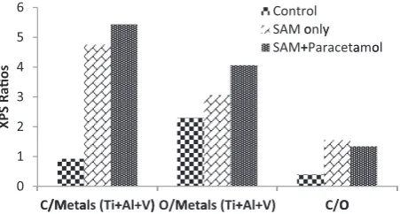
Fig. 8shows the ratio of carbon and oxygen to the underlying metals (titanium + aluminium and vanadium) and the ratio of carbon to oxy-gen for all three samples (control, SAM attached and Paracetamol coat-ed). It can be observed that the carbon and oxygen concentration increased after SAM attachment and functionalisation confirming the attachment of the Paracetamol to the tail group of 16-PhDA monolayers. A comparison of the C 1s spectra obtained for the control, 16-PhDA SAM coated and SAMs functionalised with Paracetamol is shown in
Fig. 9. High-resolution spectrum of C 1s for the control sample was deconvoluted into two components. Peaks formed at 285.3 eV and 286.2 eV were assigned to C\C and C\O of hydrocarbon contaminants. Adsorption of hydrocarbon as contaminants to metal oxide surfaces has been discussed in literature and the major source for these contami-nants is adventitious carbon in air, solvents (used for cleaning) and from hydrocarbons (containing proteins and oils) due to manual handling[35–38].
The C 1s spectrum for the SAM coated Ti6Al4V surface was deconvoluted into three components. The peaks observed at 284.9 eV, 286.5 eV and 289.3 eV were assigned to C\C, C\O and C_O[23]. A sig-nificant increase in the relative C\C peak intensity compared to the control surface was due to the 16 membered carbon chain [HOOC (CH2)15PO(OH)2] in the 16-PhDA molecule. The C\O and C_O are also from the 16-PhDA molecule, present in the head and tail group of the chain. The significant change in the intensity of C\C, and C\O and the introduction of C_O peak confirms the modification of SLM fabricated Ti6Al4V surface with 16-PhDA monolayers.
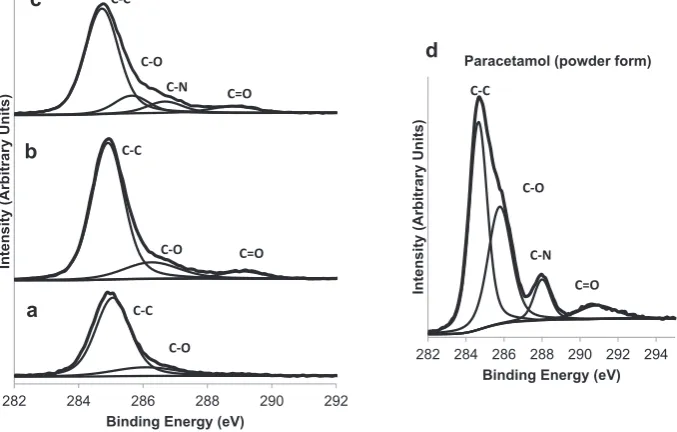
On characterising the Ti6Al4V surface functionalised with Paraceta-mol, the deconvoluted C 1s spectrum rendered four components at 284.7 eV, 285.8 eV, 286.9 eV and 289.1 eV. These peaks can be assigned to C\C, C\O, C\N and C_O respectively[17,23]. Comparing this to the SAM coated surface, the peak intensity of C\C and C_O has changed. Also the introduction of C\N peak at 286.9 eV was observed. This C\N peak is from the amide group in Paracetamol.
Paracetamol powder in its pure form (as purchased) was char-acterised using XPS to obtain the spectrum for C 1s region (Fig. 9d). The deconvoluted spectrum showed the existence of four components C\C (284.7 eV), C\O (286 eV), C\N (288.2 eV) and C_O (291.2 eV) [23,39]. These peaks can be assigned to carbon atoms in hydrocarbon, hydroxyl, amide and ester groups of Paracetamol. The XPS results ob-tained for Paracetamol coated Ti6Al4V surface was in good agreement with the results obtained for the C 1s region of Paracetamol powder. However, a small change in the peak binding energies was noted and this could be due to errors in peak integration during deconvolution and might also be due to minor calibrating errors due to sample charging.
The O 1s region spectrum obtained using XPS for the control sample (Fig. 10a) was deconvoluted into two components, metal oxide at 530.5 eV and oxygen atoms in O\C at 532.1 eV[40–42]. The metal oxide peak observed at 530.5 eV is likely to be mostly due to titanium oxide; however, a small proportion of aluminium and vanadium oxides is also possible due to the presence of these elements in Ti6Al4V alloy. In agreement with the C 1s spectrum, the O 1s spectrum also showed the existence of O\C species on the control sample as contaminants.
On deconvoluting the O 1s spectrum for the 16PhDA SAM coated Ti6Al4V surface, three components were obtained (Fig. 10b). The peaks formed at 530.4 eV, 531.9 eV and 533.2 eV were assigned to metal oxides, O\C and O_C respectively[41–43]; however, there is also the possibility for a small contribution from aluminium oxide for the peak observed at 531 eV. The relative intensity of metal oxide peak at 530.4 eV for the SAM coated sample can be observed to decrease and the O_C peak at 533.2 eV to increase when compared to the control sample. The introduction of O_C at 533.2 eV should mostly derive from the carboxylic group at the terminal end of the 16-PhDA molecule. These observed changes compared with the control sample can be at-tributed to the adsorption of 16-PhDA monolayers on to the SLM fabri-cated Ti6Al4V surfaces.
C O N Ti Al V P Cl S
16-PhDA SAM 51.1 ± 2.3 32.9 ± 1.4 2.0 ± 0.5 8.7 ± 1.1 1.8 ± 0.7 0.3 ± 0.1 3.3 ± 0.5 0 0 SAM + Paracetamol 48 ± 1.8 35.8 ± 1.6 4.5 ± 0.7 7.8 ± 1.3 1.1 ± 0.5 0 1.2 ± 0.4 0.8 ± 0.2 0.9 ± 0.1
XPS
Raos
0 1 2 3 4 5 6
C//MMetaals (Ti++Al+V)) O/MMetaals (Ti++Al+V)
Co SA SA
ntr M o M+
ol only +Pa
C/O
y rac
O
[image:6.595.41.563.78.126.2]etaamool
[image:6.595.56.282.596.717.2]Intensity (Arbitrary Units)
Binding Energy (eV)
C-C
C-C C-C
C-O C=O C-O
C=O C-O
C-N
c
b
a
282 284 286 288 290 292
282 284 286 288 290 292 294
Intensity (Arbitrary Units)
Binding Energy (eV)
C-C
C-O
C-N
C=O
Paracetamol (powder form)
[image:7.595.124.463.65.281.2]d
Fig. 9.High resolution spectra of carbon 1s region obtained for (a) control, (b) SAM attached, (c) Paracetamol coated samples, (d) shows the C 1s region for Paracetamol powder characterised using XPS.
Intensity (Arbitrary Units)
Binding Energy (eV)
O-C O=C
Paracetamol (powder form)
d
527 529 531 533 535 537
526 528 530 532 534 536
Intensity (Arbitrary Units)
Binidng Energy (eV)
O-Metal
c
b
a
O-C O=C
O-C O=C
[image:7.595.122.465.329.518.2]O-C O-Metal O-Metal
Fig. 10.XPS spectra obtained for the oxygen 1s region for (a) control, (b) SAM attached, (c) Paracetamol coated samples, (d) shows the O 1s region for Paracetamol powder characterised using XPS.
Intensity (Arbitrary Units)
Binding Energy (eV) N-C
N-C N-C
N-O
N-O N-O
c
b
a
396 398 400 402 404 406 396 398 400 402 404
Intensity (Arbitrary Units)
Binding Energy (eV) N-C
Paracetamol (powder form)
d
[image:7.595.120.464.570.727.2]After functionalising SAMs with Paracetamol, the deconvoluted O 1s XPS spectrum rendered three components metal oxide (530.8 eV), O\C (532.1 eV) and O_C (533.4 eV) and are shown inFig. 10a. Although the O\C and O_C are expected only from Paracetamol, a small contribu-tion from the underlying monolayers is possible. The XPS characterisa-tion of Paracetamol powder for the O 1s region rendered two components including O\C (531.7 eV) and O_C (533.1 eV) on deconvolution [41] as shown in Fig. 10d. On comparing the deconvoluted O 1s peaks obtained for the Paracetamol coated surface with the O 1s peaks obtained for the Paracetamol powder, both the peaks were in good agreement. However, there was a slight variation in the binding energies. This further confirms the attachment of Paracet-amol to the 16-PhDA adsorbed Ti6Al4V surface.
Deconvoluted N 1s spectrum showed the presence of N\C and N\O at 400.3 eV and 402.6 eV respectively on the control surface (Fig. 11a). Although nitrogen is not expected on the Ti6Al4V surface (as it is not in the composition), a small amount of nitrogen was observed. Similar to carbon, nitrogen also has strong affinity towards metal oxides and their presence on metal oxide surfaces could be from nitrogen contain-ing carbon contaminants[23]. Similar to the control sample, the SAM coated surface also showed the existence of nitrogen at 400.1 eV (N\C) and 402.3 eV (N\O) after deconvolution (Fig. 11b)[37]. This could also be from contamination of the surface as the peaks were ob-served similar to the control samples. Furthermore, the 16-PhDA SAMs do not have nitrogen in their structure.
The deconvoluted N 1s spectrum for the Paracetamol coated Ti6Al4V surface also showed two components, N\C at 400.1 eV and N\O at 401.9 eV (Fig. 11c). Although a small contribution to these peak from contaminants is possible since the other samples (control and SAM coated) were shown to have contaminants, the N\C peak is distinct and increased in relative intensity. XPS characterisation of Paracetamol powder for the N 1s region showed a single N\C peak at 400.3 eV (Fig. 11d).
Wettability of a surface is influenced by its surface roughness,film thickness and its chemical composition/functional group[44]. The
as-fabricated SLM surface was rough and this may have affected the wetta-bility. Hence a mechanically polished surface was used. Static contact angles obtained for the control, 16-PhDA SAM attached and Paraceta-mol coated surfaces are shown inFig. 12.Fig. 13shows the contact angle values obtained graphically. It can be observed that the control and 16-PhDA SAM deposited surfaces were highly wettable whereas the Paracetamol coated surface was more hydrophobic. The wettability of the surface cleaned control surface was due to the presence of metal oxides which impart a high surface energy. Wettability of the 16-PhDA SAM coated surfaces was due to the presence of carboxylic acid in their terminal group. Since the SAM coated surface was functionalised with Paracetamol, it would be expected that a methyl group (\CH3) is ex-posed as the terminal group leading to a lower energy surface. In agree-ment with this, the measured contact angles showed the Paracetamol coated surface to be more hydrophobic with a contact angle higher than 90°. Thus, both the XPS characterisation and surface wettability measurements are consistent with the attachment of Paracetamol to the 16-PhDA SAMs and functionalisation of SLM fabricated Ti6Al4V sur-face with Paracetamol.
4. Conclusion
16-PhDA phosphonic acid SAMs were successfully attached to a SLM fabricated Ti6Al4V surface. The 16-PhDA SAMs were stable on the sur-face for more than 28 days when immersed in Tris-buffer solution be-fore significant desorption from the surface was observed. Changes observed in the surface chemistry of the Ti6Al4V surface at each stage through XPS characterisation and surface wettability measurements are consistent with the functionalisation of monolayers with Paraceta-mol. This study showed the attachment and stability of 16-PhDA SAMs on SLM fabricated Ti6Al4V surface and the successful immobilisa-tion of Paracetamol to 16-PhDA SAMs. However, nitrogen and carbon alone cannot be used as a key element to confirm the Paracetamol at-tachment, since they are present on the surfaces as a contaminant and may affect the result. Hence, Paracetamol may not be the ideal drug of choice to prove the immobilisation of the drug to SAMs. The use of drugs with other elements that are unlikely to be present in the atmo-sphere as contaminants could be used to prevent contamination affect-ing the results in future studies.
Acknowledgement
This work was supported by the Engineering and Physical Sciences Research Council (EPSRC; UK) under grant EP/I033335/2.
References
[1] I. Drstvensek, N.I. Hren, T. Strojnik, T. Brajlih, B. Valentan, Applications of rapid
prototyping in cranio-maxilofacial surgery procedures, Int. J. Biol. Biomed. Eng. 2 (2008) 29–38.
[2] R. Bibb, D. Eggbeer, P. Evans, Rapid prototyping technologies in soft tissue facial
[image:8.595.81.530.53.162.2]prosthetics: current state of the art, Rapid Prototyp. J. 16 (2010) 130–137. Fig. 12.Contact angles obtained for SLM fabricated Ti6Al4V surfaces after a) cleaning b) 16-PhDA SAM attachment, c) Paracetamol attachment.
Contact
Angle
(
°
) 1 1
Contact
Angle
(
)
0 20 40 60 80 00 20
0 0 0 0 0 0 0
C 24
Con 4
ntrool S S SAM
Sam 3
M A mp
35
Atta ple
ach Ty hed ype d e
S SAAM++P
95
Para 5
aceetaamol
[image:8.595.53.285.583.717.2][3] S.J. Hollister, T.L. Bergman, Biomedical Applications of Integrated Additive/Subtrac-tive Manufacturing, World Technology Evaluation Centre, 2004, pp. 55–62.
[4] L.C. Zhang, D. Klemm, J. Eckert, Y.L. Hao, T.B. Sercombe, Manufacture by selective
laser melting and mechanical behavior of a biomedical Ti\24Nb\4Zr\8Sn alloy, Scr. Mater. 65 (2011) 21–24 (Acta Materialia Inc.).
[5] S. Hoeges, M. Lindner, W. Meiners, R. Smeets, Bioresorbable implants using selective
laser melting, Int. Solid Freeform Fabrication Symposium, University of Texas, Austin, 2010, pp. 908–920.
[6] D.G. Castner, B.D. Ratner, Biomedical surface science: foundations to frontiers, Surf.
Sci. 500 (2002) 28–60.
[7] B.D. Ratner, A.S. Hoffman, F.J. Schoen, J.E. Lemons, in: B.D. Ratner, A.s. Hoffman, F.J.
Schoen, J.E. Lemons (Eds.), Biomaterials Science: An Introduction to Materials in Medicine, second ed.Elsevier, London, 2004, pp. 10–813.
[8] G. Mani, M.D. Feldman, D. Patel, C.M. Agrawal, Coronary stents: a materials
perspec-tive, Biomaterials 28 (2007) 1689–1710.
[9] M. Zilberman, R.C. Eberhart, Drug-eluting bioresorbable stents for various
applica-tions, Annu. Rev. Biomed. Eng. 8 (2006) 153–180.
[10]Y. Levy, D. Mandler, J. Weinberger, A.J. Domb, Evaluation of drug-eluting stents'
coating durability—clinical and regulatory implications, J. Biomed. Mater. Res. B Appl. Biomater. 91 (2009) 441–451.
[11] G. Mani, D.M. Johnson, D. Marton, M.D. Feldman, D. Patel, A.a. Ayon, et al., Drug
de-livery from gold and titanium surfaces using self-assembled monolayers, Biomate-rials 29 (2008) 4561–4573.
[12]N. Torres, S. Oh, M. Appleford, D.D. Dean, J.H. Jorgensen, J.L. Ong, et al., Stability of
antibacterial self-assembled monolayers on hydroxyapatite, Acta Biomater. 6 (2010) 3242–3255.
[13]S.R. Paital, N.B. Dahotre, Calcium phosphate coatings for bio-implant applications:
materials, performance factors, and methodologies, Mater. Sci. Eng. R Rep. 66 (2009) 1–70.
[14]R. Schmidmaier, M. Simsek, M. Baumann, P. Emmerich, G. Meinhardt, Synergistic
antimyeloma effects of zoledronate and simvastatin, Anticancer Drugs 17 (2006) 621–630.
[15]R.M. Stone, M.R. O'Donnell, M.a. Sekeres, Acute myeloid leukemia, Hematol. Am.
Soc. Hematol. Educ. Program (2004) 98–117.
[16] N. Faucheux, R. Schweiss, K. Lützow, C. Werner, T. Groth, Self-assembled monolayers
with different terminating groups as model substrates for cell adhesion studies, Bio-materials 25 (2004) 2721–2730.
[17]G. Mani, N. Torres, S. Oh, Paclitaxel delivery from cobalt–chromium alloy surfaces
using self-assembled monolayers, Biointerphases 6 (2011) 33–42.
[18]A. Ulman, Formation and structure of self-assembled monolayers, Chem. Rev. 96
(1996) 1533–1554.
[19] A. Mahapatro, D.M. Johnson, D.N. Patel, M.D. Feldman, A.a. Ayon, C.M. Agrawal, The
use of alkanethiol self-assembled monolayers on 316L stainless steel for coronary artery stent nanomedicine applications: an oxidative and in vitro stability study, Nanomedicine 2 (2006) 182–190.
[20]N.K. Chaki, M. Aslam, J. Sharma, K. Vijayamohanan, Applications of self-assembled
monolayers in materials chemistry, J. Chem. Sci. 113 (2001) 659–670.
[21]D.K. Schwartz, Mechanisms and kinetics of self-assembled monolayer formation,
Annu. Rev. Phys. Chem. 52 (2001) 107–137.
[22] R. Bhure, T.M. Abdel-Fattah, C. Bonner, F. Hall, A. Mahapatro, Stability of phosphonic
self assembled monolayers (SAMs) on cobalt chromium (Co\Cr) alloy under oxida-tive conditions, Appl. Surf. Sci. 257 (2011) 5605–5612.
[23] G. Mani, C.E. Macias, M.D. Feldman, D. Marton, S. Oh, C. Mauli Agrawal, Delivery of
paclitaxel from cobalt–chromium alloy surfaces without polymeric carriers, Bioma-terials 31 (2010) 5372–5384.
[24] J.C. Palmaz, Intravascular stents in the last and the next 10 years, J. Endovasc. Ther.
11 (Suppl. 2) (2004) II200–II206.
[25] I. Gibson, D.W. Rosen, B. Stucker, Additive Manufacturing Technologies, Springer US,
Boston, MA, 2010.
[26] M. Lindner, S. Hoeges, W. Meiners, K. Wissenbach, R. Smeets, R. Telle, et al.,
Manufacturing of individual biodegradable bone substitute implants using selective laser melting technique, J. Biomed. Mater. Res. A 97 (2011) 466–471.
[27]J. Kruth, B. Vandenbroucke, J. Van Vaerenbergh, I. Naert, Rapid manufacturing of
dental prostheses by means of Selective Laser Sintering/Melting, J. Dent. Technol. (2007) 24–32.
[28] I. Yadroitsev, I. Smurov, Surface morphology in selective laser melting of metal
pow-ders, Phys. Procedia 12 (2011) 264–270.
[29] M. Rombouts, J.P. Kruth, L. Froyen, P. Mercelis, Fundamentals of Selective Laser
Melting of alloyed steel powders, CIRP Ann. - Manuf. Technol. 55 (2006) 187–192.
[30] J. Kruth, B. Vandenbroucke, J. Van Vaerenbergh, P. Mercelis, Benchmarking of
differ-ent SLS/SLM Processes as Rapid Manufacturing Techniques, Int. Conf. Polym. Mould. Innov. Ghent, Belgium, 2005, pp. 1–7.
[31]C. Kaufmann, G. Mani, D. Marton, D. Johnson, C.M. Agrawal, Long-term stability of
self-assembled monolayers on electropolished L605 cobalt chromium alloy for stent applications, J. Biomed. Mater. Res. A 98B (2011) 280–289.
[32] C.-W. Hsu, H.-R. Liou, W.-F. Su, L. Wang, Self-assembled monolayers of
2-(thienyl)hexylphosphonic acid on native oxide surface of silicon fabricated by air–liquid interface-assisted method, J. Colloid Interface Sci. 324 (2008) 236–239.
[33]X. Yu, H. Hantsche, Some aspects of charging effect in monochromatized focussed
XPS, Fresenius J. Anal. Chem. 346 (1993) 233–236.
[34] J. Vaithilingam, R. Goodridge, R. Hague, S. Christie, S. Edmondson, Surface modifi
ca-tion of selective laser melted structures using self-assembled monolayers for bio-medical applications, 23rd Annual International Solid Freeform Fabrication Symposium, 2012, pp. 316–325.
[35] B. Kasemo, J. Lausmaa, Biomaterial and implant surfaces: on the role of cleanliness,
contamination, and preparation procedures, J. Biomed. Mater. Res. 22 (1988) 145–158.
[36] D.D. Deligianni, N. Katsala, S. Ladas, D. Sotiropoulou, J. Amedee, Y.F. Missirlis, Effect
of surface roughness of the titanium alloy Ti–6Al–4V on human bone marrow cell response and on protein adsorption, Biomaterials 22 (2001) 1241–1251.
[37] A.P. Dementjev, A. De Graaf, M.C.M. Van De Sanden, K.I. Maslakov, X-ray
photoelec-tron spectroscopy reference data for identification of the C3 N4 phase in carbon–
nitrogenfilms, Diamond Relat. Mater. 9 (2000) 1904–1907.
[38]Y. Fu, H. Du, S. Zhang, W. Huang, XPS characterization of surface and interfacial
structure of sputtered TiNifilms on Si substrate, Mater. Sci. Eng. A 403 (2005)
25–31.
[39] J. Szewczenko, W. Walke, K. Nowinska, J. Marciniak, Corrosion resistance of Ti–6Al–
4 V alloy after diverse surface treatments, Corrosion 41 (2010) 360–371.
[40]W. Chrzanowski, J. Szewczenko, Influence of the anodic oxidation on the
physico-chemical properties of the Ti6Al4V ELI alloy, J. Mater. Process. Technol. 162–163 (2005) 163–168.
[41] M. Ask, J. Lausmaa, B. Kasemo, Preparation and surface spectroscopic
characteriza-tion of oxidefilms on Ti6Al4V, Appl. Surf. Sci. 35 (1989) 283–301.
[42] E. Chang, T.M. Lee, Effect of surface chemistries and characteristics of Ti6Al4V on the
Ca and P adsorption and ion dissolution in Hank's ethylene diamine tetra-acetic acid solution, Biomaterials 23 (2002) 2917–2925.
[43] T. Hanawa, S. Hiromoto, A. Yamamoto, D. Kuroda, K. Asami, XPS characterization of
the surface oxidefilm of 316L stainless steel samples that were located in
quasi-biological environments, Bioengineering 43 (2002) 3088–3092.
[44] K. Cai, M. Frant, J. Bossert, G. Hildebrand, K. Liefeith, K.D. Jandt, Surface
functional-ized titanium thinfilms: zeta-potential, protein adsorption and cell proliferation,
Colloids Surf. B: Biointerfaces 50 (2006) 1–8.
Mr Jayasheelan Vaithilingamis afinal year PhD student in the Additive Manufacturing and 3D Printing Research Group at the University of Nottingham, UK. His PhD is exploring the potential to functionalise additively manufactured surfaces using therapeutics. He holds undergraduate and masters degrees in Pharmaceutical Engineering, and has a particular interest in additive manufacturing technologies for pharma-ceutical and other medical applications.
Mr Samuel Kilsbyis afinal year Chemistry PhD student at Loughborough University, UK. His PhD is researching the use of functional polyesters as drug delivery systems and as a UV curing ink for 3D printing technology. He received his M.Chem. degree (with industrial studies) in Chemistry from Loughborough University in 2011.
Dr Ruth D Goodridgeis an Assistant Professor in Additive Manufacturing & 3D Printing at the University of Notting-ham, UK. She gained a BMedSc degree (BirmingNotting-ham, UK) and PhD (Leeds) before being awarded a 2 year JSPS Postdoc-toral Fellowship to perform research at NAIST, Japan. She has worked in thefield of AM for 14 years, primarily focusing on material development and understanding of material-processing relationships for both single and multiple materi-al systems. She has a particular interest in the use of AM for medical applications and is an investigator in the EPSRC Cen-tre for Innovative Manufacturing in Additive Manufacturing.
the University of Sheffield, UK, before being appointed as a Lecturer in Polymer Science and Engineering at Loughbor-ough University, UK. He is currently a Lecturer in Polymer Physics at the University of Manchester, UK, where his research interests include surface modification, controlled radical polymerisation and new materials for Additive Manufacturing and 3D Printing.