R E S E A R C H A R T I C L E
Open Access
Optimisation of the foot-and-mouth disease virus
2A co-expression system for biomedical
applications
Ekaterina Minskaia
*, John Nicholson and Martin D Ryan
Abstract
Background:Many biomedical applications require the expression or production of therapeutic hetero-multimeric proteins/protein complexes: in most cases only accomplished by co-ordinated co-expression within the same cell. Foot-and-mouth disease virus 2A (F2A) and‘2A-like’sequences are now widely used for this purpose. Since 2A mediates a co-translational‘cleavage’at its own C-terminus, sequences encoding multiple proteins (linked via 2As) can be concatenated into a single ORF: a single transgene. It has been shown that in some cases, however, the cleavage efficiency of shorter versions of F2A may be inhibited by the C-terminus of certain gene sequences immediately upstream of F2A. This paper describes further work to optimise F2A for co-expression strategies.
Results:We have inserted F2A of various lengths in between GFP and CherryFP‘reporter’proteins (in reciprocal or tandem arrangements). The co-expression of these proteins and cleavage efficiencies of F2As of various lengths were studied byin vitrocoupled transcription and translation in rabbit reticulocyte lysates, western blotting of HeLa cell lysates and fluorescence microscopy.
Conclusions:Optimal and suboptimal lengths of F2A sequences were identified as a result of detailed‘fine-tuning’ of the F2A sequence. Based on our data and the model according to which 2A activity is a product of its
interaction with the exit tunnel of the ribosome, we suggest the length of the F2A sequence which is not‘sensitive’ to the C-terminus of the upstream protein that can be successfully used for co-expression of two proteins for biomedical applications.
Keywords:Protein co-expression, Bicistronic vector, Co-translational‘self-cleavage’
Background
Many biomedical applications rely on the ability to co-express multiple proteins within the same cell. These may include co-expression of genes encoding subunits of hetero-multimeric therapeutic proteins (e.g. im-munoglobulins, cell receptors, interleukins, enzymes, transcription factors), multiple therapeutic genes for a combined and/or synergistic effects (e.g. immune-stimulatory), and co-expression of therapeutic genes plus required co-factor or marker gene (e.g. drug re-sistance, colour, fluorescence) for detection or selec-tion of transduced cells (reviewed by de Felipe [1] and Pfutzner [2]).
Various conventional approaches for protein co-expression have been utilized such as the use of multiple internal promoters, internal ribosome entry sites (IRESes), messenger RNA splicing, fusion proteins, and post-translational proteolysis. There are, however, disadvan-tages associated with each of these approaches. For ex-ample, steric effects often lead to loss of function of fused proteins. In order to avoid such problems, proteinase cleavage sites can be incorporated. In this case both the polyprotein substrate and the processing enzyme must, however, be co-expressed in the same subcellular site. When multiple promoters have been employed, interfer-ence has occurred between promoters leading to promoter suppression and rearrangement. IRES elements, identified both in viral and cellular eukaryotic mRNAs, differ in length (from 130 bp to 1 kb). The most efficient viral IRESes successfully utilized in vectors used for biomedical * Correspondence:[email protected]
Biomedical Sciences Research Complex, North Haugh, University of St. Andrews, St.Andrews, KY16 9ST Fife, Scotland, UK
purposes are about 500 bp. However, their size can cause problems in packaging within size-restricted vectors com-monly used for biomedical applications such as adeno-associated and retroviral vectors. Furthermore, expression from IRESes is dependent on various cellular binding factors which vary amongst different cell types. Critically, the protein downstream of the IRES is only expressed to ~10% of that upstream [1-7].
Picornavirus (foot-and-mouth disease virus; FMDV)
F2A and‘2A-like’sequences are now widely used for co-expression of multiple genes. The 2A region of the FMDV encodes a sequence that mediates self-processing by a translational effect variously referred to as‘ribosome skip-ping’,‘stop-go’and‘stop-carry on’translation [8,9]. Analysis of recombinant polyproteins and artificial polyprotein sys-tems in which 2A was inserted between two reporter proteins showed that the FMDV 2A oligopeptide (plus the N-terminal proline of the 2B downstream protein) co-translationally ‘self-cleaved’ at the glycyl-prolyl pair site corresponding to the 2A/2B junction (−LLNFDLLKLA-GDVESNPG↓P-). The use of longer versions of 2A with N-terminal extensions derived from FMDV capsid protein 1D upstream of 2A (~30aa in total) was reported to pro-duce higher levels of cleavage [10-13].
For simplicity, the co-translational ribosome skipping event will be referred to as‘cleavage’. The merits of this system are: (i) co-expression of proteins linked by 2A is independent of the cell type (since cleavage activity is only dependent on eukaryotic ribosomes, structurally highly conserved amongst the eukaryota), (ii) multiple proteins are co-expressed in equimolar amounts from a single transcript mRNA (single ORF) under the control of only one promoter and, (iii) 2A is smaller (54-174 bp) compared to IRES elements. This makes this unique se-quence an attractive substitute for previously used ap-proaches for co-expression of multiple genes [10,14-16]. However, it should be noted that (i) 2A remains as a C-terminal extension of the upstream product, and (ii) pro-line forms the N terminus of the downstream protein.
To date, various reporter proteins together with pro-teins requiring discrete co- and post-translational sub-cellular localization have been successfully co-expressed in in vitro and in vivo heterologous systems using F2A sequences of various lengths [11,17-24]. However, some limitations of using this strategy were reported. Firstly, whilst the longest F2A sequence (58aa) tested to date was shown to produce the most efficient cleavage, the C-terminal F2A extension of the upstream translation product may have an effect on protein confirmation and activity - such as in the production of monoclonal anti-bodies or expression of enzymes. To minimise this ef-fect, a number of laboratories have used shorter versions of F2As [11,14,19,22,25], or, incorporated a furin cleav-age site between the C-terminus of the upstream protein
and N-terminus of the 2A sequence such that the C-terminal extension is ‘trimmed’away. This approach can only be used for secreted proteins, however, since furin is primarily localised within the Golgi apparatus [22,26].
It has been reported, however, that the efficiency of F2A cleavage may be greatly inhibited in certain cases where the C-terminal sequences of the upstream gene were shown to lower F2A activity [27,28]. It was pro-posed that such inhibition could be overcome by the use of different lengths of the sequence between the C-terminal region and F2A [11]. To further investigate F2A cleavage efficiency and to optimise the 2A system for the coordinate expression of two genes from a bicistronic vector, we generated constructs encoding two fluorescent ‘reporter’ proteins, GFP and CherryFP, in different positions linked by F2A sequences of vari-ous lengths. The co-expression of these cytoplasmic proteins and the comparative cleavage efficiencies were determined bothin vivoin HeLa cells, a human cell line most commonly used in biomedical applications (west-ern blotting/fluorescence microscopy), and in vitro
(coupled transcription/translation system).
Results
Co-expression of GFP and CherryFP fluorescent proteins from pGFP-F2A-CherryFP constructs
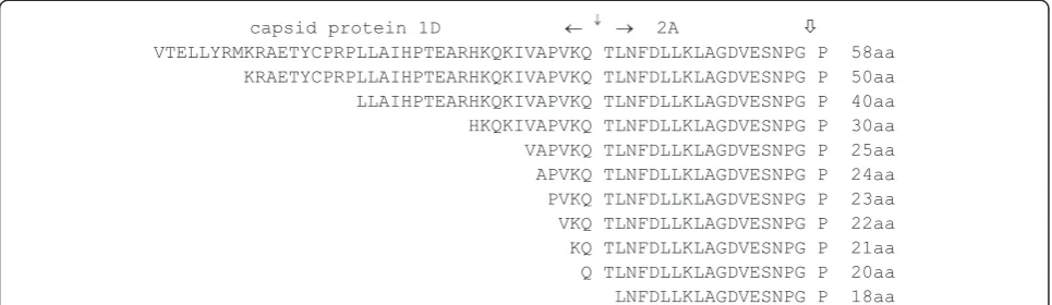
A series of constructs were produced encoding GFP and CherryFP fluorescent‘reporter’proteins under the control of both CMV and T7 promoters. Sequences encoding GFP and CherryFP were separated by either the longest F2A sequence (F2A58) or N-terminally truncated versions
of various lengths (F2A50, F240, F2A30, F2A20, and F2A18;
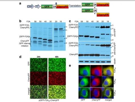
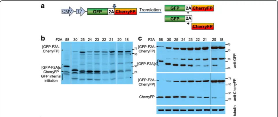
Figure 1), to establish the optimal length of F2A sequence for co-expression of two proteins and their relative clea-vage efficiencies. The expected translation products were cleaved [GFP-F2A] and CherryFP, and the uncleaved polyprotein [GFP-F2A-CherryFP] (Figure 2a).
For thein vitrotranslation studies, rabbit reticulocyte lysates were programmed with the pGFP-F2A-CherryFP series of constructs and the translation products re-solved by 12% SDS-PAGE (Figure 2b). As expected, two major translation products (of the expected molecular weights) were observed. In each track the upper slower migrating product corresponded to either [GFP-F2A58]
(34.5 kDa), [GFP-F2A50] (33.5 kDa), [GFP-F2A40] (32.3 kDa)
or [GFP-F2A30] (31.2 kDa). The second product
Kozak context (cCAUGc, indicated as‘GFP internal ini-tiation’in Figure 2b).
HeLa cells were transfected with this panel of con-structs and cell lysates analysed 30 h post transfection. Similarly, the two major cleavage products were detected by western blotting either with an anti-GFP antibody showing [GFP-F2A]s of decreasing size (Figure 2c, upper blot), or, an anti-CherryFP antibody (Figure 2c, lower blot). Uncleaved polyproteins were also detected, their accumulation increasing as the length of F2A decreased, being highest for the pGFP-F2A30-CherryFP construct.
These data, together with our in vitro analysis, demon-strated that the cleavage efficiency is slightly reduced when a shorter version (F2A30) is used, compared to that
observed for the 58aa version.
Fluorescence imaging was used to directly visualise co-expression of fluorescent proteins. To demonstrate that GFP and CherryFP proteins linked via F2A were co-expressed within the same cell, we transfected HeLa cells with the panel of pGFP-F2A-CherryFP constructs (F2A58, F2A50, F2A40 and F2A30). In each case
transfected cells efficiently co-expressed both GFP and CherryFP for up to 48 h post-transfection (Figure 2d; the images presented for pGFP-F2A30-CherryFP were
representative of all constructs), with both proteins localised to the cytoplasm of transfected cells (Figure 2e). We could not detect any effect of increasing the length of the C-terminal extension of GFP by F2A (30-58aa) upon fluorescence.
In contrast to our constructs with F2A of 58–30 amino acids, translation directed by plasmids pGFP-F2A20
-CherryFP and pGFP-F2A18-CherryFP showed three major
translation products (Figure 2b). In each gel track, the most slowly migrating band corresponded to the un-cleaved polyprotein forms [GFP-F2A20-CherryFP] and
[GFP-F2A18-CherryFP] of the expected molecular weights
(~57 kDa). The bands corresponding to the two cleavage products, [GFP-F2A20] or [GFP-F2A18] and CherryFP
could not be resolved due to the similar molecular weights. In addition, three extra minor products were ob-served in each of these translation reactions. Internal initi-ation within the upstream GFP gene (probably Met88) produced both the fastest migrating cleaved product, cor-responding to protein of ~20 kDa which was almost un-detectable on the gel (indicated in Figure 2b as ‘GFP internal initiation’) and the second slow migrating uncleaved product of ~50 kDa (highlighted with asterisk). The third slower migrating minor product corresponded to a protein of unknown identity (shown with white arrow), although we suspect that this represents a transla-tion product with a C-terminal adduct of tRNA.
Our in vivo studies were consistent with the in vitro
data: the western blotting data demonstrating that the major products were the uncleaved [GFP-F2A20-Cherry
FP] and [GFP-F2A18-CherryFP] polyproteins (Figure 2c).
Small amounts of the CherryFP cleavage product, com-pared to F2A58-30-based constructs, were still detectable
with anti-CherryFP antibody (Figure 2c, lower blot), the upstream cleavage products were, however, either al-most undetectable [GFP-F2A20] or absent [GFP-F2A18]
(Figure 2c, upper blot). It is important to note that the amounts of proteins observed in these blots is a function of detection using two quite different antibodies, raised against either GFP or CherryFP and they have different af-finities for their antigens. Interestingly, extracts of HeLa cells transfected with plasmids pGFP-F2A20-CherryFP and
pGFP-F2A18-CherryFP also contained an unexpected
[image:3.595.57.540.90.230.2]pro-tein with an estimated size of ~45 kDa (shown with white arrow), similar to that detected in reticulocyte lysates, which reacted with the anti-GFP, but not anti-CherryFP antibodies. The nature of this protein, which could be generated by a leaky scanning mechanism, or, be a product
of degradation of the uncleaved precursor is under further investigation.
Interestingly, in the case of constructs which produced a high level of cleavage, the fluorescent signals from both GFP and CherryFP were distributed throughout the cytoplasm, in the case of constructs which produced low levels of cleavage, the fluorescent signals were accumu-lated mostly around the nucleus (Figure 2e).
Tandem repeated cherry fluorescent proteins
To determine if the C-terminal region of the protein up-stream of 2A affected the cleavage activity, we substituted
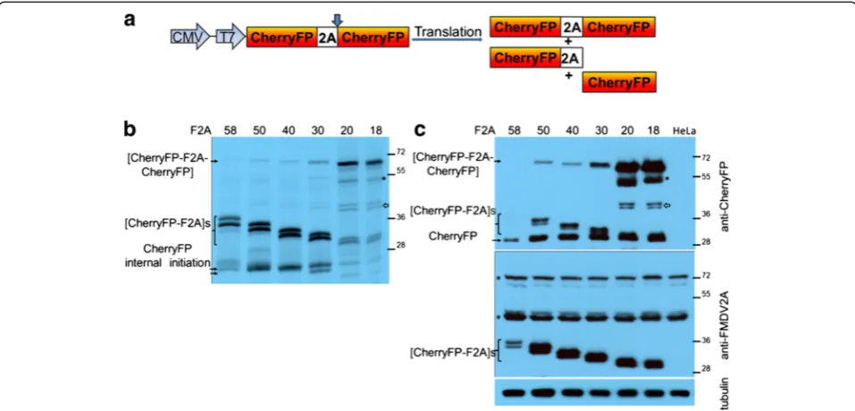
the upstream gene (GFP) with that of CherryFP (Figure 3a). Again, these sequences were linked by either F2A58, F2A50,
F2A40, F2A30, F2A20or F2A18. Similar to our previous
[image:4.595.60.535.88.447.2]re-sults, when reticulocyte lysates were programmed with constructs encoding F2As of diminishing lengths, the uncleaved polyprotein [CherryFP-F2A-CherryFP] formed a very minor translation product for F2A of 58 to 30aa (Figure 3b). In the case of these F2As, a doublet was ob-served corresponding to the [CherryFP-F2A] cleavage product. The fastest migrating product corresponded to the cleavage product downstream of F2A, CherryFP. The trans-lation profiles derived from all pCherryFP-F2A-CherryFP
constructs showed a high level of internal initiation, and produced minor translation products which corres-ponded in size to products from internal initiation at one of three start codons within the N-terminal CherryFP sequence (Met10, Met17 and Met23, with Met10 and Met23 being in more favourable Kozak con-texts (AaCAUGG, ttCAUGc and caCAUGG, respect-ively; upper-case letters indicate highly conserved bases, and lower-case letters –bases that can vary). It should be noted that internal initiation at Met10 produces the same decrease in molecular mass as that observed when the length of 2A was designed in 10aa increments.
These observations in vitro were confirmed by our
in vivoexperiments. Cells were transfected with the panel of pCherryFP-F2A-CherryFP constructs and cleavage activ-ities determined by western blotting (Figure 3c). As with the pGFP-F2A-CherryFP constructs, uncleaved [CherryFP-F2A58, 50, 40-CherryFP] polyproteins were barely detectable
by the anti-CherryFP antibodies. The major products in these cases were the [CherryFP-F2A] and CherryFP cleav-age products. Again, [CherryFP-F2A] migrated as a doub-let, with the fastest migrating product corresponding to the CherryFP cleavage product - of uniform size across the gel tracks (Figure 3c, upper blot). Although easily detectable for the 30aa version, the uncleaved form accumulated to a somewhat lesser degree than the two major cleavage
products. As we observed for thein vitrotranslations, the upstream cleavage products (now [CherryFP-F2A]) were detected by an anti-FMDV2A antibody (Figure 3c, lower blot), but the uncleaved [CherryFP-F2A-CherryFP] was not. Using this antibody, two non-specific proteins were detected both in transfected and mock-transfected cells (Figure 3c, lower blot, highlighted with asterisks). Interest-ingly, the amounts of the upstream and downstream cleav-age products detected by both CherryFP and anti-FMDV2A antibodies in HeLa cells transfected with pCherryFP-F2A58-CherryFP construct were lower than
those expressed from constructs with shorter 2As.
Similar to our data obtained for pGFP-F2A20-CherryFP
and pGFP-F2A18-CherryFP, both in reticulocyte lysates
(Figure 3b) and in transfected HeLa cells (Figure 3c), the uncleaved polyproteins [CherryFP-F2A20-CherryFP] and
[CherryFP-F2A18-CherryFP] represented the major
trans-lation products. The cleavage products upstream of F2A ([CherryFP-F2A20] and [CherryFP-F2A18]) migrated as
[image:5.595.58.537.89.319.2]doublets, as discussed above. The cleavage product down-stream of F2A, CherryFP, was of uniform size across all the gel tracks. Interestingly, it appeared that cleavage was more efficient when CherryFP was upstream of 2A, rather than GFP –shown using both anti-CherryFP (Figure 3c, upper blot) and anti-FMDV2A antibodies (Figure 3c, lower blot). We assume the higher amounts of cleaved
products arose from the changes in the C-terminal se-quences of the upstream gene (CherryFPversus GFP), as the linker sequences connecting them to F2A were identi-cal in all constructs (−RAKRSLE-). In addition, similar to the data presented for pGFP-F2A-CherryFP constructs, two extra bands were observed in both reticulocyte lysates and HeLa cells transfected with constructs containing F2As of 20 and 18 amino acids. The slow migrating band corresponded to protein produced by internal ini-tiation within the upstream Cherry gene (probably Met71; Figure 3b, highlighted with asterisk). This product was also detected in HeLa cells using CherryFP anti-bodies (Figure 3c, upper blot, highlighted with asterisk). The extra doublet (~45 kDa) we observed using reticu-locyte lysates (Figure 3b, shown with white arrow) was also detected in HeLa cell extracts using anti-CherryFP antibodies (Figure 3c, upper blot, shown with white arrow), but not anti-FMDV 2A antibodies.
Co-expression of CherryFP and GFP fluorescent proteins from pCherryFP-F2A-GFP constructs
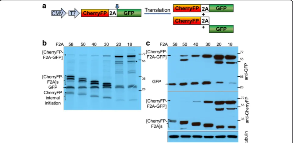
The downstream CherryFP sequences were replaced with GFP to create pCherryFP-F2A-GFP, again linked with the different lengths of F2A (Figure 4a). The pat-tern of accumulation of uncleaved polyproteins was as
observed before: an increase as the length of F2A decreases. The uncleaved polyprotein formed the major product in the case of pCherryFP-F2A20-GFP
and pCherryFP-F2A18-GFP both in reticulocyte lysates
(Figure 4b) and in HeLa cells (Figure 4c). The cleaved up-stream proteins, [CherryFP-F2A]s, expressed from all six constructs, migrated as a doublet in the case of transla-tion in vitro (Figure 4b) and in vivo - in the latter case detected by western blotting using CherryFP anti-bodies (Figure 4c, lower blot). The second major band corresponded to the downstream cleavage product, GFP (Figure 4b and 4c, upper blot). Translation profiles derived from pCherryFP-F2A-GFP constructs with F2As58-30
showed a certain level of internal initiation (probably from Met71) within the upstream CherryFP gene. This pro-duced fast migrating bands corresponding to cleaved products of decreasing molecular weights (Figure 4b, in-dicated as‘CherryFP internal initiation’).
The F2A20 and F2A18 versions showed poor cleavage:
[image:6.595.58.539.408.643.2]the GFP cleavage product was almost undetectable in western blots using the anti-GFP antibody (Figure 4c, upper blot). Internal initiation was observed in the reticu-locyte lysate translation in vitro. The faster migrating bands corresponded to N-terminally truncated [CherryFP-F2A] proteins (probably produced by initiation at Met71;
Figure 4b, indicated as‘CherryFP internal initiation’). Initi-ation at this site also accounts for products migrating somewhat more rapidly than the full-length (uncleaved) polyprotein (Figure 4, b and c, highlighted with asterisks). This extra product was observed in both reticulocyte ly-sates programmed with the plasmids encoding F2As of 20 and 18aa, and in extracts of HeLa cell transfected with these constructs. In these cases, the poor cleavage activity leads to an accumulation of uncleaved products and enabled sufficient material to accumulate to be observed directly (translation in vitro), or, be recognised by both anti-CherryFP and anti-GFP antibodies.
‘Fine-tuning’of shorter F2A sequences
To assess the utility of shorter F2A sequences for co-expression, we produced a series of constructs in which sequences encoding GFP and CherryFP ‘reporter’ pro-teins were linked by the panel of N-terminally single-residue truncated versions of F2A (F2As25-21; Figure 1).
These constructs were analysed together with the previ-ously described pGFP-F2A-CherryFP constructs contai-ning F2A58, F2A30, F2A20 and F2A18. Based upon the
data shown in Figure 2, pGFP-F2A58-CherryFP /
pGFP-F2A30-CherryFP were used as exemplars of highly
effi-cient 2A cleavage and pGFP-F2A20-CherryFP /
pGFP-F2A18-CherryFP as exemplars of inefficient cleavage.
Analysis of the translation products showed three major bands which corresponded to the three predicted translation
products and their molecular weights: uncleaved [GFP-F2A-CherryFP] polyprotein and the two major cleavage products, [GFP-F2A] and CherryFP (Figure 5b). While the intensities of the latter bands were much higher in reticulocyte lysates programmed with constructs con-taining F2As of 30, 25, 24 and 23aa (in comparison with F2As of 22-18aa), the translation products derived from the four constructs encoding shorter 2A sequences were mostly uncleaved. Translation profiles derived from all constructs showed a low level of internal initiation (probably from Met88) within the upstream GFP gene. The fast migrating band corresponded to N-terminally truncated cleaved products [GFP-F2A] and slow mig-rating band corresponded to N-terminally truncated uncleaved polyprotein (Figure 5b, highlighted with asterisk). Again, the additional translation product of ~ 45KDa was observed (more prominent with F2A ver-sions of 25aa and shorter), but was absent in constructs with F2A30and F2A58. This additional translation
prod-uct was detected by anti-GFP (Figure 5c, upper blot, shown with white arrow), but not CherryFP anti-bodies (Figure 5c, lower blot). From F2A25, the intensity
[image:7.595.57.539.442.645.2]of this band increased as the length of F2A decreased. In the cases of F2A of 23aa and longer, the translation products upstream of F2A ([GFP-F2A]) and down-stream ([CherryFP]) were easily detected by anti-GFP (Figure 5c, upper blot) and anti-CherryFP antibodies (Figure 5c, lower blot), respectively. In the case of
F2As22-20, cleavage products were barely detectable,
whilst for F2A18no cleavage products could be detected.
Discussion
Initial characterization of the FMDV 2A region by Ryanet al. [10] showed that while the FMDV 2A/2B cleavage ac-tivity was mediated by residues located within the 19 amino acid version of F2A, N-terminal truncation of F2A reduced cleavage efficiency. Ryan and Drew [14] further described the construction of plasmid (pCAT2AGUS) in which a 19 amino acid version of F2A was inserted be-tween the reporter genes chloramphenicol acetyl trans-ferase (CAT) and β-glucuronidase (GUS). Analyses of
in vitro translation products derived from a series of constructs in which sequences were progressively de-leted from the N-terminal region of F2A showed that activity was ablated in versions with less than 13 resi-dues (−LKLAGDVESNPGP-). It was concluded that residues comprising the N-terminal portion of the F2A region ‘whilst being influential were not critical for cleavage activity’[14].
According to the model for 2A-mediated cleavage, resi-dues that may influence the activity are predicted to reside within the exit tunnel of the ribosome. The use of longer versions of F2A with N-terminal extensions derived from capsid protein 1D (to a total of ~30aa) were reported to produce higher levels of cleavage [10-13]. Specifically, in a series of constructs based upon pGFP2AGUS, N-terminal extension of 2A by 5aa of 1D (equivalent to F2A25)
im-proved cleavage, but extension by 14, 21 and 39aa (equiva-lent of F2A of 35, 40 and 58aa) produced complete cleavage and an equal stoichiometry of both translation productsin vitro[11].
These observations are consistent with ourin vitro and
in vivodata on cleavage efficiency in constructs expressing GFP and CherryFP fluorescent ‘reporter’ proteins sepa-rated by F2As of 58, 50, 40 and 30aa. For all constructs tested, the in vitroanalyses showed the polyprotein was ef-ficiently cleaved, the upstream protein bearing the F2A ‘tail’at its C-terminus. In transfected HeLa cells, however, small amounts of uncleaved polyprotein were detected by western blotting (using anti-GFP / anti-CherryFP, but not anti-FMDV2A, antibodies) in constructs encoding the F2A30version. Swapping the gene order had no effect on
cleavage efficiency when longer versions of 2As were used, showing that for these particular lengths, cleavage is not sensitive to the C- terminal sequence of the upstream pro-tein - since this has already transited through the exit tun-nel of the ribosome.
Further experiments demonstrated that the cleavage efficiency was gradually reduced with each N-terminal amino acid deletion, shown by both our in vitro and
in vivo data when pGFP-F2A-CherryFP constructs with
F2As of 25aa to 18aa were used. The length of F2A of
23aa was demonstrated to be the most efficient of the shorter versions of 2A, and deletion of just one amino acid from its N-terminus resulted in a substantial de-crease in cleavage efficiency, whereas truncation to F2A18 essentially ablated cleavage: data apparently at
variance with those previously reported for F2A20, where
cleavage was efficient [10, 11; discussed below]. Cleavage efficiency was improved somewhat in constructs where the upstream gene was CherryFP (rather than GFP).
The exit tunnel of the ribosome accommodates 30–40 amino acids. For FMDV, the C-terminal region of pro-tein 1D (immediately upstream of 2A) must have evo-lved to interact with the exit tunnel such that F2A (18aa long) mediates cleavage highly efficiently. In the case of artificial polyproteins, the ‘authentic’ upstream context of 2A is replaced with foreign gene or linker sequences which may create an unfavourable environment for ribo-some ‘skipping’: their presence inside the exit tunnel may adversely affect the interaction of F2A with the ribosome. Low cleavage efficiency may arise, therefore, due to (i) the nature of the C-terminal region of the up-stream gene, or, (ii) the manner in which it is linked to the shorter versions of F2A.
The data presented here is in agreement with many pre-viously published studies that successfully used F2A and ‘2A-like’sequences in a wide range of heterologous sys-tems, notably, adoptive cell cancer gene therapies, genetic engineering of human stem cells and the co-expression of transcription factors for the induction of pluripotent stem cells [11,19,20,22-25,29-35]. Our findings also help explain previously published studies in which incomplete cleavage was observed. To avoid using longer 2A sequences (which remain attached at the C-terminus of the upstream pro-tein), researchers used shorter versions of F2A. Conflicting cleavage efficiencies have been reported for 2As of the same lengths [11,14,19,22,25,27]. For example, Furleret al. [18] showed that within the context of a poliovirus vector, 2A-mediated cleavage was incomplete when F2A24 was
used. Fanget al. [19] described similar results for the con-stitutive expression of full-length two-chain antibodies from a single ORF by linking the heavy and light antibody chains with F2A24. Different cleavage efficiencies
depend-ing on protein arrangement were observed when GFP, EGFP and hrGFP were co-expressed with cytochrome P450 2B1 (CYP2B1) via F2A20[36]. In addition, Donnelly
and co-workers, using F2A20 in GFP2AGUS and
GUS2-AGFP, also reported the presence of small amounts of uncleaved polyprotein [11]. With F2A19, cleavage was
reported to be less efficient (70%) in [CFP-2A-PAC] con-text, with some uncleaved polyprotein still present [37], while de Felipe and co-workers [17] used an F2A18version
58aa [17]. Finally, Ma and Mitra used F2A17to co-express
GUS and CAT proteins in different arrangements and demonstrated that [GUS-2A-CAT] and [GUS-2A-GUS] were cleaved to completion, while [CAT-2A-GUS] was not [38].
Interestingly, we show here that pGFP-F2A20-CherryFP
and pGFP-F2A18-CherryFP produced poor cleavage both in vitroand in vivo, apparently at variance with our own previously published data [10,11]. In these studies, the pGFP2AGUS construct contained the same upstream GFP gene and the same F2A20sequence and produced
ef-ficient cleavage. Sequence comparison of pGFP-F2A20
-CherryFP and pGFP2AGUS constructs shows that the dif-ferences are only observed in the sequence immediately upstream of 2A (RAKRSLE vs SGSRGAC, respectively). Ironically, this seven amino acid stretch is a part of neither F2A nor C terminus of the upstream GFP, but is actually composed of two different restriction sites introduced for cloning purposes during the construction of both plas-mids. The effect of the differences in this particular region on the efficiency of 2A-mediated cleavage is the subject of a separate report [39].
Swapping the order of fluorescent ‘reporter’ proteins in our expression systems had no effect on the general pattern of cleavage efficiency. However, while two genes could be effectively co-expressed (irrespective of pos-ition) when linked by a longer version of F2A (30aa and longer), we did observe a small effect for the shorter F2A20and F2A18versions.
Conclusions
In summary, F2As are used widely in biotechnology and biomedicine and we wished to identify the source(s) of ap-parent discrepancies in the literature with regards to cleav-age efficiency. While constructs containing F2A of 58-23aa in length exhibited enhanced abilities for cleavage compared to the constructs with F2A of 22aa and shorter, with least efficient 2As being those of 20aa and 18aa, we suggest that researchers use F2A30if opting for a shorter
sequence. This 2A proved to be most optimal in terms of both length and cleavage efficiency, and, in our hands, was unaffected by the sequence of the upstream gene. A more extensive characterization of sequence requirements be-tween the C-terminal region of the upstream protein and the N-terminal part of F2A is underway to test the specu-lation that this particularly interesting region of the se-quence may have certain effect on 2A-mediated cleavage. Ultimately, the fine-tuning of the F2A sequence can help researchers in their efforts to efficiently co-express various multiple proteins for successful biomedical strategies.
Methods
The following reagents were obtained from commercial suppliers: Pfu polymerase, T4 DNA ligase, restriction
enzymes, JM109 competent cells, Quick TnTrabbit re-ticulocyte lysate kit (Promega, Madison, WI, USA); S35 methionine (MP Biomedicals, Santa Ana, CA, USA); EDTA-free protease inhibitor cocktail (Sigma-Aldrich Company Ltd, Dorset, UK); pcDNA3.1, anti-RFP, rabbit purified polyclonal antibody against Red Fluorescent Protein (1:1000 dilution), GFP, mAb 3E6, anti-Green Fluorescent Protein, mouse monoclonal antibody (1:1000 dilution), anti-β-tubulin, mouse monoclonal antibody (1:2000 dilution), Lipofectamine2000 transfec-tion reagent, iBlot Gel Transfer Stacks, nitrocellulose (Life Technologies Ltd, Paisley, UK); secondary HRP-conjugated polyclonal goat anti-mouse and anti-rabbit antibodies (Dako UK Ltd, Ely, UK); rabbit anti-FMDV 2A antiserum raised against the synthetic peptide NH2-LLNFDLLKLAGDVESNPGP-COOH; EZ-ECL Chemilu-minescence Detection Kit for HRP (Geneflow Ltd, Elmhurst, UK); VECTASHIELD mounting medium with DAPI (VECTOR Laboratories Ltd, Peterborough, UK); Oligonucleotides were purchased from Integrated DNA Technologies (Coralville, Iowa, USA).
Construction of plasmids
pGFP-F2As-CherryFP constructs (F2As of 58, 50, 40, 30, 25–20, and 18aa, sequences shown in Figure 1). To make pGFP-F2A58-CherryFP construct, F2A58sequence, obtained
by digesting pSV7 plasmid (kindly provided by Sandra Varte) with NsiI and ApaI restriction enzymes, was cloned into pJC3 plasmid (pcDNA3.1-based with T2A linking GFP and CherryFP genes) in place of T2A sequence using these restriction sites. pGFP-F2A58-CherryFP construct was then
used as a template to produce deletions of F2A. Forward oligonucleotides incorporating XhoI restriction site: (5′
-CTCGAGAAGAGGGCCGAAACATACTGTCCAAGG-3′
for F2A50, 5′-CTCGAGTTGCTGGCAATCCACCCAAC
TGAAGCC-3′ for F2A40, 5′-GCGCTCGAGCACAAAC
AGAAAATTGTGGCACCGGTG-3′ for F2A30, 5′-GCG
CTCGAGGTGGCACCGGTGAAACAGACTTTG-3′ for
F2A25, 5′-GCGCTCGAGGCACCGGTGAAACAGACTTT
G-3′for F2A24, 5′-GCGCTCGAGCCGGTGAAACAGAC
TTTGAATTTTG-3′for F2A23, 5′-GCGCTCGAGGTGA
AACAGACTTTGAATTTTGAC-3′for F2A22, 5′-GCGC
TCGAGAAACAGACTTTGAATTTTGACCTTC-3′ for
F2A21, 5′-GCGCTCGAGCAGACTTTGAATTTTGACC
TTCTCAAG-3′for F2A20, 5′-GCGCTCGAGTTGAATT
TTGACCTTCTCAAGTTGGCG-3′for F2A18) and a
re-verse oligonucleotide incorporating NotI restriction site (5′ -TAGAAGGCACAGTCGAGGC-3′) were used to amplify F2As-CherryFP with 2As of various lengths. The PCR pro-ducts were digested with XhoI and NotI restriction enzymes and cloned into pGFP-F2A58-CherryFP in place of F2A58
-CherryFP sequence using XhoI and NotI restriction sites.
constructs were digested with BamHI and NsiI restriction enzymes to substitute GFP sequence with that of CherryFP. In order to do that, pJC3 plasmid was used as a template to amplify CherryFP gene using forward (5′ -GCGGGATCCATGGTGAGCAAGGGCGAGGAGGATA AC -3′) and reverse (5′-GCGATGCATTTTGTACAAT TCATCCATGCCGCCG -3′) primers incorporating, re-spectively, BamHI and NsiI restriction sites. PCR products were digested with BamHI and NsiI restriction enzymes and ligated into prepared vectors.
pCherryFP-F2As-GFP constructs (F2As of 58, 50, 40, 30, 20 and 18aa).Respective pCherryFP-F2As-CherryFP constructs were digested with ApaI and EcoRI restric-tion enzymes to substitute downstream CherryFP gene with that of GFP. pJC3 plasmid was used as a template to amplify GFP gene using forward (5′-GCGGGGCCC
GATATCGTGTCCAAAGGGGAAGAG-3′) and reverse
(5′-GCGGAATTCTTACTTATACAGCTCGTCCATGC
CGAG-3′) primers incorporating ApaI and EcoRI re-striction sites. PCR products were digested with ApaI and EcoRI restriction enzymes and ligated into pre-pared vectors.
Transcription and translation
Plasmid constructs were used to programme QuickTnT
Coupled Transcription/ Translation reticulocyte lysate systems (Promega) and the reactions repeated three times. Translation reactions (10μl) were performed according to the manufacturer’s instructions. Briefly, 20 ng of plasmid DNA was used to programme rabbit reticulocyte lysates containing35S methionine (10μCi) and the reactions were incubated at 30°C for 1 h. Translation reactions were stopped by the addition of 2x protein dissociation buffer. Protein products were analysed by 12% SDS-PAGE. Con-trols of TnT reactions programmed with‘empty’ vectors produced no translation products (data not shown).
Expression in HeLa cells
HeLa cells were maintained in DMEM medium supple-mented with 10% FCS. For western blotting, the cells were transfected in 60 mm dishes (pre-plated 20 h earlier to 60% confluency) with 1.5μg of plasmid DNA and 7μl of Lipofectamine2000 (Invitrogen) in a final volume of 400μl OptiMEM. Transfection mix was added to 4 ml of antibiotic-free serum-containing medium and cells were incubated for a total of 30 h post-transfection. All trans-fections were performed in triplicate. Controls with ‘empty’ expression vector transfected cells produced no translation products (western blotting) or fluorescence (data not shown).
Western blotting
30 h post-transfection, cells were washed twice with 1 ml of PBS and harvested in 1 ml of PBS by centrifugation at
2000 rpm for 5 min. Whole-cell lysates were prepared in 70 μl of radioimmuno precipitation assay buffer, RIPA (150 mM NaCl, 10 mM Tris pH 7.4, 1% Triton X-100, 1% Na deoxycholate, 0.1% SDS, and freshly added 1/20 vo-lume of EDTA-free protease inhibitor cocktail) by incuba-tion on ice for 30 min and centrifugaincuba-tion at 12000 rpm for 20 min at 4°C. Cellular debris was discarded and supernatants containing proteins were analysed by 12% SDS-PAGE. The proteins were then transferred to a nitrocellulose membrane, which was probed with anti-GFP, anti-CherryFP or anti-FMDV 2A primary antibodies overnight after blocking in PBST containing 5% milk for 1 h. Following overnight incubation, the membranes were washed three times in PBST. Detection of bound primary antibodies was performed with respective HRP-secondary antibodies (Dako) in PBST containing 5% milk for 1 h, the membranes were washed three times with PBST, rinsed in deionised water and subjected to enhanced chemilumi-nescence by incubating it in freshly-prepared visualisation solution for 2 min. Membrane was exposed to an auto-radiograph film for 3 to 45 seconds.
Fluorescence microscopy
Transfection of HeLa cells was performed as described above for western blotting, however, 0.5 μg of plasmid DNA and 5 μl of Lipofectamine2000 in a final volume of 200 μl OptiMEM were added to 2 ml of media in each well of a 6-well plate containing coverslips. 30 h post-transfection the cells were washed twice with 1 ml of PBS, fixed with 1 ml of ice-cold 100% methanol for 10 min and washed again with 1 ml of PBS. The coverslips were moun-ted in VECTASHIELD (an antifade mounting medium, VECTOR Laboratories). The images were acquired with Deltavision microscope using 100 × objective using Resolve 3D software. Experiments were done in triplicates.
Competing interests
The authors declared that they have no competing interests.
Authors’contributions
EM carried out cloning, sequence alignment, in vitro TnTanalysis, cell transfections, Western blotting, fluorescence analysis, and drafted the manuscript. JN participated in cloning andin vitro TnTanalysis. MDR helped to draft the manuscript. All authors read and approved the final manuscript.
Funding
The project was funded by MRC grant, MRC UK.
Received: 3 May 2013 Accepted: 15 August 2013 Published: 22 August 2013
References
1. de Felipe P:Polycistronic viral vectors.Curr Gene Ther2002,2:355–378. 2. Pfutzner W:Retroviral bicistronic vectors.Drug News Perspectives2008,
31:473–480.
3. Luke G:Translating 2A research into practice.InInnovations in
Biotechnology.Edited by Agbo E. Rijeka, Croatia: InTech; 2012:161–186.
5. Wong ET, Ngoi SM, Lee CGL:Improved co-expression of multiple genes in vectors containing internal ribosome entry sites (IRESes) from human genes.Gene Ther2002,9:337–344.
6. Kozak M:A second look at cellular mRNA sequences said to function as internal ribosome entry sites.Nucleic Acids Res2005,33:6593–6602. 7. Baranick BT, Lemp NA, Nagashima J, Hiraoka K, Kasahara N, Logg CR:
Splicing mediates the activity of four putative cellular internal ribosome entry sites.Proc Natl Acad Sci USA2008,105:4733–4738.
8. Ryan MD, Donnelly MLL, Lewis A, Mehrotra AP, Wilkie J, Gani D:A model for non-stoichiometric, co-translational protein scission in eukaryotic ribosomes.Bioorganic Chem1999,27:55–79.
9. Atkins JF, Wills NM, Loughran G, Wu CY, Parsawar K, Ryan MD, Wang CH, Nelson CC:A case for‘StopGo’: reprogramming translation to augment codon meaning of GGN by promoting unconventional termination (Stop) after addition of glycine and then allowing continued translation (Go).RNA2007,13:1–8.
10. Ryan MD, King AM, Thomas GP:Cleavage of foot-and-mouth disease virus polyprotein is mediated by residues located within a 19 amino acid sequence.J Gen Virol1991,72:2727–2732.
11. Donnelly MLL, Hughes LE, Luke GA, Mendoza H, ten Dam E, Gani D, Ryan MD:The‘cleavage’activities of foot-and-mouth disease virus 2A site-directed mutants and naturally occurring‘2A-like’sequences.
J Gen Virol2001,82:1027–1041.
12. Klump H, Schiedlmeier B, Vogt B, Ryan M, Ostertag W, Baum C:Retroviral vector-mediated expression in HoxB4 in hematopoietic cells using a novel expression strategy.Gene Ther2001,8:811–817.
13. Luke GA, de Felipe P, Lukashev A, Kallioinen SE, Bruno EA, Ryan MD:The occurrence, function, and evolutionary origins of‘2A-like’sequences in virus genomes.J Gen Virol2008,89:1036–1042.
14. Ryan MD, Drew J:Foot-and-mouth disease virus 2A oligopeptide mediated cleavage of an artificial polyprotein.EMBO J1994,13:928–933. 15. de Felipe P, Hughes LE, Ryan MD, Brown JD:Co-translational,
intraribosomal cleavage of polypeptides by the foot-and-mouth disease virus 2A peptide.J Biol Chem2003,278:11441–11448.
16. Ryan MD, Donnelly MLL, Flint M, Cowton VM, Luke GA, Hughes LE, Knox C, de Felipe P:Foot-and-mouth disease virus proteinases.InFoot and Mouth
Disease Current Perspectives.Edited by Sobrino F, Domingo E. England:
Horizon Bioscience; 2004:53–76.
17. de Felipe P, Martin V, Cortes ML, Ryan M, Izquierdo M:Use of the 2A sequence from foot-and-mouth disease virus in the generation of retroviral vectors for gene therapy.Gene Ther1998,6:198–208. 18. Furler S, Paterna JC, Weibel M, Bueler H:Recombinant AAV vectors
containing the foot and mouth disease virus 2A sequence confer efficient bicistronic gene expression in cultured cells and rat substantia nigra neurons.Gene Ther2001,8:864–873.
19. Fang J, Qian J, Yi S, Harding TC, Tu G, VanRoey M, Jooss K:Stable antibody expression at therapeutic levels using the 2A peptide.Nat Biotechnol 2005,23:584–590.
20. Funston GM, Kallioinen SE, de Felipe P, Ryan MD:Expression of heterologous genes in oncolytic adenoviruses using picornaviral 2A sequences that trigger ribosome skipping.J Gen Virol2008,89:389–396. 21. Trichas G, Begbie J, Srinivas S:Use of the viral 2A peptide for bicistronic
expression in transgenic mice.BMC Biol2008,6:40.
22. Yang S, Cohen CJ, Peng PD, Zhao Y, Cassard L, Yu Z, Zheng Z, Jones S, Restifo NP, Rosenberg SA, Morgan RA:Development of optimal bicistronic lentiviral vectors facilitates high-level TCR gene expression and robust tumor cell recognition.Gene Ther2008,15:1411–1423.
23. Tan Y, Liang H, Chen A, Guo X:Co-expression of double or triple copies of the rabies virus glycoprotein gene using a‘self-cleaving’2A peptide-based replication-defective human adenovirus serotype 5 vector.Biologicals2010, 38:586–593.
24. Takata Y, Kishine H, Sone T, Andoh T, Nozaki M, Poderycki M, Chesnut J, Imamoto F:Generation of iPS cells using a BacMam expression system.
Cell Struct Funct2011,36:209–222.
25. Szymczak AL, Workman CJ, Wang Y, Vignali KM, Dilioglou S, Vanin EF, Vignali DA:Correction of multi-gene deficiencyin vivousing a single ‘self-cleaving’2A peptide-based retroviral vector.Nat biotechnol2004, 22:589–594.
26. Camper N, Byrne T, Burden RE, Lowry J, Gray B, Johnston JA, Migaud ME, Olwill SA, Buick RJ, Scott CJ:Stable expression and purification of a functional processed Fab’fragment from a single nascent polypeptide in
CHO cells expressing the mCAT-1 retroviral receptor.J Immunol Methods 2011,372:30–41.
27. de Felipe P, Ryan MD:Targeting of proteins derived from self-processing polyproteins containing multiple signal sequences.Traffic2004, 5:616–626.
28. de Felipe P, Luke GA, Brown JD, Ryan MD:Inhibition of 2A-mediated ‘cleavage’of certain artificial polyproteins bearing N-terminal signal sequences.Biotechnol J2010,5:213–223.
29. Provost E, Rhee J, Leach SD:Viral 2A peptides allow expression of multiple proteins from a single ORF in transgenic zebrafish embryos.
Genesis2007,45:625–629.
30. Hart DP, Xue S-A, Thomas S, Cesco-Gaspere M, Tranter A, Willcox B, Lee SP, Steven N, Morris EC, Stauss HJ:Retroviral transfer of a dominant TCR prevents surface expression of a large proportion of the endogenous TCR repertoire in human T cells.Gene Ther2008,15:625–631. 31. Unger C, Karner E, Treschow A, Stellan B, Felldin U, Concha H, Wendel M,
Hovatta O, Aints A, Ahrlund-Richter L, Dilber M:Lentiviral-mediated HoxB4 expression in himan embryonic stem cells initiates early hematopoiesis in a dose-dependent manner but does not promote myeloid differentiation.Stem Cells2008,26:2455–2466.
32. Karner E, Unger C, Cerny R, Ahrlund-Richter L, Ganss B, Dilber M, Wendel M: Differentiation of human embryonic stem cells into osteogenic or hematopoietic lineages: a dose-dependent effect of osterix over-expression.J Cell Psysiol2009,218:323–333.
33. Sommer CA, Stadfeld M, Murphy GJ, Hochedlinger K, Kotton DN, Mostoslavsky G:iPS cell generation using a single lentiviral stem cell cassette.Stem Cells2008,27:543–549.
34. Carey BW, Markoulaki S, Hanna J, Saha K, Gao Q, Mitalipova M, Jaenisch R: Reprogramming of murine and human somatic cells using a single polycistronic vector.Proc Natl Acad Sci2009,106:157–162.
35. Shao LS, Feng W, Sun Y, Bai H, Liu J, Currie C, Kim J, Gama R, Wang Z, Qian Z, Liaw L, Wu WS:Generation of iPS cells using defined factors linked via the self-cleaving 2A sequences in a single open reading frame.Cell Res 2009,19:296–310.
36. Lengler J, Holzmuller H, Salmons B, Gunzburg WH, Renner M:FMDV-2A sequence and protein arrangement contribute to functionality of CYP2B1-reporter fusion protein.Anal Biochem2005,343:116–124. 37. Sharma P, Yan F, Doronina VA, Escuin-Ordinas H, Ryan MD, Brown J:2A
peptides provide distinct solutions to driving stop-carry on translational recoding.Nucleic Acids Res2012,40:3143–3151.
38. Ma C, Mitra A:Expressing multiple genes in a single open reading frame with the 2A region of foot-and-mouth disease virus as a linker.Mol breed 2002,9:191–199.
39. Minskaia E, Ryan M:Protein co-expression using FMDV 2A: effect of ‘linker’residues.Biomed Res Int. in press.
doi:10.1186/1472-6750-13-67
Cite this article as:Minskaiaet al.:Optimisation of the foot-and-mouth disease virus 2A co-expression system for biomedical applications.BMC Biotechnology201313:67.
Submit your next manuscript to BioMed Central and take full advantage of:
• Convenient online submission
• Thorough peer review
• No space constraints or color figure charges
• Immediate publication on acceptance
• Inclusion in PubMed, CAS, Scopus and Google Scholar
• Research which is freely available for redistribution