FINE NEEDLE ASPIRATION CYTOLOGY AND HISTOPATHOLOGICAL CORRELATION IN
DIAGNOSING LYMPH NODE LESIONS
Khan Ishrat Younas, Ashfaq Hafiz,
Salma Yaseen, Muneera gull, Prableen Kaur and Asima Aijaz
1
PG Resident, Dep
2
Senior Resident, Department
3
Senior Resident,
4
Professor and Head,
ARTICLE INFO ABSTRACT
Introduction:
widely throughout the body. Lymph nodes lie along the course of lymphatic vessels. Most lymphadenopathy is due to a benign self
secondary to an increase in normal lymphocytes and macrophages in response to an antigen. Other less common mechanisms responsible for adenopathy include ly
diseases.
allows the pathologist to see the cells aspirated from the lesion. In contrast to large bore needle biopsy techniques, FNAC do
can be evaluated by flow cytometry or with immunologic markers. conducted at
the Department of Pathology. The study was a prospective study of 2 years i.e. from June 2016 to May 2018. Cases
subsequent biopsy were studied. with lymphadenopathy.
Male: Female ratio was 1.73:1. For Non specificity of 96.55%, positive predict
Lymphoma FNAC showed sensitivity of 85.71%, specificity of 98.84%, positive predictive value of 92.31% and accuracy of 97%.
specificity of 9
Lesions, FNAC showed sensitivity of 93.94%, specificity of 97.73%, positive predictive value of 93.94% and accuracy of 96.69%.
patient friendly in the effort to establish
Copyright © 2019, Khan Ishrat Younas et al. This is an unrestricted use, distribution, and reproduction in any medium,
INTRODUCTION
A lymph node is an oval or kidney-shaped
lymphatic system, present widely throughout the body. Lymph nodes lie along the course of lymphatic vessels. Lymph nodes have a highly cellular cortex and medulla which contains network of minute lymphatic channels (sinuses) through which lymph from afferent lymphatics is filtered, to be
hilum by efferent lymphatics (Roitt et al., adult contains upto 450 lymph nodes of which 60
in head and neck, 100 in thorax, 250 in abdomen and pelvis. Lymph nodes are particularly numerous in the neck, mediastinum,posteriorabdominalwall,abdominal
pelvis, proximal region of limbs (Susan Stranding
ISSN: 0975-833X
Article History:
Received 03rd January, 2019
Received in revised form
17th February, 2019
Accepted 21st March, 2019
Published online 29th April, 2019
Citation: Khan Ishrat Younas, Ashfaq Hafiz, Samoon Nuzhat,
lymph node lesions”, International Journal of Current Research
Key Words:
FNAC, Histopathology, Lymph node lesions.
*Corresponding author: Nuzhat Samoon
RESEARCH ARTICLE
FINE NEEDLE ASPIRATION CYTOLOGY AND HISTOPATHOLOGICAL CORRELATION IN
DIAGNOSING LYMPH NODE LESIONS
Khan Ishrat Younas, Ashfaq Hafiz, *Samoon Nuzhat, Rather Rashid, Salma gull,
Salma Yaseen, Muneera gull, Prableen Kaur and Asima Aijaz
, Department of Pathology, SKIMS Soura, Sgr, India
artment of Radiation Oncology, SKIMS Soura,
Senior Resident, Department of Pathology, SKIMS Soura, Sgr
Professor and Head, Department of Pathology SKIMS MC Sgr
ABSTRACT
Introduction: A lymph node is an oval or kidney-shaped organ
widely throughout the body. Lymph nodes lie along the course of lymphatic vessels. Most lymphadenopathy is due to a benign self-limited disease such as viral infections, and adenopathy is secondary to an increase in normal lymphocytes and macrophages in response to an antigen. Other less common mechanisms responsible for adenopathy include ly
diseases. Tubercular lymphadenitis is one of the commonest causes in developing countries. FNAC allows the pathologist to see the cells aspirated from the lesion. In contrast to large bore needle biopsy techniques, FNAC does not allow evaluation of the morphology. In some instances, aspirated cells can be evaluated by flow cytometry or with immunologic markers.
conducted at the Sher-i-kashmir Institute of Medical Sciences (SKIMS) Srinagar, Kashmir (I the Department of Pathology. The study was a prospective study of 2 years i.e. from June 2016 to May 2018. Cases presenting with lymphadenopathy where FNAC was done and underwent subsequent biopsy were studied. Results: This study included total
with lymphadenopathy. Out of 120 patients, there were 76(63.3%) males and
Male: Female ratio was 1.73:1. For Non-Hodgkin's Lymphoma FNAC showed sensitivity of 97.22%, specificity of 96.55%, positive predictive value of 92.11% and accuracy of 89.74%. For Hodgkin's Lymphoma FNAC showed sensitivity of 85.71%, specificity of 98.84%, positive predictive value of 92.31% and accuracy of 97%. For Metastatic tumors, FNAC showed sensitivity of 91.67%, specificity of 98.81%, positive predictive value of 97.06% and accuracy of 96.67%. For Benign Lesions, FNAC showed sensitivity of 93.94%, specificity of 97.73%, positive predictive value of 93.94% and accuracy of 96.69%. Conclusion: FNAC proved to be a safe, accurate,
patient friendly in the effort to establish diagnosis in patients with lymphadenopathy.
an open access article distributed under the Creative Commons Attribution medium, provided the original work is properly cited.
shaped organ of the throughout the body. Lymph nodes lie along the course of lymphatic vessels. Lymph nodes have a highly cellular cortex and medulla which contains network of minute lymphatic channels (sinuses) through which lymph from afferent lymphatics is filtered, to be collected at
et al., 2001). A young
adult contains upto 450 lymph nodes of which 60-70 are found abdomen and pelvis. Lymph nodes are particularly numerous in the neck, abdominalmesenteries, Susan Stranding, 2005).
Lymph nodes are regional, and each group of them corresponds to a particular region of the body and reflects abnormalities in that region. Common
lymph nodes are more prominent and therefore more readily noticeablearepostauricular,cervical
sub mandibular and axillary regions. The etiology varies from an inflammatory process to a malignant condition
et al., 2009). Most lymphadenopathy is due to a benign self
limited disease such as viral infections, and adenopathy is secondary to an increase in normal lymphocytes and macrophages in response to an antigen. Other less common mechanisms responsible for adenopathy include lymphadenitis, neoplasia or storage diseases. Tubercular lymphadenitis is one of the commonest causes in developing countries
International Journal of Current Research
Vol. 11, Issue, 04, pp.2876-2880, April, 2019
DOI: https://doi.org/10.24941/ijcr.35001.04.2019
Khan Ishrat Younas, Ashfaq Hafiz, Samoon Nuzhat, et al., 2019. “Fine needle aspiration cytology and histopathological correlation in
International Journal of Current Research, 11, (04), 2876-2880.
FINE NEEDLE ASPIRATION CYTOLOGY AND HISTOPATHOLOGICAL CORRELATION IN
Samoon Nuzhat, Rather Rashid, Salma gull,
Salma Yaseen, Muneera gull, Prableen Kaur and Asima Aijaz
, India
, SKIMS Soura, Sgr, India
Sgr, India
of Pathology SKIMS MC Sgr, India
organ of the lymphatic system, present widely throughout the body. Lymph nodes lie along the course of lymphatic vessels. Most limited disease such as viral infections, and adenopathy is secondary to an increase in normal lymphocytes and macrophages in response to an antigen. Other less common mechanisms responsible for adenopathy include lymphadenitis, neoplasia or storage Tubercular lymphadenitis is one of the commonest causes in developing countries. FNAC allows the pathologist to see the cells aspirated from the lesion. In contrast to large bore needle biopsy es not allow evaluation of the morphology. In some instances, aspirated cells can be evaluated by flow cytometry or with immunologic markers. Methods: This study was kashmir Institute of Medical Sciences (SKIMS) Srinagar, Kashmir (India) in the Department of Pathology. The study was a prospective study of 2 years i.e. from June 2016 to lymphadenopathy where FNAC was done and underwent This study included total of 120 patients who presented Out of 120 patients, there were 76(63.3%) males and 44 (36.7%) females. Hodgkin's Lymphoma FNAC showed sensitivity of 97.22%, ive value of 92.11% and accuracy of 89.74%. For Hodgkin's Lymphoma FNAC showed sensitivity of 85.71%, specificity of 98.84%, positive predictive value of For Metastatic tumors, FNAC showed sensitivity of 91.67%, 8.81%, positive predictive value of 97.06% and accuracy of 96.67%. For Benign Lesions, FNAC showed sensitivity of 93.94%, specificity of 97.73%, positive predictive value of FNAC proved to be a safe, accurate, inexpensive and diagnosis in patients with lymphadenopathy.
ribution License, which permits
Lymph nodes are regional, and each group of them corresponds to a particular region of the body and reflects abnormalities in that region. Common areas where swollen lymph nodes are more prominent and therefore more readily cervical,supraclavicular,inguinal, sub mandibular and axillary regions. The etiology varies from an inflammatory process to a malignant condition (Hirachand Most lymphadenopathy is due to a benign self-limited disease such as viral infections, and adenopathy is secondary to an increase in normal lymphocytes and macrophages in response to an antigen. Other less common nsible for adenopathy include lymphadenitis, Tubercular lymphadenitis is one of the commonest causes in developing countries (Shakya et
INTERNATIONAL JOURNAL OF CURRENT RESEARCH
al., 2009). Fine needle aspiration cytology (FNAC) of lymph node has become an integral part of the initial diagnosis and management of patients with lymphadenopathy due to early availability of results, simplicity, and, minimal trauma with less complication (Keith et al., 2007). FNAC is widely used as first line investigation for the diagnosis of lymphadenopathy. FNAC has been advocated as a useful method in comparison to more expensive surgical excision biopsies in developing countries with limited financial and health care resources (Das, 1999). It almost offers an accurate diagnosis for reactive lymphoid hyperplasia, infectious disease, granulomatous lymphadenitis, and metastatic malignancy. Thus, it can avoid the need for excisional biopsy in most cases and allow rapid onset of therapy (Howlett et al., 2007). FNAC allows the pathologist to see the cells aspirated from the lesion. In contrast to large bore needle biopsy techniques, FNAC does not allow evaluation of the morphology. In some instances, aspirated cells can be evaluated by flow cytometry or with immunologic markers. In every circumstance, FNAC is a test and should be interpreted with the entire clinical circumstances. False negative and false positive FNAC results are reported in almost every series. Therefore, reliance upon FNAC findings at the expense of clinical, radiographic, or other findings is unsafe (Salgarelli et al., 2009).
MATERIAL AND METHODS
This study was conducted at the Sher-i-kashmir Institute of Medical Sciences (SKIMS) Srinagar, Kashmir (India) in the Department of Pathology. The study was a prospective study of 2 years i.e. from June 2016 to May 2018. Cases presenting with lymphadenopathy where FNAC was done and underwent subsequent biopsy were studied. FNAC was performed using 20 mlsyringe attached to a 22 gauge needle. The needle was allowed to move back and forth into different parts of the lymph node several times before withdrawal. The specimens were expelled on to cover glasses, air dried and stained with May Grunwald Giemsa stain and other stains like Papanicolaou (Pap) stain. For histopathology, biopsy specimens received were fixed in 100% formalin, studied grossly, photographed, processed and studied in detail using H&E and various special stains wherever indicated.
RESULTS
This study included total of 120 patients who presented with lymphadenopathy. Out of 120 patients, there were 76(63.3%) males and 44 (36.7%) females. Male: Female ratio was 1.73:1. In this study age ranged from 5 to 80 years. Mean age was 44 years. Maximum number of cases were seen in the age group 51-60 years (28 cases, 23.33%), followed by age group 21-30 years (24 cases, 20%), age group 61-70 years (20 cases, 16.67%), age group 41-50 years (19 cases, 15.83%), age group 31-40 years (17 cases, 14.17%), age group 11-20 years (9 cases, 7.50%), age group 71-80 years (2 cases, 1.67%), age group 01-10 years (1 case, 0.83%) respectively. Maximum no. of aspirations (Table 1.) were done from Cervical nodes 74 cases (61.67%) followed by Axillary nodes 19 cases (15.83%) followed by Supraclavicular nodes 15 cases (12.50%) followed by Inguinal nodes 9 cases (7.50%) followed by Retroperitoneal lymph nodes 2 cases (1.67%) respectively. Of the 120 patients subjected to FNAC, histological diagnosis revealed 36 cases of Non-Hodgkin's Lymphoma constituting 30% of all cases, Metastatic lesions in 36 cases constituting 30% of all cases, 34
[image:2.595.334.530.122.196.2]cases turned out to be Benign constituting 28.33% of all cases, 14 cases of Hodgkin's Lymphoma constituting 11.67% of all cases.
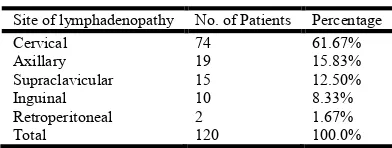
Table 1. Distribution of cases according to site of lymphadenopathy
Site of lymphadenopathy No. of Patients Percentage
Cervical 74 61.67%
Axillary 19 15.83%
Supraclavicular 15 12.50%
Inguinal 10 8.33%
Retroperitoneal 2 1.67%
Total 120 100.0%
Correlation between FNAC and histopathology (Table 2)
1. Metastatic tumors (Fig 1,2,3&4)
Out of a total of 36 cases of metastatic tumors, FNAC and histopathology were concordant in 33 cases. Two cases of metastatic poorly differentiated carcinoma were misinterpreted in FNAC as Non-Hodgkin's Lymphoma and one case of metastatic poorly differentiated carcinoma was misinterpreted as hodgkins lymphoma. One case of hodgkins lymphoma was misinterpreted as metastatic poorly differentiated carcinoma. On statistical analysis, FNAC had a sensitivity of 91.67%, specificity of 98.81%, positive predictive value of 97.06% and accuracy of 96.67% for metastatic tumors.
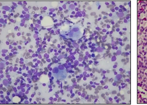
2. Hodgkin's Lymphoma (Fig. 5 &6)
Out of a total of 14 cases of Hodgkin's Lymphoma, FNAC and histopathology were concordant in 12 cases. One case of Hodgkin's Lymphoma was misinterpreted in FNAC as Granulomatous Lymphadenitis and another one as metaststic poorly differentiated carcinoma. One case of metastatic poorly differentiated carcinoma was misinterpreted as hodgkins lymphoma. On statistical analysis, FNAC had a sensitivity of 85.71%, specificity of 98.84%, positive predictive value of 92.31% and accuracy of 97% for Hodgkin's Lymphoma.
3. Non-Hodgkin's Lymphoma (Figure 7 &8)
Out of a total of 38 cases of NHL, FNAC and histopathology were concordant in 35 cases. One case of NHL was misinterpreted in FNAC as reactive lymphadenitis. Besides there were two cases of metastatic poorly differentiated carcinoma misinterpreted as NHL and one case of granulomatous lymphadenitis misinterpreted as NHL in FNAC. On statistical analysis, FNAC showed sensitivity of 97.22%, specificity of 96.43%, positive predictive value of 92.11% and accuracy of 96.67% for Non-Hodgkin's Lymphoma
4. Benign Lesions (Fig 9&10)
Out of a total of 34 cases of benign lesions, FNAC and histopathology were concordant in 31 cases. Two cases of glandular tissue inclusions were misinterpreted in FNAC as lymphoepithelial lesion and another as reactive lymphadenitis. One case of granulomatous lymphadenitis was misinterpreted in FNAC as Non-Hodgkin's Lymphoma. Besides one case of Non-Hodgkin's Lymphoma was misinterpreted in FNAC as reactive lymphadenitis and another case of Hodgkin's Lymphomawasmisinterpretedasgranulomatouslymphadenitis in FNAC.
Fig. 1. Aspiration smear from a case of metastatic deposits of adenocarcinoma in lymph node showing acinar
[image:3.595.46.296.294.495.2]malignant cells
Fig. 3. Aspiration smear from a case of metastatic deposits of malignant melanoma in lymph node showing plasmacytoid cel
[image:3.595.306.553.295.495.2]with prominent nucleoli
Fig. 5. Aspiration smear from a case of Hodgkin's lymphoma showing scattered binuclear Reed-sternberg cells w
background of lymphocytes
metastatic deposits of adenocarcinoma in lymph node showing acinar arrangement of
Fig. 2.Microphotograph of a section of metastatic deposits of adenocarcinoma showing glandular arrangemen
epithelial cells
Aspiration smear from a case of metastatic deposits of malignant melanoma in lymph node showing plasmacytoid cells
Fig. 4. Microphotograph of a section of metastatic deposits of malignant melanoma in lymph node showing
with prominent nucleoli and pigment deposits
[image:3.595.46.325.555.755.2]Aspiration smear from a case of Hodgkin's lymphoma sternberg cells with a background of lymphocytes
Fig. 6. Microphotograph of a section of
showing binuclear and mono nuclear Hodgkin cells in a cellular background of lymphocytes and eosinophil
Microphotograph of a section of metastatic deposits of adenocarcinoma showing glandular arrangement of malignant
epithelial cells
Microphotograph of a section of metastatic deposits of malignant melanoma in lymph node showing pleomorphic nuclei
t nucleoli and pigment deposits
Microphotograph of a section of Hodgkin's lymphoma showing binuclear and mono nuclear Hodgkin cells in a cellular
[image:3.595.303.555.556.753.2]On statistical analysis, FNAC had sensitivity of 91.18%, specificity of 97.67%, positive predictive value of 93.94% and accuracy of 95.83% for benign lesions. For Non-Hodgkin's Lymphoma FNAC showed sensitivity of 97.22%, specificity of 96.55%, positive predictive value of 92.11% and accuracy of 89.74%. For Hodgkin's Lymphoma FNAC showed sensitivity of85.71%,specificityof98.84%, positivepredictive value of 92.31% and accuracy of 97%. For Metastatic tumors, FNAC showed sensitivity of 91.67%, specificity of 98.81%, positive predictive value of 97.06% and accuracy of 96.67%. For Benign Lesions, FNAC showed sensitivity of 93.94%, specificity of 97.73%, positive predictive value of 93.94% and accuracy of 96.69%.
DISCUSSION
[image:4.595.41.563.73.351.2]Lymphadenopathies are common presentation of patients of all ages and both sexes (Young et al., 1981). Although the finding of lymphadenopathy sometimes raises fears about serious illness, it is usually a result of benign infectious causes. Most patients can be diagnosed on the basis of a careful history and physical examination. The causes may include various microbial infections, hematological diseases, neoplastic lesions (malignant or benign), and various connective tissue disorders (Mahbod et al., 2002). FNAC of lymph node has become an integral part of the initial diagnosis and management of patients with lymphadenopathy due to early availability of
Table 2. Correlation between histopathology and FNAC
Diagnosis Sensitivity (%) Specificity (%) PPV (%) Accuracy (%)
Non-Hodgkin's Lymphoma 97.22 96.55 92.11 89.74
Hodgkin's Lymphoma 85.71 98.84 92.31 97.00
Metastatic tumors 91.67 98.81 97.06 96.67
Benign Lesions 91.18 97.67 93.94 95.83
Fig. 7. Aspiration smear from a case of Non Hodgkin's lymphoma showing scattered monotonous population of cells with
granular chromatin and scant cytoplasm
Fig. 8. Microphotograph of a section of Non Hodgkin's lymphoma showing diffuse architectural effacement of lymph
[image:4.595.307.559.398.606.2]node with infiltration by monomorphic population of cells
Fig. 9. Aspiration smear from a case of Granulomatous lymphadenitis showing well formed granuloma in a necrotic
[image:4.595.43.295.398.605.2]background
Fig. 10. Microphotograph of a section of Granulomatous lymphadenitis showing well formed caseating granulomas with
giant cell formation
results,simplicity,and,minimaltraumawithlesscomplication. FNAC has also been advocated as a useful method in comparison to more expensive surgical excision biopsies in developing countries with limited financial and health care resources (Hafez and Tahoun, 2011). In our study malignant lymphadenopathy constituted the majority of cases. overall 86 (71.67%) of cases were malignant. Benign lesions constituted the remaining 34(28.33%) cases. In the study by Al Aiwan et al. (1996) benign cases comprised of 55.3% of all cases, while malignant involvement was observed in the remaining 44.7%. In the study by Qadri et al. (2012)profile of lymphadenopathy in kashmir valley, the cytological features were observed to be benign in 798 cases (50.5%), and malignant in 738 cases (46.7%).Thishighnumberofmalignant casesascomparedto other studies, where benign cases comprise the majority of cases, may be explained by the fact that a majority of cases which underwent FNAC were non malignant, however only those cases where on cytology, smears were suggestive of or suspicious of malignancy and subsequently underwent histopathological examination were included. Our study did not include those cases of FNAC whose subsequent histopathological examination was not done. Besides our institute is the only Regional Cancer Centre in the state due to which we receive large number of cases of malignant lymphadenopathy. Out of 86 patients with malignant lymphadenopathy (including Hodgkin's lymphoma and non-Hodgkin's lymphoma), FNAC of 81 cases were confirmed histologically, showing an overall accuracy of 91.67%, sensitivity of 95.29%, specificity of 82.86% and a positive predictive value of 93.10%. In the study by Al Aiwan et al. (1996) they found an overall accuracy of 89.6% for malignant lymphadenopathy which is almost the same as in our study (91.67%). In our study there were a total of 36 cases of non-Hodgkin's lymphoma, FNAC and histopathology were concordant in 35 cases. One case of non-Hodgkin's lymphoma was misinterpreted in FNAC as reactive lymphadenitis. Besides there were two cases of metastatic poorly differentiated carcinoma misinterpreted as non-Hodgkin's lymphoma and one case of granulomatous lymphadenitis misinterpreted as non-Hodgkin's lymphoma in FNAC. On statistical analysis, FNAC showed sensitivity of 97.22%, specificity of 96.55%, positive predictive value of 92.11% and accuracy of 89.74% for non-Hodgkin's lymphoma. In the study by Al Aiwan et al. (1996) they found an overall accuracy of 88.5% for non-Hodgkin lymphoma which is almost the same as in our study(89.74%).
In the study by Madan et al. (2014) sensitivity of FNAC in diagnosing NHL was 100% where as specificity was 95.7% which is consistent with our study. Out of a total of 14 cases of Hodgkin's Lymphoma, FNAC and histopathology were concordant in 12 cases. One case of Hodgkin's Lymphoma was misinterpreted in FNAC as Granulomatous Lymphadenitis and another one as metastatic poorly differentiated carcinoma. One case of metastatic poorly differentiated carcinoma was misinterpreted as Hodgkin's lymphoma. On statistical analysis, FNAC had a sensitivity of 85.71%, specificity of 98.84%, positive predictive value of 92.31% and accuracy of 97% for Hodgkin's Lymphoma. In the study of Al Aiwan et al. (1996), accuracy of FNAC in diagnosing Hodgkin's lymphoma was 76.9%. In the study by Madan et al. (2014), FNAC showed a sensitivity of 78.6% in diagnosing HL whereas the specificity was 100% which is consistent with our study. Out of a total of 36 cases of metastatic tumors, FNAC and histopathology were concordant in 33 cases. Two cases of metastatic poorly
differentiated carcinoma were misinterpreted in FNAC as Non-Hodgkin's Lymphoma and one case of metastatic poorly differentiated carcinoma was misinterpreted as Hodgkin's lymphoma. One case of Hodgkin's lymphoma was misinterpreted as metastatic poorly differentiated carcinoma. On statistical analysis, FNAC had a sensitivity of 91.67%, specificity of 98.81%, positive predictive value of 97.06% and accuracy of 96.67% for metastatic lesions. In the study of Al Aiwan et al. (1996)Accuracy of FNA in diagnosing metastatic tumours was 96.0% which is almost the same as in our study (96.67%). In the study by Singh et al. (2017), sensitivity and specificity of FNAC in metastatic carcinoma proved to be 97.5% and 100% respectively which is consistent with our study.
Conclusion
FNAC proved to be a safe, accurate, inexpensive and patient friendly in the effort to establish diagnosis in patients with lymphadenopathy
REFERENCES
Al-Aiwan, Nada A., et al. 1996. "Fine needle aspiration cytology versus histopathology in diagnosing Iymph node Iesions." Eastern Mediterranean Health Journal, 2.2, 321.
Das DK. 1999. Value and limitation of fine-needle aspiration cytology in diagnosis and classification of lymphomas: a review. Diagn Cytopathol., 21:240–9.
Hafez NH, Tahoun NS. 2011. Reliability of fine needle aspiration cytology (FNAC) as a diagnostic tool in cases of cervical lymphadenopathy. J Egyptian National Cancer Institute, 23, 105-14.
Hirachand S, Lakhey M, Akhter J, Thapa B. 2009. Evaluation of fine needle aspiration cytology of lymph nodes in Kathmandu Medical College, Teaching hospital. Kathmandu Univ Med J., 7(26):139– 42.
Howlett DC, Harper B, Quante M, Berresford A, Morley M, Grant J. 2007. Diagnostic adequacy and accuracy of fine needle aspiration cytology in neck lump assessment: results from a regional cancer network over a one year period. J Laryngol Otol., 121(6):571–9
Keith VE, Harsharan SK, Jerald GZ. 2007. Fine needle aspiration biopsy of lymph nodes in the modern era: reactive lymphadenopathies. Pathol Case Rev., 12(1):27–35.
Madan M, Kaur P, Manjari M, Sharma M. 2014. FNAC as a Diagnostic Tool in the Evaluation of Lymphadenopathy-A Tertiary Hospital Experience. Global Journal of Medical Research, 24.
Mahbod G, Koasri F, Tafreshi MA. 2002. Fine needle aspiration cytology in diagnosis of nonthyroidal neck masses. Acta Medica Iranica, 40(1):49-51.
Qadri SK, Hamdani NH, Shah P, Lone MI, Baba KM. 2012. Profile of lymphadenopathy in Kashmir valley: a cytological study. Asian Pacific Journal of Cancer Prevention, 13(8):3621-5.
Roitt I, Brostoff J, Male D. 2001. Structure of lymph node. Immunology. 6th edition. London: Mosby, page 35.
Salgarelli AC, Cappare P, Bellini P, Collini M. 2009. Usefulness of fine needle aspiration in parotid diagnostics. Oral Maxillofac Surg., 13(4):18590.
Shakya, G., Malla, S., Shakya, K. N., Shrestha, R. 2009. A Study of FNAC of Cervical Lymph Nodes. JNHRC; 7(1):1-5.
Singh A, Bhambani P, Nema SK. 2017. Diagnostic accuracy of FNAC in diagnosis for causes of lymphadenopathy: a hospital based analysis. International Journal of Research in Medical Sciences, 27;1(3):271-7.
Susan Stranding, 2005. Gray's Anatomy: Anatomical basis of clinical practice. 39th edition. New York: Churchill Livingstone.
Young JE, Archibald SD, Shier KJ. 1981. Needle aspiration cytologic biopsy in head and neck masses. Am J Surg., 142(4):484-9.