Fibroblast-specific inhibition of TGF-
bb
1
signaling attenuates lung and tumor fibrosis
Ying Wei, … , Bradley J. Backes, Harold A. Chapman
J Clin Invest.
2017;
127(10)
:3675-3688.
https://doi.org/10.1172/JCI94624
.
TGF-
b
1 signaling is a critical driver of collagen accumulation and fibrotic disease but also a
vital suppressor of inflammation and epithelial cell proliferation. The nature of this
multifunctional cytokine has limited the development of global TGF-
b
1 signaling inhibitors
as therapeutic agents. We conducted phenotypic screens for small molecules that inhibit
TGF-
b
1–induced epithelial-mesenchymal transition without immediate TGF-
b
1 receptor
(T
b
R) kinase inhibition. We identified trihydroxyphenolic compounds as potent blockers of
TGF-
b
1 responses (IC
50~50 nM), Snail1 expression, and collagen deposition in vivo in
models of pulmonary fibrosis and collagen-dependent lung cancer metastasis. Remarkably,
the functional effects of trihydroxyphenolics required the presence of active lysyl oxidase–
like 2 (LOXL2), thereby limiting effects to fibroblasts or cancer cells, the major LOXL2
producers. Mechanistic studies revealed that trihydroxyphenolics induce auto-oxidation of a
LOXL2/3–specific lysine (K731) in a time-dependent reaction that irreversibly inhibits
LOXL2 and converts the trihydrophenolic to a previously undescribed metabolite that
directly inhibits T
b
RI kinase. Combined inhibition of LOXL2 and T
b
RI activities by
trihydrophenolics resulted in potent blockade of pathological collagen accumulation in vivo
without the toxicities associated with global inhibitors. These findings elucidate a
therapeutic approach to attenuate fibrosis and the disease-promoting effects of tissue
stiffness by specifically targeting T
b
RI kinase in LOXL2-expressing cells.
Research Article
Cell biology
Pulmonology
Find the latest version:
Introduction
Tissue fibrosis is a major cause of human morbidity and mortality worldwide (1, 2). TGF-β1 signaling through its heterotetrameric receptor complex of 2 receptor types, TβRII and TβRI, is a well-known driver of collagen expression and tissue accumulation important to wound repair (3). Exaggerated TGF-β1 signaling is also strongly implicated in numerous fibrotic diseases, including those involving liver, heart, and lung (4–7). For example, approx-imately 80% of the upregulated genes in lungs of patients with idiopathic pulmonary fibrosis are reported to be direct or indirect TGF-β1 target genes (8). Pathological collagen accumulation, and its promoting effects on tissue stiffness, are also strongly impli-cated in cancer progression (9–11). TGF-β1 signaling is both an initiator and a driver of tissue stiffness because accumulation of collagen and other matrix proteins promotes integrin-dependent latent TGF-β1 activation and further extracellular matrix deposi-tion (12). Enhanced stiffness is thought to promote tumor cell β1
integrin activation, leading to more invasive tumor phenotypes and metastasis, consistent with the strong correlation of TGF-β1 signaling with poor cancer prognosis (9, 13, 14). For these and oth-er reasons thoth-ere has been much intoth-erest in TGF-β1 signaling as a therapeutic target (15–17).
Although attractive as a target, the critical roles of TGF-β1 in suppressing inflammation and epithelial proliferation give pause to the idea of global inhibition of TGF-β1 signaling (18). Indeed, systemic inhibition of TGF-β1 can lead to the development of squamous skin tumors and autoreactive immunity (18–21). In addition, chronic administration of several small-molecule inhib-itors of TGF-β1 receptor (TβR) kinases has led to enhanced skin and colonic inflammation and abnormalities in cardiac valves (22, 23). To minimize adverse consequences, an approach of blocking TGF-β1 activation in specific cell types using the unique pathway of αvβ6-dependent latent TGF-β1 activation has
devel-oped and is currently in clinical trial (24). But this integrin is pri-marily expressed in epithelia of lung, kidney, and skin (25). In an attempt to develop a more circumscribed inhibitor of TGF-β1 signaling centered on suppression of collagen accumulation, we undertook a high-throughput, image-based phenotypic screen of small molecules that could block TGF-β1–induced epithelial- mesenchymal transition (EMT) in vitro but not directly inhibit TβRI kinase itself. We identified compounds of the ellagitannin and catechin families that met these criteria and then explored the underlying mechanisms, ultimately revealing a novel approach to fibroblast-selective inhibition of TGF-β1 signaling.
Results
Phenotypic screen identifies small molecules with antifibrotic activity in vivo. We took advantage of the dramatic phenotypic switch in A549 lung adenocarcinoma cells upon TGF-β1 stimulation resulting in TGF-β1 signaling is a critical driver of collagen accumulation and fibrotic disease but also a vital suppressor of inflammation
and epithelial cell proliferation. The nature of this multifunctional cytokine has limited the development of global TGF-β1 signaling inhibitors as therapeutic agents. We conducted phenotypic screens for small molecules that inhibit TGF-β1– induced epithelial-mesenchymal transition without immediate TGF-β1 receptor (TβR) kinase inhibition. We identified trihydroxyphenolic compounds as potent blockers of TGF-β1 responses (IC50 ~50 nM), Snail1 expression, and collagen deposition in vivo in models of pulmonary fibrosis and collagen-dependent lung cancer metastasis. Remarkably, the functional effects of trihydroxyphenolics required the presence of active lysyl oxidase–like 2 (LOXL2), thereby limiting effects to fibroblasts or cancer cells, the major LOXL2 producers. Mechanistic studies revealed that trihydroxyphenolics induce auto-oxidation of a LOXL2/3–specific lysine (K731) in a time-dependent reaction that irreversibly inhibits LOXL2 and converts the trihydrophenolic to a previously undescribed metabolite that directly inhibits TβRI kinase. Combined inhibition of LOXL2 and TβRI activities by trihydrophenolics resulted in potent blockade of pathological collagen accumulation in vivo without the toxicities associated with global inhibitors. These findings elucidate a therapeutic approach to attenuate fibrosis and the disease-promoting effects of tissue stiffness by specifically targeting TβRI kinase in LOXL2-expressing cells.
Fibroblast-specific inhibition of TGF-
β
1 signaling
attenuates lung and tumor fibrosis
Ying Wei,1 Thomas J. Kim,1 David H. Peng,2 Dana Duan,3 Don L. Gibbons,2 Mitsuo Yamauchi,4 Julia R. Jackson,1 Claude J. Le Saux,1,5 Cheresa Calhoun,5 Jay Peters,5 Rik Derynck,3 Bradley J. Backes,1 and Harold A. Chapman1
1Department of Medicine, UCSF Cardiovascular Research Institute, San Francisco, California, USA. 2Departments of Thoracic/Head and Neck Medical Oncology and Molecular and Cellular Oncology,
The University of Texas MD Anderson Cancer Center, Houston, Texas, USA. 3Department of Cell and Tissue Biology, UCSF, San Francisco, California, USA. 4Oral and Craniofacial Health Sciences, University of
North Carolina at Chapel Hill, Chapel Hill, North Carolina, USA. 5Department of Pulmonary and Critical Care, University of Texas Health Science Center at San Antonio, San Antonio, Texas, USA.
Conflict of interest: The authors have declared that no conflict of interest exists. Submitted: April 18, 2017; Accepted: July 18, 2017.
The Journal of Clinical Investigation
R E S E A R C H A R T I C L Edeterminants of TGF-β1–induced EMT suppression in A549 cells by other polyphenol family members and found that only polyphe-nols with at least 1 trihydroxyphenolic motif in their primary struc-ture inhibited Snail1 expression and their potency of inhibition correlated with the number of trihydroxyphenolic units (Supple-mental Figure 1, B–D). This point is best illustrated by a comparison of epicatechin (EC) and epigallocatechin (EGC), which are struc-loss of E-cadherin expression and induction of fibronectin (26).
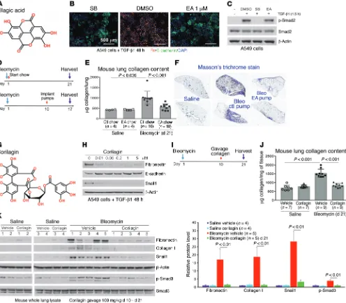
[image:3.585.43.539.56.491.2]Several small-molecule libraries totaling approximately 40,000 compounds composed of both diverse and bioactive compounds were screened. We identified ellagic acid (EA) as one compound meeting our criteria (Figure 1, A–C, and Supplemental Figure 1A; supplemental material available online with this article; https:// doi.org/10.1172/JCI94624DS1). We next examined the structural
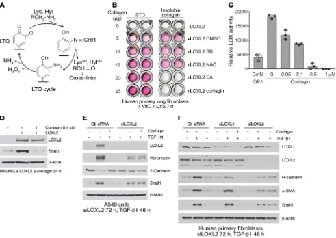
of lysine primary amines (refs. 33, 34, and Figure 3A). The catalytic site quinone oxidizes the primary amines of collagen lysine (Lys) and hydroxylysine (Hyl) residues, releasing the respective aldehydes, Lysald and Hylald, creating an intermediate aminophenol followed by
release of H2O2 and NH3, completing the LTQ cycle. Lysald and Hylald
in monomeric collagens then undergo a series of spontaneous con-densation reactions that result in the formation of intra- and intermo-lecular covalent collagen cross-links (35).
We first established an assay of collagen cross-linking induced by recombinant LOXL2 (36, 37) and found that corilagin and all other trihydroxyphenolics tested prevented cross-linking (IC50 = 10 nM; Supplemental Figure 3, A–C) whereas an inhibitor of TGF-β1 signaling (SB431542) and an antioxidant (N-acetylcysteine) had no effect (Figure 3B). LOXL2 enzymatic activity toward a model substrate was assessed monitoring H2O2 release. Corilagin blocked this activity with an IC50 of approximately 50 nM (Figure 3C). LOXL2-induced stabilization of Snail1 protein was also blocked by corilagin (Figure 3D). To test the possibility that the putative anti-oxidant activity of trihydrophenolics could either account for the observed inhibition of TGF-β1 signaling (38) or directly neutralize H2O2 in our LOXL2 enzyme assay, we defined the concentrations of corilagin that neutralized H2O2 activity in vitro (Supplemen-tal Figure 4). We observed no inhibition of H2O2 interaction with a reporter substrate by corilagin at ≤10 μM, whereas vitamin C neutralized H2O2 at submicromolar concentrations, confirming that direct antioxidant scavenging activity could not account for our corilagin findings.
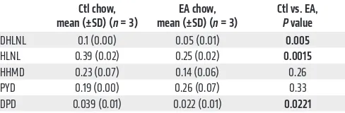
To assess inhibition of LOXL2 activity in vivo, we performed biochemical analyses of collagens obtained from primary 344SQ tumors of mice treated with EA or control chow (refs. 35, 39, and Figure 2A). Accumulation of links including 2 reducible cross-links, HLNL and DHLNL, and a nonreducible, mature cross-link, deoxypyridinoline (DPD), was all significantly decreased in prima-ry tumors of mice fed EA chow, consistent with the altered collagen organization in the primary tumors demonstrated by quantitative analysis of second-harmonic generation (Table 2 and Figure 2I), and confirming inhibition of cross-linking activity by trihydroxy-phenolics in vivo. Long-term EA chow treatment (6 months) did not affect mouse total bone mineral density or collagen content in the aorta (Supplemental Figure 3, D–F), indicating that not all LOX family members were affected by the active compounds.
LOXL2 activity confers inhibition of TGF-β1 signaling by trihy-droxyphenolics. To further interrogate the impact of LOXL2 inhibi-turally identical aside from a dihydroxy- rather than the
trihydroxy-phenolic motif in EC (Supplemental Figure 1C). EC had no activity in our in vitro assays, whereas EGC was a potent inhibitor of Snail1 and fibronectin in TGF-β1–stimulated A549 cells.
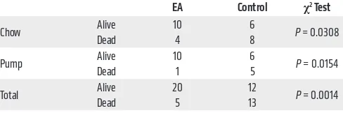
EA was then tested for its antifibrotic activity in vivo by either ad libitum feeding of chow composed of 2% wt/wt raspberry extract rich in EA and EA precursors given to mice or adminis-tration of EA using osmotic pump (days 10–17) after intratracheal bleomycin (Figure 1D). We found that either treatment substan-tially improved survival (Table 1) and inhibited collagen accumu-lation (Figure 1, E and F). Because EA is poorly soluble, a more soluble trihydroxyphenolic-containing compound, corilagin, with an IC50 for EMT of approximately 50 nM (Figure 1, G and H), was given daily by gavage beginning 10 days after intratracheal bleo-mycin (Figure 1I). At day 21 these mice exhibited marked attenua-tion of bleomycin-induced total lung collagen, fibronectin, Snail1, and p-Smad3 (Figure 1, J and K). The average circulating level of corilagin 2 hours after the last dose was about 80 nM (Supple-mental Figure 1E). EA-rich chow and corilagin had no effect on immune cell numbers or markers of injury (Supplemental Figure 2). Collectively, these findings demonstrate that trihydroxypheno-lic compounds attenuate TGF-β1–induced Snail1 and EMT markers in vitro as well as collagen accumulation in vivo and do so at low nanomolar levels. Members of this polyphenol family have previ-ously been shown to inhibit TGF-β1 signaling at micromolar levels in vitro and fibrosis in vivo but by unclear mechanisms (27, 28).
To test the efficacy of EA in a second in vivo model of tissue fibrosis, we examined the occurrence of metastatic lung nodules in mice injected subcutaneously 5 weeks earlier with syngene-ic KrasG12D/p53R172H metastatsyngene-ic lung cancer cells (344SQ), known to metastasize as a function of the cross-linked fibrillar collagen content of the primary tumors (Figure 2A) (29). Con-sumption of EA-rich chow following tumor implantation mark-edly reduced the numbers of metastatic lung nodules (Figure 2, B and C). Although primary tumor volume or weight was unchanged (Figure 2, D and E), immunohistochemistry showed significantly reduced collagen I expression within the primary tumors treated with EA chow (Figure 2F). Furthermore, immunoblotting of these tumor extracts also revealed attenuated total fibronectin and col-lagen I expression, and decreased Smad activation, assessed by p-Smad3 (Figure 2G). Interestingly, visualizing collagen in situ by second-harmonics microscopy, we observed that the primary tumor collagen in mice fed EA chow was not only reduced but also exhibited more curved structures, suggesting less cross-linking (Figure 2, H and I, and ref. 9).
[image:4.585.303.547.96.177.2]LOXL2 is identified as the target of trihydroxyphenolic-containing compounds. We next turned to underlying mechanisms that could account for the activities and potency of the polyphenolic com-pounds. Because of the striking inhibition of Snail1 expression by several trihydroxyphenolic compounds (Figure 1H and Supplemental Figure 1B), as well as the altered collagen cross-linking structure in primary 344SQ tumors (Figure 2I), we explored the hypothesis that lysyl oxidase–like 2 (LOXL2) was their target. LOXL2 has previously been linked to Snail1 accumulation in tumor cells (30), and its expres-sion is potently induced by both hypoxia and TGF-β1 (31, 32). LOXL2, like all mammalian copper-dependent LOX enzymes, utilizes an intrinsically generated quinone, termed LTQ, to mediate oxidation
Table 1. The survival of bleomycin-treated mice by day 21 of EA chow and day 17 of EA pump treatment
EA Control χ2 Test
Chow Alive 10 6 P = 0.0308
Dead 4 8
Pump Alive 10 6 P = 0.0154
Dead 1 5
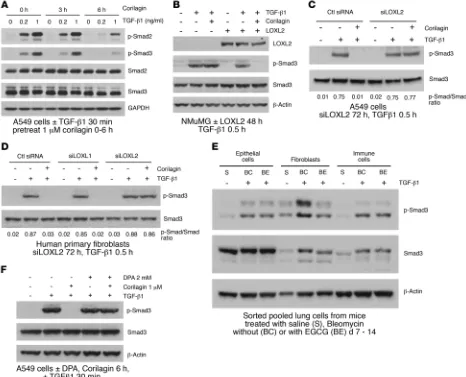
Smad activation and Snail1 induction (Figure 4F and Supplemental Figure 5, D–F). Together these data indicate that trihydroxypheno-lic compounds selectively target proximal TGF-β1 signaling and Snail1 accumulation only in cells expressing LOXL2 and do so by a mechanism requiring the presence of active LOXL2.
To further investigate the specificity of trihydroxyphenolics for TGF-β1 signaling, we asked whether corilagin inhibited other kinases at 1–10 μM, well above the IC50 for its inhibitory effects on TGF-β1 signaling (Figure 1H). In a screen of 82 purified kinas-es conducted at the Km for ATP binding for each kinase, only the tyrosine kinases EGFR and PDGFRβ were inhibited more than 50% at 1 μM (Supplemental Figure 8A). However, when we specifically tested the inhibitory effects of corilagin at 1 μM on either EGFR or PDGFRβ activities of intact cells, neither were inhibited by corilagin, implying that the cell-free kinase screen is more sensitive than the inhibition of these enzymes in intact cells (Supplemental Figure 8, B and C). These data confirm that corilagin is not a nonspecific kinase inhibitor, at least at concentrations less than or equal to 1 μM.
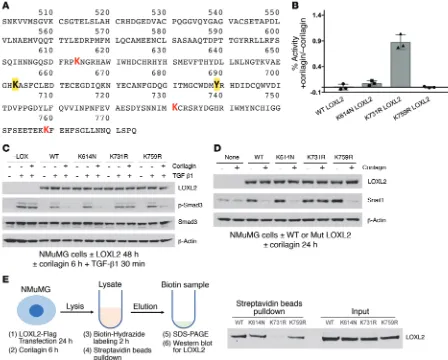
Trihydroxyphenolics induce auto-oxidation of LOXL2 lysine 731. Given the structural similarities between the trihydroxyphenolic motif and LTQ (Figure 3A), the results described above raised the possibility that the trihydroxyphenolic motif operates as an LTQ-like mimic, leading to its metabolism by LOXL2 and generating an inhibitor of TGF-β1 signaling. To begin testing this hypothe-sis we inspected the LOX catalytic domain for lysines specific to LOXL2 and not found in LOXL1 (Figure 5A), whose silencing had no impact on corilagin responsiveness (Figure 3F and Figure 4D). tion on TGF-β1 responses, we suppressed LOXL2 levels with RNAi
in A549 cells. Surprisingly, rather than inhibiting TGF-β1, silenc-ing LOXL2 completely abrogated the inhibitory effects of corilagin on TGF-β1–induced EMT in A549 cells (Figure 3E). Further, LOXL2 but not LOXL1 silencing in fibroblasts completely prevent-ed the corilagin inhibitory effects on the TGF-β1–induced mesen-chymal proteins N-cadherin, α-smooth muscle actin, and Snail1 (Figure 3F). These findings revealed that the corilagin mechanism of action was not simply LOXL2 inhibition, prompting us to revisit corilagin effects on TGF-β1 signaling.
Because trihydroxyphenolic compounds did not block TGF-β1– induced p-Smad generation in short-term assays (Figure 1C), we considered the possibility that impaired TGF-β1 signaling was due to defective p-Smad nuclear import, but immunostaining revealed no blockade of Smad2/3 nuclear translocation within 2 hours of compound treatment. However, longer preincubation of cells with corilagin (or other trihydroxyphenolics) for several hours com-pletely suppressed Smad activation (Figure 4A and Supplemental Figure 5, A–C), implying that a degree of ongoing p-Smad gener-ation is required for regulgener-ation of the TGF-β1 gene targets studied here (Figure 1H). Similarly, expression of LOXL2 in NMuMG cells that normally express very low levels of LOXL2 conferred corilagin responsiveness and blockage of p-Smad3 following 6-hour pre-treatment (Figure 4B). Conversely, silencing LOXL2 in A549 cells and primary lung fibroblasts completely blocked corilagin effects on p-Smad3 generation (Figure 4, C and D). Consistent with a critical role for LOXL2 in proximal TGF-β1 signaling, a survey of numerous cell lines revealed a direct correlation of LOXL2 mRNA levels with the degree of inhibition of TGF-β1–induced Smad acti-vation by corilagin (Supplemental Figure 6). To further test this principle in vivo, epithelial, fibroblast-rich mesenchymal, and immune cells were isolated by flow cytometry from pools of normal lungs and lungs of mice exposed 14 days earlier to bleomycin, then immediately stimulated with TGF-β1 and tested for their degree of p-Smad accumulation as a marker of TGF-β1 signaling (Figure 4E). There was a marked attenuation of p-Smad3 in the fibro-blast-rich lung fraction of bleomycin-exposed mice also given oral epigallocatechin-3-gallate (EGCG; 100 mg/kg daily) but no dis-cernible inhibition of p-Smad3 in either the epithelial or immune cell fractions. A second experimental design in which the isolated fractions from pools of normal lungs and lungs of mice exposed 14 days earlier to bleomycin fed with control or EA chow demon-strated the same pattern (Supplemental Figure 7), confirming the cell selectivity of trihydroxyphenolics on TGF-β1 signaling in vivo. Finally, we confirmed that inhibition of active LOXL2 by the copper chelator d-penicillamine (DPA) also abrogated corilagin effects on
Figure 2. EA-rich diet attenuates 344SQ lung tumor metastasis and primary tumor collagen cross-linking. (A) Implantation and treatment in syngeneic lung cancer model. Metastatic 344SQ tumor cells were subcutaneously injected in syngeneic mice at 12 weeks old and treated with ctl or EA chow for 5 weeks. (B) Representative pictures of lung metastasis of 344SQ tumors treated with ctl or EA chow. (C) Quantification of lung metastasis of 344SQ tumors treated with ctl or EA chow (n = 14 mice per group). (D)Total volume of 344SQ tumors treated with ctl or EA chow (n = 14 mice per group).(E)Total tumor weight (grams) of ctl or EA chow–treated 344SQ primary tumors (n = 14 mice per group). (F) Collagen I immunohistochemistry (IHC) of 344SQ primary tumors treated with ctl or EA chow. Scale bars: 50 μm.(G) Ctl or EA chow–treated 344SQ primary tumors were lysed and immunoblotted for fibronectin, collagen I, β-actin, p-Smad3, and total Smad3. Quantification of bands normalized to β-actin was pooled from 10 mice per group. Data represent mean ± SD. Each protein was analyzed by 1-way ANOVA with a Tukey post hoc test. (H) Representative second-harmonic generation (SHG) images from 344SQ primary tumors treated with ctl or EA chow. Scale bars: 50
[image:6.585.303.547.573.653.2]μm.(I)Quantification of curvature ratio for individual collagen fibers imaged by SHG microscopy of primary 344SQ tumor tissues treated with ctl (n = 102 collagen fibers per sample) or EA chow (n = 141 collagen fibers per sample). Data for C–E and I are expressed as mean ± SD. P value by unpaired 2-tailed t test.
Table 2. Collagen cross-links in 344SQ tumors treated with ctl or EA chow (moles per mole of collagen)
Ctl chow,
mean (±SD) (n = 3) mean (±SD) (EA chow, n = 3) Ctl vs. EA, P value
DHLNL 0.1 (0.00) 0.05 (0.01) 0.005
HLNL 0.39 (0.02) 0.25 (0.02) 0.0015
HHMD 0.23 (0.07) 0.14 (0.06) 0.26 PYD 0.19 (0.00) 0.26 (0.07) 0.33 DPD 0.039 (0.01) 0.022 (0.01) 0.0221
LOXL2 mediates the conversion of Lys to Lysald, which is ultimately processed
to HHMD. LOXL2 also mediates the conversion of Hyl to Hysald. Hylald can then
be converted into DHLNL and subsequently PYD, Hylald can be converted to
The Journal of Clinical Investigation
R E S E A R C H A R T I C L EWe then established and expressed Flag-tagged point mutants of each of 3 LOXL2/3–specific lysines, converting each to the corre-sponding LOXL1 residues: K614N, K731R, and K759R. Each of the point mutants had comparable enzyme activity to WT LOXL2 when expressed in NMuMG cells (Supplemental Figure 9A), and all except the K731 mutant were completely inhibited by corilagin (Figure 5B). Cells expressing K731R LOXL2 were completely resistant to inhibition of TGF-β1 signaling (Figure 5C) and Snail1 stabilization (Figure 5D) by corilagin, whereas the other mutants were indistinguishable from WT. Because lysine auto-oxidation by LOX family enzymes is critical to LTQ generation, we asked whether K731 was auto-oxidized to an aldehyde in the presence of corilagin. Incubation of a biotin hydrazide that covalently links to free aldehydes with immunoprecipitated WT and mutant LOXL2 confirmed that each enzyme except the K731R mutant developed
an aldehyde when mixed with corilagin, implying that K731 but not other lysines is converted to an aldehyde during metabolism of corilagin by LOXL2 (Figure 5E).
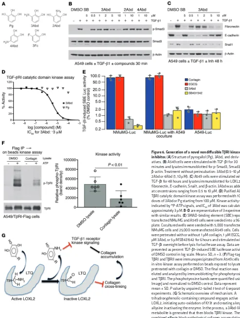
A novel TβRI kinase inhibitor is generated inside LOXL2-expressing cells. We next screened compounds structurally similar to the inter-mediate aminophenol known to appear during the LTQ cycle (Fig-ure 3A) for direct TGF-β1 inhibition. A catechol containing an amino group at position 3 (3Abd, 3-aminobenzene-1,2-diol) but not at either position 2 (2Abd) or 4 (4Abd) was found to be a potent inhibitor of TGF-β1–induced Smad3 activation and Snail1 expression without preincubation and regardless of LOXL2 expression (Figure 6, A–C, and Supplemental Figure 9B). We confirmed that 3Abd, but not con-trol pyrogallol or 2Abd, directly blocked the kinase activity of recom-binant TβRI catalytic domain with an IC50 of approximately 3 μM (Figure 6D and Supplemental Figure 9C), consistent with the
inhibi-Figure 3. Identification of LOXL2 as the target of EA and corilagin; requirement for active LOXL2 for corilagin-induced inhibition of EMT and Snail expression. (A) LTQ cycle. LTQ converts lysine to allysine and yields an aminophenol intermediate. Subsequent hydrolysis release allysine and the original cofactor, producing hydrogen peroxide and ammonia as side products. (B) Primary human lung fibroblasts cultured in the presence of vitamin C and dextran sulfate were treated with recombinant human LOXL2 and different inhibitors for 7 days. The insoluble cross-linked collagen was extracted and measured by Sircol assay. SB, SB431542, TβRI inhibitor; NAC, N-acetylcysteine, antioxidant. STD, standard. (C) Recombinant human LOXL2 was incubated with 2 mM d-penicillamine (DPA) or different concentrations of corilagin (0–1 μM) for 1 hour, and LOX activity was measured. Data represent mean ± SD; n = 3. (D) NMuMG cells overexpressing human LOXL2 were incubated with or without 0.5 μM corilagin for 24 hours, lysed, and immunoblot-ted for LOXL2, Snail1, and β-actin. (E) A549 cells transfected with siRNA to LOXL2 were stimulated with TGF-β1 or left unstimulated for 48 hours in the presence or absence of 1 μM corilagin. The lysates were immunoblotted for LOXL2, fibronectin, E-cadherin, Snail1, and β-actin. (F)Primary human lung fibroblasts transfected with siRNAs to LOXL1 or LOXL2 were stimulated with TGF-β1 or left unstimulated for 72 hours in the presence or absence of 1
[image:7.585.40.531.54.402.2]tory profile of 3Abd in cells (Figure 6C). In cells overexpressing TβRI we also observed that 3Abd but not control 3Fc (3-Fluorocatechol) blocked the kinase activity of immunoprecipitated TGF-β receptors (Supplemental Figure 9D). Notably, 3Abd is structurally distinct from any of the known low–molecular weight inhibitors of TβRI (18).
To further define the mechanism of TGF-β1 inhibition by tri-hydroxyphenolic compounds, we asked whether secreted LOXL2 generated active 3Abd-like metabolites. Overnight coculture of corilagin-treated A549 cells with corilagin-nonresponsive NMuMG cells (Figure 4B) expressing a Smad3 reporter (12X CAGA) (40) revealed indistinguishable TGF-β1–induced reporter activation with or without 5-fold excess A549 cells in coculture, indicating that the generation of a diffusible inhibitor was unlikely (Figure 6E). In addition, lysates of corilagin-treated A549 cells,
but not that of untreated cells, inhibited the kinase activity of immunoprecipitated TGF-β receptors (Figure 6F). These results point to an intracellular origin of a trihydroxyphenolic metabo-lite(s) inhibiting TβRI kinase (Figure 6G). While we demonstrated that the small fragment 3Abd that would result from a trihydroxy-phenolic acting through an LTQ-like mechanism (and not pyrogal-lol) directly inhibits TβRI kinase (Figure 6D), future studies will be needed to isolate the exact inhibitory metabolite(s) present within trihydroxyphenol-treated LOXL2-expressing cells.
Discussion
These studies reveal, for the first time to our knowledge, a pathway of inhibition of the TGF-β1–induced collagen program selective to the cells that are most accountable for pathological collagen
[image:8.585.57.523.57.434.2]The Journal of Clinical Investigation
R E S E A R C H A R T I C L Ethat of previously described extracellular LOXL2 inhibitors as well as global LOX inhibitors, accounting for the lack of negative impact of long-term exposure to EA-rich compounds on bone or vascular collagen content (Supplemental Figure 3, D–F, and refs. 34, 41). Maintenance of musculoskeletal and vessel wall collagen and elastin integrity depends on LOX and LOXL1, not LOXL2 (42–45). Although selective, the combined inhibition of LOXL2 and TGF-β1 signaling in fibroblast-like cells results in potent in vivo antifibrotic activity that has untapped but promising potential as a therapeutic approach for chronic diseases such as pulmonary fibrosis dominated by progressive collagen accumulation.
The combined inhibition of LOXL2 and TGF-β1 signaling in fibroblast-like cells could be expected to impact biomarkers of col-lagen turnover in vivo. Indeed, we observed an increase in urinary levels of pyridinoline (PYD) and DPD, nonreducible end products of cross-linked collagen metabolism, in mice 10–21 days after sition, tissue fibroblasts and fibroblast-like tumor cells. The
[image:9.585.63.511.56.416.2]sur-prising finding that such selectivity depends on active LOXL2/3 and appears to operate in a cell-autonomous manner largely abro-gates inhibition by trihydroxyphenolic-containing compounds of TGF-β1 signaling in epithelial or immune cells that don’t express LOXL2/3. This selectivity likely avoids the toxicities of long term general TGF-β1 inhibition in chronic disease processes such as fibrosis and cancer progression. Indeed, we have observed no adverse events in mice on the trihydroxyphenolic-rich diet (EA chow) for at least 6 months, including the absence of skin inflam-mation and discernible lesions in cardiac valves. Likewise, none of the compounds tested here had any negative effects on cell viabil-ity in vitro at concentrations up to 10 μM of trihydroxyphenolics or 50 μM of 3Abd (Supplemental Figure 10). The selective inhibition of intracellular and extracellular LOXL2/3 by trihydroxyphenolics also distinguishes this mechanism of LOX family inhibition from
Figure 6. Generation of a novel nondiffusible TβRI kinase inhibitor. (A) Structure of pyrogallol (Pg), 3Abd, and deriv-atives. (B) A549 cells were stimulated with TGF-β1 for 30 minutes and lysates immunoblotted for p-Smad3, Smad3, and
β-actin. Treatment without preincubation: 3Abd (0.5–10 μM); 2Abd or 4Abd (1, 10 μM). (C) A549 cells were stimulated with TGF-β1 for 48 hours and lysates immunoblotted for LOXL2, fibronectin, E-cadherin, Snail1, and β-actin. 3Abd was added at concentraions ranging from 0.5 to 10 μM. (D)Purified ALK5/ TβRI catalytic domain kinase assay was performed with 10 doses of 3Abd or Pg starting from 100 μM. Kinase activity was indicated by 33P-ATP signals, and IC
50 of 3Abd was calculated as
approximately 3 μM. B–D are representative of 3 experiments with similar results. (E) SMAD-binding element (SBE) reporter– transfected NMuMG and A549 cells were seeded into a 96-well plate. Cocultured wells were seeded with 5,000 transfected NMuMG cells and 25,000 nontransfected A549 cells. Cells were pretreated with or without 1 μM corilagin, 1 μM EGCG, 10
The Journal of Clinical Investigation
R E S E A R C H A R T I C L E(100-21) was from PeproTech. 3-Aminobenzene-1,2-diol (3Abd; W4593) was from Aurum Pharmatech Inc. 2-Aminobenzene-1,3-di-ol (2Abd; 23488), 4-aminobenzene-1,2-di2-Aminobenzene-1,3-di-ol (4Abd; 31975), and 3- fluorocatechol (CL8492) were from AstaTech Inc. Human recombi-nant LOXL2 (2639-AO-010) was purchased from R&D Systems.
Cell culture. Human or mouse cell lines were purchased from
ATCC and grown in DMEM or RPMI 1640 medium supplemented with l-glutamine and 10% FBS (Hyclone). Human and mouse lung fibroblasts were isolated from crude whole-lung single-cell suspen-sion cultures on Petri dish in DMEM supplemented with l-glutamine and 10% FBS for 2 weeks. Mouse type II alveolar epithelial cell isola-tion and culture were performed as previously described (57). All the cell lines in the laboratory are periodically tested for mycoplasma con-tamination. Only the mycoplasma-free cells are used for experiments.
High-throughput screen and high-content imaging analysis. A549
cell–based screening for inhibitors of TGF-β1–induced EMT from small-molecule libraries was performed in 384-well plate format, and the images were captured and analyzed using GE IN Cell 2000 as described previously (26).
Immunofluorescence. Cultured cells and 5- to 7-μm cryosections were fixed in 4% paraformaldehyde and stained with various antibod-ies and IgG isotype controls. Where indicated in the figure legends, mosaic images were generated from multiple ×20 images captured on a Zeiss Axio upright fluorescent microscope and tiled using 10% image overlap by Axiovision 4.7 software (Zeiss).
Masson’s trichrome stain. For histological assessment of lung collagen,
frozen sections of the left lung were stained using Masson’s trichrome stain kit (22-110-648, Thermo Fisher Scientific). The whole section was imaged with a Zeiss Axio upright microscope and tiled using 10% image overlap into a single panoramic by Axiovision 4.7 software (Zeiss).
Immunoblot. Pulverized tissue and cells were lysed in RIPA buffer
(150 mM NaCl, 50 mM Tris, pH 8.0, 1% Triton X-100, 0.5% sodium deoxycholate, 0.1% SDS, supplemented with protease and phospha-tase inhibitors) and analyzed by immunoblotting. Densitometry was quantified using NIH ImageJ software. See complete unedited blots in the supplemental material.
Bleomycin fibrosis model. Eight-week-old C57BL/6 mice were
intratra-cheally instilled with saline or 1.9 units/kg of bleomycin (Sigma-Aldrich). Mice were implanted with Alzet osmotic pumps (1007D, DURECT Corp.) loaded with EA salt (24 mg/kg/d, days 10–17), fed with red raspberry diet (EA chow, days 0–21), or gavaged with corilagin (100 mg/kg, days 10–21). Controls were treated with control pump, control diet, or vehicle in the same formulation. Red raspberry diet (TD.130761) and red control diet (TD.150279) was custom-made by Envigo. The lungs were lavaged, and then embedded in OCT compound for imaging or snap-frozen in liquid nitrogen for protein extraction or hydroxyproline assay.
Syngeneic in vivo tumorigenesis and metastasis assays. KrasG12D/
p53R172H metastatic lung cancer cells (344SQ) were subcutaneously injected in the right flanks of male, syngeneic 129/Sv mice at 3 months of age and allowed to form tumors for 5–6 weeks (29). The mice were fed with red raspberry diet or control diet. After euthanasia, tumors were measured and lung metastatic nodules were quantified. Pri-mary tumor tissues were snap-frozen and analyzed by Western blot. Some primary tumors were formalin fixed, paraffin embedded, and sectioned for immunohistochemistry or second-harmonic generation imaging. The investigators were completely blinded to drug treat-ment and outcome assesstreat-ment.
bleomycin injection, and this increase was suppressed by treat-ment of the mice with corilagin (Suppletreat-mental Figure 11A). Con-sistent with these findings, we observed increased mean urinary PYD/DPD levels in 2 cohorts of patients with idiopathic pulmo-nary fibrosis (Supplemental Figure 11B), suggesting that a signal from fibrotic lungs is present in most of these patients and may enable tracking of collagen turnover and drug responses in vivo.
The biological pathway identified here uses trihydroxy- containing polyphenols at concentrations achievable by dietary ingestion. Indeed, foods rich in this class of compounds, such as EGCG (the major polyphenol in green tea), have been consumed as therapeutics for decades (46). Yet these compounds are not generally thought of as workable drugs because of their potential for pro- and antioxidant reactions that could negatively impact pathways, such as drug metabolism, sensitive to such reactions (47). The designation of these polyphenols as antioxidants and as reactive compounds, however, largely stems from prior stud-ies that have used micromolar levels of polyphenols to achieve in vitro “antioxidant” or signaling inhibition in multiple cell sys-tems (48), even though blood levels above approximately 150 nM have not been documented for dietary polyphenols consumed by humans (49–53). As well, we observed no neutralization of H2O2 oxidant activity by corilagin at concentrations below 10 μM (Sup-plemental Figure 4). We believe the LOXL2-dependent interac-tion with trihydroxyphenolics is a singular example of a relevant protective pathway activated by nanomolar levels of the relevant ellagitannin and catechin subclasses, possibly contributing to the observed beneficial effects of green tea and other trihydroxyphe-nolic-rich diets in numerous population studies (54–56).
Methods
Reagents. Ellagic acid (EA; E2250), epigallocatechin gallate (EGCG;
cence plate reader (BMG LabTech FLUOstar) at excitation (Ex)/emis-sion (Em) = 540/590 nm. Sample buffer or medium alone without LOXL2 was used for determination of the background fluorescence.
Collagen cross-link analysis. Snap-frozen primary 344SQ tumors
were pulverized in liquid nitrogen using a Spex Freezer Mill (Spex), washed with cold PBS and cold distilled water, lyophilized, and weighed. Aliquots were reduced with standardized NaB3H
4 and hydro-lyzed with 6N HCl. The hydrolysates were then subjected to amino acid and cross-linking analyses using liquid chromatography–tandem mass spectrometry (LC–MS/MS) as described previously (59). The terms DHLNL, HLNL, and HHMD represent both the unreduced and reduced forms. The mature trivalent cross-links, PYD and DPD, were simultaneously analyzed by their fluorescence. All cross-links were quantified as the moles per mole collagen based on the value of 300 residues of hydroxyproline per collagen molecule.
Urinary PYD/DPD measurements. Pooled urine from each of 3–5
mice for each time point after bleomycin in a cohort of mice treated with vehicle or corilagin (100 mg/kg) beginning on day 10 after bleomycin was collected. Urine specimens were also collected from 2 cohorts of idiopathic pulmonary fibrosis patients and controls at 2 sites: The Uni-versity of Texas Health Science Center at San Antonio and UCSF. All consenting patients with physician-established diagnosis of idiopathic pulmonary fibrosis followed in the respective interstitial lung disease (ILD) programs were included in sample collection. PYD/DPD levels from all the samples were measured using MicroVue EIA Assay Kit (catalog 8010) and MicroVue Creatinine EIA Kit (catalog 8009, Quidel Corp.) along with PYD/DPD standards, and the results were normal-ized relative to urinary creatinine. The statistical significance of human urine samples was analyzed using Mann-Whitney U test.
Bone mineral density measurement. Bone mineral density of mice
treated with red raspberry diet or control diet up to 6 months was mea-sured using dual-energy X-ray absorptiometry (DEXA) scan. DEXA scans were performed using the Lunar PIXImus Densitometer (GE Medical Systems) at the UCSF animal facility. The PIXImus Densitom-eter was calibrated before each testing using a quality control phantom following the manufacturer’s instructions.
Elastic Van Gieson stain. Aortas isolated from mice treated with
red raspberry diet or control diet up to 6 months were embedded in paraffin (n = 3 per group). Sections (5 μm) were cut every 30 μm along the aortas (starting from the proximal end). Selected sections were stained with Miller’s Elastica Van Gieson stain.
Site-directed mutagenesis of LOXL2. Site-directed mutagenesis was
performed to generate K614N, K731R, and K759R point mutations using Phusion Site-Directed Mutagenesis Kit (F541, Thermo Fisher Scientific) according to the manufacturer’s instructions. A pcDNA3-hLOXL2-Flag plasmid containing the cDNA fragment of WT human LOXL2 fused with a Flag tag at the C-terminus (gift from Amparo Cano, Instituto de Investigaciones Biomédicas, Universidad Autónoma de Madrid/Consejo Superior de Investigaciones Científicas, Madrid, Spain) (30) was used as the template DNA (30). Mutations were con-firmed by DNA sequencing. The primers and their complementary strands used were: K614N forward 5′ -GACTTCCGGCCTAATA-ATGGCCGC-3′, K614N reverse 5′ -GGACTGGCCATTGTTGTG-GATCTG-3′; K731R forward 5′ -ACAACATCATACGATGCAGGAG-CC-3′, K731R reverse 5′-TGGAGTAATCGGATTCTGCAACCT-3′; K759R forward 5′-ACGGAAAAACGTTTTGAGCACTTCA-3′, and K759R reverse 5′-CTCTTCGCTGAAGGAACCACCTAT-3′.
Immunohistochemistry and second-harmonic generation micros-copy. Paraffin-embedded tissue sections were rehydrated, blocked
with goat serum, and probed for collagen I. Tissues were subse-quently washed and probed with HRP-conjugated secondary anti-bodies, and signal was attained by developing with a DAB reagent. Collagen cross-linking alteration was evaluated by second-harmon-ic generation msecond-harmon-icroscopy. Tissues stained by H&E were visualized using a Zeiss LSM 7 MP Multiphoton Microscope at an excitation wavelength of 800 nm, and collagen fiber signals were detected at 380–430 nm using bandpass filters. Collagen linearity was calculat-ed as a ratio of the total length versus the end-to-end length of the individual collagen fiber.
Collagen content. Lung or aorta collagen content was
evaluat-ed using hydroxyproline assay (58). Briefly, whole left lung tissue or aorta was hydrolyzed in 1 ml 12N HCl at 110°C for 24 hours, the hydroxyproline was detected by incubation with chloramine T and
p-dimethylaminobenzaldehyde, and the absorbance was measured
at 550 nm. Each sample was run in triplicate. Collagen content in lung or aorta tissues was expressed as micrograms of collagen per lung or aorta and was converted from micrograms of hydroxyproline.
Bronchoalveolar lavage. After the trachea was exposed, a 20-gauge
catheter was inserted into the trachea through a small incision. One milliliter cold PBS was instilled into the mouse lungs followed by gentle aspiration, and this was repeated 2 more times. All the bronchoalveolar lavage fluid (BALF) was centrifuged, and the cell pellet was resuspended in erythrocyte (rbc) lysis buffer (Sigma-Aldrich) followed by recentrif-ugation. Cell number was counted using a hemocytometer. Cell types of BALF were determined by morphology following Diff-Quik stain of cytospin slides. About 500 cells were counted for each sample in order to determine the cell types. Macrophages account for more than 80% of the cells in BALF and were collected by centrifugation. After centrifugation, the supernatant was collected to measure total protein content using the BCA assay (23225, Pierce), while the cell pellet was lysed for immunoblot-ting or RNA isolation.
Plasma level of corilagin. Plasma level of corilagin in C57BL/6 mice
2 hours following the last oral administration at day 21 was analyzed by Quintara Discovery. Blood samples (~500 μl/sample) were collected via cardiac puncture. Samples were placed in tubes containing heparin sodium and stored on ice until centrifuged for plasma.
Preparation of insoluble cross-linked collagen. Fibroblasts were
cul-tured on a 10-cm dish until confluent. The medium was then changed to DMEM containing 5% FBS, 100 μM l-ascorbic acid with 500 kDa dextran sulfate at 100 μg/ml, and 50 ng/ml recombinant human LOXL2 for 7 days (36, 37). The cell layer was extracted with 0.5 M ace-tic acid and 0.1 mg/ml pepsin overnight at 4°C. The leftover insoluble fraction was further extracted and the insoluble cross-linked collagen measured using Sircol Insoluble Collagen Assay kit (S1000, Biocolor) according to the manufacturer’s instruction.
LOX activity assay. LOX activity of recombinant human LOXL2 or
conditioned medium collected from cells expressing LOXL2 was mea-sured using a Fluorometric Lysyl Oxidase Activity Assay Kit (ab112139, Abcam) following a protocol provided by the manufacturer. Briefly, 50
fluores-The Journal of Clinical Investigation
R E S E A R C H A R T I C L Eincubated for 1 hour at 4°C with the following primary antibodies: rat anti–mouse CD45–APC-Cy7 (1:100; BD, 557659), rat anti–mouse PeCAM–PE (1:100; BD, 553373), rat anti–mouse EpCAM–Alexa Fluor 488 (1:250; BioLegend, 118210), and viability dye Sytox blue (1:1,000; Thermo Fisher Scientific, S34857). Cell sorting was performed on BD FACS Aria cytometers. EpCAM+ and CD45+ cells were collected, respectively. EpCAM/CD45/CD31–triple-negative cells were collected as mesenchymal cells. Each of the 3 cell types sorted from saline con-trol, bleomycin concon-trol, and bleomycin EGCG groups (n = 5) were lysed and blotted for p-Smad3 and total Smad3.
Protein and lipid kinase screen. Protein kinase assays were conducted
using the KinaseProfiler service of Eurofins Pharma Discovery Services UK Ltd. The kinase of interest was incubated with the test compound in assay buffer containing substrate, 10 mM magnesium acetate, and [γ-33P-ATP]. The reaction was initiated by the addition of the Mg/ATP mix. After incubation at room temperature, the reaction was stopped by the addition of a 3% phosphoric acid solution. An aliquot of the reaction was then spotted onto a filtermat and washed in phosphoric acid fol-lowed by a rinse in methanol prior to drying and scintillation counting. Results were expressed in relation to controls containing DMSO only in place of test compound. The ATP concentration in each assay was within 15 μM of the determined apparent Km for ATP.
Lipid kinase assays were conducted using the KinaseProfiler ser-vice of Eurofins Pharma Discovery Serser-vices UK Ltd. The kinase of interest was incubated in assay buffer containing substrate and Mg/ ATP. The reaction was initiated by the addition of the Mg/ATP solu-tion. After incubation for 30 minutes at room temperature, the reac-tion was stopped by the addireac-tion of stop solureac-tion containing EDTA and a biotinylated form of the reaction product. Finally, detection buffer was added, containing europium-labeled anti-GST monoclo-nal antibody, a GST-tagged lipid binding domain, and streptavidin- conjugated allophycocyanin. The plate was then read in time-resolved fluorescence mode, and the homogeneous time-resolved fluores-cence (HTRF) signal was determined according to the formula HTRF = 10,000 × (Em665nm/Em620nm).
H2O2 scavenging assay. The oxidant scavenging activity of corilagin
and vitamin C was measured using Amplex Red Hydrogen Peroxide/ Peroxidase assay kit (Thermo Fisher Scientific, A22188) following the manufacturer’s instructions. H2O2 (0.5 μM) samples were incubated with different concentrations of corilagin (0–100 μM) and vitamin C (0–10 μM) for 2 hours at room temperature before Amplex Red/HRP reaction and the plate read at Ex/Em 540/590 nm with a fluorescence plate reader (BMG LabTech FLUOstar). Compound vehicle was used for determination of the background fluorescence. Experiments were performed in triplicate, and data are presented as means ± SD.
Cell viability assay. AlamarBlue cell viability reagent (Thermo
Fisher Scientific, DAL1025) was used to assess cell viability. A549 or human primary lung fibroblasts (20,000 cells) in complete RPMI or DMEM medium were seeded into a 96-well tissue culture plate overnight; then the cells were exposed to different compounds for 72 hours. AlamarBlue reagent (1:10) was added to the cells, followed by 2 hours of incubation at 37°C, and absorbance at 570 nm was measured. Experiments were performed in triplicate using different batches of cells, and data are presented as means ± SD.
Statistics. Descriptive statistics are reported as means ± SD. For
evaluation of differences between compound treated and control groups, the unpaired 2-tailed Student’s t test or Mann-Whitney U
Biotin hydrazide derivatization of carbonylated LOXL2. NMuMG
cells were transiently transfected with WT or mutant human LOXL2-Flag in a 10-cm dish, and 24 hours later the cells were treated with 1
μM corilagin for 6 hours at 37°C before lysis in 50 mM HEPES, 100 mM NaCl, 2 mM EDTA, 0.5% Triton-100 plus protease inhibitor cocktail, 10 mM NaF, 1 mM Na3VO4. The lysates were incubated with 2.5 mM EZ-Link Hydrazide-LC-Biotin in the dark for 2 hours at room temperature. Biotin hydrazide–bound proteins were captured using streptavidin–magnetic beads (Pierce) on a rotary mixer at 4°C over-night. The beads were washed 3 times with lysis buffer and eluted with sample buffer for 10 minutes at 70°C. Biotin hydrazide–linked carbo-nylated LOXL2 and total input LOXL2 were detected by LOXL2 poly-clonal antibody or Flag monopoly-clonal antibody (M2) blot.
In vitro TGF-β receptor kinase assay. A549 cells were transiently
cotransfected with Flag-tagged human TGF-β receptor I and II. After 24 hours the cells were lysed in 1% NP-40 lysis buffer (1% NP-40, 20 mM Tris, pH 7.6, 200 mM NaCl plus protease inhibitor cocktail, 10 mM NaF, 1 mM Na3VO4), and the type I and II receptors were immunopre-cipitated using anti-Flag antibody and Protein G–Agarose (Roche). The beads were washed 3 times with kinase buffer (0.01% Triton X-100, 25 mM HEPES, pH 7.4, 2 mM MnCl2, 10 mM MgCl2, 20 μM DTT, 0.1 mM NaF, 0.1 mM Na3VO4). The kinase reactions were initiated by addition of 0.1 mM ATP in the presence or absence of inhibitors or lysate from A549 cells pretreated with corilagin (1:10 dilution into kinase reaction). The kinase reactions were terminated by addition of an equal volume of 2× sample buffer. The TGF-β receptor kinase activity was analyzed by SDS-PAGE and immunoblotting with anti-phosphotyrosine mono-clonal antibody 4G10 and anti-Flag antibody.
ALK5/TβRI catalytic domain kinase assay. ALK5/TβRI kinase assays using purified catalytic domain were performed by Reaction Biology Corp. The reaction buffer contained 20 mM HEPES, pH 7.5, 10 mM MgCl2, 1 mM EGTA, 0.02% Brij35, 0.02 mg/ml BSA, 0.1 mM Na3VO4, 2 mM DTT, and 200 μM N-acetylcysteine. In brief, ten 3-fold series dilu-tions of 3Abd and control pyrogallol or 2Abd starting at 100 μM were delivered into kinase reaction mixture with kinase, cofactors, and sub-strate. After 20 minutes of incubation at room temperature, 33P-ATP was delivered into the mixture to initiate the reaction. Kinase activity was detected 2 hours later by P81 filter-binding method.
Coculture SMAD-binding element reporter. NMuMG and A549 cells
were transiently transfected with pGL(CAGA)12Luc using Turbofect reagent as specified by the manufacturer (Thermo Fisher Scientific). The transfected cells were seeded into a 96-well plate in triplicates 24 hours after transfection. Cocultured wells were seeded with transfected NMuMG cells and nontransfected A549 cells (1:5 ratio). The cells were pretreated with or without 1 μM corilagin, 1 μM EGCG, 10 μM 3Abd, or 5 μM SB431542 for 6 hours before stimulation with TGF-β1 overnight. The cells were lysed and luciferase activity was measured using the luciferase assay kit (E4030) from Promega.
Quantitative reverse transcription PCR analysis. Total RNA (1 μg of each sample isolated using RNeasy Kit, 74004, Qiagen) was reverse transcribed using SuperScript III (18080-051, Invitrogen) and assayed for gene expression using Platinum Quantitative PCR SuperMix-UDG (11730-025, Invitrogen). β-Actin, Gapdh, and S9 were used as internal controls, and all the data were normalized by β-actin. The primer and probe sequences are listed in Supplemental Table 1.
Mouse lung cell sorting and analysis. Mouse lung single-cell
provided expertise in chemical structure-activity relationship and support on experimental design. HAC designed and oversaw the study and wrote the manuscript.
Acknowledgments
This work was supported by NIH PO1 HL108794 (to HAC), Cancer Prevention Research Institute of Texas (CPRIT) grant RP120713 P2 (to DLG), and CPRIT Graduate Scholar Training Grant RP140106 (to DHP). The authors thank Steve Chen from UCSF Small Molecule Discovery Center for assistance with high-throughput screening, Reaction Biology Corp. for perform-ing the TβRI catalytic domain kinase assay, Eurofins Pharma Dis-covery Services UK Ltd. for performing protein and lipid kinase assays, David Morgan at Pliant Therapeutics for assistance with the kinase screen, and Julie Ren from Quintara Discovery for ana-lyzing LC-MS/MS data. We thank Amparo Cano from Instituto de Investigaciones Biomédicas, Madrid, Spain, for LOXL2 construct. We also thank Ying Xi, Kevin Tan, and Alexis Brumwell for techni-cal assistance, Joyce Lee from the University of Colorado (Denver, Colorado, USA) for providing human urine samples for analysis, and Anoop M. Nambiar for recruiting human subjects.
Address correspondence to: Ying Wei or Harold A. Chapman, Pulmonary and Critical Care Division, University of California at San Francisco, 513 Parnassus Avenue, San Francisco, California 94143-0130, USA. Phone: 415.514.1210; E-mail: ying.wei@ucsf. edu (Y. Wei); hal.chapman@ucsf.edu (H.A. Chapman).
test was used assuming equal variance. Comparisons among mul-tiple treatments were performed via 1-way ANOVA for repeated measures. Differences between groups were assessed with Tukey’s multiple comparison tests. A P value less than 0.05 was accepted as significant. Two-tailed Spearman’s rho calculation was used to assess the correlations. The survival of bleomycin-treated mice was analyzed by χ2 test.
Study approval. All mice were maintained under specific pathogen–
free conditions at UCSF according to IACUC protocol AN109566 and at MD Anderson Cancer Center according to IACUC protocol 00001271. For human subjects, written informed consent was obtained from each patient in accordance with the ethics guidelines for research in the US (protocols 10-02400 and 12-09662, approved by the IRB committee of UCSF, and protocol HSC20110086H, approved by the IRB commit-tee of The University of Texas Health Science Center at San Antonio).
Author contributions
YW designed and performed experiments, analyzed the data, and wrote the manuscript. TJK performed most of the in vitro and in vivo experiments with the lung fibrosis model. DHP performed in vivo experiments with the tumor metastasis model and analyzed data. DD performed in vitro TGF-β1 signaling experiments and conducted kinase assays. DLG provided expertise in tumor metas-tasis. MY analyzed collagen cross-links in tumors. JRJ assisted with in vivo experiments. CJL, CC, and JP measured mouse and human urinary PYD and analyzed data. RD provided expertise in the TGF-β1 pathway and critical revision of the manuscript. BJB
1. Thannickal VJ, Zhou Y, Gaggar A, Duncan SR. Fibrosis: ultimate and proximate causes. J Clin Invest. 2014;124(11):4673–4677.
2. Friedman SL, Sheppard D, Duffield JS, Violette S. Therapy for fibrotic diseases: nearing the starting line. Sci Transl Med. 2013;5(167):167sr1. 3. Bartram U, Speer CP. The role of transforming
growth factor beta in lung development and disease. Chest. 2004;125(2):754–765.
4. Han CY, et al. Hepcidin inhibits Smad3 phosphor-ylation in hepatic stellate cells by impeding ferroportin-mediated regulation of Akt. Nat Commun. 2016;7:13817.
5. Hu C, et al. Regulation of TGFbeta1-mediated collagen formation by LOX-1: studies based on forced overexpression of TGFβ1 in wild-type and lox-1 knock-out mouse cardiac fibroblasts. J Biol Chem. 2008;283(16):10226–10231.
6. Sheppard D. Transforming growth factor β: a central modulator of pulmonary and airway inflammation and fibrosis. Proc Am Thorac Soc. 2006;3(5):413–417.
7. Principe DR, et al. TGFβ signaling in the pancre-atic tumor microenvironment promotes fibrosis and immune evasion to facilitate tumorigenesis. Cancer Res. 2016;76(9):2525–2539.
8. Kaminski N, et al. Global analysis of gene expres-sion in pulmonary fibrosis reveals distinct pro-grams regulating lung inflammation and fibrosis. Proc Natl Acad Sci U S A. 2000;97(4):1778–1783. 9. Levental KR, et al. Matrix crosslinking forces
tumor progression by enhancing integrin signaling. Cell. 2009;139(5):891–906. 10. Cox TR, et al. LOX-mediated collagen
crosslinking is responsible for fibrosis-enhanced metastasis. Cancer Res. 2013;73(6):1721–1732. 11. Egeblad M, Rasch MG, Weaver VM. Dynamic
interplay between the collagen scaffold and tumor evolution. Curr Opin Cell Biol. 2010;22(5):697–706. 12. Sheppard D. Integrin-mediated activation of
latent transforming growth factor β. Cancer Metastasis Rev. 2005;24(3):395–402. 13. Chang TT, Thakar D, Weaver VM. Force-
dependent breaching of the basement mem-brane. Matrix Biol. 2017;57-58:178–189. 14. Vázquez PF, et al. TGF-β specifically enhances the
metastatic attributes of murine lung adenocarci-noma: implications for human non-small cell lung cancer. Clin Exp Metastasis. 2013;30(8):993–1007. 15. Fernandez IE, Eickelberg O. The impact of TGF-β
on lung fibrosis: from targeting to biomarkers. Proc Am Thorac Soc. 2012;9(3):111–116. 16. Yingling JM, Blanchard KL, Sawyer JS.
Devel-opment of TGF-beta signalling inhibitors for cancer therapy. Nat Rev Drug Discov. 2004;3(12):1011–1022.
17. Neuzillet C, et al. Targeting the TGFβ pathway for cancer therapy. Pharmacol Ther. 2015;147:22–31. 18. Akhurst RJ, Hata A. Targeting the TGFβ
signal-ling pathway in disease. Nat Rev Drug Discov. 2012;11(10):790–811.
19. Travis MA, Sheppard D. TGF-β activation and function in immunity. Annu Rev Immunol. 2014;32:51–82.
20. Mordasky Markell L, Pérez-Lorenzo R, Masiuk KE, Kennett MJ, Glick AB. Use of a TGFβ type I receptor inhibitor in mouse skin carcinogenesis reveals a dual role for TGFβ signaling in tumor
promotion and progression. Carcinogenesis. 2010;31(12):2127–2135.
21. Lacouture ME, et al. Cutaneous keratoacanthomas/ squamous cell carcinomas associated with neu-tralization of transforming growth factor β by the monoclonal antibody fresolimumab (GC1008). Cancer Immunol Immunother. 2015;64(4):437–446. 22. Anderton MJ, et al. Induction of heart valve
lesions by small-molecule ALK5 inhibitors. Toxicol Pathol. 2011;39(6):916–924. 23. Herbertz S, et al. Clinical development of
gal-unisertib (LY2157299 monohydrate), a small molecule inhibitor of transforming growth factor-β signaling pathway. Drug Des Devel Ther. 2015;9:4479–4499.
24. Blackwell TS, et al. Future directions in idio-pathic pulmonary fibrosis research. An NHLBI workshop report. Am J Respir Crit Care Med. 2014;189(2):214–222.
25. Bandyopadhyay A, Raghavan S. Defining the role of integrin alphavbeta6 in cancer. Curr Drug Tar-gets. 2009;10(7):645–652.
26. Xi Y, et al. Inhibition of epithelial-to-mesenchymal transition and pulmonary fibrosis by methacy-cline. Am J Respir Cell Mol Biol. 2014;50(1):51–60. 27. Sriram N, Kalayarasan S, Manikandan R,
Aru-mugam M, Sudhandiran G. Epigallocatechin gallate attenuates fibroblast proliferation and excessive collagen production by effectively intervening TGF-β1 signalling. Clin Exp Pharma-col Physiol. 2015;42(8):849–859.
The Journal of Clinical Investigation
R E S E A R C H A R T I C L E29. Peng DH, et al. ZEB1 induces LOXL2-mediated collagen stabilization and deposition in the extra-cellular matrix to drive lung cancer invasion and metastasis. Oncogene. 2017;36(14):1925–1938. 30. Peinado H, et al. A molecular role for lysyl oxidase-
like 2 enzyme in snail regulation and tumor pro-gression. EMBO J. 2005;24(19):3446–3458. 31. Erler JT, et al. Lysyl oxidase is essential
for hypoxia-induced metastasis. Nature. 2006;440(7088):1222–1226.
32. Sethi A, Mao W, Wordinger RJ, Clark AF. Trans-forming growth factor-β induces extracellular matrix protein cross-linking lysyl oxidase (LOX) genes in human trabecular meshwork cells. Invest Ophthalmol Vis Sci. 2011;52(8):5240–5250. 33. Finney J, Moon HJ, Ronnebaum T, Lantz M, Mure
M. Human copper-dependent amine oxidases. Arch Biochem Biophys. 2014;546:19–32. 34. Kagan HM, Trackman PC. Properties and
func-tion of lysyl oxidase. Am J Respir Cell Mol Biol. 1991;5(3):206–210.
35. Yamauchi M, Sricholpech M. Lysine post- translational modifications of collagen. Essays Biochem. 2012;52:113–133.
36. Chen CZ, et al. The Scar-in-a-Jar: studying poten-tial antifibrotic compounds from the epigenetic to extracellular level in a single well. Br J Pharma-col. 2009;158(5):1196–1209.
37. Chen CZ, Raghunath M. Focus on collagen: in vitro systems to study fibrogenesis and antifibrosis state of the art. Fibrogenesis Tissue Repair. 2009;2:7. 38. Hecker L, et al. NADPH oxidase-4 mediates myo-fibroblast activation and fibrogenic responses to lung injury. Nat Med. 2009;15(9):1077–1081. 39. Chen Y, et al. Lysyl hydroxylase 2 is secreted
by tumor cells and can modify collagen in the extracellular space. J Biol Chem. 2016;291(50):25799–25808.
40. Dennler S, Itoh S, Vivien D, ten Dijke P, Huet S, Gauthier JM. Direct binding of Smad3 and Smad4 to critical TGFβ-inducible elements in the
pro-moter of human plasminogen activator inhibitor- type 1 gene. EMBO J. 1998;17(11):3091–3100. 41. Raghu G, et al. Efficacy of simtuzumab versus
placebo in patients with idiopathic pulmonary fibrosis: a randomised, double-blind, controlled, phase 2 trial. Lancet Respir Med. 2017;5(1):22–32. 42. Hornstra IK, Birge S, Starcher B, Bailey AJ,
Mecham RP, Shapiro SD. Lysyl oxidase is required for vascular and diaphragmatic development in mice. J Biol Chem. 2003;278(16):14387–14393. 43. Mäki JM, et al. Inactivation of the lysyl oxidase
gene Lox leads to aortic aneurysms, cardiovas-cular dysfunction, and perinatal death in mice. Circulation. 2002;106(19):2503–2509. 44. Mäki JM, Sormunen R, Lippo S, Kaarteenaho-
Wiik R, Soininen R, Myllyharju J. Lysyl oxidase is essential for normal development and function of the respiratory system and for the integrity of elastic and collagen fibers in various tissues. Am J Pathol. 2005;167(4):927–936.
45. Liu X, et al. Elastic fiber homeostasis requires lysyl oxidase-like 1 protein. Nat Genet. 2004;36(2):178–182.
46. Peter B, Bosze S, Horvath R. Biophysical charac-teristics of proteins and living cells exposed to the green tea polyphenol epigallocatechin-3- gallate (EGCg): review of recent advances from molecular mechanisms to nanomedicine and clinical trials. Eur Biophys J. 2017;46(1):1–24. 47. Mereles D, Hunstein W. Epigallocatechin-3-
gallate (EGCG) for clinical trials: more pitfalls than promises? Int J Mol Sci. 2011;12(9):5592–5603. 48. Zhang HM, Zhao L, Li H, Xu H, Chen WW, Tao
L. Research progress on the anticarcinogenic actions and mechanisms of ellagic acid. Cancer Biol Med. 2014;11(2):92–100.
49. Stoner GD, et al. Pharmacokinetics of anthocy-anins and ellagic acid in healthy volunteers fed freeze-dried black raspberries daily for 7 days. J Clin Pharmacol. 2005;45(10):1153–1164. 50. Mertens-Talcott SU, Jilma-Stohlawetz P, Rios J,
Hingorani L, Derendorf H. Absorption, metab-olism, and antioxidant effects of pomegranate (Punica granatum l.) polyphenols after ingestion of a standardized extract in healthy human volun-teers. J Agric Food Chem. 2006;54(23):8956–8961. 51. Seeram NP, Henning SM, Zhang Y, Suchard M,
Li Z, Heber D. Pomegranate juice ellagitannin metabolites are present in human plasma and some persist in urine for up to 48 hours. J Nutr. 2006;136(10):2481–2485.
52. Lee MJ, et al. Pharmacokinetics of tea catechins after ingestion of green tea and (–)-epigallocate-chin-3-gallate by humans: formation of different metabolites and individual variability. Cancer Epide-miol Biomarkers Prev. 2002;11(10 pt 1):1025–1032. 53. Nagle DG, Ferreira D, Zhou YD.
Epigallo-catechin-3-gallate (EGCG): chemical and biomedical perspectives. Phytochemistry. 2006;67(17):1849–1855.
54. Liu J, et al. Association of green tea consumption with mortality from all-cause, cardiovascular dis-ease and cancer in a Chinese cohort of 165,000 adult men. Eur J Epidemiol. 2016;31(9):853–865. 55. Lustosa BB, et al. Green tea (Cammellia
sin-ensis) attenuates ventricular remodeling after experimental myocardial infarction. Int J Cardi-ol. 2016;225:147–153.
56. Kuriyama S, et al. Green tea consumption and mortality due to cardiovascular disease, cancer, and all causes in Japan: the Ohsaki study. JAMA. 2006;296(10):1255–1265.
57. Chapman HA, et al. Integrin α6β4 identifies an adult distal lung epithelial population with regenerative potential in mice. J Clin Invest. 2011;121(7):2855–2862.
58. Henderson NC, et al. Targeting of αv integrin identi-fies a core molecular pathway that regulates fibrosis in several organs. Nat Med. 2013;19(12):1617–1624. 59. Yamauchi M, Shiiba M. Lysine hydroxylation