ISSN Online: 2160-5629 ISSN Print: 2160-5440
DOI: 10.4236/oju.2017.711025 Nov. 17, 2017 212 Open Journal of Urology
The Usage of Ultrasound in Diagnosing
Retrocaval Ureter
Kimassoum Rimtebaye
1*, Herve Edouard Moby Mpah
2, Franklin Danki Silong
3,
Arya Zarif Agah Tashkand
1, Miangnal Kaboro
1, Lamine Niang
4, Serigne Magueye Gueye
41Hôpital Général de Reference Nationale, N’Djaména, Tchad 2Hôpital Laquintinie, Douala, Cameroun
3Hôpital Protestant de N’Gaoundéré, N’Gaoundéré, Cameroun 4Hôpital Général Grand Yoff, Dakar, Senegal
Abstract
Introduction: Retrocaval ureter is a rare congenital anomaly concerning the inferior vena cava and the ureter, where the infra-renal segment of the vena cava is in front of the ureter embryologically normal. This anomaly is more frequent in the male sex. Its symptoms are dominated by right lumbar pain, which is managed surgically as dictated by its intensity. Observations: The cases involved three male patients aged 42 years, 38 years and 39 years respec-tively. These patients had consulted for intermittent right lumbar pain similar to that of renal colic. The patients’ general condition was satisfactory. Renal function was normal. Urinary tract ultrasound, IVU and scanning were the main imagery examinations used in diagnosing retrocaval ureter, confirmed by exploratory lumbotomy. Ureteral anastomosis in front of the vena cava was carried out in the 3 patients during simple surgical procedure that resulted in disappearance of the lumbar pain. Conclusion: Retrocaval ureter is a rare congenital malformation, often encountered in the male sex. Clinical symp-toms are dominated by right lumbar pain similar to renal colic. Surgical treatment is dictated by the intensity of the pain.
Keywords
Ureter, Vena Cava, Embryology, Renal Colic, Laparoscopy
1. Introduction
Retrocaval ureter was first described in 1893 by Hochstetter [1], as a rare conge-nital anomaly concerning the inferior vena cava and the ureter whereby the infrarenal segment of the vena cava is located behind the embryologically nor-How to cite this paper: Rimtebaye, K.,
Mpah, H.E.M., Silong, F.D., Tashkand, A.Z.A., Kaboro, M., Niang, L. and Gueye, S.M. (2017) The Usage of Ultrasound in Diagnosing Retrocaval Ureter. Open Jour-nal of Urology, 7, 212-218.
https://doi.org/10.4236/oju.2017.711025
Received: October 14, 2017 Accepted: November 14, 2017 Published: November 17, 2017
Copyright © 2017 by authors and Scientific Research Publishing Inc. This work is licensed under the Creative Commons Attribution International License (CC BY 4.0).
DOI: 10.4236/oju.2017.711025 213 Open Journal of Urology mal ureter [2] [3] [4]. The incidence is 0.9 to 2 per 1000 with a male-dominated predominance, corroborated by a sex ratio ranging from 2.8 to 4 per various studies [2] [3] [4] [5]. Retrocaval ureter is usually associated with a certain de-gree of ureteral obstruction. It is mainly manifested by pain in the right flank (except in the case of situs inversus, infection of the urinary system and renal li-thiasis) given the increasing number of complications, it is mostly treated by surgical intervention. Ureteral obstruction usually leads to functional deteriora-tion of the kidney in adulthood. Treatment is dictated by presented symptoms, while surgical procedure needs to be conservative as possible. When available, laparoscopy could be a better replacement to surgery due to its less invasive character. As observed in the three cases of retrocaval ureter and by some au-thors, emphasis is placed on the importance of medical imaging in the diagnos-tic procedure, and adequate management of the anomaly [2] [3] [4] [5].
2. Patients and Method: Observations
Observation 1DOI: 10.4236/oju.2017.711025 214 Open Journal of Urology
Figure 1. Inverted capital “S” image on IVU excretion image.
Figure 2. Intraoperative image of a retrocaval ureter of case 1.
OBSERVATION 2
[image:3.595.212.540.264.484.2]DOI: 10.4236/oju.2017.711025 215 Open Journal of Urology
Figure 3. Image of net cessation of excretion of the contrast fluid from the ureter just
be-fore passing it from the vena cava to the scanner.
Figure 4. Intraoperative image of a retrocaval ureter of case 2.
“double J”. The surgical procedure was simple while removal of the JJ probe was conducted after 3 months, resulting in the disappearance of the lumbar pain and a normal UIV.
Observation 3
[image:4.595.209.540.330.499.2]DOI: 10.4236/oju.2017.711025 216 Open Journal of Urology lumbar ureter while the contralateral kidney secreted and excreted within physi-ological time frames. The scanner made it possible to suspect a right retrocaval ureter. Patient’s informed consent was obtained for the chirurgical management and the using of photographies only for scientific aim. A right exploratory lum-botomy confirmed the diagnosis of a type II retrocaval ureter [Figure 5]. Sur-gery, consisted to the resection and an end-to-end anastomosis of the ureter in front of the vena cava on probe “double J” tutor [Figure 6]. The surgical proce-dure was simple: removal of the JJ probe was carried out 3 months later by ul-trasound, leading to the disappearance of the right lumbar pain and a normal UIV.
3. Comment: Discussion
[image:5.595.208.540.280.446.2]Retrocaval ureter (RCU) is a rare congenital malformative anomaly with an
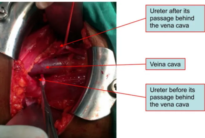
Figure 5. Intraoperative image of a retrocaval ureter of case 3.
[image:5.595.213.512.473.701.2]DOI: 10.4236/oju.2017.711025 217 Open Journal of Urology incidence ranging from 0.9 to 2 per 1000. The male sex is most affected with a sex ratio of 2.8 men to 1 woman. This prevalence is reported by several authors [2] [3] [4] [5]. This was also noted in our study, with the 3 male patients and an average age of 40 years at diagnosis. Retrocaval ureter is often the cause of mul-tiple complications including: renal colic, urinary tract infection, renal insuffi-ciency, lithiasis. Of all the complications, renal colic is by far the most frequent [2]-[8]. Renal colic is generally presented by intermittent crisis which may be similar to a nephrotic syndrome as the attacks are close and very unbearable. Our three patients suffered from years of renal colic that led to unsuccessful multiple consultations and medical treatments. Failure in the medical treatment of renal colic justifies initiation of a morphological assessment, in this case: ul-trasound, UIV and scanner whose results must be the subject of rigorous inter-pretation.
Ultrasonography often reports a pyelo-calicial dilation when performed dur-ing renal colic attacks, it may be normal and should not interrupt the continua-tion of the examinacontinua-tions. It should be noted that the results gotten from ultra-sound performed during or away from the crisis may be discordant from that performed away from the renal colic crisis. UIV, besides its advantage in pro-viding information on the functions of the left epsilateral kidney, provides two crucial information, on adequate interpretation of the images. An opacification of right ureter reversed in S majuscule and an upstream pyelocalyceal dilation of the ureter. The scanner’s superior imaging confirmed the UIV’s results with bet-ter clarity. In medically advanced countries where MRI is easily accessible, diag-nosis is even easier.
The treatment of CRU is surgical and dictated by renal colic pain intensity es-pecially when pain is unbearable and may lead to a decrease in the patients’ ac-tivities. Open surgery, though presenting satisfactory results should give way to minimally invasive surgery like laparoscopy, where available [7]-[14].
4. Conclusion
Retrocaval ureter is a rare congenital malformation often encountered in the male sex. The clinical symptoms are dominated by right renal colic affecting el-derly patients generally between the ages of 30 and 40 years. Medical Imagery such as, ultrasound, intravenous Urography, CT scanning and MRI, are major contributions to diagnosis. Radical surgical treatment, formerly known as open surgery, must give way to minimally invasive surgery presented by laparoscopy, where available.
References
[1] Hochstetter, F. (1893) Beitrage zur entwicklungsgeschichte des venen-systems der amnioten: III. [Contribution to the Developmental History of the Vein System of Amniotes: III.] SaugerMorphJahrb, 20, 542-542.
An-DOI: 10.4236/oju.2017.711025 218 Open Journal of Urology nales de Chirurgie, 126, 156-158. https://doi.org/10.1016/S0003-3944(00)00481-8
[3] Tembely, A., Diarra, A., Berthé, H., Diakité, M.L. and Ouattara, K. (2014) Uretere Retrocave: Deux Nouvelles Observations à L’hôpital Du Point G A Bamako. [Ure-tere Retrocave: Two New Observations at Point G Hospital in Bamako.] African Journal of Urology, 20, 104-107. https://doi.org/10.1016/j.afju.2013.11.007
[4] Hadzi-Djokic, J., Basic, D., Dzamic, Z., Aćimovic, M. and Markovic, Z. (2009) Uretère rétrocave: à propos de 16 cas. [Retrocaval Ureter Based on a Series of 16 Cases.] Progrès en Urologie, 19, 33-38. https://doi.org/10.1016/j.purol.2008.09.047
[5] Ichikawa, T., Kawada, S., Yamashita, T., Niwa, T., Iino, M., Koizumi, J., et al. (2014) A Case of Right Double Inferior Vena Cava with Circumcaval Ureter. Japanese Journal of Radiology, 32, 421-424. https://doi.org/10.1007/s11604-014-0312-2
[6] Muthusami, P. and Ramesh, A. (2013) Appearances of the Circumcaval Ureter on Excretory Urography and MR Urography: A Single-Center Case Series. Indian Jounal of Radiology and Imaging, 23, 81-85.
https://doi.org/10.4103/0971-3026.113621
[7] Ratkal, J.M., Jadhav, R. and Naique Dessai, R.R. (2016) Circumcaval Ureter—The Paradigm Shift in Diagnosis and Management. Indian Journal of Surgery, 78, 37-40.
https://doi.org/10.1007/s12262-015-1352-2
[8] Bhattacharjee, S., Sanga, S., Gupta, P. and George, R.A. (2016) Retrocaval Ureter or Preureteral Vena Cava: Lest We Forget This Rare Cause of Hydronephrosis. Medi-cal Journal Armed ForcesIndia, 72, 77-79.
https://doi.org/10.1016/j.mjafi.2016.08.004
[9] Li, H.Z., Ma, X., Qi, L., Shi, T.P., Wang, B.J. and Zhang, X. (2010) Retroperitoneal laparoscopic ureteroureterostomy for retrocaval ureter: report of 10 cases and lite-rature review. Urology, 76, 873-876. https://doi.org/10.1016/j.urology.2009.12.056
[10] Zhang, J., Liu, B., Song, N., Hua, L. and Wang, Z. (2014) Retroperitoneal Laparos-copic Ureteroureterostomy for Retrocaval Ureter: A Report of 15 Cases. Surgical Practice, 18, 37-41. https://doi.org/10.1111/1744-1633.12045
[11] Cardoza, F., Shambhulinga, C.K. and Rajeevan, A.T. (2016) Retrocaval Ureter and Contra Lateral Renal Agenesis—A Case Report and Review of Literature. I nterna-tional Brazilian Journal of Urology : Official Journal of the Brazilian Society of Urology, 42, 842-844. https://doi.org/10.1590/S1677-5538.IBJU.2015.0549
[12] Tengue, K., Botcho, G., Kpatcha, T.M., Adabra, K., Sewa, E., Leloua, E., et al. (2016) Prise en charge de l’uretère rétrocave au Togo: A propos de 3 observations (Man-agement of the Retrocaval Ureter in Togo about 3 Cases.] African Journal of Urol-ogy, 22, 279-283. https://doi.org/10.1016/j.afju.2016.03.008
[13] El Harrech, Y., Ghoundale, O., Kasmaoui, E.H. and Touiti, D. (2016) Transperito-neal Laparoscopic Pyelopyelostomy for Retrocaval Ureter without Excision of the Retrocaval Segment: Experience on Three Cases. Advances in Urology, Article ID 5709134, 4 p. https://doi.org/10.1155/2016/5709134
[14] Kumar, S., Singh, S. and Garg, N. (2015) Right Sided Double Inferior Vena Cava with Obstructed Retrocaval Ureter: Managed with Single Incision Multiple Port Laparoscopic Technique Using “Santosh Postgraduate Institute Tacking Ureteric Fixation Technique”. Korean Journal of Urology, 56, 330-333.