Movement coordination during Sit-to-Stand in low back
persons
SHAFIZADEHKENARI, Mohsen <http://orcid.org/0000-0002-7524-1058>
Available from Sheffield Hallam University Research Archive (SHURA) at:
http://shura.shu.ac.uk/12457/
This document is the author deposited version. You are advised to consult the
publisher's version if you wish to cite from it.
Published version
SHAFIZADEHKENARI, Mohsen (2016). Movement coordination during Sit-to-Stand
in low back persons. Human Movement, 17 (2), 107-111.
Copyright and re-use policy
See
http://shura.shu.ac.uk/information.html
Sheffield Hallam University Research Archive
MoveMent coordination during Sit-to-Stand
in low back pain people
doi: 10.1515/humo-2016-0012
* Corresponding author.
MohSen Shafizadeh
Sheffield Hallam University, Sheffield, United Kingdom
AbSTrACT
Purpose. The purpose of this study was to compare the inter-joint coordination during sit-to-stand (STD) and stand-to-sit (SIT) execution between healthy people and people with low back pain. Methods. Fifteen healthy adults (age = 45.14 ± 5.18 years) and fifteen age-matched (age = 46.17 ± 8.26 years) people with chronic low back pain were selected voluntarily. They performed three repetitions of STD and SIT movement patterns in their preferred pace. Motion analysis system was used for measuring 3-dimensional (3D) angular displacement of hip, knee and ankle joints during execution of movement patterns. Decomposition indices were analysed and were compared between two groups through Hotelling T2 Multivariate Analysis of Variance (MANOVA)
and follow-up Analysis of Variance (ANOVA). Results. The results showed that there is a significant difference (T2 = 18.32,
F14, 5 = 8.33, p < 0.05) between the groups on decomposition indices. The ANOVA follow-up results showed that there are significant
differences between two groups on decomposition indices of the whole pattern of STD (F1, 18 = 7.96, p < 0.05), whole pattern of
SIT (F1, 18 = 5.37, p < 0.05), the first-half phase of STD (F1, 18 = 7.26, p < 0.05) and the first-half phase of SIT (F1, 18 = 6.33, p < 0.05).
Conclusions. People with low back pain have dis-coordination in the function of different body parts, and results in pausing of one segment while the other segment moves independently. This knowledge may help in the development of rehabilitation strategies for movement in this population.
Key words: decomposition index, inter-joint coordination, low back pain
Introduction
Low back pain [LbP] is common in many developed countries [1–4]. According to a national survey in the UK [1] it is reported that 40% of adults have experienced back pain lasting more than one day in the previous 12 months. In addition, it is reported that 15% of peo-ple with back pain said they were in pain throughout the year. The European Union Commission study [2] in 2007 reported that 67 million people of the European countries had experienced pain in their lower or upper back in the previous week. Strine and Hootman [3], based on the 2002 National Health Interview Survey in the USA comprising adults over 18 years, reported that 34 million people suffered from low back pain. Fernández-de-las-Peñas et al. [4] in a recent report on Spanish population reported that 1-year prevalence of low back pain in adults over 16 years was approximately 20%.
Low back pain has physical, psychological, social and economic consequences for the individual. It is believed that adults with low back pain exhibit more psychologi-cal distress, engage in more risky health behaviours than adults without back pain [3] and are more likely to ex-perience depression and other physical complaints such as arthritis and osteoporosis [4, 5].
Some surveys reported that in the UK 12.5% of all sick days were found to be related to low back disorders. In Sweden it is estimated that 13.5% of sick days were the result of lower back problems [6]. The economic cost of back pain on society in the Netherlands has been estimated to be 1.7% of the gross national product [7]. In another survey in the UK it is reported that the direct health care cost of back pain in 1998 was 1632 million, of which approximately 35% relates to services pro-vided in the private sector [8].
Physical and behavioural consequences of low back pain are interrelated so that behavioural changes often are accompanied with physical limitations in painful regions. In a severe level of back pain, it can result in movement disability that ultimately may lead to suf-ferers avoiding their daily activities or occupations in the short or long term [9]. Since mechanical stressors in the workplace are the most important cause of low back incidence in the developed countries and its mani-festations are physical complaints in different forms such as back ache, back pain, muscle soreness, muscle stiffness and limited joint range of motion due to pain [10].
Keefe and block [11] labelled the pain behaviours in low back persons into 4 categories including guard-ing, bracguard-ing, rubbing and grimacguard-ing, which were later expanded by McDaniel et al. [12] into 8 categories includ-ing guardinclud-ing, bracinclud-ing, grimacinclud-ing, sighinclud-ing, rigidity, self-stimulation, passive and active rubbing.
scien-M. Shafizadeh, Movement coordination during Sit-to-Stand in low back pain people
tists investigating low back pain. Keefe and block [11] defined guarding as abnormal stiff, interrupted, or rigid movement while moving from one position to another. This behaviour is observable in movements such as sitting, standing, reclining, walking or other movement patterns that require shifting from one position to an-other. McDaniel et al. [12] later revised the original characteristics that were defined by Keefe and block. They assumed that the guarding cannot occur during a stationary position such as sitting, standing and re-clining. They included other features in their definition for guarding which were hesitation in the execution of movement that was different from movement under-taken at a slow velocity. Guarding that is considered to be an adaptive mechanism in response to acute pain in people with low back pain [13] is accompanied with in-creased muscle activity during flexion-extension tasks and walking [14–17] and restricted optimal trunk move-ment [18, 19]. These two guarding features that are known as muscle stiffness and joint rigidity are responsible for stabilising the spine via changes in the reflex control of trunk muscles [20].
Coordination between different body parts or muscle groups is necessary in order to control the multi-joint movement in a fluent manner. This synergy [21] might be deteriorated by factors such as pain, muscle stiffness, decreased joint range of motion [22, 23] and neurologi-cal problems [24] which may eventually result in the lack of coordination between different body parts. Silfies et al. [22] demonstrated that in a standing reach task lumbar-pelvic coordination was more separated in time and more variable in people with chronic low back pain compared to healthy participants. This lack of coordi-nation was attributed to freezing the motion of the lumbar spine in the subjects with low back pain [21, 22, 25] in contrast to healthy people who simultane-ously moved their lumbar spine and pelvis in the same direction during trunk bending [26].
Previous studies [23, 25] have shown that inter-joint coordination is altered in the lumbar spine and hips during sit-to-stand (STD) and stand-to-sit (SIT) in people with LbP. The method used to compute joint coordina-tion in these studies was the relative phase, quantified by subtracting the phase angle (inverse tangent of angular velocity relative/angular displacement) of one joint from the other [29]. Positive or negative values of relative phase represent the earlier onset, or delay of movement, in one joint relative to other joint. For example, if relative phase between hip to lumbar spine is negative, the hip move-ment is delayed until after onset of the lumbar spine movement. relative phase is an indicator of positional changes in coordination of two joints rather than a time parameter of joint coordination. An alternative method for representing joint coordination is the decomposition index. This is defined as an index of dis-coordination between two segments in terms of smooth or hesitant movement on the basis of timing [24]. It shows whilst one segment is moving another segment is stopping.
This index is applicable for studying the pain behaviours such as hesitation in guarding behaviour.
There are no previous studies which have investigated joint motion based on the decomposition index in a pop-ulation with low back pain, thus the aim of this study was to compare movement coordination between the lumbar spine and hip joints using this method in par-ticipants with and without low back pain.
Material and methods
Participants
Fifteen adult (age = 46.17 ± 8.28 years) subjects (male = 7, female = 8) with chronic low back pain and 15 age-matched (age = 45.14 ± 5.18) asymptomatic healthy people (male = 7, female = 8) were selected voluntarily. All subjects completed an informed consent form, the recent Physical Activity Questionnaire (rPAQ) and the Visual Analog Scale (VAS) prior to participation in this study. They suffered from chronic pains in low back area and were inactive in the past year according to their responses in questionnaires. The research Com-mittee of the University approved all stages of study.
Instrument
An 8-camera motion analysis system (Simi motion, co) was used to calculate angular displacement during STD and SIT according to a standard protocol. For the purpose of this study only lumbar spine and thigh markers were analysed for calculating movement co-ordination. Markers were placed on the body on the second sacral vertebra (S2), right and left Anterior Supe-rior Iliac Spine (ASIS). right and left thigh wands and markers were placed nearly 15 cm above the patella.
Procedure
SIT, the eccentric contraction and negative power are produced, whereas in the second phase of STD and SIT the concentric contraction and positive power are pro-duced [25].
Data analysis
Inter-joint coordination
Angular velocities of hip and lumbar spine joints were computed through dividing of angular displacement (de-gree) of flexion-extension (frontal) axis to time (second). The instantaneous velocity was computed for each frame number in order to acquire the detailed changes in move-ment sequence. Decomposition index values as indi-cators of inter-joint coordination were the percentage of STD and SIT time during which movement was decom-posed. A joint was considered to pause when its angular velocity dropped below 5º/s [24]. Average decomposition index values (%) were calculated for the lumbar-hip joint pair in each phase of STD, SIT and whole STD and SIT when one joint was moving while the other joint paused.
Statistical analysis
Descriptive statistics include mean and standard de-viation. Hotelling’s T2 MANOVA test was used to com-pare movement coordination between healthy and patient groups. If the results were significant, follow-up ANOVA tests were used to find the between-group differences in decomposition indices of STD and SIT and their phases. Confidence interval value was set at 95% and two-sided.
Results
Figure 2 demonstrates the mean decomposition index changes in different phases of STD and SIT. According to the results, decomposition index changed differently be-tween two groups so that for low back pain persons’ de-composition indices of the first-half phase were higher
than the second-half phase in STD and SIT, but for healthy group the second-half phase had higher score than the first-half phase for both STD and SIT.
The Hotelling T2 test result showed that there is a sig-nificant difference (T2 = 18.32, F
14, 5 = 8.33, p < 0.05) in decomposition indices between the low back pain group and the healthy one. ANOVA follow-up results showed that there are significant differences between the two groups for decomposition indices of whole pat-tern of STD (F1, 28 = 7.96, p < 0.05), whole pattern of SIT (F1, 28 = 5.37, p < 0.05), the first-half phase of STD (F1, 28 = 7.26, p < .05) and the first-half phase of SIT (F1, 28 = 6.33,
p < 0.05). Low back pain people had significantly higher decomposition indices relative to healthy group in whole STD (21.16 vs. 15.35), whole SIT (22.18 vs. 18.95), the first-half of STD (21.35 vs. 16.04), and the first-first-half of SIT (23.04 vs. 13.18).
Discussion
The aim of this study was to examine the effects of chronic low back pain on movement coordination in the lumbar spine and hip joints during two functional movement abilities including STD and SIT. Our find-ings showed that there were significant differences between low back pain people and healthy ones in decomposition indices of STD, SIT and the first-half phases of STD and SIT. These findings are indicative of the lack of synergy between movements of two joints that move independently due to lack of coordination. On the other hand, while the hip joint flexed lumbar joint paused and vice versa. These findings also support the findings of previous studies about the incidence of hesitation due to pain in low back pain people [11, 12].
[image:4.595.309.550.100.284.2]Silfies et al. [22] showed that lumbar-pelvic coordi-nation was more separated in time and more variable in people with chronic low back pain. Shum et al. [23] have demonstrated that low back pain people showed
Figure 2. Decomposition index of control group and low back pain group in different phases of STD and SIT
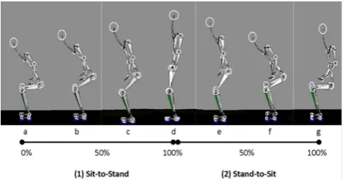
Sit-to-Stand Phases Stand-to-Sit a and b represent seat-off phase or the first-half of STD; c and d represent
[image:4.595.40.287.102.230.2]stand-up phase or the second-half of STD; d and e represent sit-down phase or the first-half of SIT; f and g represent seat-on or the second-half of SIT
M. Shafizadeh, Movement coordination during Sit-to-Stand in low back pain people
different lumbar-hip coordination relative to healthy people. In fact, the contribution of the lumbar spine in STD and SIT movements was reduced due to immobility in these joints induced to protect the spine against pain. Shum et al. [25] in another study have revealed that muscle moment reduction in the lumbar spine in the sagittal plane is the reason why STD and SIT strategies change in low back pain people. They minimise the trunk motion and thereby reduce the muscle moment on the joint that in turn changes inter-joint coordination. Another study [30] showed a decreased power flow from the pelvis to the lower limbs in low back pain people during STD. The present findings also showed dis-coordination of joints due to pausing of one joint whilst the other joint is moving. The method of current study was different from pre-vious studies [22, 25] that measured inter-joint coordi-nation through relative phase as an indicator of phase difference between paired-joints such as hip and lumbar spine joints. relative phase is an indicator of positional changes in coordination (leading or lagging joint into de-gree) rather than time parameter (pausing one joint for a millisecond). In fact, guarding behaviour as a form of muscle stiffness or joint freezing [14–20] that is ob-servable in low back pain people resulted in limitation in trunk or thigh movements and it led to inter-joint dis-coordination.
The additional data analysis of decomposition index of the lumbar and hip joints showed different contribu-tion of them in inducing dis-coordinacontribu-tion in healthy and low back pain groups. In healthy group the pausing percentage in the lumbar and hip joints in entire move-ments were 77% and 22%, respectively (lumbar to hip ratio: 3.5), whereas in low back pain group the pausing percentage for lumbar and hip joints were 60% and 42%, respectively (lumbar to hip ratio:1.42). Thus, the hip joint slightly (25% less than in healthy people) contributed to body weight transfer in low back pain people. These find-ings are important as they show to what extent a hesi-tant movement is shared between two different body parts so that STD and SIT could be executed.
In addition, as Figure 2 shows that the decomposition index for low back pain people in different phases of STD and SIT are different – in the first-half of STD and SIT they demonstrated more pausing than in the second-half. This pattern was different in healthy people who showed more pausing in the second-half of STD and SIT. Shum et al. [25] revealed that muscle powers are differ-ent in differdiffer-ent phases of STD and SIT, namely in the first-half phase the muscle work is negative because the type of muscular contraction is eccentric. It seems that keeping the trunk upright during seat-off phase to peak lumbar spine flexion (a, b and c in Figure 1) due to pain-ful condition deteriorates inter-joint coordination by reducing the fluent motion and converting it into a hesi-tant movement. Again during SIT movement, the type of muscle contraction in first-half phase is eccentric that will interrupt the joints’ synergy which caused more pausing during movement execution.
reduction in the angular velocity of both lumbar and hip joints during STD and SIT have been demonstrated in previous studies [23, 31] and were explained as a pre-ventive mechanism against pain that is caused by muscle contraction and high levels of acceleration. Difficulty in transferring the muscle force from the pelvis to the lower limbs causes an interruption in the execution of closed kinetic chain that in turn is responsible for trans-ferring the force from the upper to lower body parts [30]. These findings suggest that reducing angular velocity in the lumbar spine is helpful to reduce the angular moment between two joints and subsequently prevents the risk of losing balance. but reducing it beyond the normal values relative to hip movement is a preventive mecha-nism that is observable in low back pain people that could change the mechanics of movement into hesitant behav-iours. Thus, in rehabilitation programmes of low back pain, emphasising on a constant and fluent motion and prevention from hesitant movement reduce the pres-sure on the lumbar spine through efficient utilisation of the hip in coordination with the lumbar spine by means of a closed kinetic chain.
Future studies should investigate the possible mecha-nisms of hesitation behaviours through electromyo-graphy [EMG] study to confirm the biomechanical findings that have been revealed in the present study.
In conclusion, low back pain causes dis-coordination in the function of different body parts and results in pausing in one segment while the other segment moves independently. Therapeutic exercises that emphasise coordinative movement of the pelvis and the hip joints could reduce dis-coordination due to freezing in move-ment segmove-ments.
References
1. Department of Health Statistics Division. The prevalence of back pain in Great britain in 1998. Government Sta-tistical Service, London 1999.
2. European Commission, Special Eurobarometer, 272e, Nov 2007.
3. Strine T.W., Hootman J.M., US national prevalence and correlates of low back and neck pain among adults. Arthritis Rheum, 2007, 57 (4), 656–665, doi: 10.1002/art.22684. 4. Fernández-de-las-Peñas C., Hernández-barrera V., Alonso-blanco C., Palacios-Ceña D., Carrasco-Garrido P., Jimé-nez-Sánchez S. et al., Prevalence of neck and low back pain in community-dwelling adults in Spain: a popula-tion-based national study. Spine (Phila Pa 1976), 2011, 36 (3), 213–219, doi: 10.1097/brS.0b013e3181d952c2. 5. Sloan T.J., Gupta r., Zhang W., Walsh D.A., beliefs about
the causes and consequences of pain in persons with chronic inflammatory or noninflammatory low back pain and in pain-free individuals. Spine (Phila Pa 1976), 2008, 33 (9), 966–972, doi: 10.1097/brS.0b013e31816c8ab4. 6. Andersson G.b., Epidemiological features of chronic
low-back pain. Lancet, 1999, 354 (9178), 581–585, doi: 10.1016/ S0140-6736(99)01312-4.
8. Maniadakis N., Gray A., The economic burden of back pain in the UK. Pain, 2000, 84 (1), 95–103, doi: 10.1016/ S0304-3959(99)00187-6
9. Ehrlich G.E., Low back pain. Bull World Health Organ, 2003, 81 (9), 671–676, doi: 10.1590/S0042-96862003000900010.
10. Punnett L., Prüss-Utün A., Nelson D.I., Fingerhut M.A., Leigh J., Tak S. et al., Estimating the global burden of low back pain attributable to combined occupational expo-sures. Am J Ind Med, 2005, 48 (6), 459–469, doi: 10.1002/ ajim.20232.
11. Keefe F.J., block A.r., Development of an observation method for assessing pain behaviour in chronic low back pain patients. Behav Ther, 1982, 13 (4), 363–375, doi: 10.1016/S0005-7894(82)80001-4.
12. McDaniel L.K., Anderson K.O., bradley L.A., Young L.D., Turner r.A., Agudelo C.A. et al., Development of an ob-servation method for assessing pain behavior in rheu-matoid arthritis patients. Pain, 1986, 24 (2), 165–184, doi: 10.1016/0304-3959(86)90039-4.
13. Verbunt J.A., Seelen H.A., Vlaeyen J.W., van de Heijden G.J., Heuts P.H., Pons K. et al., Disuse and deconditioning in chronic low back pain: concepts and hypotheses on con-tributing mechanisms. Eur J Pain, 2003, 7 (1), 9–21, doi: 10.1016/S1090-3801(02)00071-X.
14. Ahern D.K., Follick M.J., Council J.r., Laser-Wolston N., Litchman H., Comparison of lumbar paravertebral EMG patterns in chronic low back pain patients and non-patient controls. Pain, 1988, 34 (2), 153–160, doi: 10.1016/0304-3959(88)90160-1.
15. Watson P.J., booker C.K., Main Ch.J., Evidence for the role of psychological factors in abnormal paraspinal activity in patients with chronic low back pain. J Musculoskelet Pain, 1997, 5 (4), 41–56, doi: 10.1300/J094v05n04_05. 16. Geisser M.E., Haig A.J., Wallborn A.S., Wiggert E.A.,
Pain-related fear, lumbar flexion, and dynamic EMG among persons with chronic musculoskeletal low back pain.
Clin J Pain, 2004, 20 (2), 61–69.
17. van der Hulst M., Vollenbroek-Hutten M.M., rietman J.S., Hermens H.J., Lumbar and abdominal muscle activity during walking in subjects with chronic low back pain: support of the “guarding” hypothesis. J Electromyogr Ki-nesiol, 2010, 20 (1), 31–38, doi: 10.1016/j.jelekin.2009.03.009. 18. Hodges P.W., Moseley G.L., Pain and motor control of the
lumbopelvic region: effect and possible mechanisms. J Electromyogr Kinesiol, 2003, 13 (4), 361–370, doi: 10.1016/S1050-6411(03)00042-7.
19. Lund J.P., Donga r., Widmer C.G., Stohler C.S., The pain-adaptation model: a discussion of the relationship be-tween chronic musculoskeletal pain and motor activity.
Can J Physiol Pharmacol, 1991, 69 (5), 683–694, doi: 10.1139/y91-102.
20. Hodges P., van den Hoorn W., Dawson A., Cholewicki J., Changes in the mechanical properties of the trunk in low back pain may be associated with recurrence. J Biomech, 2009, 42 (1), 61–66, doi: 10.1016/j.jbiomech.2008.10.001. 21. Shumway-Cook A., Woollacot M.H., Motor control: trans-lating research into clinical practice. 3rd Edition, Williams
& Wilkins, Philadelphia 2007.
22. Silfies S.P., bhattacharya A., biely S., Smith S.S., Giszter S., Trunk control during standing reach: a dynamical system analysis of movement strategies in patients with mechani-cal low back pain. Gait Posture, 2009, 29 (3), 370–376, doi: 10.1016/j.gaitpost.2008.10.053.
23. Shum G.L., Crosbie J., Lee r.Y., Effect of low back pain on the kinematics and joint coordination of the lumbar spine and hip during sit-to-stand and stand-to-sit. Spine (Phila Pa 1976), 2005, 30 (17), 1998–2004, doi: 10.1097/01. brs.0000176195.16128.27.
24. Earhart G.M., bastian A.J., Selection and coordination of human locomotor forms following cerebellar dam-age. J Neurophysiol, 2001, 85 (2), 759–769. Available from: http://jn.physiology.org/content/85/2/759.long.
25. Shum G.L., Crosbie J., Lee r.Y., Three-dimensional ki-netics of the lumbar spine and hips in low back pain patients during sit-to-stand and stand-to-sit. Spine (Phila Pa 1976), 2007, 32 (7), 211–219, doi: 10.1097/01. brs.0000259204.05598.10.
26. Lee r.Y., Wong T.K., relationship between the move-ments of the lumbar spine and hip. Hum Mov Sci, 2002, 21 (4), 481–494, doi:10.1016/S0167-9457(02)00117-3. 27. Weiss P., Stelmach G.E., Hefter H., Programming of a
movement sequence in Parkinson’s disease. Brain, 1997, 120 (1), 91–102, doi: 10.1093/brain/120.1.91.
28. 28. bennett K.M., Marchetti M., Ivoine r., Castiello U., The drinking action of Parkinson’s disease subjects. brain, 1995, 118 (4), 959–970, doi: 10.1093/brain/118.4.959. 29. burgess-Limerick r., Abernethy b., Neal r.J., relative
phase quantifies interjoint coordination. J Biomech, 1993, 26 (1), 91–94, doi: 10.1016/0021-9290(93)90617-N. 30. Shum G.L., Crosbie J., Lee r.Y.,Energy transfer across the
lumbosacral and lower- extremity joints in patients with low back pain during sit-to-stand. Arch Phys Med Rehabil, 2009, 90 (1), 127–135, doi: 10.1016/j.apmr.2008.06.028. 31. Marras W.S., Wongsam P.E., Flexibility and velocity of
the normal and impaired lumbar spine.Arch Phys Med Rehabil, 1986, 67 (4), 213–217. Available from: https:// www.researchgate.net/profile/William_Marras/publi- cation/20216816_Flexibility_and_velocity_of_the_nor-mal_and_impaired_lumbar_spine/links/00b495240b 0bcc1f75000000.pd.
Paper received by the Editor: July 23, 2015 Paper accepted for publication: May 25, 2016
Correspondence address
Mohsen Shafizadeh
Academy of Sport and Physical Activity Faculty of Health and Wellbeing Sheffield Hallam University Sheffield, UK, S10 2bP