non-steroidal anti-inflammatory drugs
BJARNASON, Ingvar, SCARPIGNATO, Carmelo, HOLMGREN, Erik,
OLSZEWSKI, Michael, RAINSFORD, Kim and LANAS, Angel
Available from Sheffield Hallam University Research Archive (SHURA) at:
http://shura.shu.ac.uk/17716/
This document is the author deposited version. You are advised to consult the publisher's version if you wish to cite from it.
Published version
BJARNASON, Ingvar, SCARPIGNATO, Carmelo, HOLMGREN, Erik, OLSZEWSKI, Michael, RAINSFORD, Kim and LANAS, Angel (2018). Mechanisms of damage to the gastrointestinal tract from non-steroidal anti-inflammatory drugs.
Gastroenterology, 154 (3), 500-514.
Copyright and re-use policy
See http://shura.shu.ac.uk/information.html
Mechanisms of Damage to the Gastrointestinal Tract From Non-steroidal Anti-inflammatory Drugs
Ingvar Bjarnason, Carmelo Scarpignato, Erik Holmgren, Michael Olszewski, Kim D. Rainsford, Angel Lanas
PII: S0016-5085(17)36666-0 DOI: 10.1053/j.gastro.2017.10.049
Reference: YGAST 61566
To appear in: Gastroenterology
Accepted Date: 31 October 2017
Please cite this article as: Bjarnason I, Scarpignato C, Holmgren E, Olszewski M, Rainsford KD, Lanas A, Mechanisms of Damage to the Gastrointestinal Tract From Non-steroidal Anti-inflammatory Drugs, Gastroenterology (2018), doi: 10.1053/j.gastro.2017.10.049.
M
AN
US
CR
IP
T
AC
CE
PT
ED
Mechanisms of Damage to the Gastrointestinal Tract From Non-steroidal
Anti-inflammatory Drugs
Short title: Pathogenesis of NSAID induced GI damage
Ingvar Bjarnason1, Carmelo Scarpignato2, Erik Holmgren3, Michael Olszewski3, Kim
D Rainsford4, Angel Lanas5,
1
Professor of Digestive Diseases,Department of Gastroenterology, King’s College
Hospital, London, UK.
2
Professor of Pharmacology and Therapeutics, Associate Professor of
Gastroenter-ology, Consultant Clinical Pharmacologist & Gastroenterologist,
Clinical Pharmacology & Digestive Pathophysiology Unit, Department of Clinical and
Experimental Medicine, University of Parma, Italy
3
Research Fellows, Department of Gastroenterology, King’s College Hospital,
Lon-don, UK
4
Emeritus Professor of Biomedical Sciences, Biomedical Sciences, Biomedical
Re-search Centre, Sheffield Hallam University, Sheffield, UK
5
Professor of Medicine, Chairman Department of Gastroenterology, Vice Dean for
Research Affairs, University of Zaragoza School of Medicine, IIS Aragón, CIBERehd,
Zaragoza, Spain.
M
AN
US
CR
IP
T
AC
CE
PT
ED
2
Abbreviations: NSAIDs: Non steroidal anti-inflammatory drugs; COX:
Cyclooxygen-ase; pKa: Logarithmic transformed acid dissociation constant; ATP: Adenosine
tri-phosphate; NADH: Nicotinamide adenine dinucleotide; MPTP: mitochondrial
mem-brane transition pore; 6-MNA: 6-methoxy naphthalene acetic acid; TLR: Toll-like
Re-ceptor
Address for Correspondence:
Professor Ingvar Bjarnason MD, MSc, FRCPath, FRCP, DSc
Professor of Digestive Diseases
Department of Gastroenterology
King’s College Hospital
Denmark Hill
London E5 9RS
Ingvarbjarnason@mac.com
Ingvar.bjarnason@nhs.net
Tel 44-7784589003
Fax: 4-2032996474
Disclosures: None of the authors have any financial, personal or professional
con-flicts of interest in relation to this paper. Professors Bjarnason, Scarpignato,
Rains-ford and Lanas have received lecture fees, travel support, research grants and sat
on advisory panels of a number of Pharmaceutical companies, some of which are
involved with NSAIDs or drugs to prevent or heal their adverse effects on the
intesti-nal tract.
Transcript Profiling: None
Writing assistance: Professor Brian Callingham, Department of Pharmacy, Queens
College, University of Cambridge read and commented on the final version of the
manuscript.
Authors contributions: The study concept originated with IB. EH and MO wrote the
first draft and carried out the literature search while IB supervised them and did most
M
AN
US
CR
IP
T
AC
CE
PT
ED
3
M
AN
US
CR
IP
T
AC
CE
PT
ED
4 ABSTRACT
Non-steroidal anti-inflammatory drugs (NSAIDs) can damage the gastrointestinal
tract, causing widespread morbidity and mortality. Although mechanisms of damage
involve the activities of prostaglandin-endoperoxide synthase 1 (PTGS1 or COX1)
and PTGS1 (COX2), other factors are involved. We review mechanisms of
gastroin-testinal damage induction by NSAIDs, via COX-mediated and COX-independent
processes. NSAIDs interact with phospholipids and uncouple mitochondrial oxidative
phosphorylation, which initiates biochemical changes that impair function of the
gas-trointestinal barrier. The resulting increase in intestinal permeability leads to
low-grade inflammation. NSAID’s inhibition of COX enzymes, along with luminal
aggres-sors, results in erosions and ulcers, with potential complications of bleeding, protein
loss, stricture formation, and perforation. We propose a model for NSAID-induced
damage to the gastrointestinal tract that includes these complex, interacting, and
in-ter-dependent factors. This model highlights the obstacles for the development of
safer NSAIDs.
M
AN
US
CR
IP
T
AC
CE
PT
ED
5More than 30 million people take non-steroidal anti-inflammatory drugs (NSAIDs)
each day (1). This number has grown significantly with increasing use of over the
counter and prescription NSAIDs, low-dose aspirin and following reports of their
po-tential anti-neoplastic effects. The efficacy of NSAIDs as anti-inflammatory
analge-sics is not in doubt, but their adverse events are problematic. These relate mainly to
cardiovascular, renal, hepatic, and the gastrointestinal tissues. The cardiovascular
adverse events have recently received much attention (2, 3), but the frequency and
severity of the gastrointestinal damage continues to cause concern. Accordingly the
range of gastroduodenal ulcer rates range from 5% to 80% in short-term endoscopy
studies (4) and from 15% to 40% in long-term users (5). NSAIDs also damage the
small intestine (6)—as many as 70% of long-term users of NSAIDs have small
intes-tinal inflammation, and 30% have erosions or ulcers (7). The gastric and small bowel
damage is associated with various management problems and at times life
threaten-ing complications, such as bleedthreaten-ing, strictures and perforations.
There have been many studies of the pathogenesis of NSAID-induced
gastro-intestinal damage. NSAIDs inhibit prostaglandin-endoperoxide synthase 1 (PTGS1
or COX1) and COX2, which have been believed to mediate the gastrointestinal
damage (8-10). NSAID-induced decreases in mucosal levels of prostaglandins
(driv-en by inhibition of COX1) correlate with gastric and small bowel damage (11-13),
which can be attenuated by administration of exogenous prostaglandins (14-18).
Since COX2 is not constitutively expressed in the gastrointestinal tract COX2
selec-tive inhibitors are perceived as safer than conventional NSAIDs (14, 15, 19, 20).
Proposed mechanisms of damage to the stomach involve prostaglandin mediated
increased gastric acid secretion, decreased mucus and bicarbonate secretion,
de-creased cell proliferation, and dede-creased mucosal blood flow (21-24). These are all
actions that are detrimental to mucosal defense and healing, but the observed
changes were only modest (21, 23, 25-30) and the damage seemed to lack an
initia-tive action. Furthermore, decreased mucosal prostaglandins have been fund to be
less important in the pathogenesis of the small bowel damage (11, 31, 32).
Further studies showed that gastric and small bowel mucosal prostaglandins
could be decreased by 95%–98% without mucosal damage (33-35), confirmed in
COX1-knockout mice (35-37). Short-term loss or inhibition of COX2 does not cause
damage, but small bowel damage is evident in mice and humans exposed to
M
AN
US
CR
IP
T
AC
CE
PT
ED
6gastric and small bowel lesions, albeit somewhat less severe than that the lesions
caused by conventional acidic NSAIDs (36).
So, inhibition of COX does not seem to be the only mechanism of NSAID-induced
gastrointestinal damage. We review the prostaglandin-independent mechanisms of
NSAIDs and how these interact with the consequence of alterations in prostaglandin
levels as a consequence of COX inhibition. We provide a model in which COX
inhibi-tion is one of several important factors in the pathogenesis of gastrointestinal
dam-age (see Figure 1). Our model considers the effects of the specific biochemical
“topi-cal” effects of NSAIDs (i.e. the effects that occur by direct contact between the
NSAIDs in the lumen and mucosal epithelium following oral ingestion and/or biliary
excretion of the drugs, as opposed to topical skin application) and the consequential
increase in intestinal permeability and intestinal inflammation. These initiate damage
and inhibition of COX1 and COX2 aggravate it, along with luminal aggressors,
lead-ing to development of erosions and ulcers (42, 43).
BIOCHEMICAL EFFECTS OF NSAIDS
The biochemical actions common to all conventional NSAIDs are their “topical”
ef-fects, and inhibition of COX1 and COX2. These biochemical actions are brought
about by the physicochemical properties that NSAIDs share (44-46), namely being
lipid soluble weak acids (see Figure 2). This combination provides them with
deter-gent action (interaction with phospholipids), uncoupling of oxidative phosphorylation,
and non-covalent inhibition of COX1 and COX2. These biochemical activities depend
on the same physical and chemical characteristics, so changing these will change all
the pharmacologic actions. For example, esterification of NSAIDs (47) causes loss of
their “topical” effects and at the same time their ability to inhibit the COX enzymes.
Interactions between NSAIDs and phospholipids
NSAIDs interact with the intestinal mucus layer and the cell surface phospholipid
bi-layer. There are subtle differences in mucus thickness and composition in different
regions of the gastrointestinal tract (19, 48). The role of mucus is to act as a lubricant
between the surface epithelium and the luminal contents, restricting access of large
hydrophilic molecules, digestive enzymes, and bacteria to the surface epithelium. In
the stomach, mucus also buffers luminal acids. The production and secretion of
M
AN
US
CR
IP
T
AC
CE
PT
ED
7in the stomach and bile and bacteria in the small bowel) and the surface epithelium
mediated by numerous factors such as inflammatory cytokines and prostaglandins.
Mucus serves as a matrix for phospholipids that maintain gastrointestinal
in-tegrity (49). Like NSAIDs, phospholipids are amphiphilic molecules, with a
hydro-philic polar head group and a hydrophobic tail region. The integrity of the mucus
lay-er can be assessed by various methods (50). NSAIDs decreased the hydrophobicity
in the gastroduodenal mucosa (51), an effect seen also after parenteral
administra-tion via the biliary excreadministra-tion of the drug (52). The interaction between NSAIDs and
phospholipids compromises the hydrophobic lining, which leads to mucosal
expo-sure to luminal aggressors (acid and pepsin in the stomach and bacteria and bile in
the small intestine).
The concept of a hydrophobic barrier attributed to phospholipids and the
bind-ing of NSAIDs to dipalmitoylphosphatidylcholine (the dominant phospholipid in the
gastrointestinal-tract), in vitro and in vivo (49, 53), led to a series of studies
investi-gating the effect of orally co-administrated phospholipids with NSAIDs, and other
tox-ic compounds, with a view to diminishing their toxtox-icity. Combining NSAIDs with the
phospholipid phosphatidylcholine protects against NSAID-induced gastric (49, 54)
and small bowel (55) damage in short-term rodent studies. Lichtenberger et al
demonstrated decreased gastric toxicity of the otherwise damaging combination of
aspirin and a COX2-selective agent, if the aspirin was co-administered with a
phos-pholipid (56).
These and other animal studies provided the platform for testing the safety of
NSAIDs combined with phospholipids in humans. Volunteers were given aspirin or a
combination of aspirin and phospholipid (650 mg aspirin/day for 3 days). The number
of gastric erosions (assessed during endoscopy) was significantly lower in volunteers
given aspirin and phospholipid (mean 2.8 ± 4.3) than aspirin alone (mean 8.8 ± 10.8;
both drugs reduced mucosal prostaglandin content to the same extent (57). In a
separate study, healthy volunteers given aspirin (325 mg/day for 7 days) or the same
amount of aspirin combined with phosphatidylcholine, had a significant decrease in
gastric ulcers, from 17.6% in volunteers given aspirin to 5.1% in volunteers given
as-pirin with phosphatidylcholine (58). In a 6-week study of patients with osteoarthritis,
the combination of ibuprofen and phosphatidylcholine was associated with significant
M
AN
US
CR
IP
T
AC
CE
PT
ED
8(2400 mg) alone, but only in patients older than 55 years (59). These studies
demonstrated greater gastric tolerability of combinations of aspirin and phospholipid,
in the short-term, in humans, in which damage is more likely to be caused by the
physicochemical properties of NSAIDs than their effect on COX1 or COX2 (4).
Uncoupling mitochondrial oxidative phosphorylation
Mitochondria are the main source of ATP in cells. Mitochondrial ATP synthesis takes
place by integrated biochemical-physiological-physical processes (60) (see Figure
3).
Whatever the cause of uncoupling there is a cascade of detrimental
down-stream effects: water flows into the matrix, causing characteristic and
pathognomon-ic swelling of mitochondria. There is release of intra-mitochondrial Ca2+ into
cyto-plasm with depletion of reduced glutathione, depletion of NAD(P)H2, generation of
superoxide anion (O2–) and release of pro-apoptogenic proteins (61). Free radicals
accumulate within the mitochondria setting up a vicious cycle as this activates
un-coupling proteins in the inner mitochondrial membrane (62). The unun-coupling
ulti-mately leads to depletion of cellular ATP levels, with loss of integrity of the
intercellu-lar junctions in the gastrointestinal tract (leading to increased mucosal permeability)
(63), and ultimately apoptosis and cell death (64).
Well before the understanding that NSAIDs inhibited the COX enzyme(s) it
was evident that NSAIDs were uncouplers of mitochondrial oxidative phosphorylation
(65, 66). Adams et al screened possible anti-inflammatory agents based on their
un-coupling properties and several (such as ibuprofen, naproxen and indomethacin)
have been marketed on that basis. However, the idea of the uncoupling action of
NSAIDs as a mechanism for their therapeutic actions became obsolete when the
prostaglandin hypothesis gained momentum.
A few reports describe uncoupling of mitochondrial oxidative phosphorylation
in the gastric mucosa following aspirin (67, 68). Using the technique of selective
sub-cellular marker enzyme analyses of small bowel mucosa following administration of
NSAIDs in animals (69) showed a significant change in the brush border marker
en-zyme, compatible with the interaction of NSAIDs with phospholipids, and the
mito-chondrial marker enzymes. Electron microscopic changes of uncoupling were
uncou-M
AN
US
CR
IP
T
AC
CE
PT
ED
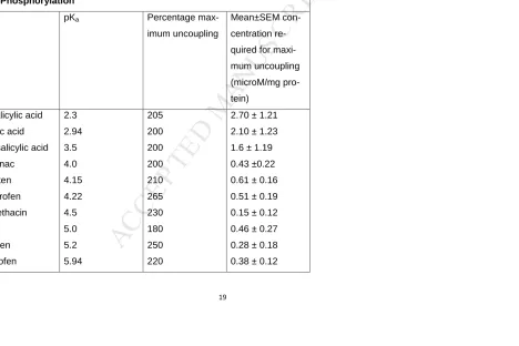
9pling of conventional acidic (carboxylic or enolic acids) NSAIDs relates to their pKa
values (see Table 1) (70). Drugs that are purported to be safer such as paracetamol
(non acidic analgesic), nabumetone (a non-acidic NSAID pro-drug (71), and
esteri-fied non-acidic pro-NSAIDs (see Figure 2), such as nitro-butyril flubiprofen are not
uncouplers in vitro (69).
Micromolar to millimolar concentrations of NSAIDs have the ability to
uncou-ple mitochondrial oxidative phosphorylation in vitro (42, 69, 72-76), due to ion
trap-ping during absorption (see Figure 4). COX2-selective agents also uncouple
oxida-tive phosphorylation in vitro and in cell systems, but with lower potency than that of
acidic NSAIDs (76, 77). The uncoupling by NSAIDs was demonstrated by electron
microscopy in the small bowel of mice given conventional acidic NSAIDs (42, 69,
73-75, 78, 79) and similar changes are also found in gastric biopsies from patients (67,
68, 80-83). No studies have assessed the possible prevention of uncoupling brought
about by NSAIDs.
INHIBITION OF COX1 AND 2 AND ROLE OF PROSTAGLANDINS
The 3-dimensional structure of the COX enzymes reveals the active site of both COX
isoforms to be at the end of a hydrophobic channel. NSAIDs inhibit the enzyme by
blocking the entrance of arachidonic acid to this channel and thereby denying
sub-strate access to the active site (84, 85). The COX1 and 2 channels differ.
Conven-tional NSAIDs have access to both channels and form an ionic bond via their
car-boxyl or enolic group (86). The COX1 channel is smaller than the channel in COX2
and does not accommodate COX2-selective agents, but a side pocket in the COX2
enzyme has a polar binding site (87) for the aryl sulfonamide and sulfone moieties of
the COX2-selective agents.
The most damaging consequence of decreased prostaglandin production with
COX inhibition could be the effects on the microcirculation. Regulation and
mainte-nance of the intestinal microcirculation is complex involving several interacting
bio-chemical mechanisms. The most relevant mediators are prostaglandins,
leukotri-enes, nitric oxide, and hydrogen sulphide. NSAID-induced prevention of
physiologi-cal compensatory increases in blood flow (leading to tissue hypoxia) following injury
is well described. The effects of nitric oxide and hydrogen sulphide are remarkably
in-M
AN
US
CR
IP
T
AC
CE
PT
ED
10creased mucus secretion, and a modest decrease of gastric acid secretion (88, 89).
Targeting these processes with nitric oxide donors such as nitro-glycerine,
nitroprus-side, nitric-oxide-NSAIDs, and hydrogen sulphite NSAIDs can reduce the
gastroin-testinal damage due to NSAIDs in laboratory animals (27, 90-93). Presumably these
effects counteract the reduced microvascular blood flow (94) consequent to
NSAID-induced decreased prostaglandins (95). Proof-of-concept endoscopic studies of
healthy volunteers showed that nitric oxide donors and NSAIDs reduced
gastroduo-denal damage, compared with NSAIDs (96, 97), but the results of a longer-term
clin-ical trial did not show statistclin-ically significant differences (98).
Another vascular effect of NSAIDs involves NSAID-induced expression of
neutrophil adhesion molecules within the endothelium (common to most intestinal
inflammatory conditions) (27, 29, 93, 99). Neutrophil accumulation could
mechanical-ly compromise microvascular blood flow. Nitric oxide and hydrogen sulphite are, like
prostaglandins, inhibitors of leucocyte adhesion to the vascular endothelium (100).
However, vascular effects are probably not the primary or initiating event in
NSAID-induced gastrointestinal damage. The effects on the vasculature cannot
ac-count for the selective localization of the macroscopic damage (101-104) within the
gastrointestinal tract nor the mesenteric rather than the anti-mesenteric location of
small bowel ulcers. The damage also differs macroscopically and microscopically
from ischemic damage. The suggestion that neutrophil adhesion to the vessel wall (a
COX2-mediated effect) is a primary event in the damage is difficult to reconcile with
the fact that COX2 is not constitutively expressed in the gastrointestinal tract.
Fur-thermore, neutrophil adhesion to the intestinal vessel wall does not automatically
in-dicate damage as neutrophils require a chemoattractant for activation-degranulation
and hence damage (105, 106).
Consequences of the biochemical effects of NSAIDs
Studies on COX-knockout mice have increased our understanding of the
conse-quences of COX1 and COX2 deficiency. Absence or selective inhibition of COX1 (by
the non-acidic COX1 inhibitor, SC-560) reduced levels of prostaglandins by 95% or
more, which was not associated with increased intestinal permeability, inflammation,
or ulcers (35, 36). Neither was short-term, selective deletion or inhibition of COX2
M
AN
US
CR
IP
T
AC
CE
PT
ED
11consequences of the “topical” effects and dissociated these from the consequences
of COX inhibition. These studies were done by comparing key pathophysiological
events in the damage, namely the “topical” effect (in vitro and in vivo uncoupling),
prostaglandin levels, intestinal permeability, and inflammation following the use of
selective drugs. This provides convincing evidence that the “topical” effects
(phos-pholipid-NSAID interaction and uncoupling) initiate gastrointestinal damage, but only
with COX1 inhibition (in association with luminal aggressive factors), does this lead
to mucosal erosions and ulcers. The compounds and their effects can be
catego-rized as follows (see Table 2):
• Selective uncouplers (dinitrophenol [DNP] or R-flurbiprofen) can increase intestinal permeability associated with mild inflammation, but do not
signifi-cantly alter mucosal prostaglandin levels, and do not cause mucosal
ulcer-ation.
• Uncouplers (conventional acidic NSAIDs) that inhibit COX enzymes are as-sociated with increased intestinal permeability, inflammation, and ulcers.
• COX2-selective agents such as celecoxib do not uncouple oxidative phos-phorylation (nimesulide with a Pka of 6.4, despite showing uncoupling
activ-ity, behaves like celecoxib—possibly because the uncoupling effect in vivo
affects only a few mitochondria). These agents are not associated with
in-creased intestinal permeability, inflammation or ulcers.
Collectively these studies, together with studies of knockout mice, have provided
compelling evidence that uncoupling of mitochondrial oxidative phosphorylation
(along with the NAID-phospholipid interaction) increases intestinal permeability and
low-grade inflammation. Decreased mucosal prostaglandin production and the
mu-cosal aggressors lead to more severe inflammatory and ulcerative damage, perhaps
via effects on the microcirculation.
The findings from COX2-knockout mice are more difficult to explain. These
mice have normal mucosal levels of prostaglandin, but half have normal intestinal
permeability and no inflammation or intestinal ulcers, and the other half develop
small intestinal inflammation and ulcers or die because of ulcer perforation. Similar
findings were seen with long-term administration of a selective COX2 inhibitor to
M
AN
US
CR
IP
T
AC
CE
PT
ED
12TISSUE REACTION AND ROLE OF LUMINAL AGGRESSORS
The tissue reaction is characterized by inflammation and the presence of erosions
and ulcers and this appears to be driven by COX inhibition and the luminal
aggres-sive factors. The luminal aggressors differ between the stomach (acid, pepsin, and H
pylori) and small bowel (bile and commensal bacteria). The importance of gastric
lu-minal aggressors is widely appreciated, but the same does not hold true for small
bowel aggressors. Our review focuses on effects in the small bowel.
Role of acid and H pylori in NSAID-induced gastropathy
The importance of gastric acid in the damage of NSAID-induced gastro-duodenal
damage in humans is amply demonstrated clinically in the reduced incidence of
damage (short and long-term) and serious ulcer outcomes when NSAIDs are
co-administered with proton pump inhibitors (107, 108) or high dose histamine
receptor-2 inhibitors (109). In the context of the current pathogenic model the macroscopic
damage in the stomach is principally due to back diffusion of acid due to the
im-paired barrier function (brought about by the “topical” effects) induced by NSAIDs
and amplified by the prostaglandin dependent effects induced by NSAIDs. The
fre-quent finding of chemical gastritis (reactive gastritis) in antral biopsies in patients on
NSAIDs (110), who do not have H pylori infection, can be considered as the
conse-quence of the “topical” effect of these drugs. In this context, the mucosal
inflammato-ry reaction is weak compared to that seen in patients infected by H pylori.
The effects of H pylori infection in the pathogenesis of NSAID-associated
gas-tric ulcers is controversial. H pylori does not seem to mediate development of
short-term NSAID-induced gastric damage in humans (4), although it may affect gastric
adaptation to short-term administration of aspirin (111). Gastric damage induced by
long-term NSAIDs or aspirin occurs in addition to the gastritis induced by H pylori
in-fection, which occurs early in life. H pylori induces gastric mucosal lesions by
inter-acting with the immune response (112). The intrinsic virulence factors of each
specif-ic H pylori strain may induce a weak or a strong host immune cytokine-mediated
in-flammatory response, which is genetically determined. Patients infected by H pylori
may develop pangastritis or antral predominant gastritis, which affect acid secretion
secre-M
AN
US
CR
IP
T
AC
CE
PT
ED
13tion whereas antral predominant gastritis is associatd with increased acid secretion
due to a decrease in somatostantin and increased gastrin secretion (113, 114).
Therefore, the type of gastritis associated with H pylori may explain the contradictory
results obtained in different clinical studies (113, 114). H pylori exacerbates
aspirin-induced gastric damage associated with normal or increased gastric acid secretion
but reduces the damage in patients who became hyposecretors (115). A
meta-analysis concluded that NSAIDs and H pylori infection were independent but additive
risk factors for development of peptic ulcer, when taken long term, and separately in
the ulcer complication of bleeding (116).
Role of bile in NSAID-induced enteropathy
Bile contributes to intestinal and gastric damage caused by NSAIDs (23, 117), but
the biochemical mechanisms have not been established. The severity of
NSAID-enteropathy correlates to the amount of the drug excreted in bile and with the extent
of enterohepatic circulation (117, 118). Bile duct ligation almost completely abolishes
the small intestinal macroscopic damage following NSAIDs (119, 120).
Bile and the NSAIDs excreted in bile play have complex roles in the
patho-genesis of NSAID-induced small intestinal damage. Conventional NSAIDs cause
small intestinal lesions in rats regardless of whether they are given orally or
paren-terally, but drugs such as aspirin and 6-MNA (the active component of the non-acidic
pro-NSAID nabumetone), which are not excreted in bile, do not, when given
paren-terally (121). This indicates that the combination of NSAIDs and bile are more toxic
than either alone. When certain bile acids (taurocholic acid, taurodeoxycholic acid
and glycocholic acid) were co-administered with indomethacin, the incidence and
severity of gastric and small bowel damage was significantly increased in rats (122,
123).
Bile collected from rats given indomethacin that was then infused into small
intestinal loops of untreated rats (124) reduced the hydrophobicity of the mucosa and
caused ileal bleeding. These effects were abolished when phosphatidylicholine was
added to the bile (from the indomethacin treated rats) prior to instillation into the
small bowel. Furthermore certain bile acids caused identical damage and this was
again reversed by addition of equimolar phosphatidylcholine. It was suggested that
M
AN
US
CR
IP
T
AC
CE
PT
ED
14competing for the available protective phosphatidylicholine molecules. Increased
amounts of unbound bile acids could therefore increase the indomethacin-induced
(macroscopic) damage. Dial et al similarly showed that bile was cytotoxic following
indomethacin administration but this effect was reversed when phosphatidylcholine
was added to the bile-indomethacin mixture (125) again emphasising the
NSAID-phospholipid interaction. Furthermore, although primary and secondary bile acids
have differential potential to cause damage to intestinal epithelial cßells, they also
act as effector molecules that activate nuclear and G-protein-coupled receptors;
col-lectively known as bile acid-activated receptors, these help maintain intestinal
integri-ty (126).
Bile therefore appears to have an important role in the pathogenesis of small
bowel damage. It has been shown to maintain and disrupt intestinal integrity. The
choice of the bile acids used in a study is important because bile acids differ in their
gastrointestinal tolerability (122, 127). For example, taurochendeoxycholic acid
in-creases intestinal inflammation caused by indomethacin, whereas ursodeoxycholic
acid reduces the damage (128, 129) and chenodeoxycholic acid may be neutral
(130)
The effects of diclofenac on bile excretion have been investigated in
consid-erable detail. Diclofenac is metabolized by the liver and the major biliary metabolite,
diclofenac acyl glucuronide, is excreted by a specific hepatocanalicular conjugate
export pump. Rats deficient in this transporter have normal bile composition and
flow, but do not excrete diclofenac or its conjugate into bile (131). These rats had
significantly less small bowel damage when given diclofenac orally or parenterally.
Furthermore, bile containing diclofenac glucuronide increased small bowel damage
in normal rats, and transferase-deficient rats over and above diclofenac and bile
mixed together. Moreover, increasing the activity of glucuronosyltranferase, which
increases glucuronidaton of diclofenac, increased small bowel damage. This
indi-cates that biotransformation of diclofenac (acyl glucuronide or its oxidative
metabo-lites) accounts for a significant part of its small bowel toxicity. Of note is the fact that
most carboxylic acid-NSAIDs are metabolised to acyl-glucoronides in a similar
fash-ion. Although these conjugates are reactive in their own right, they are also
deconju-gated by bacterial beta-glucuronidase yielding aglycone, which is believed to be
beta-M
AN
US
CR
IP
T
AC
CE
PT
ED
15glucoronidase (133), researchers gave mice diclofenac (intraperitoneally), with or
without pre-administration of a specific inhibitor of bacterial beta-glucoronidase. The
inhibitor reduced the number of small bowel erosions and ulcers significantly. Similar
results were obtained when indomethacin and ketoprofen were used (134).
The interaction between biliary excretion of NSAIDs and intestinal bacterial
deconjugation (which may be enhanced by concomitant treatment with proton pump
inhibitors (135)) possibly provides an explanation for the mid and distal small bowel
location of NSAID-enteropathy. However, it is important to remember that there are
significant differences between species in the extent of enteric hepatic circulation of
carboxylic NSAIDs (relatively low in humans) (136), although all seem to be
associ-ated with NSAID-enteropathy to a similar extent in humans (137). In particular there
is very little, if any, biliary excretion of ibuprofen or its metabolites in humans (138),
but this NSAID is still associated with enteropathy.
The practical implications from the experiments in animals (119, 120, 139) are
that co-administration of a bile-binding resin, such as cholestyramine, with NSAIDs
might reduce or prevent some of the small bowel damage. Co-administration of a
specific inhibitor of bacterial beta-glucoronidase with NSAIDs might also prevent
damage, but this has not yet been tested in clinical trials.
Role of bacteria in NSAID-induced enteropathy
It is difficult to dissociate the effect of intestinal bacteria on the metabolism of
NSAID-conjugates and formation of secondary bile acids to their more direct role to cause or
increase inflammation in NSAID-enteropathy. Nevertheless, germ-free rats and rats
given antimicrobial agents do not develop small bowel ulcers when they are given
indomethacin (140). Indomethacin-induced enteropathy in mice is associated with
numerous alterations in the number and type of bacteria (135, 140, 141) The precise
and specific bacterial alterations (true increases, relative shifts, etc.) and effects are
well documented, but probably not relevant to humans, because their microbiomes
differ substantially.
The mechanisms of interactions between the effects of NSAIDs on the
micro-biome and human cells could be mediated by lipopolysaccharide, a bacterial protein
that binds to and activates toll-like receptor 4 (TLR4). TLR4 signalling activates
im-M
AN
US
CR
IP
T
AC
CE
PT
ED
16portant effector cells in the macroscopic damage due to NSAIDs, demonstrated by
the findings that neutropenic mice do not develop macroscopic lesions in response
to NSAIDs (144). These findings might offer therapeutic possibilities, such as
inhibit-ing TLR4 or interferinhibit-ing with neutrophil functions.
The effects of intestinal bacteria on induction of enteropathy by NSAIDs has
been studied in humans. A capsule enteroscopy study in volunteers showed that
co-administration of the poorly absorbed anti-microbial rifaximin with NSAIDs prevented
development of erosions and ulcers (145). Patients with established NSAID
enterop-athy, metronidazole reduced inflammation and bleeding but did not affect intestinal
permeability (146).
An alternative approach is to reduce or prevent NSAID-induced small bowel
damage with probiotics, although results from studies of probiotics have been
incon-sistent. In a clinical trial, the probiotic VSL-3 prevented the small bowel damage due
to indomethacin (50 mg/day), assessed by fecal levels of calprotectin (147). In
pa-tients taking aspirin and a proton pump inhibitor who had iron-deficiency anaemia,
the probiotic Lactobacillus casei significantly reduced mucosal damage, based on
capsule endoscopy analysis, compared with controls (148). However, many
addi-tional studies must be performed before specific probiotics can be recommended for
prevention or treatment of NSAID-enteropathy in humans.
Future Directions
Prevention and treatment of the adverse events of NSAIDs on the gastrointestinal
tract requires knowledge of mechanisms of pathogenesis of the lesions. The
com-plexities of the pathways to this damage have been evident for a long time, but have
not received much attention, presumably because the effects of inhibiting COX
en-zymes offer simple and logical explanation for the damage. This hypothesis led to
development of the COX2-selective agents with increased gastrointestinal safety.
However, studies of knockout mice (especially COX1- and COX2-knockout mice)
and development of drugs with highly specific actions increased our understanding
of the effects of NSAIDs. We now recognize that inhibition of COX1 or COX2 does
not solely account for the gastrointestinal damage induced by NSAIDs. NSAIDs have
“topical” effects that damage intestinal cells by disrupting membrane and mucus
M
AN
US
CR
IP
T
AC
CE
PT
ED
17NSAIDs increase intestinal permeability in patients (149), leading to low-grade
intestinal inflammation. Disruption of the intestinal barrier is associated with many
human small bowel diseases that are distinctively different to the damage seen with
NSAIDs (43).NSAIDs also have microvascular effects that aggravate inflammation
and lead to macroscopic damage, such as erosions and ulcers in the stomach and
the small bowel. It should be noted that these observations relate to the
pathogene-sis of damage, but not necessarily the clinical adverse effects. Clinically serious
gas-tric and small bowel ulcer events of perforation and bleeding involve separate clinical
and co-morbidity factors (150).
Our model emphasizes the multi-stage complexities of the pathogenesis and
the numerous interactive and ongoing synergistic factors that intensify or modulate
the damage. For example, the increased intestinal permeability that is brought about
by the “topical” effects of NSAIDs is intensified because of the inflammatory
re-sponse (to luminal aggressors) and the microvascular effects of COX inhibition, etc.
Conventional NSAIDs cause maximum intestinal damage whereas the various
com-binations of the biochemical actions observed experimentally, such as selective
inhi-bition or absence of COX1 and 2 (without the “topical” effect), “topical” effect
com-bined with COX1 absence or inhibition (without COX2 involvement), “topical” effect
combined with COX2 absence or inhibition (without COX1 involvement) can increase
tolerability, but do not fully prevent intestinal damage.
In patients, strategies to alter or minimize a single biochemical effect of
NSAIDs, such by co-administration of a phospholipid, esterification of NSAIDs (with
or without the addition of nitric oxide or hydrogen sulfite moieties), or use of selective
COX2 inhibitors (which spare COX1 and reduces the “topical” effect) does not
re-move their toxicity. Altering the physical and chemical properties of NSAIDs to alter
their efficacy or tolerability is impractical, because the same physicochemical
proper-ties of NSAIDs mediate their “topical” effects and effects on COX enzymes.
Strate-gies to interfere with their non-biochemical actions, such as the luminal aggressors,
could be a more realistic approach for reducing NSAID-induced small bowel damage
in patients. By analogy inhibition of gastric acid secretion prevents and heals
NSAID-associated ulcers.
The current model is largely based on findings from rodents, which have
M
AN
US
CR
IP
T
AC
CE
PT
ED
18
not least the gastrointestinal tract microbiome. Furthermore, in these studies,
NSAIDs were administered to the animals at doses that are an order of magnitude
higher than doses taken by patients, and the compounds used to solubilize NSAIDs
given to animals are toxic. Extrapolation of data from animal studies to humans
therefore requires great care. However, some aspects of the damage show
remark-able similarities, such as the increase in intestinal permeability seen with NSAIDs,
the localization of NSAID enteropathy to the mid to distal small bowel, similar
re-sponses to some therapeutic interventions, etc. Animal experiments are a
conven-ient way to explore pathogenic processes, but findings must be confirmed in human
studies.
Many view the clinical importance of NSAID-induced gastropathy to the
exclu-sion of NSAID-induced enteropathy and, moreover, there have been very few
at-tempts to minimize the incidence or clinical impact of NSAID-induced enteropathy.
This may be because of selective funding for research into the treatment of
NSAID-induced gastropathy, but also because NSAID enteropathy has been perceived as
being asymptomatic and benign. However, most patients with NSAID-induced
enter-opathy bleed from the small bowel (146, 151), which frequently leads to an iron
defi-ciency anemia (152), occasional hypoalbuminemia, diaphragm disease (6), and even
death from intestinal perforation with peritonitis (153). Increasing understanding of
the mechanisms of NSAID-induced damage to the small bowel, should stimulate
M
AN
US
CR
IP
T
AC
CE
PT
ED
19Table 1. Relationship Between pKa and Uncoupling of Mitochondrial
Oxi-dative Phosphorylation
Drug pKa Percentage
max-imum uncoupling
Mean±SEM
con-centration
re-quired for
maxi-mum uncoupling (microM/mg pro-tein) Nitrosalicylic acid Salicylic acid Acetylsalicylic acid Diclofenac Naproxen Flurbiprofen Indomethacin 6-MNA Ibuprofen Ketoprofen 2.3 2.94 3.5 4.0 4.15 4.22 4.5 5.0 5.2 5.94 205 200 200 200 210 265 230 180 250 220
2.70 ± 1.21
2.10 ± 1.23
1.6 ± 1.19
0.43 ±0.22
0.61 ± 0.16
0.51 ± 0.19
0.15 ± 0.12
0.46 ± 0.27
0.28 ± 0.18
M
AN
US
CR
IP
T
AC
CE
PT
ED
20
Piroxicam
Azapropazone
6.3
6.3
215
210
0.20 ± 0.11
0.02 ± 0.02
Notes: Data derived from in vitro experiments with conventional NSAIDs.
The maximum degree of respiration stimulation was similar among the NSAIDs
tested, but the concentration needed for maximum stimulation differed. The
more acidic the NSAID the higher concentration required for maximum
M
AN
US
CR
IP
T
AC
CE
PT
ED
[image:23.595.107.768.161.497.2]21
Table 2. Results of Studies of Uncoupling and Other Factors That Contribute to Small Bowel Damage From
NSAIDs
Reference Drug Uncoupling Mucosal
Level of
PGE2
Intestinal
vitro vivo Permeability Inflammation Ulcers
(75) Flurbiprofen + + + +
NO-flurbiprofen 0 + + +
(73) DNP + + +10% + + 0
R-flurbiprofen + + –12% + + 0
R + S flurbiprofen + + –92% + + +
S- flurbiprofen + + –89% + + +
(78) Indomethacin + + a Reduction of
71%–96%
M
AN
US
CR
IP
T
AC
CE
PT
ED
22
Nimesulide + + b 0–75% 0 0 0
(154) DNP + + +12% + + 0
Indomethacin + + –89% + + +
Aspirin + 0 –88% 0 0 0
Aspirin + DNP + + –81% + + +
(79) Indomethacin + + –90% + +
Celecoxib 0 0 0% 0 0 0
(36) COX1-/- –97% 0 0 0
COX1+/+ + SC560 –97% 0 0 0
COX2-/- (50%)
(50%)
96%
94%
0
+
0
+
0
+
DNP, dintirophenol; SC560, selective non-acidic inhibitor of COX1; Cox1+/+, full-length Cox1 gene in mice; Cox1–/–,
homozygous disruption of Cox1 gene in mice; Cox2–/–, homozygous disruption of Cox2 gene in mice. Approximately
15% of Cox2–/– mice die from small bowel perforation; 50% of mice had normal intestinal permeability and no intestinal
inflammation and 50% had small bowel ulcers.
M
AN
US
CR
IP
T
AC
CE
PT
ED
23
+b, 10%–30% of the mitochondria have uncoupling determined by electron microscopy
Mucosal levels PGE2: percentages indicate increase (+) or decrease (–) from control level
Permeability (measured by 51CrEDTA) and inflammation (fecal level of calprotectin): 0, unchanged; +, increased
M
AN
US
CR
IP
T
AC
CE
PT
ED
24 FIGURE LEGENDS
Figure 1. Mechanisms of Gastrointestinal Damage by NSAIDs
In our model, the interaction between NSAIDs and phospholipids and uncoupling of oxidative phosphorylation damage
intestinal cells and increase gastrointestinal permeability. Inhibition of COX reduces microvascular blood flow, and
lu-minal aggressive factors modify and amplify this reaction, leading to inflammation, erosions, and ulcers. Principal lulu-minal
aggressors are acid and pepsin in the stomach and acid, bile, and bacteria in the small bowel.
Figure 2. Structures of Conventional NSAIDs and Derivatives
Conventional NSAIDs are usually lipid-soluble molecules (often benzene derivatives) with an acidic carboxylic group. The
analgesic paracetamol has no anti-inflammatory activity and does not cause gastrointestinal damage because it lacks the
acidic moiety. Derivatives of flurbiprofen, such as nitric oxide flurbiprofen and flurbiprofen dimer (thought to cause less
intestinal damage than flurbiprofen) are non-acidic because of the esterification of the carboxylic moiety.
Nabumetone, a pro-NSAID that causes minimal gastrointestinal damage, becomes anti-inflammatory only after
conver-sion in the liver into the active component MNA, which is acidic.
Figure 3. Mechanism of Uncoupling Actions of NSAIDs
High-energy intermediates feed into the respiratory chain; as energy is released, it is used to pump out hydrogen ions
into the inter-mitochondrial membrane space. Normally these hydrogen ions re-enter via a channel (ionopore) that is
mitochondri-M
AN
US
CR
IP
T
AC
CE
PT
ED
25
al membrane and create similar ionopores that allow hydrogen ions to enter the inner mitochondrial matrix, thereby
by-passing the ATP synthase. The uncoupling (that is, uncoupling the hydrogen gradient from the ATPase activities) by
NSAIDs leads to cell dysfunction from decreased levels of ATP, calcium release into the cytosol, etc.
Figure 4. Ion Trapping Hypothesis for NSAIDs
The intracellular concentration of an NSAID in the stomach depends on the interaction between the pKa of the NSAID
and luminal pH as well as the rate of exit from the cell, which also depends on the pKa of the drug. Furthermore, lipid
sol-ubility, size, and metabolism of the NSAIDs and protein binding have roles in absorption-trapping. The more acidic the
NSAID, the more it depends on a low gastric pH (an uncharged NSAID partitions through the surface cell membrane
more effectively that a charged one) for entry into the epithelial cells; once inside, it is again charged (cytosol has a pH of
7.4) and it accumulates to reach a greater concentration than NSAIDs with pKas that are closer to neutral. Uncoupling
potency appears to be directly proportional to the pKa of the NSAID. For example, after an oral dose of aspirin (pKa of
3.5) the drug does not enter the gastric mucosal cells when the gastric lumen is neutral (pH 7.0) because it is fully
ion-ised. However, at a gastric pH of 2, for example, it is uncharged and easily partitions into the cells. Inside the cell, it is
ful-ly ionized because of the intercellular pH (7.4). It can therefore not pass into the circulation, and intracellular
concentra-tions increase to the micromolar range required for uncoupling. A less-acidic NSAID with a pKa of 6.4 is less dependant
on the luminal pKa for its entry into the gastric cells. However, because it is only partially ionized at the intracellular pH of
7.4, it is absorbed into the circulation and the intracellular concentrations may only be modestly high in comparison with
M
AN
US
CR
IP
T
AC
CE
PT
ED
26
NSAIDs more effectively than with less acidic NSAIDs. Because of the enormous surface area of the small intestine, the
M
AN
US
CR
IP
T
AC
CE
PT
ED
27 REFERENCES
1. Singh G, Triadafilopoulos G. Epidemiology of NSAID induced gastrointestinal complications. J Rheumatol. 1999;56
Supplement:18-24.
2. Bhala N, Emberson J, Merhi A, et al. Vascular and upper gastrointestinal effects of non-steroidal anti-inflammatory
drugs: meta-analyses of individual participant data from randomised trials.
Coxib and traditional NSAID Trialists' (CNT) Collaboration. Lancet. 2013;382:769-79.
3. Nissen SE, Yeomans ND, Solomon DH, et al. Cardiovascular Safety of Celecoxib, Naproxen, or Ibuprofen for Arthritis.
N Engl J Med. 2016;375:2519-29.
4. Bjarnason I, Scarpignato C, Takeuchi K, et al. Determinants of the short-term gastric damage caused by NSAIDs in
man. Aliment Pharmacol Ther. 2007;26:95-106.
5. Geis GS, Stead H, Wallemark CB, et al. Prevalence of mucosal lesions in the stomach and duodenum due to chronic
use of NSAID in patients with rheumatoid arthritis or osteoarthritis, and interim report on prevention by misoprostol of
diclofenac associated lesions. J Rheumatol Suppl 1999;28. Suppl:11-4.
6. Bjarnason I, Hayllar J, Macpherson AJ, et al. Side effects of nonsteroidal anti-inflammatory drugs on the small and
M
AN
US
CR
IP
T
AC
CE
PT
ED
28
7. Maiden L, Thjodleifsson B, Theodors A, et al. A quantitative analysis of NSAID-induced small bowel pathology by
capsule enteroscopy. Gastroenterology. 2005;128:1172-8.
8. Vane JR. Inhibition of prostaglandin synthesis as a mechanism of action of aspirin-like drugs. Nature. 1971;231:232-5.
9. Whittle BJ. Arachidonic acid metabolites and the gastro-intestinal toxicity of anti-inflammatory agents. Prostaglandins.
1981;21 Suppl:113-8.
10. Vane JR. Towards a better aspirin. Nature. 1994;367:215-6.
11. Whittle BJR. Temporal relationship between cyclooxygenase inhibition, as measured by prostacyclin biosynthesis,
and gastrointestinal damage induced by indomethacin in the rat. Gastroenterology. 1981;80:94-8.
12. Peskar BM. On the synthesis of prostaglandins by human gastric mucosa and its modification by drugs. Biochem
Biophys acta. 1977;487:307-14.
13. Strub KM, Muller RK. Relation between ulcerogenic activity of various NSAID and their potency as inhibitors of
prostaglandin synthesis in vivo. Agents Actions. 1979;4 Supplement:245-54.
14. Graham DY, Agrawal NM, Roth SH. Prevention of NSAID-induced gastric ulcer with misoprostol: multicenter double
M
AN
US
CR
IP
T
AC
CE
PT
ED
29
15. Silverstein FE, Graham GY, Senior JR, et al. Misoprostol reduces serious gastrointestinal complications in patients
with rheumatoid arthritis receiving nonsteroidal anti-inflammatoy drugs. Ann Int Med. 1995;123:241-9.
16. Roberts A. Cytoprotection by prostaglandins. Gastroenterology. 1975;77:761-7.
17. Jiranek GC, Kimmey MB, Saunders DR, et al. Misoprostol reduces gastroduodenal injury from one week of aspirin:
An endoscopic study. Gastroenterology. 1989;96:656-61.
18. Bardhan KD, Bjarnason I, Scott DL, et al. The prevention and healing of acute NSAID-associated gastroduodenal
mucosal damage by misoprostol. Br J Rheumatol. 1993;32:990-5.
19. Laine L, Takeuchi K, Tarnawski A. Gastric mucosal defense and cytoprotection: bench to bedside. Gastroenterology.
2008;165:41-60.
20. Rostom A, Muir K, Dubé C J, et al. Ga-strointestinal safety of cyclooxygenase-2 inhibitors: a Cochrane Collaboration
systematic review. Clin Gastroenterol Hepatol. 2007;5:818-28.
21. Wallace JL. Prostaglandins, NSAIDs, and gastric mucosal protection: why doesn't the stomach digest itself? Physiol
Rev 2008;88:1547-65.
22. Whittle BJR. Unwanted effects of aspirin and related agents on the gastrointestinal tract. In: Vane JR, Botting RM
M
AN
US
CR
IP
T
AC
CE
PT
ED
30
23. Whittle BJR. Mechanism underlying gastric mucosal damage induced by indomethacin and bile salt, and the actions
of prostaglandins. Br J Pharmacol. 1977;60:455-60.
24. Whittle BJR. Protective mechanisms of the gastric mucosa. In Gustsavsson S, Kumar D, Graham DY, (eds.) The
Stomach. Churchill Livingstone, Edinburgh. 1992:81-101.
25. Wallace JL. Nonsteroidal anti-inflammatory drugs and gastroenteropathy: the second hundred years.
Gastroenterology. 1997;112:1000-16.
26. Wallace JL, McKnight GW. The mucoid cap over superficial gastric damage in the rat. A high-pH microenvironment
dissipated by nonsteroidal anti-inflammatory drugs and endothelin. . Gastroenterology 1990;99:295-304.
27. Wallace JL, Caliendo G, Santagada V, et al. Gastrointestinal safety and anti-inflammatory effects of a hydrogen
sulfide-releasing diclofenac derivative in the rat. Gastroenterology 2007;132:261-71.
28. Asako H, Kubes P, Wallace J, et al. Modulation of leukocyte adhesion in rat mesenteric venules by aspirin and
salicylate. Gastroenterology. 1992;103:146-52.
29. McCafferty DM, Granger DN, Wallace JL. Indomethacin-induced gastric injury and leukocyte adherence in arthritic
M
AN
US
CR
IP
T
AC
CE
PT
ED
31
30. Whittle BJR, Kaufman GL, Moncada S. Vasoconstriction with thromboxane A2 induces ulceration of gastric mucosa.
Nature. 1981;292:472-4.
31. Wallace JL. The 1994 Merk Frosst Award. Mechanism of nonsteroidal anti-inflammatory drug (NSAID) induced
gastrointestinal damage-potential for development of gastrointestinal tract safe NSAIDs. Can J Physiol Pharmacol.
1994;72:1493-8.
32. Syer SD, Blackler RW, Martin R, et al. NSAID enteropathy and bacteria: a complicated relationship. J Gastroenterol.
2015;50:387-93.
33. Ligumski M, Golanska EM, Hansen DG, et al. Aspirin can inhibit gastric mucosal cyclo-oxigenase without causing
lesions in the rat. Gastroenterology. 1983;84:756-61.
34. Ligumski M, Sestieri M, Karmeli F, et al. Rectal administration of nonsteroidal antiinflammatory drugs.
Gastroenterology. 1990;98:1245-9.
35. Langenbach R, Morham SG, Tiano HF, et al. Prostaglandin synthase 1 gene disruption in mice reduced arachidonic
acid-induced inflammation and indomethacin-induced gastric ulceration. Cell. 1995;83:483-92.
36. Sigthorsson G, Simpson RJ, Walley M, et al. COX-1 and 2, intestinal integrity, and pathogenesis of nonsteroidal
M
AN
US
CR
IP
T
AC
CE
PT
ED
32
37. Wallace JL, McKnight W, Reuter BK, et al. NSAID-induced gastric damage in rats: requirement for inhibition of both
cyclooxygenase 1 and 2. Gastroenterology. 2000;119:706-14.
38. Takeuchi K, Smale S, Premchand P, et al. Prevalence and mechanism of nonsteroidal anti-inflammatory
drug-induced clinical relapse in patients with inflammatory bowel disease. Clin Gastroenterol Hepatol 2006;4:196-202.
39. Morham SG, Langenbach R, Loftin CD, et al. Prostaglandin synthase 2 gene disruption causes severe renal
pathology in the mouse. Cell. 1995;83:473-82.
40. Sigthorsson G, Crane R, Simon T, et al. COX-2 specific inhibition with rofecoxib 25 or 50 mg OD does not increase
intestinal permeability: a controlled study with placebo and indomethacin 50 mg TID. Gut. 2000;47:527-32.
41. Maiden L, Thjodleifsson B, Seigal A, et al. Long-term effects of nonsteroidal anti-inflammatory drugs and
cyclooxygenase-2 selective agents on the small bowel: a cross-sectional capsule enteroscopy study. Clin Gastroenterol
Hepatol. 2007;5:1040-5.
42. Somasundaram S, Hayllar J, Rafi S, et al. The biochemical basis of NSAID-induced damage to the gastrointestinal
tract: A review and a hypothesis. Scand J Gastroenterol. 1995;30:289-99.
43. Bjarnason I, Takeuchi K, Bjarnason A, et al. The G.U.T. of gut. Scand J Gastroenterol 2004;39:807-15.
M
AN
US
CR
IP
T
AC
CE
PT
ED
33
45. Brune K, Graf P, Rainsford KD. Biodistribution of Acidic Anti-Inflammatory Drugs: A Clue to the Understanding of
their Effects and Side-Effects Drug Exp Clin Res. 1977;2:155-68.
46. Rainsford KD. Structure-Activity Relationships of Non-Steroidal Anti-Inflammatory Drugs. I. Gastric Ulcerogenic
Activity. Agents and Actions,. 1978;8:587-605.
47. Rainsford KD, Whitehouse MW. Anti-inflammatory antipyretic salicylic acid esters, with low gastric ulcerogenic
activity. Agents Actions. 1980;10:451-6.
48. Varum FJ, Veiga F, Sousa JS, et al. An investigation into the role of mucus thickness on mucoadhesion in the
gastrointestinal tract of pig Eur J Pharm Sci. 2010;40:335-41.
49. Lichtenberger LM, Wang Z-M, Romero JJ, et al. Non-steroidal anti-inflammatory drugs (NSAIDs) associate with
zwitterionic phospholipids: Insight into the mechanism and reversal of NSAID-induced gastrointestinal injury. Nature
Medicine. 1995;1:154-8.
50. Lichtenberger LM. The hydrophobic barrier properties of gastrointestinal mucus. Annu Rev Physiol. 1995;57:565-83.
51. Goddard PJ, Hills BA, Lichtenberger LM. Does aspirin damage canine gastric mucosa by reducing its surface
M
AN
US
CR
IP
T
AC
CE
PT
ED
34
52. Lugea A, Antolin M, Mourelle M, et al. Deranged hydrophobic barrier of the rat gastroduodenal mucosa after
parenteral nonsteroidal anti-inflammatory drugs. Gastroenterology. 1997;112:1931-9.
53. Lichtenberger LM, Zhou Y, Jayaraman V, et al. Insight into NSAID-induced membrane alterations, pathogenesis and
therapeutics: characterization of interaction of NSAIDs with phosphatidylcholine. Biochem Biophys acta.
2012;182:994-1002.
54. Lichtenberger LM, Ulloa C, Romero JJ, et al. Nonsteroidal anti-inflammatory drug and phospholipid prodrugs:
combination therapy with antisecretory agents in rats. Gastroenterology. 1996;111:990-5.
55. Lim YJ, Phan TM, Dial EJ, et al. In vitro and in vivo protection against indomethacin-induced small intestinal injury by
proton pump inhibitors, acid pump antagonists, or indomethacin-phosphatidylcholine. Digestion. 2012;86:171-7.
56. Lichtenberger LM, Romero JJ, Dial EJ. Surface phospholipids in gastric injury and protection when a selective
cyclooxygenase-2 inhibitor (Coxib) is used in combination with aspirin. Br J Pharmacol. 2007;150:913-9.
57. Anand BS, Romero JJ, Sanduja SK, et al. Phospholipid association reduces the gastric mucosal toxicity of aspirin in
human subjects. Am J Gastroenterol. 1999;94:1818-22.
58. Cryer B, Bhatt DL, Lanza FL, et al. Low-dose aspirin-induced ulceration is attenuated by aspirin-phosphatidylcholine:
M
AN
US
CR
IP
T
AC
CE
PT
ED
35
59. Lanza FL, Marathi UK, Anand BS, et al. Clinical trial: comparison of ibuprofen-phosphatidylcholine and ibuprofen on
the gastrointestinal safety and analgesic efficacy in osteoarthritic patients. Aliment Pharmacol Ther. 2008;28:431-42.
60. Tyler DD. Respiratory enzyme systems of mitochondria In: The mitochondria in health and disease. VCH Publishers,
New York. 1991.
61. Zamzami N, Susin SA, Marchetti P, et al. Mitochondrial control of nuclear apoptosis. J Exp Med. 1996;183:1533-44.
62. Sivalingam N, Basivireddy J, Balasubramanian KA, et al. Curcumin attenuates indomethacin-induced oxidative stress
and mitochondrial dysfunction Arch Toxicol. 2008;82:471-81.
63. Madara JL. Tight junction dynamics: is paracellular transport regulated? Cell. 1988;53:497-8.
64. Masubuchi Y, Saito H, Horie T. Structural requirements for the hepatotoxicity of nonsteroidal anti-inflammatory drugs
in isolated rat hepatocytes. J Pharmacol Exp Ther 1998;287:208-13.
65. Adams SS, Cobb R. A possible basis for the inflammatory activity of salicylates and other non-hormonal
anti-rheumatic drugs. Nature. 1958;181:773-4.
66. Adams SS, Cliffe EE, Lessel B, et al. Some biological properties of 'ibufenac', a new anti-rheumatic drug. Nature.
M
AN
US
CR
IP
T
AC
CE
PT
ED
36
67. Glarborg-Jorgensen T, Weis-Fogh US, Neilsen HH, et al. Salicylate- and aspirin-induced uncoupling of oxidative
phosphorylation in mitochondria isolated from the mucosal membrane of the stomach. Scand J Lab Invest.
1976;36:649-53.
68. Spenny JG, Bhown M. Effect of prostaglandin acid on gastric mucosa II. Mucosal ATP and phosphocreatinine
content and salicylic effects on mitochondrial metabolism. Gastroenterology. 1977;73:995-9.
69. Somasundaram S, Rafi S, Hayllar J, et al. Mitochondrial damage: A possible mechanism of the "topical" phase of
NSAID-induced injury to the rat intestine. Gut. 1997;41:344-53.
70. Mahmud T, Rafi SS, Scott DL, et al. Nonsteroidal antiinflammatory drugs and uncoupling of mitochondrial oxidative
phosphorylation. Arth Rheum. 1996;39:1998-2003.
71. Roth SH. Endoscopy-controlled study of the safety of nabumetone compared with naproxen in arthritis therapy. Am J
Med. 1987;83:25-30.
72. Basivireddy J, Vasudevan A, Jacob M, et al. Indomethacin-induced mitochondrial dysfunction and oxidative stress in
M
AN
US
CR
IP
T
AC
CE
PT
ED
37
73. Mahmud T, Somasundaram S, Sigthorsson G, et al. Enantiomers of flurbiprofen can distingush key
pathophysiological steps of NSAID-enteropathy in the rat by steroselective inhibition of cyclooxygenase. Gut.
1998;43:775-82.
74. Somasundaram S, Macpherson AJ, Hayllar J, et al. Enterocyte mitochondrial damage due to NSAID in the rat. Gut.
1992;33 (Suppl 1):S5.
75. Somasundaram S, Rafi S, Jacob M, et al. Intestinal tolerability of nitroxybutyl-flurbiprofen in rats. Gut.
1997;40:608-13.
76. Krause MM, Brand MD, Krauss S, et al. Nonsteroidal antiinflammatory drugs and a selective cyclooxygenase 2
inhibitor uncouple mitochondria in intact cells. Arthritis Rheum. 2003;48:1438-44.
77. Fornai M, Antonioli L, Colucci R, et al. NSAID-induced enteropathy: are the currently available selective COX-2
inhibitors all the same? J Pharmacol Exp Ther. 2014;348:86-95.
78. Sigthorsson G, Jacob M, Wrigglesworth JM, et al. A comparison of indomethacin and nimesulide, a selective
cyclooxygenase-2 inhibitor, on key pathophysiological steps in the pathogenesis of nsaid enteropathy in the rat. Scand J
M
AN
US
CR
IP
T
AC
CE
PT
ED
38
79. Tibble JA, Sigthorsson G, Foster R, et al. Comparison of the intestinal toxicity of celecoxib, a selective COX-2
inhibitor, and indomethacin in the experimental rat. Scand J Gastroenterol. 2000;35:802-7.
80. Kawai K, Shiojiri HS, Fukushima H, et al. The inhibition of mitochondrial respiration by indomethacin, a non-steroidal
anti-inflammatory agent possessing inhibitory effect on prostaglandin biosynthesis. Res Commun Chem Path Pharmacol.
1984;48:267-74.
81. McDougall, Markham A, Cameron I, et al. The mechanism of inhibition of mitochondrial oxidative phosphorylation by
the non-steroidal anti-inflammatory agent diflunisal. Biochem Pharmacol. 1983;32:2595-8.
82. Mehlman MA, Tobin RB, Sporn EM. Oxidative phosphorylation and respiration by rat liver mitochondria from aspirin
treated rats. Biochem Pharmacol. 1972;21:3279-85.
83. Tokumitsu Y, Lee S, Ui M. In vitro effects of nonsteroidal antiinflammatory drugs on oxidative phosphorylation in rat
liver mitochondria. Biochem Pharmacol. 1977;26:2101-6.
84. Picot D, Loll PJ, Garavito RM. The x-ray crystal structure of the membrane protein prostaglandin H2 synthase-1.
Nature. 1994;367:243-9.
85. Kurumbail RG, Stevens AM, Gierse JK, et al. Structural basis for selective inhibition of cycoloxygenase-2 by
M
AN
US
CR
IP
T
AC
CE
PT
ED
39
86. Vane JR, Botting RM. Formation and actions of prostaglandins and their inhibition of their synthesis. In Therapeutic
roles of selective COX-2 inhibitors. Eds. Vane JR and Botting RM. William Harvey Press, Burlington Press, Foxton,
Cambridge. 2001:1-47.
87. Kurumbail RG, Kiefer JR, Marnett LJ. Cyclooxygenase enzymes: catalysis and inhibition. Curr Opin Struct Biol.
2001;11:752-60.
88. Papapetropoulos A, Foresti R, Ferdinandy P. Pharmacology of the 'gasotransmitters' NO, CO and H2S: translational
opportunities. Br J Pharmacol. 2015;172:1395-6.
89. Martín MJ, Jiménez MD, Motilva V. New issues about nitric oxide and its effects on the gastrointestinal tract. Curr
Pharm Dis. 2001;7:881-908.
90. Jansson EA, Petersson J, Reinders C, et al. Protection from nonsteroidal anti-inflammatory drug (NSAID)-induced
gastric ulcers by dietary nitrate. Free Radic Biol Med. 2007;42:510-8.
91. Lanas A, Bajador E, Serrano P, et al. Nitrovasodilators, low-dose aspirin, other nonsteroidal antiinflammatory drugs,
and the risk of upper gastrointestinal bleeding. N Engl J Med. 2000;343:834-9.
92. Fiorucci S, Antonelli E, Distrutti E, et al. Inhibition of hydrogen sulfide generation contributes to gastric injury caused
M
AN
US
CR
IP
T
AC
CE
PT
ED
40
93. Wallace JL, Caliendo G, Santagada V, et al. Markedly reduced toxicity of a hydrogen sulphide-releasing derivative of
naproxen (ATB-346). Br J Pharmacol 2010;159:1236-46.
94. Miura S, Suematsu M, Tanaka S, et al. Microcirculatory disturbance in indomethacin-induced intestinal ulcer. Am J
Physiol 1991;26:G213-9.
95. Davies NM, Roseth AG, Appleyard CB, et al. NO-naproxen versus naproxen: Ulcerogenic, analgesic and
anti-inflammatory effect. Aliment Pharmacol Ther. 1997;11:69-79.
96. Fiorucci S, Santucci L, Gresele P, et al. Gastrointestinal safety of NO-aspirin (NCX-4016) in healthy human
volunteers: a proof of concept endoscopic study. Gastroenterology 2003;124:600-7.
97. Hawkey CJ, Jones JI, Atherton CT, et al. Gastrointestinal safety of AZD3582, a cyclooxygenase inhibiting nitric oxide
donator: proof of concept study in humans. Gut. 2003;52:1537-42.
98. Lohmander LS, McKeith D, Svensson O, et al. A randomised, placebo controlled, comparative trial of the
gastrointestinal safety and efficacy of AZD3582 versus naproxen in osteoarthritis. Ann Rheum Dis. 2005;64:449-56.
99. Wallace JL, Elliott SN, Del Soldato P, et al. Gastrointestinal-sparing anti-inflammatory drugs: The development of
M
AN
US
CR
IP
T
AC
CE
PT
ED
41
100. Zanardo RC, Brancaleone V, Distrutti E, et al. Hydrogen sulfide is an endogenous modulator of leukocyte-mediated
inflammation. FASEB J. 2006;20:2118-20.
101. Anthony A, Dhillon AP, Nygard G, et al. Early histological features of small intestinal injury induced by indomethacin.
Aliment Pharmacol Ther. 1993;7:29-40.
102. Anthony A, Pounder RE, Dhillon AP, et al. Vascular anatomy defines sites of indomethacin induced jejunal
ulceration along the mesenteric margin. Gut. 1997;41:763-70.
103. Kelly D, Piasecki C, Anthony A, et al. Early indomethacin lesions in rat jejunum: reduced focal blood flow and
shortening of villi preceed ulceration. Gut. 1998;42:366-73.
104. Nygard G, Anthony A, Piasecki C, et al. Acute indomethacin-induced jejunal injury in the rat: Early morphological
and biochemical changes. Gastroenterology. 1994;106:567-75.
105. Weiss GJ. Tissue destruction by neutrophils. N Eng J Med. 1989;320:365-76.
106. Wilkinson PC. Leucocyte locomotion: determinants of locomotor capacity, chemotaxis and chemokinesis. In Peters
TJ (ed). The cell biology of inflammation in the gastrointestinal tract. 1990:15-27.
107. Scheiman JM, Yeomans ND, Talley NJ, et al. Prevention of ulcers by esomeprazole in at-risk patients using
M
AN
US
CR
IP
T
AC
CE
PT
ED
42
108. Yeomans ND, Tulassay Z, Juhász L, et al. A comparison of omeprazole with ranitidine for ulcers associated with
nonsteroidal antiinflammatory drugs. Acid Suppression Trial: Ranitidine versus Omeprazole for NSAID-associated Ulcer
Treatment (ASTRONA