organic papers
o3420
Gao and Ng C10H10O5 doi:10.1107/S1600536806027577 Acta Cryst.(2006). E62, o3420–o3421 Acta Crystallographica Section E
Structure Reports Online
ISSN 1600-5368
3-(4-Carboxyphenoxy)propionic acid
Shan Gaoaand Seik Weng Ngb*
aCollege of Chemistry and Materials Science,
Heilongjiang University, Harbin 150080, People’s Republic of China, andbDepartment of Chemistry, University of Malaya, Kuala Lumpur 50603, Malaysia
Correspondence e-mail: [email protected]
Key indicators
Single-crystal X-ray study
T= 295 K
Mean(C–C) = 0.002 A˚
Rfactor = 0.046
wRfactor = 0.153
Data-to-parameter ratio = 15.2
For details of how these key indicators were automatically derived from the article, see http://journals.iucr.org/e.
Received 13 July 2006 Accepted 17 July 2006
#2006 International Union of Crystallography
All rights reserved
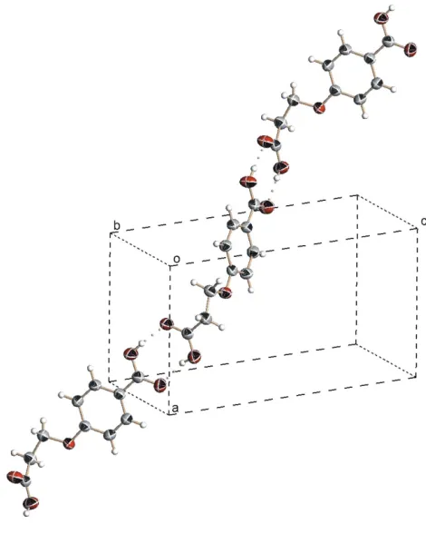
The molecules of the title compound, C10H10O5, are linked by
O—H O hydrogen bonds into a linear chain along [201].
Comment
The crystal structures of 2- and 3-carboxyphenoxyacetic acid have been reported previously. The 2-isomer exists as a zigzag chain polymer hydrogen-bonded through a pair of hydrogen bonds (Byriel et al., 1991), whereas the 3-isomer exists as a monohydrate that displays a three-dimensional hydrogen-bonded network (Gu et al., 2004). The structure of the 4-isomer has not been reported to date, as the compound does not furnish single crystals. Interest in the 4-isomer is extended to the title compound, (I), which has an extra methylene linkage.
The title compound is anhydrous (Fig. 1). A pair of hydrogen bonds (Table 1) links adjacent molecules into a linear chain along [201] (Fig. 2).
Experimental
4-Carboxyphenoxyacetic acid was synthesized in a manner analogous to that used for synthesizing 3-carboxyphenoxyacetic acid (Guet al., 2004), but with 4-hydroxybenzoic acid in place of 3-hydroxybenzoic acid and 3-chloropropionic acid in place of chloroacetic acid. Colourless crystals of (I) separated from the aqueous solution after several days.
Crystal data
C10H10O5
Mr= 210.18
Monoclinic,P21=c
a= 7.813 (4) A˚ b= 9.032 (6) A˚ c= 13.631 (9) A˚ = 94.92 (2) V= 958 (1) A˚3
Z= 4
Dx= 1.457 Mg m 3
MoKradiation = 0.12 mm1
T= 295 (2) K Plate, colourless 0.420.170.08 mm
Data collection
Rigaku R-AXIS RAPID IP diffractometer
!scans
Absorption correction: multi-scan (ABSCOR; Higashi, 1995) Tmin= 0.722,Tmax= 0.937
(expected range = 0.763–0.991)
9176 measured reflections 2188 independent reflections 1359 reflections withI> 2(I) Rint= 0.035
Refinement
Refinement onF2
R[F2> 2(F2)] = 0.046 wR(F2) = 0.153
S= 1.00 2188 reflections 144 parameters
H atoms treated by a mixture of independent and constrained refinement
w= 1/[2
(Fo2) + (0.0912P)2]
whereP= (Fo2+ 2Fc2)/3
(/)max= 0.001
max= 0.23 e A˚
3
min=0.20 e A˚
[image:2.610.320.555.71.365.2]3
Table 1
Hydrogen-bond geometry (A˚ ,).
D—H A D—H H A D A D—H A
O1—H1O O4i
0.83 (1) 1.82 (2) 2.618 (2) 162 (5) O5—H5O O2ii
0.84 (1) 1.85 (1) 2.676 (2) 173 (5)
Symmetry codes: (i)xþ1;yþ3 2;z
1
2; (ii)x1;yþ 3 2;zþ
1 2.
Carbon-bound H atoms were positioned geometrically, with C—H = 0.93 or 0.97 A˚ , and were included in the refinement in the riding-model approximation, withUiso(H) = 1.2Ueq(C). The acid H atoms were located in a difference Fourier map and were refined with a distance restraint of O—H = 0.82 (1) A˚ ; their displacement para-meters were freely refined.
Data collection: RAPID-AUTO(Rigaku, 1998); cell refinement:
RAPID-AUTO; data reduction: CrystalStructure (Rigaku/MSC, 2002); program(s) used to solve structure: SHELXS97 (Sheldrick, 1997); program(s) used to refine structure:SHELXL97(Sheldrick, 1997); molecular graphics:ORTEPII(Johnson, 1976); software used to prepare material for publication:SHELXL97.
The authors thank the National Natural Science Foundation of China (grant No. 20101003), the Scientific Fund for
Remarkable Teachers of Heilongjiang Province (grant No. 1054 G036) and the University of Malaya for supporting this study.
References
Byriel, K. A., Smith, D. E. & Kennard, C. H. L. (1991).Aust. J. Chem.44, 1459– 1464.
Gu, C.-S., Liu, J.-W., Huo, L.-H., Zhao, H., Zhao, J.-G. & Gao, S. (2004).Acta Cryst.E60, o760–o761.
Higashi, T. (1995).ABSCOR. Rigaku Corporation, Tokyo, Japan.
Johnson, C. K. (1976).ORTEPII. Report ORNL-5138. Oak Ridge National Laboratory, Tennessee, USA.
Rigaku (1998).RAPID-AUTO. Rigaku Corporation, Tokyo, Japan. Rigaku/MSC (2002). CrystalStructure. Rigaku/MSC Inc. The Woodlands,
Texas, USA.
[image:2.610.45.294.72.179.2]Sheldrick, G. M. (1997). SHELXL97 and SHELXS97. University of Go¨ttingen, Germany.
Figure 1
The molecular structure of (I). Displacement ellipsoids are drawn at the 50% probability level and H atoms are shown as spheres of arbitrary radii.
Figure 2
supporting information
sup-1 Acta Cryst. (2006). E62, o3420–o3421
supporting information
Acta Cryst. (2006). E62, o3420–o3421 [https://doi.org/10.1107/S1600536806027577]
3-(4-Carboxyphenoxy)propionic acid
Shan Gao and Seik Weng Ng
3-(4-Carboxyphenoxy)propionic acid
Crystal data
C10H10O5
Mr = 210.18 Monoclinic, P21/c Hall symbol: -P 2ybc
a = 7.813 (4) Å
b = 9.032 (6) Å
c = 13.631 (9) Å
β = 94.92 (2)°
V = 958 (1) Å3
Z = 4
F(000) = 440
Dx = 1.457 Mg m−3
Mo Kα radiation, λ = 0.71073 Å Cell parameters from 5947 reflections
θ = 3.0–27.5°
µ = 0.12 mm−1
T = 295 K Plate, colourless 0.42 × 0.17 × 0.08 mm
Data collection
Rigaku R-AXIS RAPID IP diffractometer
Radiation source: fine-focus sealed tube Graphite monochromator
ω scans
Absorption correction: multi-scan (ABSCOR; Higashi, 1995)
Tmin = 0.722, Tmax = 0.937
9176 measured reflections 2188 independent reflections 1359 reflections with I > 2σ(I)
Rint = 0.035
θmax = 27.5°, θmin = 3.0°
h = −10→8
k = −11→11
l = −17→17
Refinement
Refinement on F2 Least-squares matrix: full
R[F2 > 2σ(F2)] = 0.046
wR(F2) = 0.153
S = 1.00 2188 reflections 144 parameters 2 restraints
Primary atom site location: structure-invariant direct methods
Secondary atom site location: difference Fourier map
Hydrogen site location: inferred from neighbouring sites
H atoms treated by a mixture of independent and constrained refinement
w = 1/[σ2(F
o2) + (0.0912P)2] where P = (Fo2 + 2Fc2)/3 (Δ/σ)max = 0.001
Δρmax = 0.23 e Å−3 Δρmin = −0.20 e Å−3
Fractional atomic coordinates and isotropic or equivalent isotropic displacement parameters (Å2)
x y z Uiso*/Ueq
O1 0.7570 (2) 0.4302 (2) 0.2068 (1) 0.0650 (5)
O3 0.3768 (2) 0.4912 (2) 0.3441 (1) 0.0454 (4)
O4 −0.0388 (2) 0.9064 (2) 0.6092 (1) 0.0628 (5)
O5 −0.2541 (2) 0.8132 (2) 0.5119 (1) 0.0608 (5)
C1 0.5972 (2) 0.4410 (2) 0.1810 (1) 0.0402 (4)
C2 0.4826 (2) 0.3328 (2) 0.2264 (2) 0.0461 (5)
C3 0.3230 (2) 0.4016 (2) 0.2608 (2) 0.0432 (5)
C4 0.2544 (2) 0.5667 (2) 0.3894 (1) 0.0380 (4)
C5 0.0802 (2) 0.5598 (2) 0.3607 (1) 0.0426 (5)
C6 −0.0322 (2) 0.6431 (2) 0.4101 (1) 0.0434 (5)
C7 0.0258 (2) 0.7344 (2) 0.4873 (1) 0.0392 (4)
C8 0.2008 (2) 0.7377 (2) 0.5162 (1) 0.0441 (5)
C9 0.3143 (2) 0.6542 (2) 0.4687 (1) 0.0441 (5)
C10 −0.0943 (2) 0.8237 (2) 0.5398 (1) 0.0432 (5)
H1O 0.804 (7) 0.494 (4) 0.174 (4) 0.21 (2)*
H5O −0.325 (4) 0.856 (5) 0.544 (3) 0.18 (2)*
H2A 0.5466 0.2857 0.2820 0.055*
H2B 0.4493 0.2564 0.1785 0.055*
H3A 0.2656 0.4617 0.2089 0.052*
H3B 0.2442 0.3255 0.2790 0.052*
H5 0.0396 0.4995 0.3086 0.051*
H6 −0.1493 0.6379 0.3913 0.052*
H8 0.2414 0.7976 0.5686 0.053*
H9 0.4308 0.6560 0.4894 0.053*
Atomic displacement parameters (Å2)
U11 U22 U33 U12 U13 U23
O1 0.0463 (8) 0.0904 (12) 0.0569 (10) −0.0101 (8) −0.0033 (7) 0.0161 (9)
O2 0.0430 (8) 0.0649 (10) 0.0663 (10) 0.0041 (7) 0.0112 (7) 0.0194 (8)
O3 0.0385 (7) 0.0534 (8) 0.0446 (8) 0.0016 (6) 0.0059 (6) −0.0088 (6)
O4 0.0530 (9) 0.0779 (11) 0.0566 (10) 0.0052 (8) 0.0004 (7) −0.0228 (8)
O5 0.0387 (8) 0.0706 (10) 0.0733 (11) 0.0035 (7) 0.0067 (7) −0.0202 (8)
C1 0.0406 (9) 0.0434 (10) 0.0368 (10) 0.0016 (8) 0.0047 (8) −0.0052 (8)
C2 0.0492 (11) 0.0406 (11) 0.0497 (12) −0.0001 (9) 0.0117 (9) −0.0005 (9)
C3 0.0409 (10) 0.0468 (10) 0.0425 (11) −0.0033 (8) 0.0065 (8) −0.0028 (8)
C4 0.0360 (9) 0.0422 (10) 0.0361 (10) −0.0008 (8) 0.0049 (8) 0.0023 (8)
C5 0.0373 (9) 0.0488 (11) 0.0410 (10) −0.0017 (8) −0.0001 (8) −0.0058 (9)
C6 0.0330 (9) 0.0523 (11) 0.0446 (11) 0.0001 (8) 0.0010 (8) 0.0005 (9)
C7 0.0374 (10) 0.0423 (10) 0.0384 (10) 0.0004 (8) 0.0057 (8) 0.0017 (8)
C8 0.0401 (10) 0.0500 (11) 0.0420 (11) −0.0022 (8) 0.0011 (8) −0.0060 (9)
C9 0.0336 (9) 0.0525 (11) 0.0455 (11) −0.0014 (8) −0.0003 (8) −0.0027 (9)
C10 0.0390 (10) 0.0495 (11) 0.0413 (10) −0.0007 (9) 0.0037 (9) 0.0016 (9)
Geometric parameters (Å, º)
O1—C1 1.271 (2) C7—C10 1.469 (3)
O2—C1 1.241 (2) C8—C9 1.369 (3)
supporting information
sup-3 Acta Cryst. (2006). E62, o3420–o3421
O3—C3 1.428 (2) O5—H5O 0.84 (1)
O4—C10 1.253 (2) C2—H2A 0.97
O5—C10 1.277 (2) C2—H2B 0.97
C1—C2 1.494 (3) C3—H3A 0.97
C2—C3 1.504 (3) C3—H3B 0.97
C4—C5 1.385 (2) C5—H5 0.93
C4—C9 1.387 (3) C6—H6 0.93
C5—C6 1.375 (3) C8—H8 0.93
C6—C7 1.382 (3) C9—H9 0.93
C7—C8 1.390 (2)
C4—O3—C3 118.3 (1) C10—O5—H5O 119 (3)
O2—C1—O1 123.1 (2) C1—C2—H2A 108.8
O2—C1—C2 120.3 (2) C3—C2—H2A 108.8
O1—C1—C2 116.4 (2) C1—C2—H2B 108.8
C1—C2—C3 113.7 (2) C3—C2—H2B 108.8
O3—C3—C2 106.7 (2) H2A—C2—H2B 107.7
O3—C4—C5 124.0 (2) O3—C3—H3A 110.4
O3—C4—C9 115.7 (2) C2—C3—H3A 110.4
C5—C4—C9 120.2 (2) O3—C3—H3B 110.4
C6—C5—C4 119.4 (2) C2—C3—H3B 110.4
C5—C6—C7 121.1 (2) H3A—C3—H3B 108.6
C6—C7—C8 118.6 (2) C6—C5—H5 120.3
C6—C7—C10 121.1 (2) C4—C5—H5 120.3
C8—C7—C10 120.3 (2) C5—C6—H6 119.4
C9—C8—C7 121.1 (2) C7—C6—H6 119.4
C8—C9—C4 119.6 (2) C9—C8—H8 119.5
O4—C10—O5 122.4 (2) C7—C8—H8 119.5
O4—C10—C7 120.0 (2) C8—C9—H9 120.2
O5—C10—C7 117.6 (2) C4—C9—H9 120.2
C1—O1—H1O 106 (4)
O2—C1—C2—C3 −48.1 (3) C5—C6—C7—C10 179.8 (2)
O1—C1—C2—C3 135.7 (2) C6—C7—C8—C9 0.9 (3)
C4—O3—C3—C2 179.9 (2) C10—C7—C8—C9 179.4 (2)
C1—C2—C3—O3 −69.8 (2) C7—C8—C9—C4 1.1 (3)
C3—O3—C4—C5 1.3 (3) O3—C4—C9—C8 177.7 (2)
C3—O3—C4—C9 −178.8 (2) C5—C4—C9—C8 −2.3 (3)
O3—C4—C5—C6 −178.6 (2) C6—C7—C10—O4 −179.6 (2)
C9—C4—C5—C6 1.5 (3) C8—C7—C10—O4 1.9 (3)
C4—C5—C6—C7 0.5 (3) C6—C7—C10—O5 0.4 (3)
C5—C6—C7—C8 −1.7 (3) C8—C7—C10—O5 −178.1 (2)
Hydrogen-bond geometry (Å, º)
D—H···A D—H H···A D···A D—H···A
O5—H5O···O2ii 0.84 (1) 1.85 (1) 2.676 (2) 173 (5)