Original Article
Degree of SUVmax correlates with Ki-67 index in
patients with breast cancer: a meta-analysis
Guohua Shen, Shuang Hu, Bin Liu, Anren Kuang
Department of Nuclear Medicine, West China Hospital, Sichuan University, No. 37 Guo Xue Xiang, Chengdu 610041, Sichuan, People’s Republic of China
Received November 2, 2016; Accepted December 20, 2016; Epub February 1, 2017; Published February 15, 2017
Abstract: Positron emission tomography (PET) imaging using the radiotracer 18F-Fluorodeoxyglucose (FDG) has been proposed as imaging biomarkers of cell proliferation. We aimed to explore the correlation of FDG uptake with the Ki-67 labeling index in patients with breast cancer. Several databases were systematically searched for all relevant literature. The quality of included studies was evaluated according to the Quality Assessment of Diagnostic Accuracy Studies (QUADAS-2) tool. The correlation coefficient (rho) and its 95% confidence interval (CI) of individual studies were meta-analyzed using a random-effects model. The sources of heterogeneity were explored by sensitivity and subgroup analysis. The pooled rho value between SUVmax and Ki-67 index was 0.40 (95% CI, 0.35-0.46), which indicated an average correlation, but with a significant heterogeneity (I2=67.4%, P<0.01). Sensitivity analysis re-vealed that a single study contributed no significant influence to the overall estimate. Study design was a potential source of heterogeneity (rho=0.36 for prospective group vs. rho=0.47 for retrospective group, P<0.05) while other two factors, including scanning modality (PET, PET/CT or both) and sample method (surgery, biopsy or both), were not. In patients with breast cancer, the correlation between 18F-FDG uptake and tumor cell proliferation is significant but at a low degree.
Keywords: Breast cancer, PET/CT, SUVmax, Ki-67, meta-analysis
Introduction
Breast cancer is the most frequently diagnosed cancer and the leading cause of cancer death in women, with an estimated 14.1 million new cancer cases and 8.2 million cancer deaths occurring in 2012 worldwide [1]. Its prognostic factors included not only some traditional histo-logic features such as tumor size, histohisto-logic grade, nodal status and vascular invasion, but also some molecular markers involved in breast cancer biology [e.g. Ki-67 proliferation index, epidermal growth factor receptor (EGFR), hor-mone receptor status, cytokeratin (CK5/6)] [2, 3]. Ki-67, as a nuclear antigen expressed in all active phases of cell cycle (G1, S, and G2) except the G0, is present in all proliferation cells and has been established as a prolifera-tion biomarker in breast cancer [4-6]. Ki-67 positivity confers a higher risk of relapse and a worse survival in patients with breast cancer and is regarded as a prognostic marker in
breast cancer [6]. However, the measurement of Ki-67 index involving immunohistochemical staining necessitates an invasive biopsy. If we find a surrogate marker that can be obtained by non-invasive means, it will make response eval-uation and prognosis assessment easier for breast cancer patients.
In breast cancer patients, positron emission tomography/computed tomography (PET/CT) with 18F-fluoro-2-deoxy-2-D-glucose (18F-FDG)
Since both Ki-67 proliferation index and FDG uptake (SUVmax) are prognostic markers of breast cancer, it is of value to analysis the asso-ciation between these two factors, further to evaluate whether calculations of tumor FDG uptake by SUVmax could provide a non-invasive metabolic parameter that is associated with the biological aggressiveness of breast cancer. In this regard, several studies reported a signifi-cant correlation between these two factors ranging from 0.4 to 0.73 [13-17] while some studies indicated no significant correlation [3, 18-20]. In consideration of the controversial conclusions on this issue, we performed a meta-analysis to pool the eligible data, and evaluated the correlation of FDG uptake and Ki-67 index, in order to provide an evidence-based conclusion.
Material and methods
Literature search and study selection
A systematic search of MEDLINE, EMBASE, the Cochrane Library and China National Knowledge Infrastructure (CNKI) (inception to November
2015) was performed for relevant articles about the relationship between 18F-FDG uptake
and Ki-67 expression in breast cancer. All searches were limited to human studies. The search strategy was based on the combination of terms related to PET, FDG, Ki-67 and breast cancer. Inclusion criteria were as follows: (1) studies limited to breast cancer; (2) studies about the relationship between Ki-67 expres-sion and FDG uptake; (3) patients who had undergone PET scans before surgery, chemo-therapy or radiochemo-therapy; and (4) tumors con-firmed by cytopathology or histopathology. Reviews, case reports, conference abstracts and letters were excluded. Studies with sample size fewer than 10 or studies with insufficient data were also excluded.
Data extraction and quality assessment
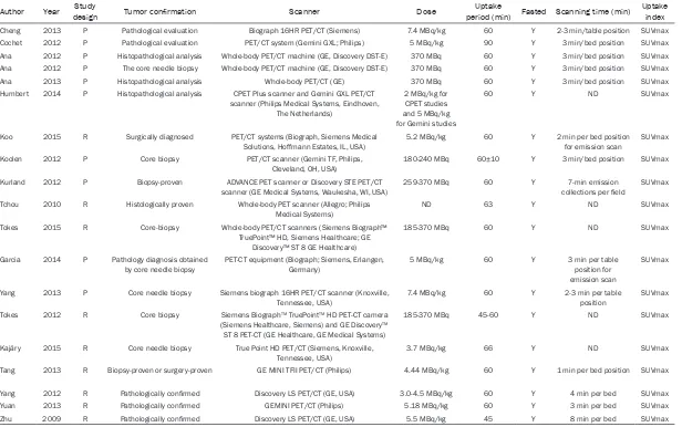
[image:2.612.91.525.71.385.2]Two reviewers independently extracted relevant data from the included studies, and the follow-ing information was recorded: first author, pub-lication year, study design, tumor type, number of patients, technical characteristics of PET
Table 1. Primary characteristics of 18F-FDG PET/CT scan
Author Year designStudy Tumor confirmation Scanner Dose period (min) Fasted Scanning time (min)Uptake Uptake index
Cheng 2013 P Pathological evaluation Biograph 16HR PET/CT (Siemens) 7.4 MBq/kg 60 Y 2-3 min/table position SUVmax
Cochet 2012 P Pathological evaluation PET/CT system (Gemini GXL; Philips) 5 MBq/kg 90 Y 3 min/bed position SUVmax
Ana 2012 P Histopathological analysis Whole-body PET/CT machine (GE, Discovery DST-E) 370 MBq 60 Y 3 min/bed position SUVmax Ana 2012 P The core needle biopsy Whole-body PET/CT machine (GE, Discovery DST-E) 370 MBq 60 Y 3 min/bed position SUVmax
Ana 2013 P Histopathological analysis Whole-body PET/CT (GE) 370 MBq 60 Y 3 min/bed position SUVmax
Humbert 2014 P Histopathological analysis CPET Plus scanner and Gemini GXL PET/CT scanner (Philips Medical Systems, Eindhoven,
The Netherlands)
2 MBq/kg for CPET studies and 5 MBq/kg for Gemini studies
60 Y ND SUVmax
Koo 2015 R Surgically diagnosed PET/CT systems (Biograph, Siemens Medical Solutions, Hoffmann Estates, IL, USA)
5.2 MBq/kg 60 Y 2 min per bed position for emission scan
SUVmax
Koolen 2012 P Core biopsy PET/CT scanner (Gemini TF, Philips,
Cleveland, OH, USA)
180-240 MBq 60±10 Y 3 min/bed position SUVmax
Kurland 2012 P Biopsy-proven ADVANCE PET scanner or Discovery STE PET/CT
scanner (GE Medical Systems, Waukesha, WI, USA) 259-370 MBq 60 Y collections per field7-min emission SUVmax Tchou 2010 R Histologically proven Whole-body PET scanner (Allegro; Philips
Medical Systems) ND 63 Y ND SUVmax
Tokes 2015 R Core-biopsy Whole-body PET/CT scanners (Siemens Biograph™ TruePoint™ HD, Siemens Healthcare; GE
Discovery™ ST 8 GE Healthcare)
185-370 MBq 60 Y ND SUVmax
Garcia 2014 P Pathology diagnosis obtained
by core needle biopsy PET-CT equipment (Biograph; Siemens, Erlangen, Germany) 5 MBq/kg 60 Y 3 min per table position for emission scan
SUVmax
Yang 2013 P Core needle biopsy Siemens biograph 16HR PET/CT scanner (Knoxville,
Tennessee, USA) 7.4 MBq/kg 60 Y 2-3 min per table position SUVmax
Tokes 2012 R Core biopsy Siemens BiographTM TruePointTM HD PET-CT camera
(Siemens Healthcare, Siemens) and GE DiscoveryTM
ST 8 PET-CT (GE Healthcare, GE Medical Systems)
185-370 MBq 45-60 Y ND SUVmax
Kajáry 2015 R Core needle biopsy True Point HD PET/CT (Siemens, Knoxville,
Tennessee, USA) 3.7 MBq/kg 66 Y ND SUVmax
Tang 2013 R Biopsy-proven or surgery-proven GE MINI TFII PET/CT (Philips) 4.44 MBq/kg 60 Y 1 min per bed position SUVmax
Yang 2012 R Pathologically confirmed Discovery LS PET/CT (GE, USA) 3.0-4.5 MBq/kg 60 Y 4 min per bed SUVmax
Yuan 2013 R Pathologically confirmed GEMINI PET/CT (Philips) 5.18 MBq/kg 60 Y 3 min per bed SUVmax
Zhu 2009 R Pathologically confirmed Discovery LS PET/CT (GE, USA) 5.5 MBq/kg 45 Y 8 min per bed SUVmax
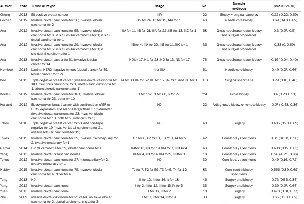
Table 2. The basic characteristics of Ki-67 immunohistochemistry
Author Year Tumor subtype Stage No. methodsSample Rho (95% CI)
Cheng 2013 ER-positive breast cancer II-IV 22 Biopsy + surgical samples 0.22 (-0.22, 0.59)
Cochet 2012 Invasive ductal carcinoma for 38; invasive lobular
carcinoma for 2 T2 for 24, T3 for 15, T4a for 1 40 Needle core biopsy 0.69 (0.48, 0.82)
Ana 2012 Invasive ductal carcinoma for 55; invasive lobular carcinoma for 6; in situ lobular carcinoma for 1; in situ ductal carcinoma for 1
IIA for 11, IIB for 21, IIIA for 22, IIIB for 13, IIIC for 1 68 Gross-needle aspiration biopsy and surgical procedures
0.3 (0.07, 0.5)
Ana 2012 Invasive ductal carcinoma for 29; invasive lobular carcinoma for 5; in situ lobular carcinoma for 1; in situ ductal carcinoma for 1
IIB for 4, IIIA for 20, IIIB for 11, IIIC for 1 36 Gross-needle aspiration biopsy
and surgical procedures 0.33 (0, 0.59)
Ana 2013 Invasive ductal cancer for 61; invasive lobular
cancer for 14 N0 for 17, N1 for 28, N2 for 13, N3 for 17 75 Gross-needle aspiration biopsy 0.19 (-0.04, 0.40)
Humbert 2014 Luminal HER2-negative tumors (ductal cancer for 49,
lobular cancer for 12) II or IIIA 61 Needle core biopsy 0.49 (0.27, 0.66)
Koo 2015 Triple-negative breast cancer (invasive ductal carcinoma for 100, mucinous carcinoma for 1, metaplastic carcinoma for 1, adenoid cystic carcinoma for 1)
IA for 30, IIA for 52, IIB for 15, IIIA for 5 and IIIB for 1 103 Surgical specimens 0.29 (0.10, 0.46)
Koolen 2012 Invasive ductal carcinoma for 181, invasive lobular
carcinoma for 23, other for 10 II for 127, III for 60, IV for 27 214 A core biopsy 0.4 (0.28, 0.51)
Kurland 2012 Biopsy-proven breast cancer with confirmation of ER or HER2 expression and lesions larger than 3 cm diameter (invasive ductal carcinoma for 23, invasive lobular carcinoma for 10, both for 2, unknown for 5)
ND 22 A diagnostic biopsy or remote biopsy -0.07 (-0.48, 0.36)
Tchou 2010 Triple negative breast cancer for 21 and non-triple negative for 19 (invasive ductal carcinoma for 21, invasive lobular carcinoma for 19)
ND 40 Surgery 0.485 (0.20, 0.69)
Tokes 2015 Invasive ductal carcinoma for 39, invasive micropapillary for 2, Invasive medullary for 1
T1c for 6, T2 for 31, T3 for 3, T4 for 2 42 Core-biopsy specimens 0.31 (0.007, 0.56)
Garcia 2014 Ductal carcinoma for 39, lobular carcinoma for 4 IIA for 13, IIB for 19, IIIA for 7, IIIB for 4 43 Core-biopsy specimens 0.408 (0.12, 0.63)
Yang 2013 Invasive ductal breast carcinomas IIA for 4, IIB for 4, IIIA for 9, IIIB for 1 18 Core-biopsy specimens 0.28 (-0.21, 0.66)
Tokes 2012 Invasive ductal carcinoma for 27, micropapillary for 2,
invasive medullary for 1 ND 30 Core-biopsy specimens 0.49 (0.16, 0.72)
Kajáry 2015 Invasive ductal carcinoma for 75, invasive lobular carcinoma for 4, other for 4
T1 for 7, T2 for 59, T3 for 5, T4 for 12 83 Core needle biopsy specimens
0.556 (0.39, 0.69)
Tang 2013 ND II for 12, III for 16, IV for 18 46 Surgery and biopsy 0.73 (0.56, 0.84)
Yang 2012 Invasive ductal carcinoma I for 2, II for 12, III for 16, IV for 5 35 Surgery and biopsy 0.39 (0.07, 0.64)
Yuan 2013 Invasive ductal carcinoma II for 16, III for 2 18 Surgery 0.473 (0.01, 0.77)
Zhu 2009 invasive ductal carcinoma for 25 cases, invasive lobular
carcinoma for 2, ductal carcinoma in situ for 3 I for 7, II for 14, III for 9 30 Surgery 0.65 (0.38, 0.82)
scan and Ki-67 measurement (imaging equip-ment, agent dose, uptake time, uptake index, and sample method), and correlation coeffi-cient (rho) value.
The methodological quality of included studies was assessed using the Quality Assessment of Diagnostic Studies-2 (QUADAS-2), which
[image:5.612.88.525.73.170.2]con-sists of four domains (patient selection, refer-ence standard, index test, and low and timing) that each requires a judgment of low, high or unclear for “Risk of bias” [21]. Three of these domains further need to be assessed in terms of concerns regarding applicability with “high”, “low” or “unclear” [21]. In our meta-analysis, the PET examination was designated as “index
Figure 2. Methodological quality of eligible studies with each item presented as percentages across all included studies.
[image:5.612.94.524.219.577.2]test” and the Ki-67 immunohistochemistry as “reference standard”.
Statistical analysis
The overall correlation coefficient was pooled based on individual Spearman correlation coef-ficient provided in each article, which was directly extracted from included studies. In cases that the rho value was not reported, it could be calculated based on the raw data using Spearman rank correlation analysis. In addition, the Pearson correlation coefficient was converted to Spearman correlation coeffi-cient based on previously published method [22]. The 95% confidence intervals (CIs) were calculated by Fisher transformation and inverse Fisher transformation [23]. Heterogeneity of included studies was evaluated by I2 index, and
it indicated the presence of significant hetero-geneity when I2 was more than 50% [24]. In
addition, the random-effect model was used to pooled analysis. We performed sensitivity anal-ysis and subgroup analanal-ysis to explore the sources of heterogeneity. The publication bias was evaluated by Begg’s test.
Statistical analysis was performed using STATA 12 software package (Stata Corporation, College Station, TX, USA). P<0.05 was consid-ered significant.
Results
Study selection and description
A total of 240 records were retrieved from the initial search. After reading the titles and abstracts, 177 articles were excluded due to duplication, irrelevant topic or article type. Sixty-three articles underwent full-text screen-ing, and 19 were eventually included for pooled analysis [3, 13-20, 25-34]. The flow chart of study selection was shown in Figure 1.
All included studies were published between 2009 and 2015, involving 1026 patients. Of these studies, ten ones [13, 15, 16, 18-20, 26, 27, 30, 32] were prospectively designed while nine [3, 14, 17, 25, 28, 29, 31, 33, 34] were retrospectively designed. The diagnosis of breast cancer was confirmed by core-biopsy or surgery in all of included studies, and the patients were in various stages. With regard to the PET scanning, only one studies [17] used PET scanner while sixteen [3, 14-16, 18, 19, 25-34] used PET/CT scanner, two [13, 20] used both. All of included studies provided SUVmax as the measurement of FDG uptake index. Some variations in acquisition and processing parameters of PET scanning such as PET scan-ner, dose and scanning time were observed among included studies. For the evaluation of cell proliferation, four studies [3, 17, 25, 31] assessed Ki-67 expression based on surgical-ly-acquired specimens while eight [13, 15, 16, 18, 20, 28, 30, 32, 34] used biopsy-acquired specimens, 5 [14, 19, 26, 27, 29] used both. The basic characteristics of included studies were presented in Tables 1 and 2.
The results of QUADAS-2 for assessing meth-odological quality
As presented in Figure 2, in six included stud-ies, the patient selection was judged to be at unclear risk of bias because they did not
[image:6.612.91.285.71.198.2]pro-Figure 4. The funnel plot of publication bias for SU-Vmax/Ki-67 correlation. The nonsignificant slope in-dicates the absence of publication bias (P=0.189).
Table 3. Results of subgroup analysis for 18
F-FDG/Ki-67 correlation in breast cancer Subgroup N Rho value (95% CI) I2 Pooled value 19 0.40 (0.35, 0.46) 67.4% Study design*
Prospective 10 0.36 (0.29, 0.43) 62.6% Retrospective 9 0.47 (0.39, 0.54) 65.4% Modality
PET 1 0.49 (0.20, 0.69)
PET/CT 16 0.41 (0.35, 0.46) 69.2% PET+PET/CT 2 0.34 (0.16, 0.52) 83.1% Sample method
[image:6.612.89.295.285.458.2]vide information about patient enrollment that is consecutive or random. Several studies [3, 17, 19, 20] only included breast cancer patients with certain subtypes such as triple-negative or hormonal receptor-positive, which might nar-row the range of patient selection, and give rise to high concern about the applicability.
We noted that in many studies, it was not clear if the interpretation of FDG uptake and Ki-67 expression were blind to each other; thus the index test and reference standard were consid-ered to be of unclear risk in these studies. In addition, several studies [17, 18, 32] did not provide enough information about PET scan-ning or Ki-67 immunohistochemistry, which led to some unclear concerns about the clinical applicability.
With regard to flow and timing, due to lack of an explicit description of the time interval between the PET scanning and immunohistochemistry, fourteen studies were at unclear risk of bias.
Meta-analysis of FDG/Ki-67 correlation
The rho values were directly extracted from most of included studies whereas for one study [30], rho was calculated based on the raw data of SUVmax and Ki-67 index. For two studies [14, 29], it was obtained by conversion of Pearson correlation coefficient.
The pooled rho value for all studies was 0.40 (95% CI, 0.35-0.46) with slightly high hetero- geneity among studies (I2=67.4%, P<0.001)
(Figure 3). Sensitivity analysis revealed that a single study contributed no significant influ-ence to the overall estimate. In addition, the Begg’s test showed that there was no signifi-cant publication bias (P=0.189) (Figure 4). As shown in Table 3, the subgroup analysis for study design revealed that the rho value of ret-rospective group was significantly higher than that of prospective group (rho=0.36 for pro-spective group vs. rho=0.47 for retropro-spective group, P<0.05) although there was still signifi-cant heterogeneity among these two subgroups (I2=62.6% for prospective group vs. I2=65.4%
for retrospective group). The results of sub-group analyses based on scanning modality (PET, PET/CT or both) and sample method (sur-gery, biopsy or both) did not show any signifi-cant difference. With regard to other factors such as tumor type and stage, we cannot
per-form the relevant subgroup analyses because of insufficient data.
Discussion
Ki-67 index has been regarded as a biomarker of proliferation activity of malignant cells in var-ious cancers [35], and a higher Ki-67 index is associated with more aggressive biological behavior and worse prognosis in breast cancer [3, 36]. As an invasive method involving biopsy or surgery, it has some drawbacks such as sample error during biopsy and the inability to perform multiple repeat procedures during or after the treatment to monitor the response or predict the prognosis. In the contrary, as a non-invasive method, PET/CT scan can be easily repeated at any point during or after treatment. Although FDG is not tumor-specific and not an indicator directly reflecting the cell prolifera-tion, its uptake is closely associated with cell proliferation for the reason that glycolytic metabolism involves in the proliferation pro-cess by providing energy and some molecules [37]. Many studies have focused on evaluating tumor proliferation based on the PET imaging [38-40]. In this study, we investigated whether a correlation existed between tumor 18F-FDG
uptake on PET/CT and cell proliferation activity, expressed as SUVmax and Ki-67 index respec-tively, for patients with breast cancer. The results showed that for breast cancer, FDG uptake and Ki-67 index displayed an average correlation (rho=0.40), which is a relatively low level [41]. In addition, the heterogeneity among studies was slightly high and its sources need-ed to be further explorneed-ed.
interpretation of PET/CT scan and Ki-67 immu-nohistochemistry, information whether using blind method was not obtained in many arti-cles. This condition might lead to interpreting bias for the reason that knowledge of the previ-ously performed examination may influence the judgment or interpretation of later one. In majority of included studies, the time interval between index test (PET/CT scan) and refer-ence standard (Ki-67 immunohistochemistry) was not clearly stated, which might be a poten-tial source of heterogeneity. If the PET/CT scan was performed after core biopsy, the time interval should be more than 7 days to avoid false positive accumulation of FDG caused by existence of inflammatory cells, as well as less than 1 month to avoid disease progression [15, 16, 38]. If the PET/CT scan was performed before biopsy or surgery, the interval also need-ed to be controllneed-ed.
In addition, subgroup analysis revealed that only study design (retrospective design vs. pro-spective design) contributed significantly to the overall estimate (rho=0.36 for prospective group vs. rho=0.47 for retrospective group,
P<0.05) while other two factors, including scan-ning modality (PET, PET/CT or both) and sample method (surgery, biopsy or both), did not. If a study was retrospective, a potential risk may exist that researchers have known results of PET/CT imaging or Ki-67 index in advance; thus, pooled rho value of retrospective sub-group was significantly higher than that of pro-spective subgroup. Although further analysis for the factors such as tumor subtypes or stag-es was not performed, they were still consid-ered to introduce some bias. Finally, based on the result of Begg’s test, the publication bias was not significantly observed among included studies.
Although it has been proved to be an effective marker for prognosis in breast cancer [6, 45], Ki-67 has a main disadvantage that is the high degree of interobserver variability in its assess-ment [46]. The measureassess-ment of Ki-67 can vary due to several factors including human error, tumor area selection, specific antibody and analysis method [3, 47]. A recent study showed that for determination of the Ki-67 index, differ-ent methods yielded differdiffer-ent results with 67% of examined tumor in inconsistent grading [48]. Another study also demonstrated that the Ki-67 labeling index differed according to the
measurement methods (hot pot vs. average) and specimen types (core needle biopsy vs. surgery) [47]. In clinical practice, Ki-67 index was assessed only in a few micrometer thick sections that are representative samples not the entire tumor. If the tumor is in high intra-tumoural heterogeneity, the concordance between selected section and whole tumor is low, and the Ki-67 index of selected sections cannot reveal the true level of proliferation activity of whole tumor [47]. A previous study showed that there were large differences of Ki-67 expression owing to intratumoral hetero-geneity, with maximum index ranging from 4.9% to 92.2%, average index ranging from 3.4% to 81.4%, respectively [49]. With regard to the measurement of SUVmax, it is easily calcu-lated with available commercial software. Although it is affected by the voxel size and tumor motion, it is not subject to the interob-server variability because it is not based on the delineation method [50]. For the SUVmax of small lesions, the partial volume effect might be a possible source of measurement error [50].
The present study has some limitations. First, the number of included studies was relatively small, and we cannot perform further analysis for the certain subtype of breast cancer. Further, a wide variation in Ki-67 immunohisto-chemistry and its measurement method exist-ed among includexist-ed studies, which was a main source of heterogeneity. Moreover, we only included full-text articles with sufficient data, possibly resulting in a bias; however, Begg’s test revealed no significant bias.
In conclusion, in patients with breast cancer,
18F-FDG uptake showed a positive correlation
with tumor cell proliferation; but the degree of correlation is low, which probably limits its application in clinical practice. PET/CT imaging may be a useful non-invasive tool to assess proliferation activity of breast cancer. However, our results need further validation by larger, prospective studies with improved study design, especially for those specific subtypes of breast cancer.
Acknowledgements
Disclosure of conflict of interest
None.
Address correspondence to: Dr. Anren Kuang,
Department of Nuclear Medicine, West China Hospital, Sichuan University, No. 37 Guo Xue Xiang, Chengdu 610041, China. Tel: +86-18980601582; Fax: +86 28 85422155; E-mail: kuanganren@263. net
References
[1] Torre LA, Bray F, Siegel RL, Ferlay J, Lortet-Tieu-lent J, Jemal A. Global cancer statistics, 2012. CA Cancer J Clin 2015; 65: 87-108.
[2] Colozza M, Azambuja E, Cardoso F, Sotiriou C, Larsimont D, Piccart MJ. Proliferative markers as prognostic and predictive tools in early breast cancer: where are we now? Ann Oncol 2005; 16: 1723-1739.
[3] Koo HR, Park JS, Kang KW, Han W, Park IA, Moon WK. Correlation between (18)F-FDG up-take on PET/CT and prognostic factors in tri-ple-negative breast cancer. Eur Radiol 2015; 25: 3314-3321.
[4] Gerdes J, Schwab U, Lemke H, Stein H. Produc-tion of a mouse monoclonal antibody reactive with a human nuclear antigen associated with cell proliferation. Int J Cancer 1983; 31: 13-20. [5] Gerdes J, Lemke H, Baisch H, Wacker HH,
Schwab U, Stein H. Cell cycle analysis of a cell proliferation-associated human nuclear anti-gen defined by the monoclonal antibody Ki-67. J Immunol 1984; 133: 1710-1715.
[6] de Azambuja E, Cardoso F, de Castro G Jr, Colozza M, Mano MS, Durbecq V, Sotiriou C, Larsimont D, Piccart-Gebhart MJ, Paesmans M. Ki-67 as prognostic marker in early breast cancer: a meta-analysis of published studies involving 12,155 patients. Br J Cancer 2007; 96: 1504-1513.
[7] Groheux D, Espie M, Giacchetti S, Hindie E. Performance of FDG PET/CT in the clinical management of breast cancer. Radiology 2013; 266: 388-405.
[8] Kadoya T, Aogi K, Kiyoto S, Masumoto N, Suga-wara Y, Okada M. Role of maximum standard-ized uptake value in fluorodeoxyglucose posi-tron emission tomography/computed tomo- graphy predicts malignancy grade and progno-sis of operable breast cancer: a multi-institute study. Breast Cancer Res Treat 2013; 141: 269-275.
[9] Jo JE, Kim JY, Lee SH, Kim S, Kang T. Preopera-tive 18F-FDG PET/CT predicts disease-free survival in patients with primary invasive duc-tal breast cancer. Acta Radiol 2015; 56: 1463-1470.
[10] Aogi K, Kadoya T, Sugawara Y, Kiyoto S, Shige-matsu H, Masumoto N, Okada M. Utility of (18) F FDG-PET/CT for predicting prognosis of lumi-nal-type breast cancer. Breast Cancer Res Treat 2015; 150: 209-217.
[11] Holliday DL, Moss MA, Pollock S, Lane S, Shaa-ban AM, Millican-Slater R, Nash C, Hanby AM, Speirs V. The practicalities of using tissue slic-es as preclinical organotypic breast cancer models. J Clin Pathol 2013; 66: 253-255. [12] Ohara M, Shigematsu H, Tsutani Y, Emi A,
Ma-sumoto N, Ozaki S, Kadoya T, Okada M. Role of FDG-PET/CT in evaluating surgical outcomes of operable breast cancer--usefulness for ma-lignant grade of triple-negative breast cancer. Breast 2013; 22: 958-963.
[13] Humbert O, Berriolo-Riedinger A, Cochet A, Gauthier M, Charon-Barra C, Guiu S, Desmoul-ins I, Toubeau M, Dygai-Cochet I, Coutant C, Fumoleau P, Brunotte F. Prognostic relevance at 5 years of the early monitoring of neoadju-vant chemotherapy using (18)F-FDG PET in lu-minal HER2-negative breast cancer. Eur J Nucl Med Mol Imaging 2014; 41: 416-427.
[14] Tang MD, Liu DJ, Lin DY, Zhang JP, Li SY, Cai ZH. The correlation between FDG uptake and ex-pression of Ki-67, ER, PR, HER-2 in breast can-cer. J Chin Oncol 2013; 19: 944-946.
[15] Koolen BB, Vrancken Peeters MJ, Wesseling J, Lips EH, Vogel WV, Aukema TS, van Werkhoven E, Gilhuijs KG, Rodenhuis S, Rutgers EJ, Valdés Olmos RA. Association of primary tumour FDG uptake with clinical, histopathological and mo-lecular characteristics in breast cancer pa-tients scheduled for neoadjuvant chemothera-py. Eur J Nucl Med Mol Imaging 2012; 39: 1830-1838.
[16] Cochet A, Pigeonnat S, Khoury B, Vrigneaud JM, Touzery C, Berriolo-Riedinger A, Dygai-Co-chet I, Toubeau M, Humbert O, Coudert B, Fu-moleau P, Arnould L, Brunotte F. Evaluation of breast tumor blood flow with dynamic first-pass 18F-FDG PET/CT: comparison with angio-genesis markers and prognostic factors. J Nucl Med 2012; 53: 512-520.
[17] Tchou J, Sonnad SS, Bergey MR, Basu S, To-maszewski J, Alavi A, Schnall M. Degree of tu-mor FDG uptake correlates with proliferation index in triple negative breast cancer. Mol Im-aging Biol 2010; 12: 657-662.
[19] Cheng J, Lei L, Xu J, Sun Y, Zhang Y, Wang X, Pan L, Shao Z, Zhang Y, Liu G. 18F-fluoromiso-nidazole PET/CT: a potential tool for predicting primary endocrine therapy resistance in breast cancer. J Nucl Med 2013; 54: 333-340. [20] Kurland BF, Gadi VK, Specht JM, Allison KH,
Livingston RB, Rodler ET, Peterson LM, Schubert EK, Chai X, Mankoff DA, Linden HM. Feasibility study of FDG PET as an indicator of early response to aromatase inhibitors and trastuzumab in a heterogeneous group of breast cancer patients. EJNMMI Res 2012; 2: 34.
[21] Whiting PF, Rutjes AW, Westwood ME, Mallett S, Deeks JJ, Reitsma JB, Leeflang MM, Sterne JA, Bossuyt PM; QUADAS-2 Group. QUADAS-2: a revised tool for the quality assessment of di-agnostic accuracy studies. Ann Intern Med 2011; 155: 529-536.
[22] Rupinski MT, Dunlap WP. Approximating Pear-son Product-Moment Correlations from Kend-all’s Tau and Spearman’s Rho. Educational and Psychological Measurement 1996; 56: 419-429.
[23] Chalkidou A, Landau DB, Odell EW, Cornelius VR, O’Doherty MJ, Marsden PK. Correlation be-tween Ki-67 immunohistochemistry and 18F-fluorothymidine uptake in patients with can-cer: A systematic review and meta-analysis. Eur J Cancer 2012; 48: 3499-3513.
[24] Higgins JP, Thompson SG. Quantifying hetero-geneity in a meta-analysis. Stat Med 2002; 21: 1539-1558.
[25] Zhu SG, Zou HD, Qiao GD. Research on corre-lation between fluorine-18-fluorodeoxyglucose uptake in PET-CT and tumor proliferation as-sessed by Ki-67 in breast cancer. J Int Oncol 2009; 36: 792-794.
[26] Garcia Vicente AM, Castrejon AS, Relea Calata-yud F, Munoz AP, Leon Martin AA, Lopez-Muniz IC, Del Mar Muñoz Sánchez M, Cordero García JM, Becerra Nakayo EM. 18F-FDG retention index and biologic prognostic parameters in breast cancer. Clin Nucl Med 2012; 37: 460-466.
[27] Garcia Vicente AM, Soriano Castrejon A, Relea Calatayud F, Munoz Madero V, Molina Garrido MJ, Leon Martin AA, Cordero García JM, Pilk-ington Woll JP, Chacón López-Muñiz I, Palomar Muñoz A. 18F-FDG semi-quantitative parame-ters and biological prognostic factors in locally advanced breast cancer. Rev Esp Med Nucl Imagen Mol 2012; 31: 308-314.
[28] Tokes T, Somlai K, Szekely B, Kulka J, Szentm-artoni G, Torgyik L, Galgóczy H, Lengyel Z, Györke T, Dank M. The role of FDG-PET-CT in the evaluation of primary systemic therapy in breast cancer: links between metabolic and pathological remission. Orv Hetil 2012; 153: 1958-1964.
[29] Yang H, Lin MF, Chen XG. Relationship between standardized uptake value of 18F-FDG PET/CT and Ki-67 expression in breast cancer. J Nan-chang Univ 2012; 52: 52-55,57.
[30] Yang Z, Sun Y, Xue J, Yao Z, Xu J, Cheng J, Shi W, Zhu B, Zhang Y, Zhang Y. Can Positron Emis-sion Tomography/Computed Tomography with the Dual Tracers Fluorine-18 Fluoroestradiol and Fluorodeoxyglucose Predict Neoadjuvant Chemotherapy Response of Breast Cancer? ----A Pilot Study. PLoS One 2013; 8: e78192. [31] Yuan JW, Yang J, He XH. Correlation between
combined imaging modalities of 18F-FDG PET/ CT and 3.0T MRI and expression of Ki-67 in breast cancer. Int J Radiat Med Nucl Med 2013; 37: 84-87.
[32] Garcia Garcia-Esquinas M, Garcia-Saenz JA, Ar-razola Garcia J, Enrique Fuentes Ferrer M, Fu-rio V, Rodriguez Rey C, Román JM, Carreras Delgado JL. 18F-FDG PET-CT imaging in the neoadjuvant setting for stages II-III breast can-cer: association of locoregional SUVmax with classical prognostic factors. Q J Nucl Med Mol Imaging 2014; 58: 66-73.
[33] Kajary K, Tokes T, Dank M, Kulka J, Szakall S Jr, Lengyel Z. Correlation of the value of 18F-FDG uptake, described by SUVmax, SUVavg, meta-bolic tumour volume and total lesion glycolysis, to clinicopathological prognostic factors and biological subtypes in breast cancer. Nucl Med Commun 2015; 36: 28-37.
[34] Tokes T, Szentmartoni G, Torgyik L, Somlai K, Kulka J, Lengyel Z, Györke T, Dank M. Complex-ity of Response Evaluation During Primary temic Therapy of Breast Cancer: Scoring Sys-tems and Beyond-Preliminary Results. Anticancer Res 2015; 35: 5063-5072.
[35] Gerdes J, Li L, Schlueter C, Duchrow M, Wohlenberg C, Gerlach C, Stahmer I, Kloth S, Brandt E, Flad HD. Immunobiochemical and molecular biologic characterization of the cell proliferation-associated nuclear antigen that is defined by monoclonal antibody Ki-67. Am J Pathol 1991; 138: 867-873.
[36] Inwald E, Klinkhammer-Schalke M, Hofstädter F, Zeman F, Koller M, Gerstenhauer M, Ort-mann O. Ki-67 is a prognostic parameter in breast cancer patients: results of a large popu-lation-based cohort of a cancer registry. Breast Cancer Res Treat 2013; 139: 539-552. [37] Vander Heiden MG, Cantley LC, Thompson CB.
Understanding the Warburg effect: the meta-bolic requirements of cell proliferation. Sci-ence 2009; 324: 1029-1033.
[39] Ikenaga N, Otomo N, Toyofuku A, Ueda Y, Toyo-da K, Hayashi T, Nishikawa K, Tanaka M. Stan-dardized uptake values for breast carcinomas assessed by fluorodeoxyglucose-positron emission tomography correlate with prognostic factors. Am Surg 2007; 73: 1151-1157. [40] Buck A, Schirrmeister H, Kuhn T, Shen C,
Kalk-er T, KotzKalk-erke J, DankKalk-erl A, Glatting G, Reske S, Mattfeldt T. FDG uptake in breast cancer: cor-relation with biological and clinical prognostic parameters. Eur J Nucl Med Mol Imaging 2002; 29: 1317-1323.
[41] Landis JR, Koch GG. The measurement of ob-server agreement for categorical data. Biomet-rics 1977; 33: 159-174.
[42] Rakha EA, Reis-Filho JS, Ellis IO. Basal-like breast cancer: a critical review. J Clin Oncol 2008; 26: 2568-2581.
[43] Koo HR, Park JS, Kang KW, Cho N, Chang JM, Bae MS, Kim WH, Lee SH, Kim MY, Kim JY, Seo M, Moon WK. 18F-FDG uptake in breast can-cer correlates with immunohistochemically de-fined subtypes. Eur Radiol 2014; 24: 610-618. [44] Basu S, Chen W, Tchou J, Mavi A, Cermik T, Cz-erniecki B, Schnall M, Alavi A. Comparison of triple-negative and estrogen receptor-positive/ progesterone receptor-positive/HER2-negative breast carcinoma using quantitative fluo-rine-18 fluorodeoxyglucose/positron emission tomography imaging parameters. Cancer 2008; 112: 995-1000.
[45] Zhang G, Xie W, Liu Z, Lin C, Piao Y, Xu L, Guo F, Xie X. Prognostic function of Ki-67 for patho-logical complete response rate of neoadjuvant chemotherapy in triple-negative breast cancer. Tumori 2013; 100: 136-142.
[46] Urruticoechea A, Smith IE, Dowsett M. Prolifer-ation marker Ki-67 in early breast cancer. J Clin Oncol 2005; 23: 7212-7220.
[47] Yamamoto S, Chishima T, Mastubara Y, Adachi S, Harada F, Toda Y, Arioka H, Hasegawa N, Ka-kuta Y, Sakamaki K. Variability in measuring the ki-67 labeling index in patients with breast cancer. Clin Breast Cancer 2015; 15: e35-e39. [48] Goodell PP, Krasinskas AM, Davison JM, Hart-man DJ. Comparison of methods for prolifera-tive index analysis for grading pancreatic well-differentiated neuroendocrine tumors. Am J Clin Pathol 2012; 137: 576-582.
[49] Muller W, Schneiders A, Meier S, Hommel G, Gabbert HE. Immunohistochemical study on the prognostic value of MIB-1 in gastric carci-noma. Br J Cancer 1996; 74: 759-765. [50] Chalkidou A, Landau D, Odell E, Cornelius V,