Original Article
Protein kinase TTK promotes proliferation and
migration and mediates epithelial-mesenchymal
transition in human bladder cancer cells
Feiran Chen1, Peikang Wu1, Hailong Hu1, Dawei Tian1, Ning Jiang1,2, Changli Wu1,3
1Department of Urology, The Second Hospital of Tianjin Medical University, Tianjin, China; 2Tianjin Key Laboratory
of Urology, Tianjin Institute of Urology, The Second Hospital of Tianjin Medical University, Tianjin, China; 3
Depart-ment of Urology, Sino-Singapore Eco-city Hospital of Tianjin Medical University, Tianjin, China
Received August 22, 2018; Accepted September 16, 2018; Epub October 1, 2018; Published October 15, 2018
Abstract: Aim: To investigate the expression level of TTK in bladder cancer, and its role in the proliferation and migration. To investigate the relationship between TTK and epithelial-mesenchymal transition (EMT). Patients and Methods: We compared the expression level of TTK between human bladder cancer tissues and normal bladder epithelial tissues from 70 patients using immunohistochemistry, qRT-PCR and western blotting. Subsequently, we conducted cell viability and cell migration experiments to investigate the effect of TTK on bladder cancer cells. Fur-thermore, we used qRT-PCR to detect the biomarkers of EMT to examine the relationship between TTK and EMT. Results:The expression level of TTK was significantly higher in bladder cancer tissues as compared to the adjacent noncancerous tissues (P < 0.001). The qRT-PCR, immunohistochemistry, and western blotting also showed the same trend. Furthermore, cell viability and cell migration assays showed that TTK promoted proliferation and migra-tion of human bladder cancer cells, and mediated EMT. Conclusion:This study showed that high expression of TTK can promote proliferation and migration, and mightmediate the EMT process in human bladder cancer cells.
Keywords: Bladder cancer, TTK, proliferation, migration, EMT
Introduction
Bladder cancer is the second most commonly diagnosed genitourinary neoplasm, with appro- ximately 360,000 new cases diagnosed world-wide and 145,000 deaths each year [1, 2].The majority of bladder cancer (75%) cases are cla- ssified as non-muscle-invasive bladder cancer (NMIBC) at the first diagnosis [3], which is char-acterized by a high possibility of recurrence and progression to muscle invasive bladder cancer (MIBC) [4]. The progression of NMIBC to MIBC is related to epithelial-mesenchymal transition (EMT), which is vital in the proliferation and migration of several cancers [5, 6].
EMT is a complex process in which epithelial cells acquire a mesenchymal phenotype and motility through a cascade of biologic events [7]. During tumorigenesis, changes in EMT reg-ulatory pathways lead to a loss of cellular adhe-sion, changes in the polarization of the cells and cytoskeleton, detachment, migration, intra-
vasation, and survival in the vascular system; extravasation, and metastasis [8]. Morpholo- gically, EMT is classically characterized by the dedifferentiation from an epithelial to mesen-chymal phenotype, marked by the decreased expression of E-cadherin, and increased expre- ssion of N-cadherin, vimentin, and cellular pro-teases [9]. Our previous study showed that EMT could be a critical step in the progression from the pre-invasive to invasive state in bladder cancer [10].
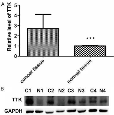
Figure 1. A. TTK expression in bladder cancer tis-sues and adjacent noncancerous tistis-sues (n = 70) was analyzed by qRT-PCR. The expression of TTK was significantly higher in bladder cancer tissues than in adjacent non-cancer tissues (***, P < 0.001). B. TTK marker was assayed in four pairs of bladder cancer and normal tissues by western blotting (C, tumor; N, normal), which showed that TTK was highly ex-pressed in human bladder cancer.
levels were found in several human cancers, including thyroid papillary carcinoma, breast cancer, gastric cancer, bronchogenic carcino-ma, and lung cancers [15-17]. Also, high levels of TTK correlated with a more aggressive histo-logic grade in breast cancers [18]. TTK plays a role in the genotoxic stress response, such as stress caused by DNA damage [19].
In this study, we assessed the differential expression of TTK between bladder cancer sues and matched adjacent non-cancerous tis-sues. We also investigated whether TTK pro-motes proliferation and migration of human bladder cancer cells, and could mediate EMT. Materials and methods
Patients and tissue specimens
This study was approved by the Ethics Com- mittee of the Second Hospital of Tianjin Medical University. Written informed consent was ob- tained from the patients for using their tissue specimens. The bladder cancer tissues and adjacent noncancerous tissues were collected from patients who underwent radical cystecto-my at the Second Hospital of Tianjin Medical
mens were snap-frozen in liquid nitrogen imme-diately after resection and stored at -80°C until use. All tissues were independently reviewed by two pathologists to determine the tumor stage and histologic grade according to the 2016 WHO classification of tumors [20].
qRT-PCR analysis
Total RNA was extracted from the tissues ac- cording to a previous protocol [21], and reverse-transcribed to cDNA using a Reverse Tran- scription kit (Roche, USA). Real-time PCR was performed using an Applied Biosystems 790- 0HT instrument with 20 μl PCR reaction mix-ture containing 10 μl of 2× Lightcycler 480 SYBR Green I Master (Roche). Reactions were incubated in a 96-well optical plate at 95°C for 10 min, followed by 45 cycles at 95°C for 15 s, and at 60°C for 60 s. Each sample was run in triplicate. A melting curve analysis was con-ducted to validate the specific generation of the expected PCR product. The primers for amplifi-cation of TTK were as follows: 5’-TCCCCAGC- GCAGCTTTCTGTAGA-3’ (forward) and 5’-CCAG- TCCTCTGGGTTGTTTGCCAT-3’ (reverse). N-cad- herin primers were: forward, 5’-TAAACTGCCT- GGCCGAATC-3’ and reverse 5’-TCCTTCTGCCTC- TATGACCTG-3’. E-cadherin primers were: for-ward, 5’-CAACGACCCAACCCAAGAA-3’ and re- verse 5’-CCGAAGAAACAGCAAGAGCA-3’. ZEB1 primers were: forward, 5’-AACTGCTGGCAAGAC- AAC-3’ and reverse 5’-TTGCTGCAGAAATTCTTC- CA-3’. Human GAPDH was amplified as an inter-nal control using the forward primer 5’-CTCG- CTTCGGCAGCACA-3’ and reverse primer 5’-AA- CGCTTCACGAATTTGCGT-3’. The data were cal-culated by 2-ΔΔCT method [22].
Immunohistochemistry and western blotting
Figure 2. The expression of TTK in normal human bladder urothelium and urothelial carcinoma. (A) and (C) show low expression in normal human urothelium; while (B) and (D) show high expression in human urothelial carcinoma.
Total tissue and cell proteins were extracted using cell lysate buffer with 1% protease/phos-phatase inhibitor cocktail. Equal amount of pro-teins were separated by 10% SDS-PAGE and transferred to nitrocellulose membranes. The membranes were blocked with 5% non-fat milk in PBS buffer and then incubated with primary antibodies overnight at 4°C. The protein bands were scanned after incubation with secondary antibodies conjugated with ECL and quantita-tively measured with Quantity One software.
Cell lines and cell culture
Human bladder cancer cell lines EJ, T24 and 5637 were purchased from the American Type Culture Collection (Manassas, VA, USA). The
cells were cultured in RPMI 1640 medium with 10% fetal bovine serum (FBS; Gibco, USA) and grown at 37°C in an atmosphere of 95% air and 5% CO2.
Cell migration assay
Figure 3. A. Western blotting showed that TTK expression was down-regulated in human bladder cancer cell lines (EJ, T24 and 5637) after transfection with TTK-siRNA for 48 h. B. Images of migration of different human bladder cancer cell lines in transwell assay. The number of migrated cells in the histograms represented mean values per field (from three fields, mean ± SD) (***, P = 0.0013).
were fixed with 4% paraformaldehyde for 15 min and stained with 0.5% crystal violet for at least 30 min. Cells in the upper chamber were removed using cotton swabs. Cells on the lower surface of the membrane were counted and photographed in at least three random micro-scopic fields (magnification, ×40). The experi-ments were independently repeated three times.
Cell viability analysis
Bladder cancer cells (EJ, T24 and 5637) were seeded in 96-well plates, with 2.0×10⁴ cells per well. The cells were incubated for 0, 24, 48,
72, 96 h. The 3-(4,5-dimethylthiazol-2-yl)-2,5-di- phenyltetrazolium bromide (MTT) assay was used to analyze the cell viability as previously described [23].
Statistical analysis
Figure 4. The effects of TTK on cell proliferation. Blad-der cancer cell lines were transfected with NC-siRNA and TTK-siRNA for 24, 48, and 72 h. The cell growth was measured by MTT assay. The y-axis represents cell viability based on measured OD560 values. The x-axis represents time course.
Results
TTK expression is increased in human bladder cancer
We first used 70 pairs of bladder cancer sues and matched adjacent noncancerous tis-sues to analyze the expression level of TTK by qRT-PCR. The results showed that TTK mRNA expression was higher in the bladder cancer tissues than in the paired adjacent non-tumor specimens (Figure 1A, ***, P < 0.001).
Next, we conducted immunohistochemistry and western blotting with bladder cancer and normal bladder tissues, and found that the expression of TTK was significantly higher in
bladder cancer tissues than in normal bladder tissues (Figures 1B, 2).
Down-regulation of TTK inhibits proliferation and migration of human bladder cancer cells
To examine the effects of TTK on bladder can-cer cell growth and proliferation, we conducted siRNA-mediated knockdown experiments in bladder cancer cell lines (T24, 5637 and EJ). The knockdown efficiency of the siRNA was measured by western blotting (Figure 3A), with NC-siRNA as a control. The results showed that TTK-siRNA could down-regulate TTK expres-sion. In cell migration with transwell assay, transfection of TTK-siRNA in T24, 5637 and EJ cells significantly decreased cell migration (P < 0.01, Figure 3B). Furthermore, the TTK-siRNA group showed lower cell viability as compared to the control group (P < 0.01, Figure 4). These results suggested that TTK contributes to tumorigenesis by promoting growth and migra-tion of bladder cancer cells.
TTK could promote EMT in bladder cancer cells
We compared the expression of EMT biomark-ers between the control group and the si-TTK group. N-cadherin and ZEB1 showed signifi-cantly lower expression in the si-TTK group as compared to the control group. In contrast, E-cadherin was increased in the si-TTK group (P < 0.01, Figure 5). These data suggested that TTK may be involved in EMT.
Discussion
In this study, we evaluated the expression level of TTK in bladder cancer tissues and normal bladder epithelial tissues. The results showed that TTK had a higher expression in bladder cancer tissues than in normal bladder epithelial tissues. Similar findings have been reported in thyroid papillary carcinoma, breast cancer, gas-tric cancer, bronchogenic carcinoma, and lung cancers [15-17, 23, 27]. This finding suggested that TTK acts as an oncogene, and could acti-vate some oncogenic signals. Our findings have also shown that down-regulation of TTK could decrease the proliferation and migration of human bladder cancer cells, which could be because TTK is an essential kinase for proper distribution of chromosomes in mitosis, and could cause severe chromosomal segregation defects that lead to cancer cell death [28]. Numerous potent TTK inhibitors have been reported as cancer therapeutic targets in clini-cal trials [29].
A prominent characteristic of cancer is inva-sion, and EMT is known to play a role in invasion and metastasis [30-33]. The present study showed that down-regulation of TTK led to decreased expression of N-cadherin and ZEB1, while E-cadherin expression was increased. The results suggested that inhibiting TTK could inhibit the EMT process in human bladder can-cers. This is because activation of TTK can pro-mote transforming growth factor-beta (TGF-b)-independent Smad signaling [34], which is one of the primary pathways involved in EMT. Our future study will focus on the molecular mecha-nism of TTK-mediated EMT in bladder cancer. In summary, our study showed that TTK was highly expressed in human bladder cancer, and this could promote proliferation, migration, and EMT.
Acknowledgements
This study was supported by the Science foun-dation of Tianjin (No. 16JCZDJC34400).
Disclosure of conflict of interest
None.
Address correspondence to: Changli Wu and Ning Jiang, Department of Urology, The Second Hospital of Tianjin Medical University, 23 Pingjiang Road, Hexi District, Tianjin 300211, China. Tel: +86-13820197998; Fax: +86-21-64085875; E-mail: wujygc2003@163.com (CLW); jiangningbear@163. com (NJ)
References
[1] Ploeg M, Aben KK and Kiemeney LA. The pres-ent and future burden of urinary bladder can-cer in the world. World J Urol 2009; 27: 289-293.
[2] Ferlay J, Soerjomataram I, Ervik M, Dikshit R, Eser S, Mathers C, Rebelo M, Parkin DM, For-man D, Bray F. GLOBOCAN 2012 v1.1, Cancer Incidence and Mortality Worldwide: IARC Can-cer Base No. 11. 2012.
[3] Chen W, Zheng R, Baade PD, Zhang S, Zeng H, Bray F, Jemal A, Yu XQ and He J. Cancer statis-tics in China, 2015. CA Cancer J Clin 2016; 66: 115-132.
[4] Fidler IJ. Critical determinants of metastasis. Semin Cancer Biol 2002; 12: 89-96.
[5] Soloway MS. Overview of treatment of superfi -cial bladder cancer. Urology 1985; 26: 18-26. [6] Gorin MA, Ayyathurai R and Soloway MS.
Diag-nosis and treatment of bladder cancer: how can we improve? Postgrad Med 2012; 124: 28-36.
[image:6.612.91.521.73.188.2][7] Prat A and Perou CM. Deconstructing the mo-lecular portraits of breast cancer. Mol Oncol 2011; 5: 5-23.
[8] Taube JH, Herschkowitz JI, Komurov K, Zhou AY, Gupta S, Yang J, Hartwell K, Onder TT, Gup-ta PB, Evans KW, Hollier BG, Ram PT, Lander ES, Rosen JM, Weinberg RA and Mani SA. Core epithelial-to-mesenchymal transition interac-tome gene-expression signature is associated with claudin-low and metaplastic breast can-cer subtypes. Proc Natl Acad Sci U S A 2010; 107: 15449-15454.
[9] Scimeca M, Antonacci C, Colombo D, Bonfiglio R, Buonomo OC and Bonanno E. Emerging prognostic markers related to mesenchymal characteristics of poorly differentiated breast cancers. Tumour Biol 2016; 37: 5427-5435. [10] Coradini D, Boracchi P, Ambrogi F, Biganzoli E
and Oriana S. Cell polarity, epithelial-mesen-chymal transition, and cell-fate decision gene expression in ductal carcinoma in situ. Int J Surg Oncol 2012; 2012: 984346.
[11] Winey M, Goetsch L, Baum P and Byers B. MPS1 and MPS2: novel yeast genes defining distinct steps of spindle pole body duplication. J Cell Biol 1991; 114: 745-754.
[12] Mattison CP, Stumpff J, Wordeman L and Win-ey M. Mip1 associates with both the Mps1 ki-nase and actin, and is required for cell cortex stability and anaphase spindle positioning. Cell Cycle 2011; 10: 783-793.
[13] Pike AN and Fisk HA. Centriole assembly and the role of Mps1: defensible or dispensable? Cell Div 2011; 6: 9.
[14] Keck JM, Jones MH, Wong CC, Binkley J, Chen D, Jaspersen SL, Holinger EP, Xu T, Niepel M, Rout MP, Vogel J, Sidow A, Yates JR 3rd and Winey M. A cell cycle phosphoproteome of the yeast centrosome. Science 2011; 332: 1557-1561.
[15] Olesen SH, Thykjaer T and Orntoft TF. Mitotic checkpoint genes hBUB1, hBUB1B, hBUB3 and TTK in human bladder cancer, screening for mutations and loss of heterozygosity. Carci-nogenesis 2001; 22: 813-815.
[16] Landi MT, Dracheva T, Rotunno M, Figueroa JD, Liu H, Dasgupta A, Mann FE, Fukuoka J, Hames M, Bergen AW, Murphy SE, Yang P, Pesatori AC, Consonni D, Bertazzi PA, Wacholder S, Shih JH, Caporaso NE and Jen J. Gene expres-sion signature of cigarette smoking and its role in lung adenocarcinoma development and sur-vival. PLoS One 2008; 3: e1651.
[17] Salvatore G, Nappi TC, Salerno P, Jiang Y, Garbi C, Ugolini C, Miccoli P, Basolo F, Castellone MD, Cirafici AM, Melillo RM, Fusco A, Bittner ML and Santoro M. A cell proliferation and chromosomal instability signature in anaplas-tic thyroid carcinoma. Cancer Res 2007; 67: 10148-10158.
[18] Yuan B, Xu Y, Woo JH, Wang Y, Bae YK, Yoon DS, Wersto RP, Tully E, Wilsbach K and
Gabriel-son E. Increased expression of mitotic check-point genes in breast cancer cells with chro-mosomal instability. Clin Cancer Res 2006; 12: 405-410.
[19] Tardif KD, Rogers A, Cassiano J, Roth BL, Cim-bora DM, McKinnon R, Peterson A, Douce TB, Robinson R, Dorweiler I, Davis T, Hess MA, Os-tanin K, Papac DI, Baichwal V, McAlexander I, Willardsen JA, Saunders M, Christophe H, Ku-mar DV, Wettstein DA, Carlson RO and Williams BL. Characterization of the cellular and antitu-mor effects of MPI-0479605, a small-molecule inhibitor of the mitotic kinase Mps1. Mol Can-cer Ther 2011; 10: 2267-2275.
[20] Chen S, Wang Y, Ni C, Meng G and Sheng X. HLF/miR-132/TTK axis regulates cell prolifera-tion, metastasis and radiosensitivity of glioma cells. Biomed Pharmacother 2016; 83: 898-904.
[21] Hudler P, Britovsek NK, Grazio SF and Komel R. Association between polymorphisms in seg-regation genes BUB1B and TTK and gastric cancer risk. Radiol Oncol 2016; 50: 297-307. [22] Liu X, Liao W, Yuan Q, Ou Y and Huang J. TTK
activates Akt and promotes proliferation and migration of hepatocellular carcinoma cells. Oncotarget 2015; 6: 34309-34320.
[23] Niittymaki I, Gylfe A, Laine L, Laakso M, Lehtonen HJ, Kondelin J, Tolvanen J, Nousiain-en K, Pouwels J, JarvinNousiain-en H, Nuorva K, Mecklin JP, Makinen M, Ristimaki A, Orntoft TF, Hauta-niemi S, Karhu A, Kallio MJ and Aaltonen LA. High frequency of TTK mutations in microsatel-lite-unstable colorectal cancer and evaluation of their effect on spindle assembly checkpoint. Carcinogenesis 2011; 32: 305-311.
[24] Society AC. Cancer figures and facts 2017. [25] Penzvalto Z, Lanczky A, Lenart J, Meggyeshazi
N, Krenacs T, Szoboszlai N, Denkert C, Pete I and Gyorffy B. MEK1 is associated with carbo-platin resistance and is a prognostic biomarker in epithelial ovarian cancer. BMC Cancer 2014; 14: 837.
[26] Xu Q, Xu Y, Pan B, Wu L, Ren X, Zhou Y, Mao F, Lin Y, Guan J, Shen S, Zhang X, Wang C, Zhong Y, Zhou L, Liang Z, Zhao H and Sun Q. TTK is a favorable prognostic biomarker for triple-nega-tive breast cancer survival. Oncotarget 2016; 7: 81815-81829.
[27] Sotillo R, Hernando E, Diaz-Rodriguez E, Ter-uya-Feldstein J, Cordon-Cardo C, Lowe SW and Benezra R. Mad2 overexpression promotes an-euploidy and tumorigenesis in mice. Cancer Cell 2007; 11: 9-23.
Inhi-bition of the spindle assembly checkpoint ki-nase TTK enhances the efficacy of docetaxel in a triple-negative breast cancer model. Ann On-col 2015; 26: 2180-2192.
[29] Bouris P, Skandalis SS, Piperigkou Z, Afratis N, Karamanou K, Aletras AJ, Moustakas A, Theo-charis AD and Karamanos NK. Estrogen recep-tor alpha mediates epithelial to mesenchymal transition, expression of specific matrix effec -tors and functional properties of breast cancer cells. Matrix Biol 2015; 43: 42-60.
[30] Cichon MA, Nelson CM and Radisky DC. Regu-lation of epithelial-mesenchymal transition in breast cancer cells by cell contact and adhe-sion. Cancer Inform 2015; 14: 1-13.
[31] Abdulla T, Luna-Zurita L, de la Pompa JL, Schleich JM and Summers R. Epithelial to mesenchymal transition-the roles of cell mor-phology, labile adhesion and junctional cou-pling. Comput Methods Programs Biomed 2013; 111: 435-446.
[32] Ho MY, Tang SJ, Chuang MJ, Cha TL, Li JY, Sun GH and Sun KH. TNF-alpha induces epithelial-mesenchymal transition of renal cell carcino-ma cells via a GSK3beta-dependent mecha-nism. Mol Cancer Res 2012; 10: 1109-1119. [33] Li CW, Xia W, Huo L, Lim SO, Wu Y, Hsu JL, Chao
CH, Yamaguchi H, Yang NK, Ding Q, Wang Y, Lai YJ, LaBaff AM, Wu TJ, Lin BR, Yang MH, Horto-bagyi GN and Hung MC. Epithelial-mesenchy-mal transition induced by TNF-alpha requires NF-kappaB-mediated transcriptional upregula-tion of Twist1. Cancer Res 2012; 72: 1290-1300.


![10 [2 (Dimethylamino)ethyl] 9 (4 methoxyphenyl) 3,3,6,6 tetramethyl 3,4,6,7,9,10 hexahydroacridine 1,8(2H,5H) dione](data:image/gif;base64,R0lGODlhAQABAIAAAP///wAAACH5BAEAAAAALAAAAAABAAEAAAICRAEAOw==)