Original Article
Spatial and temporal expression of SP-B and TGF-β1
in hyperoxia-induced neonatal rat lung injury
Dongyun Liu1, Yingzi Liu3, Liping Dou4, Mengya Sun1, Hong Jiang1, Mingji Yi2
Departments of 1Neonatal Intensive Care Unit, 2Child Healthcare, The Affiliated Hospital of Qingdao University,
Qingdao, P. R. China; 3Department of Pharmacy, Mengyin County People’s Hospital, Linyi, P. R. China; 4
Depart-ment of Neonatal Intensive Care Unit, The First Affiliated Hospital of Dalian Medical University, Dalian, P. R. China
Received October 30, 2017; Accepted November 22, 2017; Epub January 1, 2018; Published January 15, 2018
Abstract: Objective: Bronchopulmonary dysplasia (BPD) is a severe complication of extreme prematurity that can be caused by hyperoxia inhalation. SP-B and TGF-β have been reported to be implicated in the development of lung. This study aimed to reveal the spatial and temporal expression patterns of these two factors in an animal model of BPD. Methods: Newborn Sprague-Dawley (SD) rats were subjected to hyperoxia conditions to establish an animal model of BPD. The levels of SP-B, TGF-β, MDA and TAOC, as well as the activations of MAPK and PI3K/AKT pathways in lung tissues were monitored during newborn rats prolonged exposure to hyperoxia. Results: We found that hyper-oxia exposure significantly induced body weight loss of SD rats. H&E staining for morphometric analyses revealed that hyperoxia arrested alveolar development or loss of alveoli, with fewer and dysmorphic capillaries. mRNA and protein levels of SP-B and TGF-β were high expressed in hyperoxic lung tissues. The concentrations of SP-B and TGF-β in bronchoalveolar lavage fluid were also increased. All these increases begin at the 3th day of hyperoxia exposure. MDA content was increased while TAOC content was decreased in response to hyperoxia. Furthermore, hyperoxia activated p38, and deactivated PI3K and AKT expression. Conclusion: Our research demonstrated that SP-B and TGF-β1 were highly expressed in three levels: mRNA and protein levels in lung tissues, and the release of SP-B and TGF-β1 in bronchoalveolar lavage fluid, beginning at the 3th day of hyperoxia exposure.
Keywords: Bronchopulmonary dysplasia (BPD), hyperoxia, SP-B, TGF-β1, spatial and temporal patterns
Introduction
Oxygen is one of the most widely used thera-pies in the care of preterm infants as an inte-gral part of respiratory support [1]. However, recent studies have confirmed that inhalation of hyperoxia may cause lung damages [2, 3], which is the main risk factor in the occurrence of bronchopulmonary dysplasia (BPD) [4]. BPD is a severe complication of extreme prematurity that affects 12~32% of infants less than 32 weeks of gestation, with most cases occurring in extremely low birth weight infants [5]. In- fants who develop BPD manifest aberrant or arrested pulmonary development and can experience lifelong alterations in cardiopulmo-nary function [6]. Despite decades of promising research, current clinical management fails to reduce the incidence of BPD [7], which calls for a better understanding of BPD.
Sample collection
At the 3th, 7th and 14th day, 5 newborn rats were randomly selected form each group and were weighted. Rats were anesthetized by intraperitoneal (IP) injection with chloral hy- drate (3 mL/kg; Qingdao Yulong Seaweed, Qingdao, China), and were killed by a blow to the head. The left lung, and the middle and posterior lobes of right lung were removed. Lavages of the left lungs were performed 3 times with 0.3 mL PBS (pH 7.4). The bronchoal-veolar lavage fluids were centrifuged at 800×g for 15 min, and the supernatants were stored in -20°C before use.
The middle lobes of right lung were fixed with 4% paraformaldehyde solution for 24 h, and then were embedded in paraffin. The tissues were cut into 4~6 μm sections and used for hematoxylin and eosin (H&E) staining and immunohistochemical staining. Posterior lobes of right lung stored at -80°C were used for qRT-PCR and Western blot analyses.
Histological and immunohistochemical assess-ment
Lung lobe sections were stained by H&E for general histological examination. Alveolari- zation was assessed by performing radial alveolar count (RAC), according to the method previously described [16, 17]. Five counts were performed for each sample, and the average of 5 high-power fields was randomly selected. For immunohistochemistry, lung lobe sections were first blocked with 5% normal goat serum (LianShuo Biological, Shanghai, China) for 1 h at room temperature. Then sections were incu-bated with primer antibodies against SP-B (cat. no. ab40876) and TGF-β1 (cat. no. ab92486) (Abcam, Cambridge, MA, USA) overnight at 4°C and followed by incubation with the secondary antibody goat anti-rabbit IgG (cat. no. ab- 150077) for 4 h at room temperature. The staining was analyzed with the Image Pro Plus 6 (Media Cybernetics, Rockville, MD, USA). ELISA
SP-B and TGF-β1 concentrations in bronchoal-veolar lavage fluids were quantified by using production [10]. Additionally, they are
impor-tant mediators of the stimulatory and inhibitory cell development pathways that moderate nor-mal early lung patterning [11]. Recent studies have proposed TGF-β1 is implicated in abnor-mal lung development and fibrosis in newborn mice [10, 12], and its levels are increased in newborn lung injury [13].
Although previous studies have suggested SP-B and TGF-β1 are implicated in the develop-ment of lung, the spatial and temporal patterns of these two factor expressions have not been fully revealed in animal model of BPD. Thus, in this study, we used a well-established animal model of BPD [14], to monitor the expression levels of SP-B and TGF-β1 in lung tissues and the concentrations of SP-B and TGF-β1 in bronchoalveolar lavage fluids during newborn rats prolonged exposure to hyperoxia. The findings in this study may helpful for us to better understand the pathogenesis of BPD. Materials and methods
Animals
Thirty clean grade of pregnant Sprague-Dawley (SD) rats were purchased from the Experi- mental Animal Center of Nantong University (Nantong, China). The rats were maintained under laboratory conditions with free access to standard diet, sterile water and controlled temperature (24°C). All the animal experiments were approved by our local Animal Ethics Committee, and were conducted in accordance with the United States National Institutes of Health Guide for the Care and Use of Laboratory Animals.
Study design
their correspondingly commercial ELISA kits (CD Creative Diagnostics, NY, USA) according to the manufacturer’s prtocol. Cat. No. DEIA-BJ2273 was used for SP-B detection and cat. No. SEIA1362 was used for TGF-β1.
qRT-PCR
Total RNA from lung tissue samples was extracted by using TRIzol reagent (Invitro- gen, Carlsbad, CA, USA) according to the manu-facturer’s instructions. cDNA was synthetized by using Super M-MLV Reverse Transcriptase Kit (Bioteke Corporation, Beijing, China). qRT-PCR was performed in ABI PRISM 7500 Real-time PCR System (Applied Biosystems, Foster City, CA, USA) by using Platinum SYBR® Green®
Super Mix-UDG Kit (Life Technologies, NY, USA). Primers for TGF-β1 and SP-B were synthesized by Generay Biotech Co. Ltd. (Shanghai, China) and theirs sequences were as follows: TGF-β1 (F: 5’-AGC CCG AAG CGG AAC TAC TAT-3’ and R: 5’-AGC CCG AAG CGG ACT ACT AT-3’); SP-B (F: 5’-CTG GTC ATC AAC TAC TTC CA-3’ and R: 5’-TGT GTG TGA GAG TGA GGG TGT AAG-3’). Data were analyzed according to the classic 2-ΔΔCt method
[18], and were normalized to GAPDH expresion in each sample.
Malondialdehyde (MDA) and total antioxidant capacity (TAOC) analysis
The right lung tissue samples were homognized in cold saline at 4°C, and the homogenate was centrifuged at 800×g for 15 min, and the supernatants were collected for detection of
with thiobarbituric acid to form a red product [19]. The content of MDA in lung tissue samples were detected by using the MDA test kit (cat. No. A003-4; Jiancheng Bioengineering Re- search Institute, Nanjing, China) according to the manufacturer’s instructions. TAOC was measured by the method of ferric reducing/ antioxidant power assay by using TAOC test kit (cat. No. A015-1; Jiancheng Bioengineering Research Institute). Levels of MDA were expressed as nmol/g protein and TAOC activity was expressed as U/g protein.
Western blot
Total proteins were extracted from lung tissue samples by using lysis buffer (Beyotime, Shanghai, China). Protein samples were mixed with a sample buffer (Beyotime) and boiled for five minutes. Equal amounts of protein samples (30 µg) were dissolved in sodium dodecyl sul-fate polyacr-ylamide gel (SDS-PAGE) and trans-ferred to nitrocellulose mebrane (Millipore, Bed-ford, MA, USA). The membranes were blocked with 5% non-fat milk for 1 h at room temperature, and then were incubated with primer antibodies against SP-B (cat. no. ab40876), TGF-β1 (cat. no. ab92486), p-p38 (cat. no. ab47363), p38 (cat. no. ab170099), p-PI3K (cat. no. ab182651), PI3K (cat. no. ab191606), p-AKT (cat. no. ab38449), AKT (cat. no. ab18805) and GAPDH (cat. no. ab9485) (Abcam) overnight at 4°C. Following by incubation with the secondary antibody goat anti-rabbit IgG (cat. no. ab150077) for 1 h at room temperature, the positive signals in membranes were visualized by Super Signal Femto (Pierce, Rockford, IL, USA).
Statistical analysis
All data are provided as means ± SEM from five independent assays in triplicate. Statistical dif-ferences between control group and hyperoxia group were analyzed by SPSS version 13.0 pro-gram (SPSS Inc., Chicago, IL, USA) using one-way analysis of variance (ANOVA). P-value < 0.05 was considered as statistically significant results.
Results
[image:3.612.90.287.70.208.2]Hyperoxia exposure induces lung injury
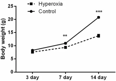
Figure 1. Changes in the body weight of newborn rats after hyperoxia exposure for 3, 7, and 14 days. n = 5 per group. Values are means ± SEM. **P < 0.01, ***P
larization and vascularization, a condition that strongly resembles “new BPD” in premature infants [2, 3]; this was also confirmed in this study. As results shown in Figure 1, following 7 and 14 days of hyperoxia exposure, body weight of newborn rats were much lower than those in control group (P < 0.01 and P < 0.001). Histopathological changes in lung tissues were monitored, and as shown in Figure 2A, lung tis-sues in control group showed uneven pink at the 3th day, and this pink changed to more uni-formed at the 7th and 14th day. The color of tissues in Hyperoxia group was rather deep than those in Control group at the 3th day, and became pale at the 7th and 14th day; in addi-tion, petechial hemorrhages were observed on the surface of the lung at the 7th and 14th day. Further, we found that hyperoxia significantly decreased RAC when compared with control group after 3, 7, and 14 days of exposure (P < 0.001; Figure 2B).
Hyperoxia up-regulates SP-B and TGF-β1
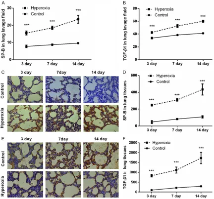
ELISA was performed to detect the concentra-tions of SP-B and TGF-β1 in bronchoalveolar lavage fluids. As shown in Figure 3A and 3B, much higher contents of SP-B and TGF-β1 were found in Hyperoxia group when compared with Control group (P < 0.001). Data from immuhist-chemical assessment were coincident with the results from Figure 3A and 3B showed that, SP-B and TGF-β1 were highly expressed after hyperoxia exposure (Figure 3C-F; P < 0.001). In addition, the expression levels of these two factors in lung tissues were also detected by qRT-PCR and Western blot analyses. As results
shown in Figure 4A-C, both the mRNA and pro-tein levels of SP-B and TGF-β1 were up-regulat-ed after hyperoxia exposure (P < 0.001). Hyperoxia increases MDA, decreases TAOC and monitors MAPK and PI3K/AKT pathways
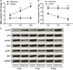
Next, we asked whether hyperoxia could alter the concentrations of MDA and TAOC in lung tis-sues. As results shown in Figure 5A and 5B, MDA content was much higher while TAOC con-tent was much lower in Hyperoxia group when compared with Control group (P < 0.01 or P < 0.001). Further, the expression levels of main factors in MAPK and PI3K/AKT pathways were detected. Western blot analycal results showed that (Figure 5C), the phosphorylation level of p38 was remarkably up-regulated, and the phosphorylation levels of PI3K and AKT were notably down-regulated following hyperoxia exposure. The total levels of p38, PI3K and AKT were unaffected.
Discussion
Supplemental oxygen is widely used in the treatment of neonatal respiratory failure [1]; however, long-term of high-concentration of oxygen may cause BPD [4]. In the present study, newborn SD rats were exposed to hyper-oxia conditions. We found that hyperhyper-oxia expo-sure significantly induced body weight loss of SD rats. Besides, H&E staining for morphomet-ric analyses revealed that hyperoxia arrested alveolar development or loss of alveoli, with fewer and dysmorphic capillaries, which are the characteristics of BPD [20]. All these suggested
that an animal model of BPD was established in newborn SD rats by using hyperoxia conditions.
SP-B is a major component of SPs that plays important functions in lung throughout life. Previous studies have pointed out that SP-B is abnormally expressed in lung diseases. Chang et al., mentioned that the serum levels of SP-B in respiratory distress syndrome (RDS) were lower than in premature infants without RDS [21], which increasing the risk of RDS in pre-term infants. The expression of SP-B is also
[image:5.612.90.522.71.474.2]injury [22]. Of contrast, in hyperoxia-induced neonatal rat lung injury, SP-B expression was increased during exposure to hyperoxia [23]. Another study reported the similar result that SP-B expression increased sharply in newborn rat lung at 7 and 14 days of hyperoxic exposure [14]. In the current study, the expression of SP-B in response to hyperoxia has been detect-ed in three levels: 1) mRNA, and 2) protein lev-els in lung tissues, and 3) the concentration in bronchoalveolar lavage fluid. Results showed that reduction of mRNA level of SP-B may decrease the transcription/translation of SP-B
Figure 3. Expression levels of SP-B and TGF-β1 in bronchoalveolar lavage fluids and lung tissues after newborn rats exposed to hyperoxia for 3, 7, and 14 days. A, B. ELISA assay for detection the concentrations of SP-B and TGF-β1 in bronchoalveolar lavage fluids. C-F. Immunohistochemistry for detection the expression levels of SP-B and TGF-β1 in lung tissues. One representative slide per group is shown. n = 5 per group. Values are means ± SEM. ***P < 0.001
release. It seems that SP-B expression reflect lung injury, that lung injury induced by pulmonary hypoplasia or in- fection reduced SP-B expres-sion, while lung injury induced by hyperoxia increased SP-B expression, providing an op- portunity for use SP-B as a biomarker candidate for dis-tinguish the cause of BPD. TGF-β1 is a secretory cyto- kine that binds its receptor TGFβR2, which initiates the TGF-β signaling involved in the regulation of branching and septation phases of lung de- velopment [24, 25]. Previous studies have reported the excessive TGF-β1 levels in BPD. For instance, TGF-β1 was increased in bronchoalveolar lavage fluid obtained from infants with chronic lung dis-ease of prematurity [26]. A high level of TGF-β1 immuno-reactivity has been observed in the walls of the peripheral lung where alveolarization is diminished by lung injury [13]. These previous studies evi-denced that the increase level of TGF-β1 is associated with newborn pulmonary injury. Our data were consistent with these previous studies, sug-gesting that TGF-β1 was highly expressed after hyperoxia exposure. But, this study pro-vided a more detailed data regarding the abundance of TGF-β1 in different spatial and temporal. That is we revealed that TGF-β1 was highly expressed in three lev-els: 1) mRNA and 2) protein levels in lung tissues, and 3) the release of TGF-β1 in bron-choalveolar lavage fluid, begin-ning at the 3th day of hyper-oxia exposure.
Figure 4. Expression levels of SP-B and TGF-β1 in lung tissues after newborn rats exposed to hyperoxia for 3, 7, and 14 days. A, B. qRT-PCR analysis for detection the mRNA level expressions of SP-B and TGF-β1. n = 5 per group. Values are means ± SEM. ***P < 0.001 vs. Control group. C. Western blotting
for detection the protein level expressions of SP-B and TGF-β1. n = 5 per group. One representative slide per group is shown.
Figure 5. Changes in MDA and TAOC contents and MAPK and PI3K/AKT pathways in lung tissues after newborn rats exposed to hyperoxia for 3, 7, and 14 days. A, B. The concentrations of MDA and TAOC in lung tissues were monitored by using commercial kits. n = 5 per group. Values are means ± SEM. ***P < 0.001 vs. Control group. C. Western blotting for detection the
[image:6.612.93.374.355.625.2]It is generally believed that MDA value reflects oxidative level, while TAOC value reflects anti-oxidative level. These two indicators are widely used in the estimation of oxidative conditions [27, 28]. Herein, the level of MDA was found to be significantly increased in response to hyper-oxia, while TAOC level was significantly reduced after hyperoxia exposure, suggesting oxidative stress was induced in lung tissues. Moreover, hyperxia activated the MAPK pathway and deactivated PI3K/AKT pathway in lung tissues. MAPK and PI3K/AKT are two cell signaling pathways implicated in the survival of pulmo-nary epithelial cells after hyperoxia exposure [29]. Our data showed that the increase of MDA, the decrease of TAOC, the activation of p38, as well as the deactivation of PI3K and AKT in hyperoxic lung were in sync with the increases of SP-B and TGF-β1 levels. Thus, we boldly propose a hypothesis: increased levels of SP-B and TGF-β1 were stimulated by oxida-tive stress, and SP-B and TGF-β1 function to lung possibly via MAPK and PI3K/AKT signaling pathways. A large number of investigations are required to verify this hypothesis.
In conclusion, our research demonstrated that SP-B and TGF-β1 were high expressed in three levels: 1) mRNA and 2) protein levels in lung tissues, and 3) the release of SP-B and TGF-β1 in bronchoalveolar lavage fluid, beginning at the 3th day of hyperoxia exposure. SP-B and TGF-β1 might be two potential biomarkers for BPD diagnose.
Acknowledgements
This work was supported by the Science and Technology Development Project in Shinan District of Qingdao [Grant Number: 2014- 14-23-YY].
Disclosure of conflict of interest
None.
Address correspondence to: Mingji Yi, Department of Child Healthcare, The Affiliated Hospital of Qingdao University, 16 Jiangsu Road, Qingdao 266003, P. R. China. E-mail: 6097hhnrmqjl@gmail. com
References
[1] Huizing MJ, Villamor-Martinez E, Vento M and
limits for preterm infants: a survey among Eu-ropean neonatal intensive care units. Eur J Pe-diatr 2017; 176: 51-56.
[2] Shivanna B, Zhang S, Patel A, Jiang W, Wang L, Welty SE and Moorthy B. Omeprazole attenu-ates pulmonary aryl hydrocarbon receptor acti-vation and potentiates hyperoxia-induced de-velopmental lung injury in newborn mice. Toxicol Sci 2015; 148: 276-287.
[3] Sharma D, Nkembi AS, Aubry E, Houeijeh A, Butruille L, Houfflin-Debarge V, Besson R, Deruelle P and Storme L. Maternal PUFA omega-3 supplementation prevents neonatal lung injuries induced by hyperoxia in newborn rats. Int J Mol Sci 2015; 16: 22081-22093. [4] Kair LR, Leonard DT and Anderson JM.
Bron-chopulmonary dysplasia. Pediatr Rev 2012; 33: 255-263; quiz 263-4.
[5] Farstad T, Bratlid D, Medbo S and Markestad T. Bronchopulmonary dysplasia - prevalence, se-verity and predictive factors in a national co-hort of extremely premature infants. Acta Pae-diatr 2011; 100: 53-58.
[6] McEvoy CT, Jain L, Schmidt B, Abman S, Ban-calari E and Aschner JL. Bronchopulmonary dysplasia: NHLBI workshop on the primary pre-vention of chronic lung diseases. Ann Am Tho-rac Soc 2014; 11 Suppl 3: S146-153.
[7] Jimenez J, Richter J, Nagatomo T, Salaets T, Quarck R, Wagennar A, Wang H, Vanoirbeek J, Deprest J and Toelen J. Progressive vascular functional and structural damage in a bron-chopulmonary dysplasia model in preterm rab-bits exposed to hyperoxia. Int J Mol Sci 2016; 17.
[8] Curley AE and Halliday HL. The present status of exogenous surfactant for the newborn. Early Hum Dev 2001; 61: 67-83.
[9] Tokieda K, Ikegami M, Wert SE, Baatz JE, Zou Y and Whitsett JA. Surfactant protein B corrects oxygen-induced pulmonary dysfunction in het-erozygous surfactant protein B-deficient mice. Pediatr Res 1999; 46: 708-714.
[10] Munger JS, Huang X, Kawakatsu H, Griffiths MJ, Dalton SL, Wu J, Pittet JF, Kaminski N, Ga-rat C, Matthay MA, Rifkin DB and Sheppard D. The integrin alpha v beta 6 binds and activates latent TGF beta 1: a mechanism for regulating pulmonary inflammation and fibrosis. Cell 1999; 96: 319-328.
[11] Warburton D, Schwarz M, Tefft D, Flores-Delga-do G, Anderson KD and CarFlores-Delga-doso WV. The mo-lecular basis of lung morphogenesis. Mech Dev 2000; 92: 55-81.
[13] Nakanishi H, Sugiura T, Streisand JB, Lonning SM and Roberts JD Jr. TGF-beta-neutralizing antibodies improve pulmonary alveologenesis and vasculogenesis in the injured newborn lung. Am J Physiol Lung Cell Mol Physiol 2007; 293: L151-161.
[14] Hou A, Fu J, Yang H, Zhu Y, Pan Y, Xu S and Xue X. Hyperoxia stimulates the transdifferentia-tion of type II alveolar epithelial cells in new-born rats. Am J Physiol Lung Cell Mol Physiol 2015; 308: L861-872.
[15] Luan Y, Zhang L, Chao S, Liu X, Li K, Wang Y and Zhang Z. Mesenchymal stem cells in com-bination with erythropoietin repair hyperoxia-induced alveoli dysplasia injury in neonatal mice via inhibition of TGF-beta1 signaling. On-cotarget 2016; 7: 47082-47094.
[16] Cooney TP and Thurlbeck WM. The radial alve-olar count method of Emery and Mithal: a re-appraisal 1--postnatal lung growth. Thorax 1982; 37: 572-579.
[17] Kunig AM, Balasubramaniam V, Markham NE, Seedorf G, Gien J and Abman SH. Recombi-nant human VEGF treatment transiently in-creases lung edema but enhances lung struc-ture after neonatal hyperoxia. Am J Physiol Lung Cell Mol Physiol 2006; 291: L1068-1078. [18] Livak KJ and Schmittgen TD. Analysis of rela-tive gene expression data using real-time quantitative PCR and the 2(-Delta Delta C(T)) method. Methods 2001; 25: 402-408. [19] Zhao YN, Wang HY, Li JM, Chen BY, Xia G,
Zhang PP and Ge YL. Hippocampal mitogen-activated protein kinase activation is associat-ed with intermittent hypoxia in a rat model of obstructive sleep apnea syndrome. Mol Med Rep 2016; 13: 137-145.
[20] Jobe AJ. The new BPD: an arrest of lung devel-opment. Pediatr Res 1999; 46: 641-643. [21] Chang HY, Li F, Li FS, Zheng CZ, Lei YZ and
Wang J. Genetic polymorphisms of SP-A, SP-B, and SP-D and risk of respiratory distress syn-drome in preterm neonates. Med Sci Monit 2016; 22: 5091-5100.
[22] Wang WN, Zhou JH, Wang P and Zhang XJ. The localization of SP-B and influences of lipopoly-saccharide on it. Eur Rev Med Pharmacol Sci 2016; 20: 2338-2345.
[23] ter Horst SA, Fijlstra M, Sengupta S, Walther FJ and Wagenaar GT. Spatial and temporal ex-pression of surfactant proteins in hyperoxia-induced neonatal rat lung injury. BMC Pulm Med 2006; 6: 8.
[24] Jankov RP and Keith Tanswell A. Growth fac-tors, postnatal lung growth and bronchopulmo-nary dysplasia. Paediatr Respir Rev 2004; 5 Suppl A: S265-275.
[25] Vicencio AG, Lee CG, Cho SJ, Eickelberg O, Chuu Y, Haddad GG and Elias JA. Conditional overexpression of bioactive transforming growth factor-beta1 in neonatal mouse lung: a new model for bronchopulmonary dysplasia? Am J Respir Cell Mol Biol 2004; 31: 650-656. [26] Kotecha S, Wangoo A, Silverman M and Shaw
RJ. Increase in the concentration of transform-ing growth factor beta-1 in bronchoalveolar la-vage fluid before development of chronic lung disease of prematurity. J Pediatr 1996; 128: 464-469.
[27] Lv WP, Li MX and Wang L. Peroxiredoxin 1 inhibits lipopolysaccharide-induced oxidative stress in lung tissue by regulating P38/JNK signaling pathway. Eur Rev Med Pharmacol Sci 2017; 21: 1876-1883.
[28] Xu B, Li C, Wang J, Wu J, Si S, Liu Z, Li J, Zhang J and Cui Y. Lung injury via oxidative stress in mice induced by inhalation exposure to rocket kerosene. Int J Clin Exp Pathol 2015; 8: 5497-5502.