Received March 30, 2019; Accepted April 25, 2019; Epub July 1, 2019; Published July 15, 2019
Abstract: Objective: Isatin has gained attention in recent years owing to its anticancer properties and is thought to
offer medical benefits. Isatin is an endogenous oxidized indole with various behavioral and metabolic properties and is commonly found in mammalian tissues and fluids. It has several plausible applications in biomedical re -search and has also been investigated as a potential anticancer agent. However, its effects on neuroblastoma (NB) cells are unclear. Here, we evaluate the effects of isatin on neuroblastoma cell metastasis and invasion and reveal the underlying mechanism. Methods: NB cell viability was evaluated with the cell counting kit (CCK)-8 assay. NB cell invasion and migration abilities were tested with transwell and wound healing experiments. The relative mRNA expression of associated molecules was detected with real-time polymerase chain reaction (RT-PCR) and quantita-tive PCR. The expression level of related proteins was detected with western blotting. Results: Isatin inhibited the proliferation, invasion, and migration of neuroblastoma cells in a dose-dependent manner. Isatin increased the expression level of H3K4m1 and phosphatase and tensin homolog (PTEN) and decreased the phosphorylation level of PTEN downstream proteins phosphoinositide 3-kinase, protein kinase B, mammalian target of rapamycin, focal adhesion kinase, and SHC. Together, these results support the potential anti-metastatic effects of isatin on NB cells.
Keywords: Isatin, neuroblastoma, PTEN, LSD1, invasion
Introduction
Neuroblastoma (NB), one of the most com- mon solid extracranial neoplasms in children, accounts for more than 7% of malignancies in patients younger than 15 years old and is responsible for around 15% of all pediatric cancer-related deaths [1, 2]. Although substan-tial improvement has been observed in the
out-comes of a few well-defined subsets of
pati-ents during the past few decades, not much improvement has been reported for those with a high-risk clinical phenotype [3, 8]. NB is a dis-ease of the sympatico-adrenal lineage of the neural crest, wherein the tumors may develop anywhere in the sympathetic nervous system.
About half of all cases are classified under a
high risk for disease relapse, and the overall survival rate is less than 40% despite intensive multimodal therapy [4, 9]. The leading cause of
death in patients with NB is metastasis, whi- ch in most cases is observed to inexplicably migrate to the bone marrow. Therefore, it is
important to understand the specific mecha -nism underlying NB invasion and metastasis and [5, 10] to discover safer and better com-pounds to inhibit NB [4, 6] invasion and me- tastasis.
Monoamine oxidase (MAO) is localized in the
outer membrane of the mitochondria and
cata-lyzes the oxidative deamination of neuroactive
As a member of the MAO family [8, 12],
lysine-specific demethylase 1 (LSD1) which specifi -cally removes the dimethyl and monomethyl
modifications of H3K4 in the presence of fla -vine adenine dinucleotide (FAD) [9, 13] is str- ongly associated with the development, inva-siveness, and metastasis of tumor cells [10, 14]. For instance, LSD1 could be recruited by the SNAG domain of Snail to the E-cadherin pro-moter for transcription suppression and EMT [11, 15]. An epigenetic marker, LSD1 overex-pression is one of the characteristics of malig-nant tumors [12, 16] and has been correlated with the malignant progression of multiple can-cers, including primary neuroblastic tumors, estrogen receptor-negative breast cancer [13, 17], and poorly differentiated NBs [14, 18]. LSD1 may also form a co-inhibitory complex with the SNAG domain of Snail and allow the recruitment of LSD1 to the phosphatase and tensin homolog (PTEN) promoter for H3K4 demethylation [15, 19] and PTEN transcription repression [16, 20].
PTEN exerts its role as a tumor suppressor th- rough the downregulation of the phosphoinosit-ide 3-kinase (PI3K)/protein kinase (AKT) signal-ing pathway, which is highly related to the inva-sion and metastasis of cancer cells [17, 18, 21, 22]. For instance, PTEN inhibits the migration and invasion of HepG2 cells by decreasing the expression of matrix metalloproteinase (MMP) via the PI3K/AKT pathway [19, 23]. PTEN also interacts with focal adhesion kinase (FAK) by reducing its tyrosine phosphorylation and neg-atively regulates the interaction between the
cell and the extracellular matrix [20, 24] to inhibit cell migration, cell spreading, and focal adhesion formation [21, 25].
Previous studies have shown that isatin is an endogenous indole that inhibits MAOB and may induce SH-SY5Y cell death in a dose- and time-dependent manner [22, 26]. Isatin was recently shown to inhibit SH-SY5Y cell migration and invasion through various pathways, especially through the downregulation of MMP-2/MMP-9 expression [23, 27]. However, the precise me- chanism involved in the anti-metastasis acti- vity of isatin is incompletely understood. We thought that the inhibition of LSD1 activity may cause H3K4 demethylation and isatin may upregulate the expression of PTEN, which would negatively regulate SH-SY5Y cell inva-sion and metastasis through the PI3K/AKT and PTEN/p-SHC/p-FAK signaling pathways. To test our assumption, we applied real-time polyme- rase chain reaction (RT-PCR), western blotting, and transwell assays and determined the mol-ecules involved in this process. The results
obtained confirm our hypothesis.
Materials and methods
Cell culture
SH-SY5Y cells (Chinese Academy of Sciences)
were cultured in Dulbecco’s modified Eagle’s
[image:2.612.93.513.74.221.2]medium (DMEM)/high glucose (HyClone, USA) supplemented with 10% fetal bovine serum (FBS; BI, California, USA) at 37°C with 5% CO2
and 98% relative humidity in a culture incuba-tor. The cells were incubated with different con-centrations (0, 50, 100, and 200 µmol/L) of isatin (99.0%; Sigma, California, USA) for 24,
48, or 72 h after reaching 80% confluency. The
cells (about 9 × 10⁵ cells/well) were harvested and used for proliferation, migration, and pro-tein analyses.
Cell viability assay
The viability of the cells was measured with a cell counting kit-8 (CCK-8) assay. The cells were
seeded in a 96-well plate in a final volume of
100 µL of complete culture medium containing 1 × 10⁴ cells/well and exposed to different con-centrations (0, 25, 50, 100, 200, 300, 400, 500, 600, and 700 µmol/L) of isatin (six wells for each concentration) for 48/72 h at 37°C. After treatment, 10 µL of CCK-8 solution was added to each well and the cells were incubat-ed at 37°C for 3 h. The absorbance of the sam-ples was measured at a wavelength of 450 nm with a microplate reader (Synergy H1; BioTek,
Vermont, USA). Each experiment was perfor- med thrice.
Cell migration and invasion assay
SH-SY5Y cells were seeded in a serum-free medium in a six-well plate and exposed to isat-in (0, 50, 100, and 200 µmol/L) for 48 h. A micropipette tip was used to scratch and cre-ate a wound each well. The cells were moni-tored during regrowth, and images were cap-tured at different time points (0, 12, 24, and 48 h). The transwell invasion assay was performed in Boyden chambers (Millipore, California, USA). About 2 × 10⁵ cells/well seeded in 200 µL of serum-free medium and pretreated with isatin for 24 h were added to the upper chamber coated with Matrigel, while the medium supple-mented with 10% FBS was added to the lower chamber. At the end of the incubation, the cells from the upper surface were completely
removed, and the filter was fixed in methanol
and stained with crystal violet. Cells invading the Matrigel were counted under an inverted microscope. Data were expressed as the
aver-Figure 2. Isatin inhibits SH-SY5Y cell invasion and migration. Isatin inhibits SH-SY5Y cell migration and invasion
(A). The effect of isatin on neuroblastoma cell migration at × 100 magnification under an inverted microscope (B). Statistical analysis of SH-SY5Y cell Matrigel invasion counted in five random fields (C). *P < 0.05, **P < 0.01 as
[image:3.612.91.524.82.339.2]age cell number in five fields and the experi -ment was repeated thrice.
Western blot analysis
SH-SY5Y cells were lysed in a radioimmunopre-cipitation assay (RIPA) buffer (Solarbio, Beijing, China) supplemented with a protease inhibitor cocktail (Sigma, Germany) on ice for 1 h and centrifuged for 20 min at 4000 × g. The super-natant was collected and the protein concen-tration was measured with bicinchoninic acid (BCA) assay (Beyotime, Jiangsu, China). DTT
from the kit was mixed with 40 μL of deionized water to obtain a 400 mM solution. The fluores
-cent 5 × master mix was mixed with 20 μL of 10 × sample buffer and 20 μL of the prepared DTT
solution. The biotinylated ladder provided with
the kit was mixed with 16 μL of deionized water, 2 μL of 10 × sample buffer, and 2 μL of the
prepared DTT solution and gently mixed. The entire volume of the ladder was transferred to a 0.6 mL tube (blue), and the 10 × sample buffer was diluted 1:100 with water to obtain 0.1 × sample buffer. The protein lysate was diluted
with 0.1 × sample buffer and the final concen
-tration of the protein sample was adjusted to 0.2 mg/mL. The samples and biotinylated lad-der were denatured at 95°C for 5 min and stored on ice. The primary antibody was diluted with antibody diluent 2 (1:50). The secondary horseradish peroxidase (HRP) conjugate was provided in the detection module and was
ready to use. About 200 μL Lumino-S and 200 μL peroxide supplied in the detection module
were mixed in a microcentrifuge tube and gen-tly pipetted and stored on ice. All the reagents were added to the wells of the plate (Figure 1A lower panel) for the immunoassay. The assay was loaded in Compass software and a capil-lary cartridge was inserted into the cartridge holder. After the change in the interior light from orange to blue, the assay plate lid was
removed, and the plate was held firmly on
bench and its evaporation seal was carefully peeled-off. Any bubbles observed in the sepa-ration matrix wells were removed with a pipette tip. The assay plate was placed on the plate holder, the door was closed, and the run was started. After run completion, the plate and cartridge were discarded. Data were collected
[image:4.612.93.524.72.377.2]and analyzed by Compass software.
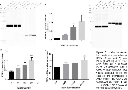
Figure 3. Isatin increases the protein expression of H3K4m1 (A and B) and PTEN (C and D) in SH-SY5Y cells after 48 h of treat-ment, as detected with a western blot analysis. Sta-tistical analysis of RT-PCR data for the expression of
Quantitative RT-PCR assay
Total RNA was extracted from the cultu-
red SH-SY5Y cells with Trizol reagent
(Solar-bio, Beijing, China). A reverse transcription kit (Transgene, Beijing, China) was used to con-struct the template cDNA for real-time PCR wi- th Trans Start Probe RT-PCR Super Mix (Tr- ansgene, Beijing, China). The data were obtain- ed on Bio-Rad (California, USA) One-Step Plus system. The primer sequences used are sh- own in Table 1.
Statistical analysis
The data were obtained from three indepen-dent experiments and represented as the mean ± standard deviation (SD). The corresponding data were compared with a one-way analysis of
variance (ANOVA) using GraphPad Prism 6 sta-tistical software (GraphPad, La Jolla, CA, USA),
and significance was set at P < 0.05.
Results
Detection of cell viability with the CCK-8 assay
Isatin inhibited the invasion and metastasis of NB cells in vitro. The growth inhibitory effects of isatin on SH-SY5Y cells were evaluated with a CCK-8 assay. As shown in Figure 1B, isatin at
various concentrations influenced the survival
rate of SH-SY5Y cells after treatment for 48/72 h. Isatin was nontoxic at concentrations below
200 μmol/L, as evident from the survival of
80% of the cells. So the concentrations of 0
μmol/L, 50 μmol/L, 100 μmol/L, 200 μmol/L
[image:5.612.94.518.71.427.2]were used in subsequent experiments.
Decrease in the invasion and migration ability of isatin-treated cells as compared with control cells
The wound healing rate was significantly lower
in isatin-treated SH-SY5Y cells than in the untreated cells. At 48 h, the wound closure was almost complete in the control cells, but the isatin-treated cells showed a noticeable wound gap (Figure 2A). In addition, isatin at 200
μmol/L concentration decreased the invasion
of cells to 20% as compared with the control cells (Figure 2B and 2C). Isatin also exerted an anti-proliferation effect on the SH-SY5Y cells (Figure 1B). All these results suggest that isatin exhibits a potentially inhibitory effect on NB cell metastasis.
Increase in the expression of H3K4m1 and PTEN
As reported in previous studies, isatin inhibits MAOA activity in SH-SY5Y cells [24, 28]. Here, we investigated whether isatin modulates LSD1 activity by evaluating the expression of its
tar-Decrease in the phosphorylation of PI3K, AKT, mTOR, FAK, and SHC
We observed a decrease in the phosphoryla-tion levels of PTEN downstream molecules, such as PI3K (Figure 4A and 4C), AKT (Figures 4B and 5D), mTOR (Figure 4E and 4F), FAK (Figure 5A and 5C), and SHC (Figure 5B and 5D), as confirmed with western blotting.
No change in the expression of LSD1
We failed to observe any change in the expres-sion of LSD1 at the protein (Figure 6A and 6B) and mRNA (Figure 6C) levels, suggesting that isatin may inhibit cell invasion not by decreas-ing the expression of LSD1 but through the inhibition of LSD1 activity.
All these results suggest that isatin may inhi- bit LSD1 activity and increase PTEN expres-sion, leading to the inhibition of SH-SY5Y cell invasion and metastasis through the PI3K/ AKT and PTEN/p-SHC/p-FAK signaling path- ways.
Figure 5. The phosphorylation level of p-SHC (A) and p-FAK (B) in SH-SY5Y cells decreased after 48 h of treatment with isatin, as detected with a west-ern blot analysis. Statistical analysis of the expression of p-SHC (C) and p-FAK protein (D). Values are expressed as the mean ± SD. *P < 0.05, **P < 0.01 as compared with control.
get protein H3K4m1 [9, 13]. The expression of the H3K4m1
protein significantly increased
in the cells incubated with 200
μmol/L of isatin as compared
with the control cells (Figure 3A and 3B), indicating that
isatin may influence SH-SY5Y
Discussion
Prior work has revealed the effectiveness of isatin in the prevention of cancer cell prolifera-tion and progression. A study by Havrylyuk [28, 32] showed that isatin exhibits a remarkable antiproliferative effect on cancer. However, these studies have either focused only on the anti-proliferation property of isatin or have not
clarified the anti-invasive mechanism isatin’s
underlying effects in NB cells. In the present study, we investigated the anti-invasive effects of isatin on NB cells and revealed the underly-ing mechanism usunderly-ing western blottunderly-ing and RT-PCR. As a result, we found that isatin treat-ment increased the expression of H3K4m1 in SH-SY5Y cells, wherein H3K4m1 acts as a sub-strate of LSD1. The expression of LSD1, how-ever, showed no change, indicating that isatin is likely to downregulate H3K4m1 expression by inhibiting the activity of LSD1 and not by decreasing LSD1 expression. The upregulation in H3K4m1 expression resulted in an increase in the level of PTEN. We also observed a decrease in the expression of the downstream molecules involved in PTEN signaling, including p-SHC, p-FAK, p-PI3K, p-AKT, and p-mTOR.
These findings indicate that isatin exerts its
anti-invasion and anti-metastasis effects on SH-SY5Y cells by increasing the expression level of H3K4m1, which then activates PTEN signaling through the inhibition of LSD1 activity.
To our knowledge, this is the first study to sys
-tematically investigate the influence of isatin
on PTEN signaling-related molecules in NB cells. However, our study has a few limitations. Although our hypotheses were statistically sup-ported by the results of biochemical experi-ments in vitro, whether the mechanism is re- producible in animals or humans is question-able. Further studies are warranted to evalu- ate the effects of isatin on tumors in animal models.
Acknowledgements
This work was supported by the National Na- tural Science Foundation of China (81472- 542, 201501-201812), the Qingdao Startup and Innovation Leader Talent Plan (13-CX-3, 201409-201709), and the “Clinical Medicine+ X” Plan, Qingdao University (201707201906). Disclosure of conflict of interest
None.
Address correspondence to: Lin Hou, Department of Biochemistry and Molecular Biology, Medical
College of Qingdao University, No. 38 Dengzhou
Road, Qingdao 266021, Shandong, China. Tel: +86-18661901982; E-mail: Qingyi001@126.com
References
[image:7.612.92.519.74.279.2][1] Maris JM. Recent advances in neuroblastoma. N Engl J Med 2010; 362: 2202-2211.
[2] Adamo A, Sese B, Boue S, Castano J, Para-monov I, Barrero MJ and Belmonte JC. LSD1 regulates the balance between self-renewal and differentiation in human embryonic stem cells. Nat Cell Biol 2011; 13: 652-659. [3] Brodeur GM, Pritchard J, Berthold F, Carlsen
NL, Castel V, Castelberry RP, De Bernardi B, Ev-ans AE, Favrot M, Hedborg F, et al. Revisions of the international criteria for neuroblastoma di-agnosis, staging, and response to treatment. J Clin Oncol 1993; 11: 1466-77.
[4] Maris JM, Hogarty MD, Bagatell R and Cohn SL. Neuroblastoma. Lancet 2007; 369: 2106-2120.
[5] Song J, Hou L, Ju C, Zhang J, Ge Y and Yue W. Isatin inhibits proliferation and induces apop-tosis of SH-SY5Y neuroblastoma cells in vitro and in vivo. Eur J Pharmacol 2013; 702: 235-241.
[6] da Silva JFM, Garden SJ and Pinto AC. The chemistry of isatins: a review from 1975 to
1999. J Braz Chem Soc 2001; 12: 273-324.
[7] Fitzgerald JC, Ufer C and Billett EE. A link be -tween monoamine oxidase-A and apoptosis in serum deprived human SH-SY5Y neuroblasto-ma cells. J Neural Transm (Vienna) 2007; 114: 807-810.
[8] Chen Y, Yang YT, Wang F, Wan K, Yarnane K, Zhang Y and Lei M. Crystal structure of human
histone lysine-specific demethylase 1 (LSD1).
Proc Natl Acad Sci U S A 2006; 103: 13956-13961.
[9] Shi Y, Lan F, Matson C, Mulligan P, Whetstine JR, Cole PA, Casero RA and Shi Y. Histone de-methylation mediated by the nuclear amine oxidase homolog LSD1. Cell 2004; 119: 941-953.
[10] Escriva M, Peiro S, Herranz N, Villagrasa P,
Dave N, Montserrat-Sentis B, Murray SA, Fran-ci C, Gridley T, Virtanen I and GarFran-cia de Herre-ros A. Repression of PTEN phosphatase by Snail1 transcriptional factor during gamma ra-diation-induced apoptosis. Mol Cell Biol 2008; 28: 1528-1540.
[11] Lin Y, Wu Y, Li J, Dong C, Ye X, Chi YI, Evers BM and Zhou BP. The SNAG domain of Snail1 func-tions as a molecular hook for recruiting
lysine-specific demethylase 1. EMBO J 2010; 29:
1803-1816.
[12] Shao GB, Huang XJ, Gong AH, Zhang ZJ, Lu RZ and Sang JR. [Histone demethylase LSD1 and its biological functions]. Yi Chuan 2010; 32: 331-338.
[13] Lim S, Janzer A, Becker A, Zimmer A, Schule R, Buettner R and Kirfel J. Lysine-specific de -methylase 1 (LSD1) is highly expressed in ER-negative breast cancers and a biomarker pre-dicting aggressive biology. Carcinogenesis 2010; 31: 512-520.
[14] Schulte JH, Lim S, Schramm A, Friedrichs N, Koster J, Versteeg R, Ora I, Pajtler K,
Klein-Hit-pass L, Kuhfittig-Kulle S, Metzger E, Schule R,
Eggert A, Buettner R and Kirfel J. Lysine-specif-ic demethylase 1 is strongly expressed in poor-ly differentiated neuroblastoma: implications for therapy. Cancer Res 2009; 69: 2065-2071. [15] Lin Y, Kang T and Zhou BP. Doxorubicin en-hances Snail/LSD1-mediated PTEN suppres-sion in a PARP1-dependent manner. Cell Cycle 2014; 13: 1708-1716.
[16] Sun G, Alzayady K, Stewart R, Ye P, Yang S, Li W
and Shi Y. Histone demethylase LSD1 regu-lates neural stem cell proliferation. Mol Cell Biol 2010; 30: 1997-2005.
[17] Yokoyama A, Igarashi K, Sato T, Takagi K, Ot-suka IM, Shishido Y, Baba T, Ito R, Kanno J, Ohkawa Y, Morohashi K and Sugawara A.
Iden-tification of myelin transcription factor 1
(MyT1) as a subunit of the neural cell
type-specific lysine-type-specific demethylase 1 (LSD1)
complex. J Biol Chem 2014; 289: 18152-18162.
[18] Stambolic V, Suzuki A, de la Pompa JL, Broth -ers GM, Mirtsos C, Sasaki T, Ruland J, Pen-ninger JM, Siderovski DP and Mak TW. Nega-tive regulation of PKB/Akt-dependent cell survival by the tumor suppressor PTEN. Cell 1998; 95: 29-39.
[19] Tian T, Nan KJ, Guo H, Wang WJ, Ruan ZP, Wang SH, Liang X and Lu CX. PTEN inhibits the migration and invasion of HepG2 cells by coor-dinately decreasing MMP expression via the PI3K/Akt pathway. Oncol Rep 2010; 23: 1593-1600.
[20] Tamura M, Gu JG, Matsumoto K, Aota S, Par-sons R and Yamada KM. Inhibition of cell mi-gration, spreading, and focal adhesions by tu-mor suppressor PTEN. Science 1998; 280: 1614-1617.
[21] Yamada KM, Araki M. Tumor suppressor PTEN: modulator of cell signaling, growth, migration and apoptosis. J Cell Sci 2001; 114: 2375-2382.
[22] Igosheva N, Lorz C, O’Conner E, Glover V and
Mehmet H. Isatin, an endogenous monoamine oxidase inhibitor, triggers a dose- and time-de-pendent switch from apoptosis to necrosis in human neuroblastoma cells. Neurochem Int 2005; 47: 216-224.
[23] Xu PP, Hou L, Ju CX, Zhang Z, Sun WY, Zhang L, Song JL, Lv YQ, Liu L, Chen ZX and Wang YH. Isatin inhibits the proliferation and invasion of SH-SY5Y neuroblastoma cells. Mol Med Rep 2016; 13: 2757-2762.
[24] Sun WY, Zhang L, Hou L, Ju CX, Zhao SM and Wei YY. Isatin inhibits SH-SY5Y neuroblastoma cell invasion and metastasis through MAO/ HIF-1a/CXCR4 signaling. Anticancer Drugs 2017; 28: 645-653.
pro-Angulo AM, Stemke-Hale K, Hauptmann M, Beijersbergen RL, Mills GB, van de Vijver MJ and Bernards R. A functional genetic approach
identifies the PI3K pathway as a major deter
-minant of trastuzumab resistance in breast
cancer. Cancer Cell 2007; 12: 395-402. [28] Havrylyuk D, Zimenkovsky B, Vasylenko O,
Gzella A and Lesyk R. Synthesis of new 4-thia
-zolidinone-, pyrazoline-, and isatin-based con -jugates with promising antitumor activity. J Med Chem 2012; 55: 8630-8641.
PTEN. Biochem Biophys Res Commun 2019; 510: 339-344.