Original Article
Enrichment and characterization of ovarian cancer
stem cells and its potential clinical application
Wenxia Wang1, Zhenbo Zhang2, Yin Zhao1, Zeng Yuan1, Xingsheng Yang1, Beihua Kong1, Wenxin Zheng3,4,5,6
1Department of Obstetrics and Gynecology, Qilu Hospital, Shandong, China; 2Department of Obstetrics and
Gynecology, Shanghai General Hospital, Shanghai, China; 3Department of Pathology, University of Arizona,
Tucson, AZ, USA; Departments of 4Pathology, 5Obstetrics and Gynecology, 6Simmmon Comprehensive Cancer
Center, University of Texas Southwestern Medical Center, Dallas, TX, USA
Received June 27, 2016; Accepted August 28, 2016; Epub October 1, 2016; Published October 15, 2016
Abstract: The cancer stem cell (CSC) theory proposes that a minor population in tumor cells with specific features, such as self-renewal and reproducible tumor phenotype could contribute to tumor relapse and chemotherapy re -sistance. Several studies have convincingly documented the existence of ovarian CSC, but questions related to the biologic behavior and specific biomarkers of ovarian CSC remain to be clarified. In the present study, we firstly established a tumor cell line with capability of regenerating tumors through serial transplantation of ovarian tu -mor tissue in non-obese/severe combined immunodeficient (SCID) mice. After separation of CD133+ cells with magnetic beads, we compared the phenotype and biologic behavior of CD133+ versus CD133- cells. It was found that the CD133+ cells were much more potent to produce colonies in semi-solid agar culture than CD133- cells. The proportion of the cells in G0/1 cell cycle is much higher in CD133+ cells than in CD133- cells. Furthermore, in vivo experiments demonstrated that the CD133+ cells were capable of repeatedly regenerate tumors in NOD/ SCID mice, while the CD133- cells were not. Compared with CD133- cells, the CD133+ cells expressed much higher levels of the stem cell markers Oct4, Sox2, Nanog and Mcl-1. Clinically, among a total of 290 ovarian epithelial cancers, increased level of CD133 expression was positively correlated with a high cancer stage and had a worse 5-year survival rate. Taken together, the results suggest that the CD133+ cells from human ovarian cancer have the characteristics of CSC, which may contribute to ovarian cancer relapse and anti-apoptotic activity. The method of ovarian CSC enrichment we established provides a feasible and practical way of ovarian cancer research in a molecular level. In addition, CD133 may be used as a prognostic marker for ovarian epithelial cancer, which may have a role for future therapeutic effect.
Keywords: Ovarian epithelial cancer, cancer stem cells, CD133
Introduction
In recent years, fast accumulating data demon
-strate that there exists a small subset of cells
within a tumor, termed cancer stem cells (CSC),
responsible for cancer development and recur
-rence [1]. Characterization of CSC in leukemia
and several other malignancies showed that
these cells are relatively quiescent and highly
express molecules mediating multi-drug resis-tance [2-4]. That is the main reason that the conventional chemotherapeutic agents are
usually effective in reducing the bulk of tumor
but unable to clear the stem cells. The residual
CSCs after chemotherapy will eventually result in disease recurrence. Thus, the finding CSC
opened a new path to explore novel therapies aiming to understand the molecular
mecha-nisms of chemoresistance and cancer recur
-rence in order to significantly improve clinical
management and cure patients with cancers [5, 6].
Ovarian epithelial cancer (OEC) is a common
malignancy and a leading cause of death in
women, with epithelial ovarian cancer being the
most frequent and aggressive subtype. Among the tumors originated from female reproductive
system, ovarian epithelial cancer is the third
commonest cancer and sits at the first place in terms of cancer mortality rate. Several research
exis-tence of CSC in ovarian cancers [7-11]. Ovarian CSCs have been defined and isolated from ovarian cancer cell lines or peritoneal fluid from ovarian cancer patients using different mark
-ers, including CD44, CD117, Myd88 and ABCG2. Among these biomarkers, the CD44+/ CD117+ immunophenotype holds promise as a defining characteristic of ovarian CSCs since these isolated cells exhibit stem cell functional
-ity well. Recently, CD133 has been document
-ed as a superb CSC marker in several non-ovar
-ian cancers including cancers of the colon, the lung, and the brain [7-11]. CD133, a biomarker,
is selectively expressed by neural stem cells, hematopoietic stem cells and epithelial
progen-itor cells. Singh et al. isolated CD133+ cells from brain tumors and compared the capabili
-ties of tumor formation after subcutaneous inoculation of CD133+ or CD133- cells. While inoculation of 105 CD133- cells failed to regen
-erate tumors in NOD/SCID mice, inoculation of only 100 CD133+ cells generated tumors easi -ly [11]. O’Brien et al. reported that all the colon
cancer-initiating cells are CD133+. In contrast, the CD133- cells accounting for the bulk of the
tumors were unable regenerate tumor [9].
Therefore, it is reasonable to speculate that the CD133+ cells may play a role in cancer devel -opment, recurrence, metastasis, and patient
survival. However, it is unknown if ovarian epi
-thelial cancers have CD133+ cells and whether they can be enriched and isolated efficiently for future applications in research as well as in
clinical management.
The main goals of this study are to enrich CD133+ cells through serial transplantation of ovarian epithelial cancer tissues into non-obese/severe combined immunodeficient (SCID) mice, to characterize if the CD133+ cells enriched from the ovarian cancer have the bio
-logic function of CSC, and to correlate if CD133+ cancers have a negative impact in
clinic.
Materials and methods
Source of human ovarian epithelial cancer stem cells
The study has been approved by Institutional
Review Board of Qilu Hospital Ethic Committee
in Shandong University, China. All primary human ovarian cancer samples were collected
in accordance with the policies of Qilu Hospital of Shandong University. Tumor samples were obtained from the operating room and immedi
-ately taken to the laboratory for processing. All
ovarian epithelial cancers were pathologically
characterized based on WHO classification sys -tem. Only the high-grade serous carcinoma
samples were used for the CSC enrichment
study. The samples were minced with scalpels to 2 mm cubic pieces and implanted into 4- to
6-week-old female NOD/SCID mice (The Jackson Laboratory, Florida, USA). The mice were monitored biweekly for tumor formation.
The mice bearing growing tumors were eutha-nized by CO2 inhalation. Tumor explants were excised aseptically, processed as above, and
implanted again into recipient female NOD/ SCID mice. Tumor explant was processed and used as a source of human tumor derived cells in 9 subsequent serial transplantation
experiments.
Cell culture and CD133+ cell isolation with magnetic beads
Cell suspensions were prepared from a portion of the tumor samples. Briefly, The specimen was mechanically dissected and filtered, red blood cells were lysed with ACK buffer, and
cells were then washed with media containing
serum and plated in RPMI 1640 supplemented with 10% heat-inactivated FBS, 20 ng/ml EGF, 1 ng/ml hydrocortisone, 5 μg/ml insulin, 100 μM β-mercaptoethanol, 10 ng/ml β-FGF, 1% penicillin/streptomycin, and 20 μg/ml gentami -cin. The cell cones were allowed to grow until
they were confluent. Then the clones were tryp -sinized and plated again. Cells were tryp-sinized
and re-suspended in PBS for FACS analysis. The CD133+ cells were separated using mag
-netic beads. Briefly, the cells were harvested
and single-cell suspension was prepared and cell number counted. The cell suspension was
centrifuged at 1000 × g for 10 minutes and the cell pellet was re-suspended in 300 μl of buffer
per 108 total cells. After adding FcR Blocking
Reagent (100 μl per 108 total cells), the cells
were incubated with CD133 MicroBeads (100 μl of per 108 total cells) for 30 minutes in the
Semi-solid agar culture to examine the clono-genic ability
The enriched CD133+ cells were assayed for their ability to form colonies in semisolid agar and compared with that of CD133- cells. Briefly,
colony cultures were established in triplicate by
plating 5.0 × 10⁶ cells in per 35 mm cell culture
dish in 1.0 ml of RPMI 1640 supplemented with 0.33% agar, 25% FCS and 2 mM L-glutamine. Colony growth was stimulated by the addition of 20 ng/ml EGF. Cultures were incubated in a humidified chamber at 37°C and
5% CO2 for 2 weeks. Clones were allowed to grow until they were confluent and were then
trypsinized, passaged to a 24-well plate,
allowed to grow to confluence, and passaged a
second time. Only the colonies that
success-fully passaged twice were deemed true clones.
The colonies were counted under a light microscope.
Flow cytometry to analyze expression of CD133 and cell cycle distribution
Cultured cells were collected, washed once
with PBS and stained with anti-CD133-PE for 30 minutes of incubation in the dark on ice. For
cell cycle analysis, the harvested cells (~106
cells) were fixed with ice-cold 70% ethanol, treated with 500 μg/ml RNase A (Sigma), and subsequently stained with 25 μg/ml propidium
iodide (Sigma). Then these cells were analyzed
by using a flow cytometer FACS Calibur.
Quantitative real-time PCR to examine the ex-pression of Oct-4, Sox2, Nanog and Mcl-1
The mRNA levels were determined by real-time
quantitative PCR using the Applied Biosystems Taqman Gene Expression kit. Total RNA from cells was isolated with the RNeasy Mini Kit, accompanied by an on-column DNase diges
-tion. The volume of each reaction was 10 μl per well, which consisted of 5 μl 2 × reaction buffer and 0.05 μl 200 × Euroscript RT (reverse tran -scriptase) enzyme and RNase inhibitor mix
from the one-step RT-qPCR MasterMix Plus, 0.5 μl 20 × Taqman Gene Expression mix together with 2 μl of 50 ng RNA as amplification template. qRT-PCR was carried out on the ABI 7900HT Fast Real-Time PCR system. The reac
-tion mixtures were incubated at 48°C for 30 minutes, 95°C for 10 minutes, followed by 40
cycles at 95°C for 15 seconds and 60°C for 1
minute. Samples were measured in triplicate.
GAPDH also was amplified as a loading control for each batch of the test. Cycle threshold (Ct)
values were used to determine the relative
amounts of the targeted genes and GAPDH
mRNA levels in the samples.
In vivo tumor formation experiment
The CD133+ and CD133- cells were collected,
counted, washed and re-suspended in PBS. Then the cells were injected subcutaneously
into NOD/SCID mice with 104-106 per mouse in
0.3 ml. The mice were monitored biweekly for tumor formation up to 8 weeks.
Tumor tissue immunohistochemical analyses
A cohort of 290 ovarian epithelial carcinoma patients hospitalized at the University of Arizona Medical Center and Arizona Cancer Center during 2000-2014 was studied after approval of Institutional Review Board (IRB).
Tumor core samples (3 mm) were obtained at
initial debulking surgery, scored according to the Federation International of Gynecology and Obstetrics (FIGO) guidelines, and were forma
-lin-fixed and paraffin-embedded. Each new par
-affin block contained 100 randomly distributed
tumor cores, with at least 2 cores per patient,
and were sectioned at 5 micron thickness as
previously described [12]. Immunohistoche-
mistry was performed on slides with tumor microarray (TMA) sections before being blocked using a solution of 1% bovine serum albumin (BSA) with 2% donkey serum in phosphate buff
-ered saline (PBS) for 1 hour at room tempera
-ture, incubated with an antibody to CD133 at 1:200 dilutions overnight in a humidified cham -ber at 4C, washed with PBS, and incubated using a secondary goat anti-rat horseradish peroxidase (HRP) conjugated antibody (Santa
Cruz biotechnology, Dallas TX) at 1:200 for 1.5
hours at room temperature. Slides were rinsed
and stained with a diaminobenzidine (DAB)
solution, counter stained with Hematoxylin
(Dako, Carpinteria CA), dehydrated, and cover-slipped before being scanned and digitized with a Scanscope (Leica, Buffalo Grove IL) and ana
-sity. For the purpose of the staining analysis,
we examined only cores in which greater than
50% of the tissue remained intact on the glass
slide. Based on this criterion we had 12 patients
for whom we calculated the PI score using a single core, and 278 in which we took the aver
-age PI score from 2 cores, for a total of 290
patients analyzed.
which were successfully passaged 4 (#8) and 5 (#5) generations, respectively. The tumor cells from these 2 “long lived” samples were collect -ed, cultur-ed, and sub-cultured. In particular,
the sample #5 was selected because of its fast in ex vivo tumor formation and better growth in cultures. The successful serial transplantation of the ovarian tumors in NOD/SCID mice sug -Table 1. Passage of ovarian epithelial cancer in NOD/SCID mice
Patient# 1st passage 2nd passage 3rd passage 4th passage 5th passage
1 0/6
2 1/6 0/6
3 2/8 0/8
4 0/8
5 5/8 6/8 8/8 8/8 8/8
6 3/8 3/8 0/8
7 1/8 0/8
8 5/8 2/8 3/8 2/8
9 0/8
[image:4.612.91.370.86.217.2]Note: The fractions indicate the number of regenerated tumors among the total engrafted mice. #indicates tumor cells derived from individual patients.
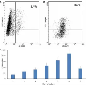
Figure 1.Isolation and enrichment of CD133+ cells from ovarian epithe -lial cancers. The CD133+ cells were isolated and accumulated with anti-CD133 coated magnetic beads. The proportions of anti-CD133+ cells were examined by FACS. A, B. Represent the proportions before and after isola -tions, respectively. C. Represents the number of CD133+ cells counted in vitro. The amount of CD133+ cells were significantly increased after the procedure (P<0.05).
Statistical analysis
All experiments were
perform-ed at least three times and data were exhibited as mean ±
SD. Statistical analysis was conducted using the chi-square
test and Student’s t-test. P<
0.05 was considered
signifi-cant.
Patient survival was calculated
from date of diagnosis to date of death, or entrance into hos -pice care when not available
and converted from days to
months by dividing the number
of days by 30. Overall survival was analyzed by Kaplan-Meier plots and statistical signifi
-cance inferred by log-rank
tests. Correlations to clinical parameters were made using
chi-square analysis. Statistical analyses were performed using the Prism 6 software (Graph-pad, La Jolla CA).
Results
Passage of ovarian epithelial cancer in NOD/SCID mice
The ovarian cancer tissues
were collected from 9 patients
with primary high-grade serous carcinomas. As shown in Table 1, the tumor tissues from 6 patients (#2, 3, and 5-8) were successfully engrafted in NOD/ SCID mice with varying tumor
growth or development. Three
tumor samples (#1, 4, and 9)
did not grow. Samples 2, 3 and 7 grew only 2 passages, while
sample #6 had 3 passages.
[image:4.612.93.368.261.534.2]gests the presence of a self-renewing cell popu -lation in the implanted tumor tissue.
Growing ovarian cancer cells in vitro
A total of 16 regenerated tumor tissue samples collected from the 4th and 5th passages of the
tumor samples was processed and cultured.
Fifteen (94%) of the samples were successfully
passaged and propagated with a similar cell
proliferation index. The majority of the cancer
cells displayed irregular shape adhering to the
flask surface. The number of cultured tumor
cells increased exponentially with a doubling
time of 18 hours (data not shown).
Expression of CD133 and isolation of the CD133+ cells
Flow cytometry was used to separate CD133+ from negative cells and the CD133+ cells were
significantly enriched (Figure 1). After separa
-tion with flow cytometry, the ratio of CD133+ to CD133- cells for each batch of the cell culture
and sub-culture was analyzed. The percentage
of CD133+ cells increased from 15% (5.4±0.43 cells per high power field) in average at the time of initial seeding to 88% (88.7±13.3 cells per high power field) on day 6. It then dropped to 36% (12.4±5.7 cells per high power field) on day 7 right before subculture. The detailed data
is summarized in Figure 1C.
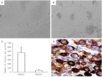
The clonogenic ability of the CD133+ cells
To test the clonogenic ability, the CD133+ or CD133- cells were plated on semi-solid agar and cultured up to 3 weeks. As expected, 105 CD133+ cells generated 468±132 colonies, with the colony-forming efficiency of 0.47%. In contrast, CD133- cells with 105 density gener
[image:5.612.94.520.73.397.2]ated only 43±19 colonies with the colony-form
-ing efficiency of 0.01%. Compared to the CD133- cells (Figure 2A), the CD133+ cells had at least 10-fold higher clonogenic capacity
(Figure 2B). That meant the CD133+ cells had a significantly more potent clonogenic ability than that of CD133- cells (Figure 2C, P<0.001). When the colonies were harvested and plated
on semi-solid agar, the CD133+ cells were able
to repeatedly generate colonies, whereas the
CD133- cells did not. A representative immuno
-cytochemistry picture of positive CD133 cells is
presented in Figure 2D. These results
demon-strated that CD133+ cells possess self-renew -al and growth potenti-al.
Cell cycle analysis of the CD133 labeled cells
After the cells with positive or negative CD133 marker expression were largely separated by flow cytometry, we further tested the cell cycle distribution of these two groups of the cells to see if they were different in cell growth status. Compared with that of the CD133- cells, the ratio of G1/G0 phase of those cells with
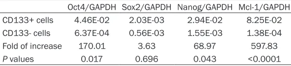
It is commonly believed that the cells
possess-ing stem cell properties express specific CSC biomarkers, such as OCT4, Sox2, Nanog and Mcl-1. To further confirm that CD133+ cells we obtained have the properties of CSC, the above biomarkers were investigated in those samples derived from the ovarian epithelial cancers by using qRT-PCR analysis. Compared to the cells without CD133 expression, the cells with posi
-tive CD133 expression showed a significantly increased copy number of these CSC related genes except Sox2. The increment ranged from 3.6 to 597.8 folds after GAPDH normalization. All the increment of the gene expression was statistically significant except Sox2. One par
-ticular gene Mcl showed about 600-fold increase of expression in the CD133+ cells
(P<0.0001). The detailed data is summarized in Table 2.
Ability of in vivo tumor formation
[image:6.612.94.519.72.242.2]To regenerate a tumor is a hallmark of tumor stem cell. To further determine the role of CD133 in tumor formation, the CD133+ and
Figure 3.Cell cycle distributions analysis of CD133 cells with flow cytometry assay. The cell cycle stage distribu -tion of the enriched CD133+ were analyzed with flow cytometry and compared with that of the CD133- cells. The proportion of cells in G1/G0 stage cells with CD133+ cells was 77.74%, which was significantly higher than those CD133- cells (56.45%) (P<0.05).
Table 2. Comparison of gene expressions assayed with qRT-PCR
Oct4/GAPDH Sox2/GAPDH Nanog/GAPDH Mcl-1/GAPDH CD133+ cells 4.46E-02 2.03E-03 2.94E-02 8.25E-02 CD133- cells 6.37E-04 0.56E-03 1.55E-03 1.38E-04
Fold of increase 170.01 3.63 68.97 597.83
P values 0.017 0.696 0.043 <0.0001
Note: all CSC markers, after normalization with GAPDH, were significantly more ex
-pressed in CD133+ cells compared to those CD133- cells except Sox2.
CD133+ immune-pheno
-type was 77.74%, which was much higher than that
[image:6.612.90.384.328.395.2]of CD133- cells (56.45%)
(Figure 3).
CD133- cells were injected into SCID mice,
respectively. The results showed that
inocula-tion of 106 CD133+ cells regenerated a tumor within 6 weeks with 100% efficiency (8/8). The efficiency rate was reduced to 25% (2/8) when
104 cells were inoculated. The tumors
regener-ated from CD133+ cells were able to be re-implanted for the next generations with a noticeable faster tumor growth. In contrast, injection of 104 CD133- cells failed to regener
-ate a tumor during a 6-week observation, and only one out of 8 inoculations was successful following an injection of 106 CD133- cells, which was significantly less efficiency of tumor generation compared with those CD133+ cells
(P<0.0001).
CD133 staining in primary ovarian epithelial cancers associated with shorter survival ana-lyzed by tumor microarray immunohistochem-istry
To confirm the trends observed in the in vitro
experimental analysis suggesting an
aggres-sive tumor growth was associated with CD133
expression, we decided to examine the protein
levels of CD133 expression in human ovarian cancer samples. Cytoplasmic expression of
CD133 protein was analyzed in a cohort of 290
ovarian epithelial cancers by
immunohisto-chemistry using a specific CD133 antibody in
an ovarian cancer tissue microarray. All cancer
samples were of epithelial origin with clinical
pathologic characteristics summarized in Table 3. All primary tumor microarray samples
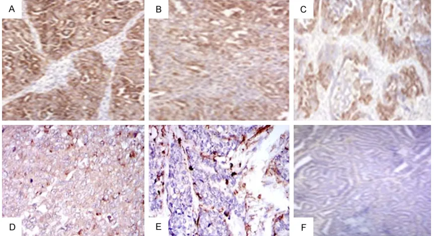
exam-ined were taken at the time of initial debulking surgery. Diffuse and focal staining was observed in the cytoplasm of glandular or
columnar epithelial cells, but not in the tumor
stroma in any of the cores. There is no nuclear staining. Representative pictures of positive and negative immunohistochemical staining of CD133 in ovarian epithelial cancers are pre -sented in Figure 4. Total intensity of positive pixels and total number of positive pixels, as determined by Imagescope software, were
combined into a positive intensity score (PI), which was averaged across the two
representa-tive cores per patient, and used for patient stratification. We chose the top and bottom 10 and 20 percent of patients to compare survival differences between low and high expressers of CD133 protein, and found a significant decrease in survival for both top 10% and 20% of patients expressing high levels of CD133, with a median survival of 28 and 27 months
when compared to patients expressing the low-est 10% (P = 0.026) and 20% (P = 0.035), with
a median survival of 47 and 50 months respec
-tively. The composition of the top and bottom 10% and 20% of patients was similar when examining clinical parameters of FIGO grade, stage, and age. Part of the data is presented in
Table 4.
Discussions
Previous studies have stated that CSC influ
-ences the progress of OECs. Residual cancer
cells, especially the CSC, have been
hypothe-sized to contribute to the relapse of OEC. Indeed, there are considerable lines of evi -dence showing that CSC promotes ovarian
can-cer cell proliferation. However, identifying ovar
-ian CSC with a specific biomarker is the main obstacle to isolate CSC. So far, some stem cell associated biomarkers have been used to iso
-late CSC, for example, CD34+ CD38- Thy- phe
-notype for acute myelogenous leukemia stem cells [4] and CD44+ CD24- and ESA+ for breast cancer stem cells [13]. Although the biomark
-ers for ovarian cancer stem cells remain elu -sive, it is believed that ovarian epithelial can-Table 3. Patient demographic in a ovarian
epithelial cancer tissue microarray of 290
patients
Characteristics Total number of cases (N = 290) Age of diagnosis
Mean 63
Medium 62.2
Range 31.5 to 92.3
FIGO Stage, n (%)
I 6 (2.0)
II 10 (3.4)
III 249 (85.9)
IV 25 (8.6)
FIGO grade, n (%)
1 10 (3.4)
2 35 (12)
3 245 (84.5)
Debulking status, n (%)
No residual 115 (39.7) Regional recurrence 78 (26.9) Never disease free 65 (22.4)
cers should have a similar CSC like properties of other epithelial cancers in the process of
cancer development and metastasis [14-17].
To find a biomarker to separate the ovarian CSC cells, we went through all references in this field and scanned most of the candidate mark
-ers, and found that CD133 could be a promis
-ing candidate marker to identify CSCs within the ovarian epithelial cancer samples. CD133
has been reported to be associated with CSCs in brain, liver, and colon cancers. There are
pre-liminary data suggest that CD133 could also be
be passaged for several generations in semi-solid agar culture and can engraft tumors repeatedly in SCID mice. These features strong
-ly suggest that the CD133+ cells enriched from ovarian cancers have all the biologic features of
the cancer stem cells.
It is commonly believed that CSCs were not
actively proliferating cells, usually with overex
-pression of multidrug resistance molecules
and anti-apoptotic mediators. Consistent with
[image:8.612.88.525.72.310.2]this result, our study showed that CD133+ cells
Figure 4.CD133 immunohistochemical staining of ovarian epithelial cancer. Based on the a positive intensity score (PI) of the cancer cells, (A) (poorly differentiated serous carcinoma) and (B) (poorly differentiated endometrioid carcinoma) represent the high PI score, (C) (moderately differentiated endometrioid carcinoma) as an intermediate PI score, while (D) (poorly differentiated clear cell carcinoma) and (E) (undifferentiated carcinoma) represent low PI score. (F) (well differentiated endometrioid carcinoma) is negative. Original magnifications 100 ×.
Table 4. Multivariate analysis using Cox proportional hazard ratio of CD133 expression in 290 patients with
ovarian epithelial cancer
Age HazardRatio 95% CILower 95% CIUpper P value Significance CD133 1.0382 0.7113 1.0534 0.0032 ** Age 1.0382 1.0143 1.0485 0.0187 * FIGO Stage 3 0.9356 0.7321 1.1033 0.4576 FIGO Grade 3 1.2631 0.7837 2.5378 0.3521
Note: CD133 = standard deviations from the mean of log2 transformed expression level of CD133 by immunohistochemistry; CI = confidence interval; Significance: *<0.05; **<0.01. The number of cases for stage I, II, and IV were too low to be calculated for the analysis. The same situ
-ation applies to the FIGO grade 1 and 2 tumors.
used to identify ovarian cancer stem
cells [18]. However, there is no method
to enrich those CD133+ cells from the
ovarian epithelial cancers. In this study,
we have identified a small subpopula
-tion of CD133+ cells from high-grade
ovarian serous cancer samples through
flow cytometry analysis and success
-fully enriched these cells by series tumor cell implantation into SCID mice. The isolated CD133+ cells showed
much more potent in vitro clonogenic
ability and in vivo tumor formation abil
-ity than that of CD133- cells. In addi
contained a higher proportion of non-cycling cells than CD133- cells. On the other hand, the
cells possessing stem cell properties also
express classical CSC markers, including Oct4, Sox2, Nanog and Mcl-1. Among the markers, Mcl-1, Oct4 and Nanog significantly overex
-pressed in CD133+ cells compared with CD133- cells, especially the Mcl-1 showed almost a 600-fold of increment in CD133+ cells. Mcl-1 is an anti-apoptotic member of the Bcl-2 family. Similar to Bcl-2, Mcl-1 can interact
with BAX and/or BAK1 to inhibit mitochondria-mediated apoptosis. Recent studies
demon-strate that Mcl-1 is essential to the survival of
hematopoietic stem cells. The human hemato-poietic stem cells seems to be dependent on
Mcl-1 to maintain the self-renewal ability, and it is found that Mcl-1 seems to be the only mem -ber selectively overexpressed in human
hema-topoietic stem cells among the Bcl-2 family [19]. In the present study, Mcl-1 was found to be overexpressed in CD133+ cells compared with CD133- cells. The significance of Mcl-1 overexpression warrants further study and could be explored for therapeutic targeting of
ovarian CSC.
Although the relationship between positive
expression of CD133 and prognosis was con -troversial in the past, in this study, we have shown that patients with ovarian epithelial
can-cer showing positive CD133 expression have a significantly worse 5-year survival than those cancers with negative or lower CD133 expres
-sion. Ferrandina et al. were the first to identify CD133 expression in human ovarian cancer tis
-sue and they found that CD133 expression did not provide additional prognostic information for ovarian cancer patients [20]. However, more recent studies [21] including one study from MD Anderson found an association between CD133 status and prognosis, which used a
400-patient cohort with a good long-term
patient follow-up data set [22]. That study showed that positive CD133 expression is
associated with various clinicopathologic
char-acteristics of primary ovarian cancer patients, with shorter disease-free survival time, and with lack of response to chemotherapy [22]. In this study, we investigated that expression of CD133 in an independent cohort of 290
patients with ovarian epithelial cancers in a tumor tissue microarray. When patients were
stratified based on expression levels to com -pare the top 10% and 20% to the bottom 10%
and 20% respectively, we found a statistically significant reduced survival in the highest
expressing patients compared to the lowest expressing patients, with a 65% and 78% reduction in median survival time respectively,
which correspond to a difference of over 24 months. The predictive power of CD133 stain -ing in cancer tissue is stronger in older patients,
suggesting it is best used for prognosis in patients above the age of 60. This is also in agreement with findings that CD133 expres -sion has prognostic value in hepatocellular car-cinoma [23] colon and rectal adenocarcar-cinoma [24], invasive ductal breast carcinoma [25], and non-small cell lung carcinoma [26].
In conclusion, CD133 is a useful and efficient biomarker to identify ovarian CSCs. Enrichment of CD133+ CSCs can be effectively generated through repeated engrafts of cancer tissue in SCID mice. The method of generating ovarian CSCs provides a feasible and practical way for future ovarian cancer research, particularly in the area of mechanism studies for
chemo-resistance and cancer recurrence. Together
with CD133, a combination with other stem cell markers such as Mcl-1 needs to be further explored for potential clinical usage. A targeted therapy by eliminating CD133+ cells may have a clinical value for ovarian cancer treatment. The immunohistochemical assessment of CD133 expression, which serves as an inde
-pendent poor survival marker, may have a
potential clinical value in predicting disease progression and prognosis in ovarian cancer.
Acknowledgements
This work was partially supported by the National High Technology Research and De-velopment Program (“863” Program) of China
(2014AA020605, 2012AA02A507), National
Natural Science Foundation of China
(812-72857, 81370074), National Science and Technology Project (2015BAI13B05), and the
Shanghai Municipal Public Health Bureau (grant number XYQ2013119), and the “Chen-xing Project” from Shanghai Jiao Tong University to Z.Z. The work was also partially supported by Mark and Jane Gibson endowment fund and UTSW Medical Center start up fund to WZ.
Disclosure of conflict of interest
Address correspondence to: Dr. Wenxin Zheng, Obstetric and Gynecology, UT Southwestern Medical Center, 6000 Harry Hines Blvd, NB6.408, USA. Tel: 214-648-1190; E-mail: wenxin.zheng@utsouthwest-ern.edu
References
[1] Werbowetski-Ogilvie TE and Bhatia M. Pluripotent human stem cell lines: what we can learn about cancer initiation. Trends Mol Med 2008; 14: 323-332.
[2] de Figueiredo-Pontes LL, Pintao MC, Oliveira LC, Dalmazzo LF, Jacomo RH, Garcia AB, Falcao RP and Rego EM. Determination of P-glycoprotein, MDR-related protein 1, breast cancer resistance protein, and lung-resistance protein expression in leukemic stem cells of acute myeloid leukemia. Cytometry B Clin Cytom 2008; 74: 163-168.
[3] Ho MM, Hogge DE and Ling V. MDR1 and BCRP1 expression in leukemic progenitors cor -relates with chemotherapy response in acute myeloid leukemia. Exp Hematol 2008; 36: 433-442.
[4] Jamieson CH. Chronic myeloid leukemia stem cells. Hematology Am Soc Hematol Educ Program 2008; 436-442.
[5] Blank U, Karlsson G and Karlsson S. Signaling pathways governing stem-cell fate. Blood 2008; 111: 492-503.
[6] Morrison R, Schleicher SM, Sun Y, Niermann KJ, Kim S, Spratt DE, Chung CH and Lu B. Targeting the mechanisms of resistance to chemotherapy and radiotherapy with the can-cer stem cell hypothesis. J Oncol 2011; 2011: 941876.
[7] Collins AT, Berry PA, Hyde C, Stower MJ and Maitland NJ. Prospective identification of tu -morigenic prostate cancer stem cells. Cancer Res 2005; 65: 10946-10951.
[8] Dalerba P, Dylla SJ, Park IK, Liu R, Wang X, Cho RW, Hoey T, Gurney A, Huang EH, Simeone DM, Shelton AA, Parmiani G, Castelli C and Clarke MF. Phenotypic characterization of human colorectal cancer stem cells. Proc Natl Acad Sci U S A 2007; 104: 10158-10163.
[9] O’Brien CA, Pollett A, Gallinger S and Dick JE. A human colon cancer cell capable of initiating tumour growth in immunodeficient mice. Nature 2007; 445: 106-110.
[10] Ricci-Vitiani L, Lombardi DG, Pilozzi E, Biffoni M, Todaro M, Peschle C and De Maria R. Identification and expansion of human colon-cancer-initiating cells. Nature 2007; 445: 111-115.
[11] Singh SK, Hawkins C, Clarke ID, Squire JA, Bayani J, Hide T, Henkelman RM, Cusimano MD and Dirks PB. Identification of human
brain tumour initiating cells. Nature 2004; 432: 396-401.
[12] Wei W, Mok SC, Oliva E, Kim SH, Mohapatra G and Birrer MJ. FGF18 as a prognostic and ther -apeutic biomarker in ovarian cancer. J Clin Invest 2013; 123: 4435-4448.
[13] Ponti D, Costa A, Zaffaroni N, Pratesi G, Petrangolini G, Coradini D, Pilotti S, Pierotti MA and Daidone MG. Isolation and in vitro propa -gation of tumorigenic breast cancer cells with stem/progenitor cell properties. Cancer Res 2005; 65: 5506-5511.
[14] Alvero AB, Chen R, Fu HH, Montagna M, Schwartz PE, Rutherford T, Silasi DA, Steffensen KD, Waldstrom M, Visintin I and Mor G. Molecular phenotyping of human ovar -ian cancer stem cells unravels the mecha-nisms for repair and chemoresistance. Cell Cycle 2009; 8: 158-166.
[15] Hosonuma S, Kobayashi Y, Kojo S, Wada H, Seino K, Kiguchi K and Ishizuka B. Clinical sig -nificance of side population in ovarian cancer cells. Hum Cell 2011; 24: 9-12.
[16] Steffensen KD, Alvero AB, Yang Y, Waldstrom M, Hui P, Holmberg JC, Silasi DA, Jakobsen A, Rutherford T and Mor G. Prevalence of epithe -lial ovarian cancer stem cells correlates with recurrence in early-stage ovarian cancer. J Oncol 2011; 2011: 620523.
[17] Zhang S, Balch C, Chan MW, Lai HC, Matei D, Schilder JM, Yan PS, Huang TH and Nephew KP. Identification and characterization of ovar -ian cancer-initiating cells from primary human tumors. Cancer Res 2008; 68: 4311-4320. [18] Curley MD, Therrien VA, Cummings CL, Sergent
PA, Koulouris CR, Friel AM, Roberts DJ, Seiden MV, Scadden DT, Rueda BR and Foster R. CD133 expression defines a tumor initiating cell population in primary human ovarian can-cer. Stem Cells 2009; 27: 2875-2883. [19] Campbell CJ, Lee JB, Levadoux-Martin M,
Wynder T, Xenocostas A, Leber B and Bhatia M. The human stem cell hierarchy is defined by a functional dependence on Mcl-1 for self-re -newal capacity. Blood 2010; 116: 1433-1442. [20] Ferrandina G, Martinelli E, Petrillo M, Prisco
MG, Zannoni G, Sioletic S and Scambia G. CD133 antigen expression in ovarian cancer. BMC Cancer 2009; 9: 221.
[21] Liang J, Yang B, Cao Q and Wu X. Association of Vasculogenic Mimicry Formation and CD133 Expression with Poor Prognosis in Ovarian Cancer. Gynecol Obstet Invest 2016; [Epub ahead of print].
[23] Sasaki A, Kamiyama T, Yokoo H, Nakanishi K, Kubota K, Haga H, Matsushita M, Ozaki M, Matsuno Y and Todo S. Cytoplasmic expres -sion of CD133 is an important risk factor for overall survival in hepatocellular carcinoma. Oncol Rep 2010; 24: 537-546.
[24] Li CY, Li BX, Liang Y, Peng RQ, Ding Y, Xu DZ, Zhang X, Pan ZZ, Wan DS, Zeng YX, Zhu XF and Zhang XS. Higher percentage of CD133+ cells is associated with poor prognosis in colon car-cinoma patients with stage IIIB. J Transl Med 2009; 7: 56.
[25] Zhao P, Lu Y, Jiang X and Li X. Clinicopathological significance and prognostic value of CD133 ex -pression in triple-negative breast carcinoma. Cancer Sci 2011; 102: 1107-1111.