Original Article
Protective effects of hydrogen-rich saline against renal
ischemia-reperfusion injury by increased expression
of heme oxygenase-1 in aged rats
Xianlin Xu, Xiaozhou He, Jipu Liu, Jiandi Qin, Jianan Ye, Min Fan
Department of Surgical Urology, The Third Affiliated Hospital of Soochow University, Changzhou 213003, Jiangsu, China
Received May 13, 2015; Accepted June 26, 2015; Epub April 1, 2019; Published April 15, 2019
Abstract: Objective: Oxygen free radicals (ROS) are considered to be one of the important factors involved in the pathophysiology of aged renal ischemia-reperfusion (I/R) injury. Hydrogen gas has been reported to alleviate I/R injury by scavenging free radicals. The aim of this study was to evaluate the effect of hydrogen-rich saline (HRS) on renal I/R injury in aged rats. Materials and methods: A rat model of renal I/R injury was induced by 45-min occlu-sion of the bilateral renal pedicles and 24-h reperfuocclu-sion. Physiological saline or HRS (8 ml/kg) was administered intraperitoneally 5 min before reperfusion. Parameters indicating renal function (blood urea nitrogen (BUN) and serum creatinine (SCr)) and those indicating oxidative stress (tissue levels of malondialdehyde (MDA) and 8-hy-droxy-deoxyguanosine (8-OHdG), tissue activities of superoxide dismutase (SOD), and tissue expression of heme oxygenase-1 (HO-1)) were measured. Results: After I/R injury, BUN, SCr, tissue levels of MDA and 8-OHdG, and gene
expression of HO-1 were all significantly increased while tissue activities of SOD were significantly decreased. HRS
reversed these changes, with the exception of HO-1 expression, which was increased further, and improved renal morphology. Conclusions: HRS improves the renal response to I/R in aged rats, possibly by reducing oxidative stress and upregulating HO-1 gene expression.
Keywords: Ischemia-reperfusion (I/R) injury, hydrogen-rich saline (HRS), heme oxygenase-1 (HO-1), reactive oxy-gen species (ROS), malondialdehyde (MDA), superoxide dismutase (SOD)
Introduction
The human lifespan has increased through medical advancements and improved health-care. However, as the elderly population has increased worldwide, the incidence of acute kidney injury (AKI) has also steadily risen [1]. Cardiovascular problems caused by age-relat-ed conditions, such as hypertension, diabetes, and ischemia-reperfusion (I/R), contribute to AKI susceptibility [2, 3]. In addition, the severity of AKI also increases with age and functional renal recovery is lower. A recent study has esti-mated that patients older than 65 years of age have an approximately 30% greater risk for non-recovery of complete renal function after AKI than those younger than 65 years old [4]. Thus, it is very important to find an effective treatment to alleviate renal I/R injury in the aging population.
Kidney aging has been recognized as a chronic process of compromised renal function and structural changes in the tubulointerstitium and glomerulus [5]. There is considerable evidence that aging occurs as a consequence of oxida-tive stress, and reacoxida-tive oxygen species (ROS) produced during cellular metabolism lead to an age-dependent increase in oxidatively modified proteins, lipids, and nucleic acids in tissue. Oxygen free radicals are considered to be one of the important factors involved in the patho-physiology of I/R.
Recently, it was demonstrated that molecular hydrogen could selectively reduce cytotoxic ROS and reactive nitrogen species (RNS), such as hydroxyl radicals and peroxynitrite, in vitro
I/R injury by reducing oxidative stress and inflammation in adult rats [7, 8]. However, the potential effect of hydrogen on renal I/R injury in aged rats has not been examined. Therefore, the present study investigated the possible therapeutic effects of HRS on renal I/R injury in aged rats.
Materials and methods
Animals
A total of 30 male Sprague-Dawley rats weigh-ing 225-250 g and 24 months of age (aged model) were used in the present study, which was approved by the Institutional Animal Care and Use Committee at Soochow University. All experiments were performed in accordance with the National Institutes of Health guidelines (NIH Publ. No. 86-23, revised 1985). Before the experiments, the animals were fed a standard rat chow, drank water ad libitum, and were housed in metabolic cages under controlled temperature in 12-h light/dark cycles for at least one week.
Surgical procedure
Animals were divided into three groups consist-ing of 10 rats each: (1) sham-operated plus physiological saline treatment; (2) renal I/R plus physiological saline treatment; and (3) renal I/R plus HRS treatment. Sham-operated animals underwent the same surgical proce-dures except that the bilateral renal pedicles were not clamped. Physiological saline or HRS (8 ml/kg) was injected intraperitoneally 5 min before reperfusion. Twenty-four hours after the initiation of renal ischemia, the rats were killed under anesthesia, blood was drawn, and the left kidneys were harvested and frozen in liquid nitrogen.
HRS production
Hydrogen was dissolved in physiological saline for 6 h under high pressure (0.4 MPa) to a supersaturated level using a hydrogen-rich, water-producing apparatus produced by our department. The saturated hydrogenated saline was stored under atmospheric pressure at 48°C in an aluminum bag with no dead vol-ume. HRS was sterilized by gamma irradiation and was freshly prepared each week, which ensured that a concentration of 0.6 mM was
maintained. Gas chromatography was used to confirm the content of hydrogen in the saline by the method described by Ohsawa et al [9]. We referenced a paper to intraperitoneal injection [8].
Analysis of renal function
Twenty-four hours after renal ischemia, blood was used to assess renal function by measur-ing blood urea nitrogen (BUN) and serum creati-nine (SCr). The samples were analyzed on a COBAS Mira chemical analyzer (Roche; Basel, Switzerland) with commercial kits from Sigma Chemical Co. (St. Louis, MO, USA).
Histopathology evaluation
Kidney tissue samples from 12-week post-isch-emic kidneys were stained with hematoxylin and eosin, and the staining was semi-quanti- tatively graded for tubulointerstitial damage (tubular dilation or atrophy and interstitial expansion with edema, inflammatory infiltrate, or fibrosis) based on a scale of 0 to 3 as follows: A score of 0 was given for a normal cortical tubulointerstitium; a score of 1 was given for mild tubulointerstitial damage affecting up to 25% of an objective field at 200× magnifica -tion; a score of 2 was given for moderate tubu-lointerstitial damage affecting 25-50% of the field; and a score of 3 was given for severe tubulointerstitial damage affecting more than 50% of the field. The examiners were blinded to the identity of each group, and ten randomly selected cortical fields were used to score each animal, with the mean score being attributed to that animal [10].
Immunohistochemistry
a negative control. Slides were then incubated with biotinylated mouse anti-rabbit IgG se- condary antibody (Maixin Biotechnology Ltd.; Fuzhou, China). Finally, slides were incubated with hydrogen peroxide-diaminobenzidine for 1 min. Sections were counterstained with hema-toxylin. Estimates of staining intensity were per-formed by a blinded observer on coded sec-tions (3-4 secsec-tions per kidney and 10-12 fields per section). The observer performed light microscopy and semiquantitatively scored the intensity of HO-1 staining in the whole section (0 = none, 1 = weak, 2 = moderate, and 3 = strong).
Measurement of antioxidant enzyme activity
and malondialdehyde (MDA) content
Kidney MDA content and superoxide dismutase (SOD) activity were determined according to the technical manuals of the detection kits (Nanjing Jiancheng Biochemistry Co.; Nanjing, China). Kidney tissue was homogenized in phosphate buffer (pH 7.4). After centrifugation at 12,000 g for 20 min, MDA content and SOD activity in the supernatant of each sample were measured using the corresponding kits. MDA content was measured by the thiobarbituric acid (TBA) assay. The method obtained measurements of the color produced during the reaction of TBA with MDA at 532 nm in a spectrophotometer; estimated MDA levels were expressed as nmol/ mg protein. SOD activity was measured using
the nitroblue tetrazoli-um (NBT) reduction assay, by following the red- uction of nitrite by a xanthine-xanthine oxid- ase system, which is a superoxide generator. One unit of SOD activity is defined as the amount that shows 50% inhibi-tion of NBT reducinhibi-tion.
Real-time reverse tran-scription polymerase chain reaction (RT-PCR)
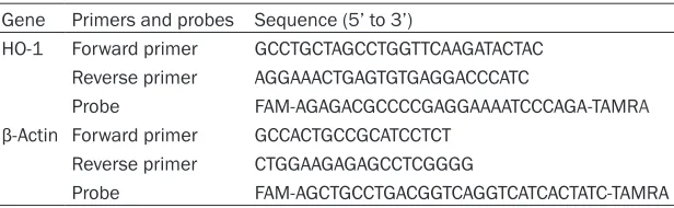
[image:3.612.92.400.83.178.2]mRNA levels were det- ermined by real-time RT-PCR. Primers and probes are listed in
Table 1. Total RNA was
Table 1. Primers and probes for rat HO-1 and β-actin Gene Primers and probes Sequence (5’ to 3’)
HO-1 Forward primer GCCTGCTAGCCTGGTTCAAGATACTAC Reverse primer AGGAAACTGAGTGTGAGGACCCATC
Probe FAM-AGAGACGCCCCGAGGAAAATCCCAGA-TAMRA
β-Actin Forward primer GCCACTGCCGCATCCTCT Reverse primer CTGGAAGAGAGCCTCGGGG
[image:3.612.90.400.212.253.2]Probe FAM-AGCTGCCTGACGGTCAGGTCATCACTATC-TAMRA
Table 2. Effect of HRS treatment on renal function
Aged Aged + I/R Aged + I/R + HRS BUN (mM) 11.3 ± 7.1 19.9 ± 5.4# 12.3 ± 5.5*
SCr (µM) 129 ± 41 227 ± 64# 147 ± 52*
Data are expressed as mean ± SD, n = 10. Both BUN and SCr levels were greater in the
aged + I/R group as compared to those of the aged group. HRS treatment significantly
decreased both BUN and SCr levels. *P < 0.01 vs. the aged + I/R group; #P < 0.01 vs. the
aged group.
Statistical analysis
Statistical analysis was performed using the GraphPad Prism 4.0 software package (GraphPad Software, Inc.; San Diego, CA, USA). All data were expressed as mean ± SD. Statistical analyses were done using one-way ANOVA followed by the Student-Newman-Keuls (SNK) t-test for multiple comparisons. A P-value less than 0.05 was considered as statistically significant.
Results
Renal function
Renal function of rats was assessed by mea-suring the BUN and SCr levels in plasma (Table 2). In the aged I/R group, BUN and SCr levels
were significantly (P < 0.05) greater than those in the aged group by nearly 2-fold, indicating the development of renal failure. In the aged + I/R + HRS group, BUN and SCr levels were sig-nificantly (P < 0.01) less than those in the aged + I/R group. In contrast, the levels of BUN and SCr in the aged + I/R + HRS and the aged groups did not significantly differ from one another (P > 0.05).
Renal oxidative stress
[image:4.612.94.521.76.419.2]MDA content was significantly greater in the aged + I/R group than in the aged group (P < 0.05; Figure 1A). HRS treatment significantly decreased MDA levels compared with the aged + I/R group. Similarly, the level of 8-OHdG in aged + I/R rats was significantly greater than that in rats not exposed to I/R (P < 0.05; Figure
Figure 1. Markers of oxidative stress in the different treatment groups. A. MDA level in the kidneys of different groups (n = 10). Data are expressed as mean ± SD. *P < 0.05 vs. the aged I/R group; #P < 0.05 vs. the aged group. B. 8-OHdG
level in the kidney in different groups (n = 10). Data are expressed as mean ± SD. *P < 0.05 vs. the aged I/R group; #P < 0.05 vs. the aged group. C. SOD activity in the kidney of different groups (n = 10). Data are expressed as mean
± SD. *P < 0.05 vs. the aged I/R group; #P < 0.05 vs. the aged group. D. mRNA level of HO-1in the kidney of different
1B). Additionally, the level of 8-OHdG in the aged + I/R + HRS group was significantly less than that in the aged + I/R group (P < 0.01;
Figure 1B).
Renal antioxidant enzymatic activities
The activity of SOD in renal tissue was signifi -cantly less in the aged + I/R group than in the aged group (P < 0.05; Figure 1C). HRS treat-ment significantly increased the SOD activity as compared to that in the aged I/R group (P < 0.05; Figure 1C).
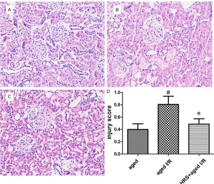
Histopathology findings
Histopathology analysis showed that tubuloint-erstitial damage presented as tubular dilation,
interstitial inflammatory infiltrate, and fibrosis in the majority of animals (Figure 2). The tubu-lointerstitial damage was greater in the aged + I/R group than in the aged group (0.80 ± 0.38 vs. 0.39 ± 0.25; P < 0.05; Figure 2). However, HRS treatment significantly decreased the score for tubulointerstitial damage (0.48 ± 0.24 vs. 0.80 ± 0.38 for the aged + I/R + HRS group vs. the aged + I/R group; P < 0.05; Figure 2).
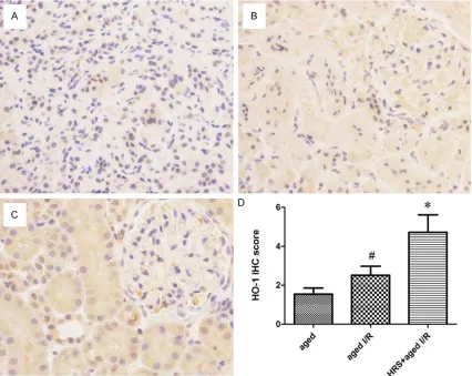
Immunohistochemical staining for HO-1
HO-1 immunostaining was localized to the prox-imal and distal tubules in the cortex and was prominent in the outer stripe region of the outer medulla (Figure 3). HO-1 immunostaining inten-Figure 2. Histology changes in the kidney of different treatment groups. Kidney tissues from rats of all groups were
fixed, embedded, sectioned, and stained with hematoxylin and eosin. A-C. Representative histology specimens from the young group, aged group, young I/R group, and aged I/R group. Original magnification: 200×. Histology changes
were observed 24 h after renal I/R injury and included tubular dilation or atrophy and interstitial expansion with
edema, inflammatory infiltrate, or fibrosis. D. Injury scores in different groups (n = 10). Data are expressed as mean
[image:5.612.91.518.70.437.2]sity in the aged + I/R + HRS group was signifi -cantly greater than that in the aged + I/R group (4.71 ± 2.54 vs. 2.51 ± 1.30, P < 0.01). HO-1 staining in the aged + I/R group did not differ significantly from that in the aged group (2.51 ± 1.30 vs. 1.53 ± 0.89, P > 0.05; Figure 3).
Renal tissue HO-1 gene expression
HO-1 mRNA levels in the aged, aged + I/R, and aged + I/R + HRS groups were determined by real-time RT-PCR using β-actin as the reference gene (Figure 1D). The HO-1 mRNA level in the aged + I/R + HRS group was significantly great -er than that of the aged + I/R group (0.00543 ± 0.0035 vs. 0.00317 ± 0.00223, P < 0.05). The levels of HO-1 mRNA in the aged + I/R and aged groups were not significantly different from one another (0.00317 ± 0.00223 vs. 0.00176 ± 0.00124, P < 0.05).
Discussion
[image:6.612.94.520.73.412.2]The present study demonstrated that intraperi-toneal injection of HRS significantly attenuated I/R-induced renal dysfunction and tissue injury, as observed by the reversal of increases in the levels of MDA and 8-OHdG and decreases in SOD activity that resulted from I/R. This improved renal function was accompanied by an increase in HO-1 expression. Because HO-1 is an inducible antioxidant enzyme, its induc-tion may be part of the mechanism underlying the protective effects afforded by HRS therapy. This finding is consistent with results from a recent study showing that HRS was able to attenuate renal I/R injury in adult rats. Compared to normal adult rats, aged rats are more susceptible to I/R-induced renal failure, which is associated with increased oxidative stress, as shown in our previous study [11]. Figure 3. HO-1 expression in the kidneys from different treatment groups. A-C. Representative HO-1 staining of
speci-mens from the aged group, aged I/R group, and aged I/R + HRS group. Original magnification: 400×. D. Immunohis -tochemistry score for HO-1 staining in different groups (n = 10). Data are expressed as mean ± SD. *P < 0.01 vs. the
Persistent oxidative stress is one of the under-lying causes or components of the aging cess [6]. Antioxidant treatment has been pro-posed to prevent aging-related general distur-bances [12]. In the kidneys, the local synthesis of ROS, at least in I/R-injured animals and cul-tured cells, seems to increase with age [13]. Antioxidant treatment may also prevent the morphological and functional aging-related changes in the kidneys. Our data demonstrate that aging aggravates ischemic acute renal fail-ure in the rat. Compared with same-age control animals, rats that underwent I/R had greater levels of MDA in renal tissue. However, renal MDA content was significantly less in those groups that received HRS as compared to those groups that received physiological saline. These results suggest that HRS is more effec-tive in aging animals than in young adult animals.
The decreased SOD activity seemed to be linked to the increased MDA levels in the aged I/R rats. SOD is an antioxidant enzyme that is important in aging, and a decrease in this parameter is correlated with advanced age. Here, we found that HRS significantly increased SOD activity compared to that in the control animals. 8-OHdG is another marker of oxidative damage that arises from the reaction of ROS with cellular DNA and may accumulate with advancing age. The present study showed that HRS treatment significantly alleviated oxidative stress following I/R injury by reducing levels of both renal MDA and 8-OHdG.
HO-1 induction is one of the most sensitive indi-cators of cellular stress [14]. Induction of HO-1 in kidney, heart, and liver grafts prior to trans-plantation provided these grafts protection after reperfusion and was associated with decreased levels of inflammation [15, 16]. Recent studies have indicated that kidneys from older animals have an impaired ability to upregulate HO-1 in response to I/R injury and exhibit a worse injury phenotype compared to young animals. Administration of the heme pre-cursor hemin to one-year-old mice robustly induced HO-1 protein expression and provided aged mice with both structural and functional protection from I/R injury [17]. Similarly, admin -istration of hydrogen gas had a protective effect on injured lung tissue by promoting the levels of HO-1 mRNA and protein [18]. Consistent with these previous studies, we found that HRS increased the levels of HO-1 mRNA and HO-1 protein in aged rat tissue,
sug-gesting that the protective effects afforded by hydrogen gas may be mediated by HO-1 induc-tion. The protection due to the increased expression of HO-1 may in turn be linked to the generation of carbon monoxide by the action of HO-1 on biliverdin and bilirubin. Carbon monox-ide has potent vasodilating effects by activat-ing guanylate cyclase and has been shown to prevent I/R injury during organ transplantation [19]. Bilirubin and biliverdin, two metabolites of heme degradation, act as scavengers of toxic oxygen radicals [20]. Moreover, HO-1 induction in grafts prior to reperfusion may eliminate free heme, which is released from the kidneys dur-ing the I/R process [21].
It is well known that mitochondria are not only the source of energy but also the major source of ROS [19]. Mitochondrial function and mor-phology are impaired in aging, as shown by declines in the membrane potential and SOD activity [22]. These changes enable accumula-tion of oxidatively damaged macromolecules with aging and render the mitochondria of aged animals more susceptible to oxidative injury [23]. Qiao et al. [24] demonstrated an aging-related increase of tubular cell apoptosis in renal I/R injury, which may be due to enhanced activation of the mitochondrial apoptosis path-way that results from the increase in oxidative stress. Although not directly tested in the pres-ent study, the findings of decreased SOD activ -ity and increased MDA levels in aged animals after renal I/R injury are consistent with mito-chondrial dysfunction occurring during the inju-ry. Furthermore, the exaggerated response in older animals as compared to that in young ani-mals suggests that mitochondrial dysfunction is also more extensive in the older animals. Hydrogen gas may protect mitochondria by scavenging ROS [13], although the exact molec-ular mechanism by which this occurs is unknown.
In conclusion, HRS has a protective effect on age-dependent renal I/R injury by reducing free radical peroxidation of lipid membranes and increasing the activity of antioxidant enzymes. The upregulation of HO-1 may play an essential role in the protective effects of HRS.
Acknowledgements
Science and Technique Foundation of Jiangsu Province, P. R. China (No. BL2012045).
Disclosure of conflict of interest
None.
Address correspondence to: Drs. Xiaozhou He and Min Fan, Department of Surgical Urology, The Third
Affiliated Hospital of Soochow University, Changzhou
213003, Jiangsu, China. Tel: 86-519-68871251;
Fax: 86-519-86621235; E-mail: fnmong@hotmail. com (XZH); [email protected] (MF)
References
[1] Xue JL, Daniels F, Star RA, Kimmel PL, Eggers PW, Molitoris BA, Himmelfarb J, Collins AJ. Incidence and mortality of acute renal failure
in Medicare beneficiaries, 1992-2001. J Am Soc Nephrol 2006; 17: 1135-1142.
[2] Esposito C, Plati A, Mazzullo T, Fasoli G, De Mauri A, Grosjean F, Mangione F, Castoldi F, Serpieri N, Cornacchia F, Dal Canton A. Renal function and functional reserve in healthy
el-derly individuals. J Nephrol 2007; 20:
617-625.
[3] De Luca L, Fonarow GC, Adams KF Jr, Mebazaa A, Tavazzi L, Swedberg K, Gheorghiade M. Acute heart failure syndromes: Clinical scenar-ios and pathophysiologic targets for therapy.
Heart Fail Rev 2007; 12: 97-104.
[4] Schmitt R, Coca S, Kanbay M, Tinetti ME, Cantley LG, Parikh CR. Recovery of kidney function after acute kidney injury in the elderly: A systematic review and meta-analysis. Am J
Kidney Dis 2008; 52: 262-271.
[5] Ding G, Franki N, Kapasi AA, Reddy K, Gibbons N, Singhal PC. Tubular cell senescence and ex-pression of TGF-1 and p21 (WAF1/C1P1) in
tubulointerstitial fibrosis of aging rats. Exp Mol Pathol 2001; 70: 43-53.
[6] Yu BP. Aging and oxidative stress: modulation by dietary restriction. Free Radic Biol Med 1996; 21: 651-668.
[7] Shingu C, Koga H, Hagiwara S, Matsumoto S, Goto K, Yokoi I, Noguchi T. Hydrogen-rich saline solution attenuates renal
ischemia-reperfu-sion injury. J Anesth 2010; 24: 569-574.
[8] Wang F, Yu G, Liu S-Y. Hydrogen-rich saline pro-tects against renal ischemia/reperfusion
inju-ry in rats. J Surg Res 2011; 167: e339-e344.
[9] Ohsawa I, Ishikawa M, Takahashi K, Watanabe M, Nishimaki K, Yamagata K, Katsura K, Katayama Y, Asoh S, Ohta S. Hydrogen acts as a therapeutic antioxidant by selectively
reduc-ing cytotoxic oxygen radicals. Nat Med 2007;
13: 688-694.
[10] Thomas GL, Yang B, Wagner BE, Savill J, El Nahas AM. Cellular apoptosis and proliferation
in experimental renal fibrosis. Nephrol Dial
Transpl 1998; 13: 2216-2226.
[11] Xu X, Fan M, He X, Liu J, Qin J, Ye J. Aging ag-gravates long-term renal ischemia-reperfusion
injury in rats. J Surg Res 2014; 187: 289-296. [12] Orr WC, Sohal RS. Extension of life-span by
overexpression of superoxide dismutase and catalase in Drosophila melanogaster. Science 1981; 116: 53-64.
[13] Ruiz-Torres MP, Gonzalez-Rubio M, Lucio-Cazan FJ, Ruiz-Villaesesa M, Rodriguez-Puyol M, Rodriguez-Puyol D. Reactive oxygen species and platelet activating factor synthesis in age-related glomerulosclerosis. J Lab Clin Med 1994; 124: 489-495.
[14] Maines MD. The heme oxygenase system: A regulator of second messenger gases. Annu
Rev Pharmacol Toxicol 1997; 37: 517-524.
[15] Tullius SG, Nieminen-Kelha M, Buelow P, Reutzel-Selke A, Martins PN, Pratschke J, Bachmann U, Lehmann M, Southard D, Iyer S, Schmidbauer G, Sawitzki B, Reinke P, Neuhaus P, Volk HD. Inhibition of ischemia/reperfusion injury and chronic graft deterioration by a sin-gle-donor treatment with cobalt-protoporphyrin for the induction of heme oxygenase-1.
Transplantation 2002; 74: 591-598.
[16] Katori M, Buelow R, Ke B, Ma J, Coito AJ, Iyer S, Southard D, Busuttil RW, Kupiec-Weglinski JW. Heme oxygenase-1 overexpression protects rat hearts from cold ischemia/reperfusion injury via an antiapoptotic pathway. Transplantation
2002; 73: 287-292.
[17] Holzen JP, August C, Bahde R, Minin E, Lang D,
Heidenreich S, Dietl KH, Spiegel HU. Influence
of heme oxygenase-1 on microcirculation after kidney transplantation. J Surg Res 2008; 148: 126-135.
[18] Huang CS, Kawamura T, Toyoda Y, Nakao A. Recent advances in hydrogen research as a therapeutic medical gas. Free Radic Res 2010;
44: 971-982.
[19] Wei YH, Lee HC. Oxidative stress, mitochondri-al DNA mutation and impairment of antioxi-dant enzymes in aging. Exp Biol Med
(Maywood) 2002; 227: 671-682.
[20] Stocker R. Antioxidant activities of bile pig-ments. Antiox Redox Signal 2004; 6: 841-849. [21] Blydt-Hansen TD, Katori M, Lassman C, Ke B,
Coito AJ, Iyer S, Buelow R, Ettenger R, Busuttil RW, Kupiec-Weglinski JW. Gene transfer-in-duced local heme oxygenase-1 overexpression protects rat kidney transplants from ischemia/ reperfusion injury. J Am Soc Nerphrol 2003;
14: 745-754.
high-energy phosphate content. J Mol Cell Cardiol
1998; 30: 661-672.
[23] Mather M, Rottenberg H. Aging enhances the activation of the permeability transition pore in mitochondria. Biochem Biophys Res Commun
2000; 273: 603-608.