Original Article
Dihydrofolate reductase as a predictor for poor
response to platinum-based chemotherapy
in epithelial ovarian cancer
Min Wang1, Ting Hu1, Ke-Yu Xie2
Departments of 1Obstetrics and Gynecology, 2Anesthesia, Second People’s Hospital of Chengdu, Chengdu
610047, Sichuan, China
Received January 24, 2019; Accepted February 22, 2019; Epub May 1, 2019; Published May 15, 2019
Abstract: Background: Platinum-based chemotherapy is the first line chemotherapy regimen for ovarian cancer patients. However, chemotherapy resistance is observed in a large proportion of patients. It is urgently needed to investigate prognostic biomarkers for chemo-sensitivity in ovarian cancer. Methods: Dihydrofolate reductase (DHFR) expression was measured by immunohistochemical staining in 108 specimens, as well as DHFR mRNA variants with qRT-PCR assays. The correlation between DHFR expression and platinum-based chemotherapy response was analyzed. The prognostic significance of DHFR expression was evaluated in ovarian cancer. Results: Positive DHFR
expression was observed in 48 specimens, which was correlated to chemotherapy resistance in ovarian cancer
patients. Elevated DHFR2 mRNA expression, rather than DHFR1, was observed in chemotherapy resistant tumors. Positive DHFR expression was correlated with higher histologic grade in ovarian cancer (P = 0.014). Kaplan-Meier analysis indicated that DHFR positive expression predicted poor disease-free survival (DFS) (P = 0.040), but not overall survival (OS) of ovarian cancer patients (P = 0.706). The prognostic value was further supported by TCGA data analysis. Cox regression analysis indicated that positive DHFR expression was an independent detrimental factor for disease progression for ovarian cancer patients (P = 0.016). Conclusion: DHFR level measurement was a valuable prognostic biomarker for chemo-sensitivity of ovarian cancer. Molecular analysis for DHFR variants will provide important evidence for chemotherapy regimen options.
Keywords: Dihydrofolate reductase, chemotherapy resistance, ovarian cancer
Introduction
Epithelial ovarian cancer is a common gyneco-logic malignancy, characterized as late-detect-ed advanclate-detect-ed stage disease and poor prognosis [1]. Platinum-based chemotherapy, such as cis-platin or carbocis-platin, is the predominant regi-men after cytoreductive surgery for advanced stage ovarian cancer [2]. However, most patients suffer recurrence within 2 years from the diagnosis in spite of initial objective response [3]. Limited chemotherapy options aer available for the patients with platinum resistant ovarian cancer [4]. Thus, effective prognostic strategies for chemoresistance are needed to improve survival.
A multifactorial procession is involved in the development of platinum-based chemotherapy resistance [5]. The resistant cells show the
typi-cal trait of increased DNA synthesis and repair [6]. Dihydrofolate reductase (DHFR) partici -pates in dihydrofolate recycling to maintain DNA synthesis and cell proliferation [7, 8]. The exploitation of inhibitors of DHFR leads to a halt of cell cycle, including methotrexate and peme -trexed for breast cancer [9, 10]. Previous data highlight elevated DHFR expression in cisplatin resistant cells of different cancers [11], but the clinical significance of DHFR in epithelial ovarian cancer has not been explored, espe-cially its role in platinum-based chemotherapy resistance.
DHFR expression for prognosis of the response to first-line chemotherapy.
Materials and methods
Patients and chemotherapy administration
We collected 108 epithelial ovarian cancer tis-sues for this retrospective study. All the pat-ients were diagnosed in our hospital from 2008 to 2014. Platinum-based chemotherapy was administered followed by cytoreductive sur -gery. Clinical information was collected form clinical documents, including age, tumor size, histologic grade, lymph node metastasis, and CA125 levels. All the patients were followed up to evaluate the chemotherapy response. Platinum-resistance was defined as suffering tumor recurrence within six months, whereas tumor recurrence aftersix months of chemo -therapy was defined as platinum-sensitive. Our study was approved by the Institutional Review Board of Second People’s Hospital of Chengdu.
Immunohistochemical staining (IHC)
Tumor specimens were collected in Pathology Department. Tumor sections were prepared from each specimen, then dewaxed and hydrat -ed as recommendation. Antigen retrieval with citrate buffer (pH 6.0) was performed in boiled water. Endogenous peroxidase blockage was performed with 0.3% H2O2 solution. Ventana Discovery XT automated staining system (Ven-tana Medical Systems, Inc., Tucson, AZ, USA) was used for IHC staining. The primary antibody for DHFR (ab82171, Abcam, Cambridge, MA) and control IgG was used in this study.
IHC score evaluation
DHFR expression was measured with a semi-quantitative system [12]. Final IHC scores were determined by staining intensity and positive proportion of tumor cells. The intensity of IHC staining were ranked in three groups (no stain-ing = 0; weak stainstain-ing = 1, middle = 2, strong = 3), the percentage of stained cells were deter -mined by the stained tumor cells in five typical high power field. The final score was obtained by multiplying the intensity score with the per-centage score of stained cells, ranging from 0 (the minimum score) to 3 (the maximum score). Reverse transcription-quantitative real-time polymerase chain reaction (qRT-PCR) Trizol®
reagent (Invitrogen, CA, USA) and PrimeScript RT Master Perfect Real Time Kit (TaKaRa, Japan) kits were used for total RNA preparation and mRNA reverse transcription. Real-time PCR analysis was performed with SYBR Green PCR master mix (Invitrogen, CA, USA). The sequ- ences of primers are as below: DHFR1: 5’-GTCATGGTTGGTTCGCTAAACTGCA-3’, 5’-ATACA-TACTTTTTTCAGAGGGAGGG-3’; DHFR2: 5’-CA-GAGAACTCAAGGAACCTCCACAAG-3’, GAACT-GCCACCAACTATCCAGAACCAT-3’; GAPDH: 5’-GTCAGTGGTGGACCTGACCT-3’, 5’-TGAGGAGGG-GAGATTCAGTG-3’.
Statistical analyses
Statistical analyses were performed with SPSS19.0 (SPSS Inc., Chicago, IL, USA) and GraphPad Prism version 5.00 (GraphPad Software, CA, USA). Differences between gro-ups were evaluated by one-way ANOVA. Kaplan-Meier analysis and Cox proportional hazards regression model were used for prognostic sig -nificance. P < 0.05 was considered significant.
Results
Elevated DHFR expression in platinum-based chemotherapy resistant ovarian cancer speci-mens
Increased transcription of DHFR2 but not DHFR1 is involved in chemotherapy resistance
To further investigate the functional role of dif -ferent DHFR variants in ovarian cancer chemo
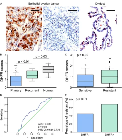
[image:3.612.92.520.66.556.2]-therapy resistance, the mRNA levels of DHFR1/2 were evaluated with tumors tissues. The qT-PCR assays were performed with 8 che -motherapy resistant tumors and 8 sensi- tive tumors. Our results showed significantly Figure 1. DHFR expression in ovarian cancer specimens. A. IHC staining for DHFR was performed on ovarian cancer specimens and oviduct tissues. Representative images of DHFR IHC staining are shown. Bar, 100 μm. B. IHC scores were compared among groups, primary and recurrent ovarian cancer specimens, as well as oviduct tissues. C. IHC
increased DHFR2 mRNA ex-pression in chemo-resistant tumors versus sensitive tu- mors, whereas no significant change was observed in DH-FR1 (Figure 2A). Further anal-ysis with TCGA data of epithe -lial ovarian cancer also indi-cated the prognostic value of DHFR2 in disease progression (Figure 2B). A higher percent-age of DHFR2 high expression was found in chemo-resistant tumor than in the sensitive tumor (Figure 2C), whereas no significant difference was ob-served in DHFR1 expression.
The correlation between DHFR expression and clinico-pathologic features
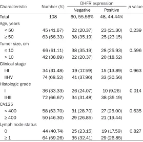
The correlation was evaluated between the IHC results of DHFR expression and clinico -pathologic parameters. Our results indicated that DHFR positive expression was asso-ciated with histologic grade (P = 0.014). The poorly differenti -ated tumors showed an in- creased percentage of posi -tive DHFR expression, as shown in Table 1. Notably, his-tologic grade 2/3 specimens showed significantly higher DHFR scores than grade-1 ones (P < 0.001, Figure 2D). However, we did not observe significant correlation in DHFR positive expression with age, tumor size, clinical stage, CA125 levels, and lymph node metastasis (Table 1).
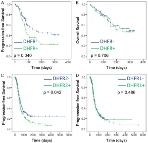
Positive DHFR expression suggests progression of ovar-ian cancer
[image:4.612.91.372.72.303.2]Survival estivation value was analyzed for the expression status of DHFR in ovarian cancer patients. Kaplan-Meier analysis showed poorer pro-gression-free survival (DFS) in Figure 2. Increased transcription of DHFR2 but not DHFR1 was involved in
chemotherapy resistance. A. qT-PCR assays for the mRNA levels of DHFR1/2, which were analyzed with chemotherapy resistant/sensitive tumors. *P < 0.01. B. ROC analysis with TCGA data of epithelial ovarian cancer for the prognostic value of DHFR2 in disease progression (AOC: 0.564, P = 0.03, 95% CI: 0.504-0.624). C. The percentage of DHFR2 high expression was compared between chemo-resistant tumor and sensitive tumor. P = 0.03. D. Histologic grade 2/3 specimens showed significantly higher DHFR scores than grade-1 ones. P < 0.001.
Table 1. Relationship between clinical characteristics and DHFR expression
Characteristic Number (%) NegativeDHFR expressionPositive p value
Total 108 60, 55.56% 48, 44.44%
Age, years
< 50 45 (41.67) 22 (20.37) 23 (21.30) 0.239 ≥ 50 63 (58.33) 38 (35.19) 25 (23.15)
Tumor size, cm
≤ 10 66 (61.11) 38 (35.19) 28 (25.93) 0.596 > 10 42 (38.89) 22 (20.37) 20 (18.52)
Clinical stage
I-II 34 (31.48) 19 (17.59) 15 (13.89) 0.963 III-IV 74 (68.52) 41 (37.96) 33 (30.56)
Histologic grade
I 36 (33.33) 26 (24.07) 10 (9.26) 0.014 II-III 72 (66.67) 34 (31.48) 38 (35.19)
CA125
< 400 58 (53.70) 31 (28.70) 27 (25.00) 0.635 ≥ 400 50 (46.30) 29 (26.85) 21 (19.44)
Lymph node status
[image:4.612.93.372.449.727.2]positive DHFR expression patients than nega
-tive ones (P = 0.040. Figure 3A), whereas no especially DHFR2 mRNA, was an effective biomarker to predict platinum-based chemothera- -Figure 3. Positive DHFR expression suggests disease progression of ovarian
cancer. (A, B) Kaplan-Meier analysis of Disease-Free Survival (A) and Overall Survival (B) for ovarian cancer patients with DHFR positive or negative express
[image:5.612.90.389.72.361.2]-ing tumors in our center. (C, D) Kaplan-Meier analysis of Disease-Free Survival (C) and Overall Survival (D) for ovarian cancer patients with DHFR2 mRNA high or low expressing tumors with TCGA data.
Table 2. Cox regression analyses of progression-free survival and overall survival for DHFR expression in ovarian cancer
Variable Analysis PFS OS
HR 95.0% CI P HR 95.0% CI P Univariate
DHFR 1.697 1.018-2.828 0.042 1.122 0.616-2.044 0.707 Multivariate
Age 1.470 0.853-2.535 0.165 1.211 0.635-2.308 0.561
Size 0.740 0.408-1.342 0.322 0.768 0.362-1.629 0.492
Stage 2.648 0.863-8.127 0.089 3.320 0.84-13.129 0.087
Grade 1.797 0.608-5.315 0.289 1.236 0.352-4.342 0.741
Ca125 0.914 0.503-1.663 0.769 0.989 0.467-2.092 0.977
LNM 2.181 0.804-5.912 0.126 2.048 0.688-6.094 0.198
DHFR 1.975 1.134-3.441 0.016 1.122 0.616-2.044 0.707
HR, hazard ratios; CI, confidence interval. The variables were compared in the following ways: Age, > 50 years vs. ≤ 50 years; size, > 10 cm vs. ≤ 10 cm; Grade, G2-3 vs. G1; CA125, < 400 vs. > 400; LNM, metastasis vs. none; DHFR, positive vs. negative.
significant difference of overall survival (OS) was observed between groups (P = 0.706. Figure 3B). Further analysis with TCGA data also confirmed that elevated DHFR2 expres -sion predicted poor PFS in ovarian cancer, but not OS (P = 0.042, 0.486, re-spectively. Figure 3C, 3D). The independent prognos-tic values of DHFR expres -sion was also analyzed with univariate and multi-variate Cox regression mo- dels. Positive DHFR expres -sion was a risk factor for PFS (HR, 1.697; 95% CI, 1.018-2.828, P = 0.042), but not for OS (HR, 1.122; 95% CI, 0.616-2.044; P = 0.707, Table 2). Multivariate analysis also supported positive DHFR expression was an independent risk factor for PFS (HR, 1.975; 95% CI, 1.134-3.441; P = 0.016), but not for OS (HR, 1.122; 95% CI, 0.616-2.044; P = 0.707, Table 2). Therefore, increased DHFR expression was an inde-pendent detrimental factor for epithelial ovarian can -cer patients.
Discussion
[image:5.612.92.386.480.640.2]py resistance. Enhanced DHFR expression was an independent prognostic biomarker for the disease progression of ovarian cancer.
The combined regimen of paclitaxel and plati -num compounds, such as cisplatin or carbopla-tin, was the first-line option for epithelial ovari -an c-ancer following surgery [16]. Chemotherapy resistance usually occurs rapidly after plati -num-based regimen treatment in epithelial ovarian cancer patients. It is still a focus to develop more effective systemic therapies for improved survival estimation [17]. In this case, it was necessary to exploit biomarkers to pre-dict chemotherapy response, which will favor ovarian cancer patients. We focused on the expression of DHFR in ovarian cancer tissues, which is a valuable biomarker to predict plati-num-based chemotherapy resistance. Elevated DHFR expression in ovarian cancer tissues was correlated with platinum-based chemotherapy resistance. Thus, the measurement of DHFR expression level provided important evidence of chemotherapy options for ovarian cancer patients.
Previous studies supported that elevated expression of folate-dependent proteins was involved in chemotherapy resistance, such as thymidylate synthase [18], DHFR [19] and phosphoribosylglycinamide formyltransferase (GART) [20], which is crucial for cell replication. The platinum-based chemotherapy resistant cell lines also showed increased DNA replica -tion and repair activity [21]. The transforma-tion of folate and 7, 8 dihydrofolate (DHF) into 5, 6, 7, 8 tetrahydrofolate (THF) is dependent on the catalytic activity of DHFR, which is an essential step in the synthesis of DNA nucleic acid bases [7]. In this study, we provided further evidence that folate-dependent proteins are involved in chemotherapy resistance and disease progres-sion of ovarian cancer. We identified that ele -vated mRNA levels of DHFR2, rather than DHFR1, was positively correlated with increased chemotherapy resistance. Furthermore, no sig-nificant correlation was observed between dis -ease progression and thymidylate synthase or GART mRNA levels (data not show). Further studies are still needed for the different func -tional role of DHFR variants, DHFR1 and 2, in disease progression of ovarian cancer, espe -cially in chemotherapy resistance.
Molecular biologic methods paved sensitive and special ways to analyze protein expression status. Immunohistochemical staining detects protein by its specific antigens, which provides evidence of protein expression levels by stain -ing percentage and intensity in cancer tissue [22]. Quantitative mRNA measurements with PCR analysis identifies mRNA transcription by specific mRNA sequence. In our study, DHFR protein expression was determined by IHC staining. DHFR expression was positively corre -lated with histologic grade, but not with other clinical parameters. Notably, the patients with DHFR positive tumors showed worse prognosis than those negative ones. Further analysis with TCGA data showed that DHFR2 mRNA exhibit -ed prognostic value for ovarian cancer. Our results suggest elevated DHFR2 expression indicates poor prognosis. However, no specific antibody for DHFR2 was validated for IHC stain -ing previously. Further analysis for IHC stain-ing with DHFR2 antibody will further prove the pathologic role of DHFR2 in ovarian cancer development and progression.
Drugs against folate metabolism are being investigated these years, which had been used as potential regimens for platinum chemother -apy-resistant cancer [4]. DHFR inhibitors have exhibited therapeutic value for cancer chemo -therapy [23], such as methotrexate and peme-trexed [24], which is already used in a broad spectrum of cancer types, including lung, colon, and pancreatic cancer [25]. Pemetrexed is shown to improve the progression free-survival in resistant ovarian cancer [26, 27]. However, notable side effects were observed in these patients, including myelosuppression and mucositis [28-30], which was caused by folate metabolism inhibition in bone marrow or gas-trointestinal tract lining. Limited patients were collected in our center for those treated with DHFR inhibitors as second or third-line therapy after platinum-based chemotherapy resis -tance. Further analysis is still needed for the therapeutic value of DHFR inhibitors in chemo-resistant ovarian cancer.
Conclusion
-sion, indicate poor prognosis in ovarian cancer. DHFR positive expression is an independent detrimental factor for ovarian cancer patients.
Acknowledgements
This research was supported by Grants from the Scientific Research of Health and Family Planning Commission, Sichuan Province 2018 (18PJ164).
Disclosure of conflict of interest
None.
Address correspondence to: Dr. Ke-Yu Xie, Depart-ment of Anesthesia, Second People’s Hospital of
Chengdu, Chengdu 610017, Sichuan, China. E-mail: xky113@sina.com
References
[1] Landen CN Jr, Birrer MJ and Sood AK. Early
events in the pathogenesis of epithelial ovari -an c-ancer. J Clin Oncol 2008; 26: 995-1005. [2] Martin L and Schilder R. Novel approaches in
advancing the treatment of epithelial ovarian cancer: the role of angiogenesis inhibition. J
Clin Oncol 2007; 25: 2894-2901.
[3] Walker JL. Intraperitoneal chemotherapy
re-quires expertise and should be the standard of care for optimally surgically resected epithelial
ovarian cancer patients. Ann Oncol 2013; 24 Suppl 10: x41-45.
[4] Li X and Wang X. The emerging roles and
thera-peutic potential of exosomes in epithelial ovar -ian cancer. Mol Cancer 2017; 16: 92.
[5] Xu X, Han L, Yang H, Duan L, Zhou B, Zhao Y, Qu J, Ma R, Zhou H and Liu Z. The A/G allele of
eIF3a rs3740556 predicts platinum-based chemotherapy resistance in lung cancer pa-tients. Lung Cancer 2013; 79: 65-72.
[6] Helleman J, Smid M, Jansen MP, van der Burg
ME and Berns EM. Pathway analysis of gene
lists associated with platinum-based chemo-therapy resistance in ovarian cancer: the big
picture. Gynecol Oncol 2010; 117: 170-176.
[7] Bhabha G, Ekiert DC, Jennewein M, Zmasek CM, Tuttle LM, Kroon G, Dyson HJ, Godzik A, Wilson IA and Wright PE. Divergent evolution of protein conformational dynamics in dihydrofo -late reductase. Nat Struct Mol Biol 2013; 20: 1243-1249.
[8] Hammes-Schiffer S. Quantum-classical simu
-lation methods for hydrogen transfer in en
-zymes: a case study of dihydrofolate reduc -tase. Curr Opin Struct Biol 2004; 14: 192-201. [9] Llado V, Teres S, Higuera M, Alvarez R,
Noguera-Salva MA, Halver JE, Escriba PV and
Busquets X. Pivotal role of dihydrofolate reduc
-tase knockdown in the anticancer activity of
2-hydroxyoleic acid. Proc Natl Acad Sci U S A 2009; 106: 13754-13758.
[10] Bai F, Yin Y, Chen T, Chen J, Ge M, Lu Y, Xie F, Zhang J, Wu K and Liu Y. Development of lipo
-somal pemetrexed for enhanced therapy
against multidrug resistance mediated by ABCC5 in breast cancer. Int J Nanomedicine 2018; 13: 1327-1339.
[11] Marverti G, Ligabue A, Paglietti G, Corona P, Piras S, Vitale G, Guerrieri D, Luciani R, Costi
MP, Frassineti C and Moruzzi MS. Collateral sensitivity to novel thymidylate synthase
inhibi-tors correlates with folate cycle enzymes im -pairment in cisplatin-resistant human ovarian cancer cells. Eur J Pharmacol 2009; 615: 17-26.
[12] Wang Q, Jiang J, Ying G, Xie XQ, Zhang X, Xu W,
Zhang X, Song E, Bu H, Ping YF, Yao XH, Wang B, Xu S, Yan ZX, Tai Y, Hu B, Qi X, Wang YX, He ZC, Wang Y, Wang JM, Cui YH, Chen F, Meng K,
Wang Z and Bian XW. Tamoxifen enhances stemness and promotes metastasis of ERal
-pha36(+) breast cancer by upregulating ALD -H1A1 in cancer cells. Cell Res 2018; 28: 336-358.
[13] Raja FA, Counsell N, Colombo N, Pfisterer J, du Bois A, Parmar MK, Vergote IB, Gonzalez-Mar
-tin A, Alberts DS, Plante M, Torri V and Leder -mann JA. Platinum versus platinum-combina-tion chemotherapy in platinum-sensitive re- current ovarian cancer: a meta-analysis using individual patient data. Ann Oncol 2013; 24: 3028-3034.
[14] Vaughan S, Coward JI, Bast RC Jr, Berchuck A,
Berek JS, Brenton JD, Coukos G, Crum CC, Drapkin R, Etemadmoghadam D, Friedlander M, Gabra H, Kaye SB, Lord CJ, Lengyel E, Levine DA, McNeish IA, Menon U, Mills GB,
Nephew KP, Oza AM, Sood AK, Stronach EA,
Walczak H, Bowtell DD and Balkwill FR. Re
-thinking ovarian cancer: recommendations for
improving outcomes. Nat Rev Cancer 2011; 11: 719-725.
[15] Kipps E, Tan DS and Kaye SB. Meeting the challenge of ascites in ovarian cancer: new av
-enues for therapy and research. Nat Rev Can -cer 2013; 13: 273-282.
[16] Markman M and Walker JL. Intraperitoneal
chemotherapy of ovarian cancer: a review, with a focus on practical aspects of treatment. J
Clin Oncol 2006; 24: 988-994.
[17] Abdul Razak AR, Li L, Bryant A and Diaz-Padilla I. Chemotherapy for malignant germ cell ovari -an c-ancer in adult patients with early stage, advanced and recurrent disease. Cochrane
Database Syst Rev 2011; 16: CD007584.
[18] Ozasa H, Oguri T, Uemura T, Miyazaki M,
-dylate synthase for resistance to pemetrexed
in lung cancer. Cancer Sci 2010; 101: 161-166.
[19] Sharma M and Chauhan PM. Dihydrofolate re
-ductase as a therapeutic target for infectious
diseases: opportunities and challenges. Fu-ture Med Chem 2012; 4: 1335-1365.
[20] Sato Y, Matsuda S, Maruyama A, Nakayama J, Miyashita T, Udagawa H, Umemura S, Yanagi-hara K, Ochiai A, Tomita M, Soga T, TsuchiYanagi-hara K and Makinoshima H. Metabolic
character-ization of antifolate responsiveness and
non-responsiveness in malignant pleural mesothe-lioma cells. Front Pharmacol 2018; 9: 1129. [21] Hamilton G. Cyclophilin a as a target of cispla
-tin chemosensitizers. Current Cancer Drug Tar -gets 2014; 14: 46-58.
[22] Jensen K, Krusenstjerna-Hafstrøm R, Lohse J, Petersen KH and Derand H. A novel quantita
-tive immunohistochemistry method for precise protein measurements directly in formalin-fixed, paraffin-embedded specimens: analyti
-cal performance measuring HER2. Mod Pathol
2017; 30: 180-193.
[23] Srinivasan B, Tonddast-Navaei S, Roy A, Zhou
H and Skolnick J. Chemical space of Esche
-richia coli dihydrofolate reductase inhibitors: New approaches for discovering novel drugs for old bugs. Med Res Rev 2019; 39: 684-705.
[24] Hopper A, Brockman A, Wise A, Gould J, Barks J, Radke JB, Sibley LD, Zou Y and Thomas S. Discovery of selective toxoplasma gondii dihy
-drofolate reductase inhibitors for the treat
-ment of toxoplasmosis. J Med Chem 2019; [Epub ahead of print].
[25] Mok TS, Wu YL, Ahn MJ, Garassino MC, Kim
HR, Ramalingam SS, Shepherd FA, He Y, Aka-matsu H, Theelen WS, Lee CK, Sebastian M,
Templeton A, Mann H, Marotti M, Ghiorghiu S,
Papadimitrakopoulou VA and Investigators A.
Osimertinib or platinum-pemetrexed in EGFR
T790M-positive lung cancer. N Engl J Med 2017; 376: 629-640.
[26] Miller DS, Blessing JA, Krasner CN, Mannel RS,
Hanjani P, Pearl ML, Waggoner SE and
Board-man CH. Phase II evaluation of pemetrexed in the treatment of recurrent or persistent plati -num-resistant ovarian or primary peritoneal
carcinoma: a study of the gynecologic oncology
group. J Clin Oncol 2009; 27: 2686-2691. [27] Chambers SK, Chow HH, Janicek MF, Cragun
JM, Hatch KD, Cui H, Laughren C, Clouser MC,
Cohen JL, Wright HM, Abu Shahin N and
Al-berts DS. Phase i trial of intraperitoneal peme -trexed, cisplatin, and paclitaxel in optimally debulked ovarian cancer. Clin Cancer Res 2012; 18: 2668-2678.
[28] Wei GL, Huang XE, Huo JG, Wang XN and Tang
JH. Phase II study on pemetrexed-based che-motherapy in treating patients with metastatic gastric cancer not responding to prior palliative chemotherapy. Asian Pac J Cancer Prev 2013; 14: 2703-2706.
[29] Wu XY, Huang XE, You SX, Lu YY, Cao J, Liu J
and Xiang J. Phase II study of pemetrexed as
second or third line combined chemotherapy in patients with colorectal cancer. Asian Pac J Cancer Prev 2013; 14: 2019-22.
[30] Tsutani Y, Miyata Y, Masuda T, Fujitaka K, Doi
M, Awaya Y, Kuyama S, Kitaguchi S, Ueda K, Hattori N and Okada M. Multicenter phase II study on cisplatin, pemetrexed, and
bevaci-zumab followed by maintenance with peme