Original Article
Arsenic trioxide inhibits proliferation of retinal pigment
epithelium by downregulating expression
of extracellular matrix and p27
Ying Su1, Feng Wang1*, Qi Hu1, Yixin Qu1, Ying Han2*
1Department of Ophthalmology, The First Affiliated Hospital of Harbin Medical University, Harbin, China; 2
Depart-ment of Geriatrics, The First Affiliated Hospital of Harbin Medical University, Harbin, China. *Equal contributors.
Received December 11, 2019; Accepted January 23, 2020; Epub February 1, 2020; Published February 15, 2020
Abstract: The present study aimed to investigate the effect of arsenic trioxide (ATO) on the proliferation of retinal pigment epithelium (RPE) and its mechanism. RPE cells were cultivated with 0.5-11 μmol/L ATO for 24, 48, and 72 h and their survival and growth were measured by MTT assay. The expression of p27 and proliferating cell nuclear antigen (PCNA) in RPE cells was detected using cell immunofluorescence and western blotting. Dose-dependency was evident in both the experimental and control groups. The 50% inhibitory concentration was obtained at a con-centration of 6 mol/L with cells treated for 3 days. The optimum concon-centration of ATO was 6 μmol/L based on the result of MTT. After the third day of ATO treatment, the number of cells was significantly lower in the experimental group compared with the control group. The expression of extracellular matrix (ECM) components decreased rela-tive to the control group. The expression of p27 and PCNA declined gradually in cells treated for 72 h at 6 μmol/L ATO compared with the control group. The difference between the experimental and control groups was significant (P=0.005). ATO has the ability to inhibit the growth and proliferation of RPE cells by regulating the expression of the ECM components’ p27 and PCNA, in a time- and dose-dependent manner. Thus, ATO may lead to an innovative method for the treatment of proliferative retinopathy.
Keywords: Retinal pigment cell, arsenic trioxide, proliferation, extracellular matrix, proliferating cell nuclear anti-gen
Introduction
Proliferative vitreoretinopathy (PVR) is the main cause of failure after rhegmatogenous retinal detachment surgery. PVR indicates a serious stage of diabetic retinopathy (DRP) and retinal vein occlusion (RVO), which are both ischemic diseases [1, 2]. Oxidative stress plays an impor-tant role in the mechanism of DRP and RVO. The contraction and traction of extensive
fibrous proliferative membranes on the surfac -es of retinal and vitreous bodi-es are key events in the pathogenesis of retinal detachment [3]. PVR is caused by contraction of epiretinal mem-branes, which contain extracellular matrix as well as various types of cells [4, 5]. PVR is a
fibrotic complication as well as a form of abnor -mal repair caused by vitreous hemorrhage, reti-nal detachment, or posterior segment ocular trauma [6-8]. Experimental evidence has
indi-cated that retinal pigment epithelia (RPE) and glial cells can differentiate and transform into
myofibroblasts, the major type of contractile
cells in epiretinal membranes [9, 10]. The com-plications of PVR include recurrence, endo-phthalmitis, and secondary glaucoma [11-13]. RPE cells form early in life and undergo minimal proliferation during life but can be activated in diseased states. RPE cells migrate and prolifer-ate when stimulprolifer-ated by pathologic damage. In
addition, they may transform into fibroblast-like
for-mation of PVR, and PCNA antisense oligonucle-otide can inhibit the expression of PCNA as well as cell proliferation in RPE cells [16-18]. Studies show that suramin can inhibit RPE cell prolifera-tion in vitro and maintain a continuous rebound suppression effect even after the withdrawal of drugs [19-21].
Arsenic trioxide (ATO) is the main active
ingredi-ent in cancer treatmingredi-ent. It has been confirmed
that ATO can inactivate some important intra-cellular enzymes such as catalase and super-oxide dismutase, which interfere with cell metabolism and inhibit cellular DNA synthesis. In recent years, studies have shown that ATO can induce apoptosis in a variety of tumor cells and has a considerable therapeutic effect, par-ticularly in acute promyelocytic leukemia, but also in gastric cancer, lung cancer, prostate cancer, and breast cancer [22, 23].
Our previous studies demonstrated that ATO
can significantly inhibit Tenon’s fibroblast prolif -eration after trabeculectomy by downregulating the expression of extracellular matrix (ECM) and PCNA [24]. In the present study, we investi-gated whether ATO can inhibit the proliferation of RPE cells and the resulting pathogenesis. Materials and methods
RPE cell culture
This experiment was approved by the First
Affiliated Hospital of Harbin Medical University
Ethics Committee and conformed to the requirements of the American Committee of Visual Science Research.
ARPE-19 cells were purchased from the American Type Culture Collection (ATCC). Cells (4*105) were cultured in 60-mm dishes with
alpha-modified Eagle’s medium (α-MEM) con
-taining 10 mL/L N1 supplements (Sigma-Aldrich Corp., St. Louis, MO), 10 mL/L MEM
nonessential amino acid, 1 mM sodium pyru-vate, and 2 mM glutamine with 20% fetal bovine serum in addition to 1% penicillin and streptomycin at 37°C in an incubator with 5% CO2. Cell culture medium was replaced 2-3 times a week. Cells from generations 8 to 12
near confluence were used for all experiments.
Drug interference; preparation for detecting cell viability by MTT assay
Cells were transferred to a 96-well plate with
200 µL in each well and adjusted to 10000
cells/well (the edge holes were filled with sterile
PBS). The holes were divided into zero holes, experimental groups, and control groups. Once the cells had adhered to the bottom of the plate, they were treated with ATO at 0, 0.5, 1, 2,
3, 4, 5, 6, 7, 8, 10, and 11 μmol/L and cultured for 72 h. MTT solution (20 μL; 5 mg/mL, 0.5%
MTT) was added to each well. Cultivation was stopped after 4 h and the culture medium was
carefully removed. Then, 150 µL DMSO was
added to each well and the well plate was oscil-lated on a shaker at low speed for 10 min to
sufficiently dissolve the crystals. The absor -bance of each well was measured at OD=490 nm for the enzyme-linked immunosorbent assay [25].
Bromodeoxyuridine (BrdU) detection
Cell viability was detected with BrdU immuno
-logical staining. To perform BrdU, RPE cells grown on a cover slip were fixed with 4% para -formaldehyde at 4°C for 30 min, then rinsed with 0.1 M phosphate buffered saline (PBS) containing 1% Triton (pH 7.4). The cells were then incubated using HCl (1N) on ice with HCl (2N) for 10 min, followed by the same method at room temperature. After culture with
anti-BrdU antibody (Santa Cruz Biotechnology, Inc., Dallas, TX) overnight, anti-BrdU-positive cells
were detected with the secondary antibody (Santa Cruz Biotechnology, Inc). The nuclei
were stained with 10 g/mL 4,6-diamidino-2-phenylindole simultaneously. BrdU-embedded
cells were analyzed and counted using an opti-cal microscope (Olympus, Tokyo, Japan) [18].
Western blot
Pre-cooled PBS (4°C; 3 mL; 0.01 M, pH 7.2 to 7.3) and 400 μL lysate containing PMSF were
added to each bottle of cells (blank control,
group A; 6 μmol/L, group B). Cells were trans
-ferred to 1.5 mL EP tubes after sitting on ice for
30 min, and the tubes were centrifuged at 6037×g for 5 min at 4°C. After the protein con-tent of each tube was measured, the sample was boiled for 5 min to denature the protein. Electrophoresis was performed for 4-5 h at 40 V. The membrane was wetted with TBS from the bottom to the top, oscillated, and blocked on the decoloration table for 1 h at room tem-perature. The primary antibody was diluted with 5% nonfat dry milk to appropriate
-nectin sc-18825, 1:500 laminin sc-74531, 1:500 of P27 sc-1641, 1:1000 of PCNA sc-56, and 1:3000 of GAPDH sc-365062) (Santa Cruz Biotechnology, Inc.) and 1:1000 collagen IV (ab6586, Abcam). After 1-2 h of incubation at room temperature, the primary antibody was washed twice with TBST for 10 min each at room temperature and once with TBS for 10 min. The secondary antibody conjugated with HRP (1:1000) (Santa Cruz Biotechnology, Inc.) was incubated at room temperature for 1-2 h, then subsequently washed with TBST twice for 10 min each at room temperature and with TBS once for 10 min. The chemiluminescence reac-tion was executed and the expression level of GAPDH served as the internal reference of the optical density analysis. Quantity One version
(Bio-Rad Laboratories Inc., Hercules, CA) was
used to obtain the band density and analyze the value compared with the control group. The experiment was repeated three times [26].
Statistical methods
SPSS14.0 statistical software (SPSS, Inc.,
Chicago, IL) was used for the analysis of each
observation by time point and group. Data are displayed as means ± standard deviation. Comparisons among different groups were ana-lyzed using one-way ANOVA. The parallels between the two groups were anlyzed by the Dunnett’s t-test. P<0.05 was considered
significant.
0, 0.5, 1, 2, 3, 4, 5, 6, 7, 8, 9, 10, and 11 mol/L
for 24, 48, and 72 h. ATO inhibited the growth of RPE cells in a dose-dependent manner because their growth was inhibited by the 50%
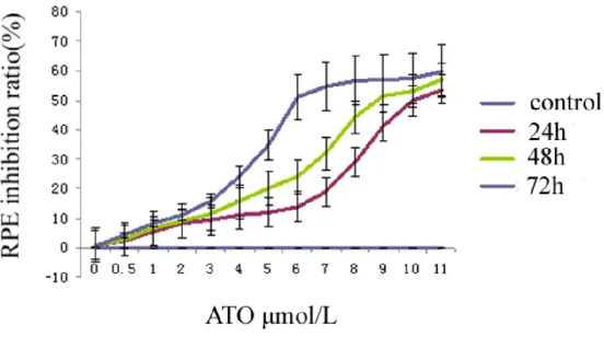
inhibitory concentration, 6 μmol/L, after 72 h as measured by BrdU (Figure 1) and MTT (Figure 2). The results suggest that ATO inhibits RPE cell growth because the results of the MTT assay showed that the survivability of RPE cells treated with ATO was reduced compared with the control group in a dose-dependent manner.
The 8 mol/L and 10 mol/L groups were not re-markably different from the 6 mol/L ATO group. Therefore, an ATO concentration of 6 mol/L was
used for the subsequent experiments.
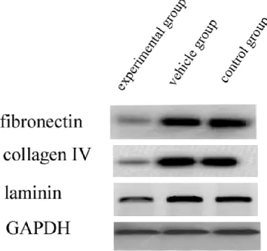
Effect of ATO treatment on the expression of fi
-bronectin, collagen IV, and laminin in RPE cells
Fibronectin, collagen IV, and laminin are the major ECM components, hence we investigated their expression by western blotting of RPE cells treated with ATO. Our results suggested a
significant reduction in the expression of ECM
components in ATO-treated RPE cells compa- red with vehicle and control groups (Figure 3, P<0.01). These data suggest that ATO may downregulate the expression of ECM compo- nents.
Decreased expression of p27 in ATO-treated RPE cells detected by western blot
[image:3.612.94.370.73.227.2]p27 expression declined in all experimental groups but not in the blank control group. The Figure 1. Inhibition of RPE cell growth with different concentrations of
ar-senic trioxide (ATO) according to MTT assay. After treating RPE cells using 0.5, 1, 2, 3, 4, 5, 6, 7, 8, 9, 10 and 11 μmol/L ATO, respectively, MTT assay revealed that RPE cell activity decreased significantly in a dose-dependent manner compared with the control group.
Results
The influence of ATO on RPE
cell proliferation
RPE cells in the control group grew well. The numbers of RPE cells in each experimental
group were not significantly dif
-ferent between the first day
Figure 2. ATO inhibited the proliferation of RPE cells according to BrdU assay. RPE cells were treated with different concentrations of ATO for different time periods and BrdU expression was detected, quantified, and plotted. BrdU-positive RPE cells in the experimental group decreased drastically compared with the control and vehicle groups.
Figure 3. Effect of ATO treatment on the expression of ECM components in RPE cells. We found that there was a major reduction in the expression of ECM com-ponents in ATO-treated RPE cells in contrast with the vehicle and control groups (P<0.01). Our results indi-cated that ATO can downregulate the expression of ECM components.
expression of p27 protein was downregulated at 24, 48, and 72 h of treatment. The
expres-sion levels were significantly different (F= 25.646, P<0.005, Figure 4).
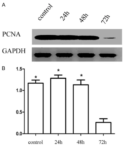
Decreased expression of PCNA protein in ATO-treated RPE cells by western blot
Our results showed that the expression of PCNA in cells treated for 24 h decreased compared with the control group. Expression levels showed a downward trend with prolonged treat-ment times. Compared with the blank control group, the expression of PCNA protein in the
experimental group decreased gradually with
time and the difference was statistically signifi -cant (F=58.141, P<0.001) (Figure 5).
Discussion
Proliferative vitreoretinopathy is a serious scar-ring process that strongly impairs visual
func-tion. In addition to RPE cells, TGF-β1/β2 is an important factor in PVR [27]. It was confirmed
[image:4.612.323.521.279.504.2] [image:4.612.93.284.281.461.2]mal transition (EMT) in proliferative membranes of PVR patients.It was reported that ATO can
inhibit TGF-β1 induced fibroblast to myofibro -blast cell differentiation by downregulation of
α-smooth muscle actin (α-SMA) and collagen I [28]. It was also confirmed that ATO can inhibit
breast cancer cells through p21 and p27 [29]. In the present study, MTT assay was used to
detect the influence of different concentrations
of ATO at different time points on RPE cells. Our
previous experiment confirmed that the dose of ATO (>11 μmol/L) and longer time (>72 h) was almost the same as 11 μmol/L for 72 h (data
not shown). Our results showed that RPE cells grew well in the blank control group. After 24 and 48 h of ATO treatment, the number of RPE cells in each group was not substantially differ-ent from that of the control group. Nonetheless, after 72 h, the number of cells in each experi-mental group was lower than that in the control group over time and with increasing
concentra-tions. This result confirmed that ATO inhibited
the proliferation of RPE cells in a time-depen-dent and dose-depentime-depen-dent manner. Moreover,
the effective concentration was 6 μmol/L which
is tenfold lower than achieved in humans. The different status of animal and human cells may cause the different concentrations of ATO.
The pathologic mechanisms of PVR include the proliferation of RPE cells and shrinkage of the ECM, while exposure of RPE cells to the vitre-ous body is a crucial event [30-32]. It was reported that RPE cells have complex and important physiologic and biochemical
func-tions. Under stimulation from inflammatory fac -tors, they can dissociate, migrate, and trans-form phenotypically, such as changing into
fibroblast-like cells, which causes traction in
retinal detachment through the secretion of collagen [33, 34].Our results also showed that
the expression of ECM proteins such as fibro -nectin, collagen IV, and laminin in RPE cells
decreased after treatment with 6 μmol/L ATO
for 3 days. The above results suggest that treat-ment with ATO may be a novel treattreat-ment for PVR.
PCNA is a suitable indicator for detecting cell proliferation [24]. p27 protein is one of the most important members of the kinase inhibi-tory protein family and inhibits the activities of various cyclin/cyclin kinase complexes. It is by far the most direct negative regulator of the cell
cycle. The fluorescence intensity expressed by
PCNA and p27 protein was detected using
immunofluorescence staining. The results dem
-onstrated that the fluorescence intensity of p27 and PCNA in RPE decreased significantly when treated at 6 μmol/L. In this study, we fur -ther detected the expression of p27 and PCNA proteins through western blot. According to the results, and comparing RPE cells treated with 6
μmol/L ATO for 24, 48, and 72 h with the blank
control group, we found that the expression of
PCNA was downregulated significantly in the
group treated for 72 h, which is roughly the
same as findings related to the relationship
between PCNA and neoplastic disease. The decreased expression of PCNA observed in this study indicated that RPE cells were arrested in the G2/M phase after treatment with ATO and their proliferation was inhibited as well. These
findings suggest that the expression level of
PCNA is indicative of the proliferative ability of RPE cells. A study examining the inhibition of glioblastoma showed that the expression of p27 protein increased where interference was present [35]. We found that p27 expression in RPE cells decreased according to the duration of time after ATO treatment. Accordingly, we
speculated that p27 may have an influence on
[image:5.612.90.288.69.303.2]RPE cell proliferation through the mechanisms Figure 5. ATO decreased the expression of PCNA
of transcription and post-transcriptional regula-tion. p27 promoted the proliferation of RPE cells by regulating the activity of proliferation proteins in RPE cells. After acting upon RPE cells, ATO inhibited the expression of p27 pro-tein and the proliferation of RPE cells. The mechanism may also relate to the cell line and
pattern of cell growth. Our result confirmed that
ATO can downregulate the expression of p27 and PCNA which are important for regulation of cell proliferation. Further study will be required to investigate whether a mutual promotion or inhibitory relationship exists between PCNA and p27.
Conclusion
This study revealed that ATO inhibited the prolif-eration of RPE cells at both the cellular and molecular level by inhibiting the expression of p27 and PCNA proteins. The limitation of our study is that the molecular mechanisms of how ATO inhibits proliferation of RPE by downregu-lating expression of extracellular matrix, p27, and PCNA will require future research. ATO shows promise as a novel therapeutic approach for the treatment of proliferative vitreous lesions and consequently the prevention of visual damage.
Acknowledgements
This work was supported by the grant of returned overseas scholars of Chinese ministry of education, the grant of returned overseas Chinese scholars of Heilongjiang province of
China (LC2013C33/H1204), the natural sci -ence grant of Heilongjiang province of China (H2018035), the innovation and development
foundation of first affiliated hospital of Harbin Medical University (2018L002).
Disclosure of conflict of interest None.
Address correspondence to: Feng Wang, Depart- ment of Ophthalmology, The First Affiliated Hospital, Harbin Medical University, Harbin 150001, China. Tel: +86-13359995660; E-mail: wangfd@126.com
References
[1] Chen X, Liu Y, Jiang Z, Zhou L, Ge J and Gao Q. Proteinkinase Cα downregulation via siRNA-PKCα released from foldable capsular vitreous body in cultured human retinal pigment epi-thelium cells. Int J Nanomedicine 2011; 6: 1303-11.
[2] Velez G, Weingarden AR, Lei H, Kazlauskas A and Gao G. SU9518 inhibits proliferative vitre-oretinopathy in fibroblast and genetically modi-fied Müller cell-induced rabbit models. Invest Ophthalmol Vis Sci 2013; 54: 1392-7.
[3] Wladis EJ, Falk NS, Iglesias BV, Beer PM and Gosselin EJ. Analysis of the molecular biologic milieu of the vitreous in proliferative vitreoreti-nopathy. Retina 2013; 33: 807-11.
[4] Saxena S, Jain A and Akduman L. Vitreopapilla- ry and vitreomacular traction in proliferative Eales’ disease. BMJ Case Rep 2012; 2012. [5] Kuhn F, Teixeira S and Pelayes DE. Late versus
prophylactic chorioretinectomy for the preven-tion of trauma-related proliferative vitreoreti-nopathy. Ophthalmic Res 2012; 48 Suppl 1: 32-7.
[6] Umazume K, Barak Y, McDonald K, Liu L, Kaplan HJ and Tamiya S. Proliferative vitreo-retinopathy in the Swine-a new model. Invest Ophthalmol Vis Sci 2012; 53: 4910-6.
[7] Salero E, Blenkinsop TA, Corneo B, Harris A, Rabin D, Stern JH and Temple S. Adult human RPE can be activated into a multipotent stem cell that produces mesenchymal derivatives. Cell Stem Cell 2012; 10: 88-95.
[8] Desjardins DM, Yates PW, Dahrouj M, Liu Y, Crosson CE and Ablonczy Z. Progressive early breakdown of retinal pigment epithelium func-tion in hyperglycemic rats. Invest Ophthalmol Vis Sci 2016; 57: 2706-13.
[9] Yang PM, Wu ZZ, Zhang YQ and Wung BS. Lycopene inhibits ICAM-1 expression and NF-κB activation by Nrf2-regulated cell redox state in human retinal pigment epithelial cells. Life Sci 2016; 155: 94-101.
[10] Kim HD, Jang SY, Lee SH, Kim YS, Ohn YH, Brinkmann R and Park TK. Retinal pigment epithelium responses to selective retina thera-py in mouse eyes. Invest Ophthalmol Vis Sci 2016; 57: 3486-3495.
[11] Gsellman L and Amini R. Patients with intravit-real gas bubbles at risk of high intraocular pressure without exceeding elevation of sur-gery: theoretical analysis. Invest Ophthalmol Vis Sci 2016; 57: 3340-7.
[12] Ghodasra DH, Fante R, Gardner TW, Langue M, Niziol LM, Besirli C, Cohen SR, Dedania VS, Demirci H, Jain N, Jayasundera KT, Johnson MW, Kalyani PS, Rao RC, Zacks DN and Sundstrom JM. Safety and feasibility of quanti-tative multiplexed cytokine analysis from of-fice-based vitreous aspiration. Invest Ophthal- mol Vis Sci 2016; 57: 3017-23.
pro-mote epithelial-mesenchymal transition. Invest Ophthalmol Vis Sci 2016; 57: 2699-705. [14] Lai K, Luo C, Zhang X, Ye P, Zhang Y, He J and
Yao K. Regulation of angiogenin expression and epithelial-mesenchymal transition by HIF-1α signaling in hypoxic retinal pigment epithe-lial cells. Biochim Biophys Acta 2016; 1862: 1594-1607.
[15] Gangalum RK, Bhat AM, Kohan SA and Bhat SP. Inhibition of the expression of the small heat shock protein αB-crystallin inhibits exo-some secretion in human retinal pigment epi-thelial cells in culture. J Biol Chem 2016; 291: 12930-42.
[16] Benhar I, Reemst K, Kalchenko V and Schwartz M. The retinal pigment epithelium as a gate-way for monocyte trafficking into the eye. EMBO J 2016; 35: 1219-35.
[17] Li KR, Yang SQ, Gong YQ, Yang H, Li XM, Zhao YX, Yao J, Jiang Q and Cao C. 3H-1,2-dithiole-3-thione protects retinal pigment epithelium cells against Ultra-violet radiation via activa-tion of Akt-mTORC1-dependent Nrf2-HO-1 sig-naling. Sci Rep 2016; 6: 25525.
[18] Jin HL and Jeong KW. Regulation of aryl hydro-carbon receptor-mediated transcription in hu-man retinal pigmented epithelial cells. Bio- chem Biophys Res Commun 2016; 472: 366-72.
[19] Eidet JR, Reppe S, Pasovic L, Olstad OK, Lyberg T, Khan AZ, Fostad IG, Chen DF and Utheim TP. The silk-protein sericin induces rapid melaniza-tion of cultured primary human retinal pigment epithelial cells by activating the NF-κB path-way. Sci Rep 2016; 6: 22671.
[20] Jun JH and Joo CK. MicroRNA-124 controls transforming growth factor β1-induced epithe-lial-mesenchymal transition in the retinal pig-ment epithelium by targeting RHOG. Invest Ophthalmol Vis Sci 2016; 57: 12-22.
[21] Yu B, Xu P, Zhao Z, Cai J, Sternberg P and Chen Y. Subcellular distribution and activity of mech-anistic target of rapamycin in aged retinal pig-ment epithelium. Invest Ophthalmol Vis Sci 2014; 55: 8638-50.
[22] Zhang G, Liu J, Zhang Y, Qu J, Xu L, Zheng H, Liu Y and Qu X. Cbl-b-dependent degradation of FLIP(L) is involved in ATO-induced autophagy in leukemic K562 and gastric cancer cells. FEBS Lett 2012; 586: 3104-3110.
[23] Li W, Wang M, Wang L, Ji S, Zhang J and Zhang C. Icariin synergizes with arsenic trioxide to suppress human hepatocellular carcinoma. Cell Biochem Biophys 2014; 68: 427-36. [24] Su Y, Jiang C, Zhang L and Wang F. Arsenic
tri-oxide inhibits proliferation of rabbit Tenon’s capsule fibroblasts after trabeculectomy by downregulating expression of extracellular ma-trix proteins. Invest Ophthalmol Vis Sci 2015; 56: 6663-70.
[25] Su Y, Cheng J, Liu H, Wang F and Zhao S. Adenovirus conducted connective tissue growth factor on extracellular matrix in trabec-ular meshwork and its role on aqueous humor outflow facility. Mol Biol Rep 2013; 40: 6091-6096.
[26] Su Y, Wang F, Yan Q, Teng Y and Cui H. Inhibition of proliferation of rabbit lens epithelial cells by S-phase kinase-interacting protein 2 targeting small interfering RNA. Mol Vis 2010; 16: 907-915.
[27] Ye F, Kaneko H, Hayashi Y, Takayama K, Hwang SJ, Nishizawa Y, Kimoto R, Nagasaka Y, Tsunekawa T, Matsuura T, Yasukawa T, Kondo T and Terasaki H. Malondialdehyde induces autophagy dysfunction and VEGF secretion in the retinal pigment epithelium in age-related macular degeneration. Free Radic Biol Med 2016; 94: 121-34.
[28] Luo F, Zhuang Y, Sides MD, Sanchez CG, Shan B, White ES and Lasky JA. Arsenic trioxide in-hibits transforming growth factor-β1-induced fibroblast to myofibroblast differentiation in vi-tro and bleomycin induced lung fibrosis in vivo. Respir Res 2014; 15: 51.
[29] Wang X, Gao P, Long M, Lin F, Wei JX, Ren JH, Yan L, He T, Han Y and Zhang HZ. Essential role of cell cycle regulatory genes p21 and p27 ex-pression in inhibition of breast cancer cells by arsenic trioxide. Med Oncol 2011; 28: 1225-54.
[30] Tran TL, Bek T, la Cour M, Prause JU, Hamann S and Heegaard S. Aquaporin-1 expression in retinal pigment epithelial cells overlying retinal drusen. Ophthalmic Res 2016; 55: 180-4. [31] Mao D, Peng H, Li Q, Wang J, Li P, Hu K, Zhang
X and Lei B. Aqueous humor and plasma adi-ponectin levels in proliferative diabetic retinop-athy patients. Curr Eye Res 2012; 37: 803-8. [32] Zhang W, Tan J, Liu Y, Li W, Gao Q and Lehmann
PV. Assessment of the innate and adaptive im-mune system in proliferative vitreoretinopathy. Eye (Lond) 2012; 26: 872-81.
[33] Xiao X, Liu Y, Guo M, Fei W, Zheng H, Zhang R, Zhang Y, Wei Y, Zheng G and Li F. pH-triggered sustained release of arsenic trioxide by poly-acrylic acid capped mesoporous silica nano- particles for solid tumor treatment in vitro and in vivo. J Biomater Appl 2016; 31: 23-35. [34] Wang X, Li D, Ghali L, Xia R, Munoz LP, Garelick
H, Bell C and Wen X. Therapeutic potential of delivering arsenic trioxide into HPV-infected cervical cancer cells using liposomal nano-technology. Nanoscale Res Lett 2016; 11: 94. [35] Gwak HS, Park MJ, Park IC, Woo SH, Jin HO,