Original Article
SIRT1 regulates C2C12 myoblast cell
proliferation by activating Wnt signaling pathway
Liang Wang1, Ruilin Xue1, Chengcao Sun1, Cuili Yang1, Yongyong Xi1, Feng Zhang1, Qiqiang He1, Suqing Wang1, Fang Zhao2, Yadong Zhang3, Dejia Li1
1Department of Occupational and Environmental Health, Wuhan University, Wuhan, China; 2Zhongnan Hospital of
Wuhan University, Wuhan, China; 3The Central Hospital of Wuhan, Tongji Medical College, Huazhong University of
Science and Technology, Wuhan, China
Received December 2, 2015; Accepted February 13, 2016; Epub March 1, 2016; Published March 15, 2016
Abstract: Sirtuin type 1 (SIRT1) is a potent NAD+ dependent deacetylase that deacetylates histone and nonhistone proteins to regulate gene expression and protein activity. Emerging evidences have indicated that SIRT1 plays a
sig-nificant role in diverse cellular processes including cell growth, differentiation, development, and physiological func -tion in muscle cells; however the signaling mechanisms involved remain to be established. In order to investigate
its potential role in muscle biological processes, we administrated C2C12 cells which were isolated from skeletal muscle tissue of dystrophic mice with SIRT1 activator resveratrol (REV), inhibitor nicotinamide (NAM) and Wnt inhibi
-tor FH535. By CCK-8, BrdU assay, real-time PCR and Western blot, we investigated whether the SIRT1 has a function in C2C12 cells by promoting β-catenin accumulation. Our results demonstrate that SIRT1 increases cell prolifera
-tion of C2C12 myoblast in a SIRT1-dependent manner. And SIRT1 significantly up-regulates the expression of cyclin D1, C-myc and Dvl2 in vitro as well as stimulates the accumulation of the Wnt/β-catenin. In conclusion, this study indicates that SIRT1 promotes the proliferation of C2C12 myoblast cells, at least partly via Wnt signaling pathway.
Keywords: SIRT1, Wnt, C2C12 myoblast cells, proliferation, muscle regeneration
Introduction
SIRT1, a well-known closest homologue of Sir2 (silent mating type information regulation 2) [1], is an NAD+-dependent histone/protein deace- tylase to regulate longevity mediated caloric restriction in model mammalian organisms [2].
SIRT1 is also an important regulatory factor of cell defense [3] and participates in a very broad
and complex array of cellular processes such as cell cycle, inflammation; energy metabolism
and DNA repair [4]. REV, which is a naturally occurring polyphenol found in grapes and red
wine [5], has been found to significantly acti -vate SIRT1. Interestingly, SIRT1 activation can
significantly improve muscular pathology in
dystrophic mdx mice [6]. And it is reported that
NAM is a potent inhibitor of SIRT1 and it can effectively inhibit SIRT1 activity both in vivo and
in vitro [7, 8].
Accumulating evidences suggest that SIRT1
may promote the cell proliferation, and activa
-tion of SIRT1 by induced growth of muscle mass of mdx mice [9]. For example, SIRT1 has been proven to regulate neurite outgrowth and cell
regeneration by repression of the mTOR
Sig-naling [10]. Moreover, overexpression of SIRT1 can promote muscle precursor cell proliferation
and cell cycle progression [11]. Associated with
the SIRT1-mediated proliferation of C2C12 cells
were the bidirectional decreases and increa-
ses in the expression of the cyclin-dependent kinase inhibitors p21 (Waf1, Cip1) and p27Kip1,
respectively [12]. Although SIRT1 can improve
the myoblast proliferation, the specific me-chanism(s) underlying remains to be clarified.
Intriguingly, Wnt signaling is a similar hot topic in muscle development and can potentially be
targeted for therapeutic treatment of musculo
-skeletal diseases [13]. In embryonic develop-ment, Wnt signaling pathway can promote the
proliferation and differentiation of embryonic
SIRT1 and C2C12 proliferation
Wnt signaling plays an important role in the
development from side portion of mesodermal stem cells to skeletal muscle cells [14, 15].
Therefore, it attaches a lot of significance to
reveal the interaction between SIRT1 and Wnt
signaling and provides another approach for the therapy of muscular dystrophy. There are three major branches of Wnt signaling pathway: the canonical Wnt signaling pathway
(Wnt/β-catenin pathway), Wnt-planar cell polarity way (Wnt-PCP pathway) and Wnt-calcium path-way (Wnt-Ca² pathpath-way) [16]. The canonical Wnt
signaling pathway regulates the amount of β-catenin into the nucleus, thereby directly reg
-ulates the presence of putative Lef1/Tcf upon binding of Wnt ligands to specific Fz-LRP5/6
complexes [17]. Wnts also signal through
β-ca-tenin-independent pathways that couple to
the Frizzled (Fzd) receptor and recruit shared
components, including axin, glycogen synthase
kinase 3 (GSK3) and disheveled (Dvl) [18]. The recent study on mechanism insights into the
specificity of interaction between mammalian
Wnt and FZD proteins have presented that
different Wnt-FZD pairs showed differential effects on phosphorylation of Dvl2 and Dvl3
[19]. Accumulation of TCF/β-catenin can poten
-tially target certain cell proliferation genes such
as c-myc, cyclin D1 [20], and the deletion of
Dvl 1/2/3 in HEK293T, MCF7 and HeLa cells
leads to lower expression of C-myc, cyclin-D1
and SIRT1 protein [21].
Here, we present for the first time a significa-nt functional link between SIRT1 and Wsignifica-nt in
C2C12 cells which has been previously impli-cated in osteoblast progenitors and breast can-cer cells [22, 23]. We co-incubated the C2C12 cells with SIRT1activator, SIRT1 inhibitor and
Wnt inhibitor, and then checked the cell growth and the expression of Wnt. Given that SIRT1 has been shown to regulate the expression of
Dvl protein to promote C2C12 cell growth, and
that Wnt signaling have a lot of analogous function in muscle development, we hypothe
-sized that Wnt signaling would be involved in SIRT1 mediated regulation of C2C12 myo
-blast cell proliferation. These results will fur -ther elucidate the relationship between SIRT1
and Wnt signaling on the regulation of muscle regeneration, and provide the basis for new methods or targets for the therapy of muscular
dystrophy.
Materials and methods
Cell culture
The C2C12 myoblast cell lines were obtained
from the school of animal science and technol
-ogy of Huazhong Agricultural University. The cells were cultured in a humidified air of 5% CO2 at 37°C in Dulbecco’s modified Eagle’s
medium (DMEM, NHyClone) supplemented wi-
th 1% penicillin/streptomycin (Gino Biomedical Technology Company) and 10% fetal bovine serum (HyClone). Different concentrations of
reagents were added to the cells culture when
the C2C12 cells grew to approximate 80% and
at least three independent repeated trials were
performed throughout the study.
Reagents and antibodies
NAM was from Beyotime Institute of Biotech-nology (Hangzhou, China) and REV was pur
-chased from Sigma-Aldrich (St. Louis, MO, USA). Immediately prior to use, a 0.1 M stock solution of REV stored at -20°C was diluted to
the desired concentration with the culture
medium and the final concentration of DMSO for all treatments was maintained at 0.2%. FH535 was from Selleck Chemicals (Texas, USA). Cell Counting Kit-8 (CCK-8) was from
Dojindo Laboratories (Kumamoto, Japan). The
antibodies used were anti-β-catenin rabbit
monoclonal (Abcam, USA), anti-SIRT1 rabbit
monoclonal (Abcam, USA), anti-β-actin mouse monoclonal (Anbo, E0012, Changzhou, China).
The secondary antibodies were purchased
from Abbkine (California, USA). Other reagents were from Goodbio technology or HyClone.
CCK-8 assay
The Cell Counting Kit-8 (CCK-8) was purchased
from Dojindo Technologies (DOJINDO, Japan).
Exponentially growing C2C12 myoblast cells (8×10³ cells/well, 100 μl) were seeded into 96-well plates 20 to 24 h prior to replacing
fresh serum free
medium containing the
indi-cated concentrations of drug. We added FH535 at first and then added NAM after 30 min in the group of the FH535 combined with NAM. After incubation for 24 h, culture medium was replaced by drug-free medium (100 μl), the effect of drug was examined by CCK-8 accordcells were further incubated away from light at 37°C for 1 to 3 h. Absorbance was measured
with excitation at 450 nm using a microplate reader (Bio-Rad).
BrdU assay
The BrdU cell proliferation ELISA kit was ob-tained from Beyotime Biotechnology (Shanghai,
China) and the cells were cultured in growth
media for 12 h followed by BrdU (5-Bromo-2-deoxyUridine) treatment for 1 h before harvest -ing. The anti-BrdU antibody (BD, 552598,) was
incubated at 4°C for 18 h, and the secondary goat anti-mouse IgG antibody conjugated with
Alexa Fluor 594. The incubation time was 2 h at
room temperature, protected from light. Nuclei were visualized by DAPI (49, 69
diamidino-2-phenylindole), and images were acquired by
Olympus microscope (IX71, Shinjuku-ku, Tokyo,
Japan).
Quantitative reverse transcription-polymerase chain reaction
Total RNA of C2C12 myoblast cells was isolat-ed with the Trizol (Invitrogen, Carlsbad, CA, USA) following the product protocol. First-strand cDNA was synthesized using PrimeScriptTM RT reagent Kit (TaKaRa, RR037A, Dalian, China).
DNA amplification was performed in StepOne
(Applied BioSystems, Foster City, CA) using
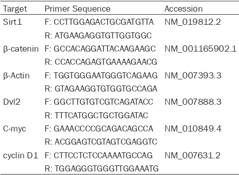
SYBR Green PCR kits (TaKaRa, RR420A, Dalian, China) for β-catenin, SIRT1, Dvl2, C-myc, cyclin D1 and β-actin. The primer sequences are list -ed in Table 1.
subjected to SDS-polyacrylamide gel
electro-phoresis and transferred to PVDF membranes (Millipore, Bedford, MA). Blocking was per
-formed in Tris-buffered saline with 5% nonfat dry milk for 2 h at room temperature. Then the
membranes were probed with a primary anti-body at 4°C overnight. The membranes were washed (6×5 min) with TBST, and incubated with secondary antibody that corresponds to
the primary antibody for 2 h at room tempera -ture. The membranes were washed as above, and HRP activity was detected using Chemi-
luminescence Reagent (Beyotime, Hangzhou, CHN) and exposure to G: BOX Chemi ×T4 Gel imaging system instrument (SYNGENE, Eng-land). Blots were quantified by densitometric analysis using the ImageJ software.
Statistical analysis
All values were presented as means _X±SEM of at least three individual experiments. To check whether difference was statistically significant, we carried out the one-way ANOVA, which was statistically informative despite the limited number of samples in each group, followed by the Bonferroni method for multiple compari
-sons (Prism 5, GraphPad Software, CA). The one-way ANOVA is based on the null hypothesis
that all groups have the same mean and
Bonferroni method was adopted for potential significant differences. BrdU-positive rate was analyzed by chi square test (SPSS v18). The dif
[image:3.629.98.334.104.277.2]-ference was considered significant if the prob -ability value was no more than 0.05.
Table 1. Primer sequences used for quantitative poly -merase chain reaction (qRT-PCR)
Target Primer Sequence Accession Sirt1 F: CCTTGGAGACTGCGATGTTA NM_019812.2
R: ATGAAGAGGTGTTGGTGGC
β-catenin F: GCCACAGGATTACAAGAAGC NM_001165902.1
R: CCACCAGAGTGAAAAGAACG
β-Actin F: TGGTGGGAATGGGTCAGAAG NM_007393.3
R: GTAGAAGGTGTGGTGCCAGA
Dvl2 F: GGCTTGTGTCGTCAGATACC NM_007888.3
R: TTTCATGGCTGCTGGATAC
C-myc F: GAAACCCCGCAGACAGCCA NM_010849.4
R: ACGGAGTCGTAGTCGAGGTC
cyclin D1 F: CTTCCTCTCCAAAATGCCAG NM_007631.2
R: TGGAGGGTGGGTTGGAAATG
Western blot
C2C12 myoblast cells were harvested in
PBS, centrifuged at 5,000 g for 10 min at
4°C and then removed the supernatant.
Samples were homogenized in ice-cold RIPA lysing buffer (Beyotime, Hangzhou, China) with 1% protease inhibitor cocktail
(Sangon Biotech, Shanghai, China) and
1% phosphatase inhibitors (Goodbio tech -nology, Wuhan, China), and incubated the
suspension on ice for 20 to 30 min. Then samples were centrifuged at 10,000 g for
10 min at 4°C. The protein concentration
of the supernatant was measured using the BCA Protein Quantification Kit (Best-Bio, Shanghai, China). Supernatant frac
SIRT1 and C2C12 proliferation
Results
SIRT1 regulates the proliferation of C2C12 myoblast cells
To elucidate the role of SIRT1 in C2C12 myo -blast cells growth control, we treated the cells
with REV and NAM. The cell proliferation index of C2C12 myoblast cells were investigated by
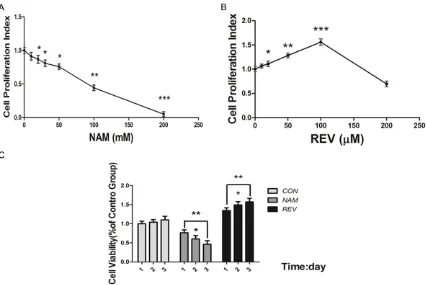
CCK-8 assay. As shown in Figure 1A, inhibition
of SIRT1 by NAM resulted in lower proliferation index than that of the untreated control group
(P<0.05). Furthermore, the results
demonstrat-ed that the cell proliferation index of C2C12
myoblast cells decreased with the
concentra-tion of NAM when it ranged from 0 to 200 mΜ, which presented in a dose dependent manner (The Pearson correlation coefficient
was 0.993). C2C12 myoblast cells number
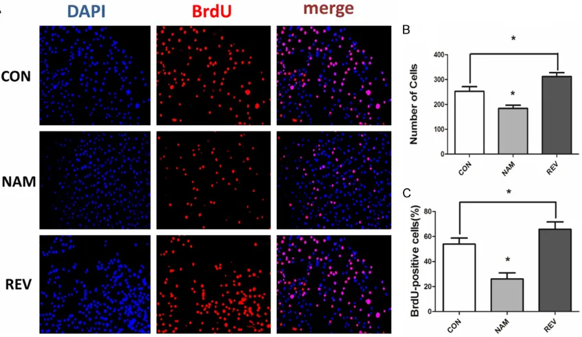
sta-bilized at increased doses of NAM at 50 mM and the BrdU positive rate reached 26.75%.
However, when NAM concentration reached
beyond 50 mM, interestingly, the cell survival rate was rather lower (P<0.01) and the cell con-dition was steadily deteriorated. Consistent
with a role for NAM in regulating cell prolifera -tion in a dose dependent manner, our results indicated that NAM inhibited cell growth in a time dependent manner. As shown in Figure 1C, cells were treated with NAM at 50 mM and
detected every 24 h. From the first day, C2C12
cells were gradually depleted and the decrease
in number was predominantly significant com -pared with the primary state.
To further confirm the effect of SIRT1 on C2C12 regeneration, we administrated different con
-centrations of REV on C2C12 myoblast cells and measured the cells growth level after 24 hours. The results showed that after treating with REV in different concentrations, the cell proliferation index was significantly higher than that of the untreated control group (P<0.05)
(Figure 1B). Meanwhile, the results demon-Figure 1. The role of SIRT1 in proliferation of C2C12 myoblast cells. A. CCK-8 analysis of C2C12 cells treated with different concentration of NAM. C2C12 cells (8×10³ cells/100 μl) were seeded in 96-well plates in triplicate, incu
-bated for 20-24 h, and 30 min later treated with different concentrations of REV or NAM for 24 h, and then subjected to CCK-8 assay to analyze proliferation of C2C12. B. CCK-8 analysis of C2C12 cells treated with REV. C. Cell prolifera
[image:4.629.102.528.79.364.2]strated that the cell proliferation index of C2C12 myoblast cells increased with REV in a dose dependent manner when the REV concen
-tration ranged from 0 to 100 µΜ (The Pearson correlation coefficient was 0.983). However, when the concentration of REV reached more than 100 μM, the cell proliferation index de-creased and the proliferation of cells was sig
-nificantly inhibited (Figure 1B). C2C12
myo-blast cells proliferation index stabilized at increased doses of REV (Figure 3B, P<0.001)
at 100 μM and the BrdU positive rate reached 65.80% (Figure 2C). Similarly, when treating
with 100 μM REV, C2C12 cells regenerated in time-dependent manner. These stimulate eff-ects of REV on C2C12 cells was statistically significant.
Inhibition of Wnt by FH535 promotes the prolif-eration of C2C12 myoblast cells
It has been demonstrated that FH535 is a
potent inhibitor of the Wnt/β-catenin signaling pathway and a significant number of evidence
has proven that FH535 inhibits proliferation of
cells including Liver Cancer Stem Cells (LCSC) and HCC cell lines [24]. Consistent with the
effect in cancer cells, our results showed that after treatment with the FH535 in different concentrations from 0 to 50 uM, the cell prolif
-eration index was significantly lower than that of the control group (P<0.05) (Figure 3A). 24 h
after treating with FH535 at 15 µM, degenera
-tion in C2C12 was remarkably evident (P<0.05)
and became worse as it developed (Figure 3B).
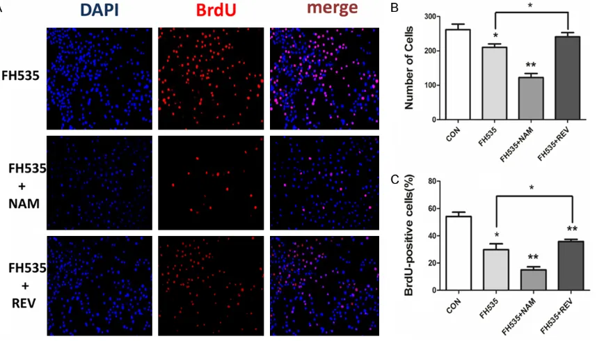
Total number of cells 201±10.27 (Figure 4B)
and BrdU positive rate (27.73±1.26%) were both significant compared to the control group
(Figure 4C).
FH535 aggravates the inhibition of C2C12 myoblast cells proliferation by regulating SIRT1
[image:5.629.106.530.81.327.2]In order to investigate the role of Wnt signal pathway in SIRT1 mediated regulation of C2C12 myoblast cell proliferation, we divided C2C12 myoblast cells into four groups: the control group, FH535 (15 μM), combination of FH535
Figure 2. BrdU assay was used to detect the proliferation level. 1×10⁵ cells were seeded in a 35 mm tissue culture
SIRT1 and C2C12 proliferation
(15 μM) with NAM (50 mM) and REV (100 μM)
respectively. We observed that FH535 acted as a negative factor on cells which had been activated by REV (Figures 3, 4). After treatment
-Figure 3. CCK-8 assay to C2C12 cells were treated with different concentrations of FH535 for 24 h. A. FH535 de
-creased the proliferation of C2C12 myoblast cells. Each value represents the mean ± standard error of the mean of triplicate determinations from three independent cell preparations. *P<0.05, **P<0.01, compared with the value of the control. B. Cell proliferation changed over time (24 h, 48 h, 72 h) in four treating groups : the control, FH535 (15 μM), NAM (50 mM) + FH535 (15 μM), REV (100 μM) + FH535 (15 μM). Each value represents the mean ± stan
[image:6.629.107.530.79.264.2]-dard error of the mean of triplicate determinations from three independent cell preparations. *P<0.05, **P<0.01, compared with the value of the control. Statistical analysis was conducted using one-way ANOVA.
[image:6.629.105.534.368.613.2]with FH535 and NAM, the cell proliferation index and BrdU positive rate were significantly lower than that of NAM alone (P<0.05) (Figures 1C, 3B). Together, these results showed that
inhibition of Wnt by FH535 may further intensi
-fy the effect of SIRT1 inhibition and aggravates the degeneration of C2C12 myoblast cells pro
-liferation by NAM. This effect may be due to that FH535 have a positive function on Wnt
expression, and that SIRT1 may ameliorate
C2C12 myoblast cell proliferation by inducing
Wnt signaling pathways.
SIRT1 inhibition decreases expression of Wnt signaling pathway and exacerbates the inhibi-tion by FH535
To examine the mechanisms underlying defec -tive regeneration in NAM treating cells, we
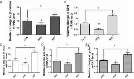
measured the expression of SIRT1 and Wnt sig -naling pathways by RT-PCR and Western Blot, respectively. According to Figure 5, NAM signifi
-cantly reduced the relative level of SIRT1 mRNA. Interestingly, the expression of
Wnt/β-catenin resulted in an overt decrease at the same time, as well as its target Dvl2, indicating
that Wnt/β-catenin may act as a downstream of SIRT1.
This hypothesis was confirmed by the later
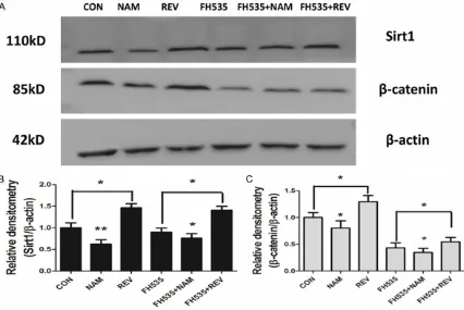
Western Blot assay, as it shown in Figure 7.
Down-regulation of SIRT1 induced an apparent loss of Wnt/β-catenin protein level, which is in accord with RT-PCT results. The expression of Wnt/β-catenin and its downstream signals were significantly lower in cells treated with
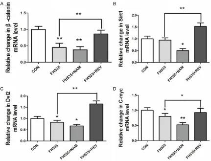
FH535 and NAM than that with NAM alone (P<0.05, Figure 6). These data demonstrated
that SIRT1 inhibition decreased expression of
Wnt signaling pathways.
SIRT1 activation increases expression of Wnt in vitro and ameliorates the suppression in-duced by FH535
Upon activation by REV, SIRT1 activators
in-crease bone mass in aged mice by preventing
β-catenin sequestration [25]. It was proven
that REV in the dose of 100 μM effectively
inhibited the regeneration and depressed
Wnt2, Wnt5a and Notch2 expression of SiHa
[image:7.629.101.530.81.335.2]and HeLa cells [26]. But in C2C12 cells, RSV
Figure 5. Regulation of SIRT1 leading to changes in mRNA expression of Wnt/β-catenin signaling. C2C12 cells were administrated with different drugs for 48 h and the expression of β-catenin, SIRT1, Dvl2, C-myc, cyclin D1 mRNA were detected by quantification real-time PCR method. Each value represents the mean ± standard error of the mean of triplicate determinations. *P<0.05, **P<0.01, ***P<0.001, compared with the value of the control. Sta
SIRT1 and C2C12 proliferation
adjusts cell cycle progression, cell cycle arrest
and primary progress of differentiation, by reg
-ulating the expression of several key cell cycle regulators, myogenic regulatory factors and muscle-specific structural proteins [27]. Inter- estingly, the researches on the connection
between C2C12 and Wnt shows that
WNT/β-catenin signaling regulates myogenesis in
sev-eral ways, and the depletion of these steps of myogenesis induces muscle growth defects
[28]. However, whether this relationship be- tween SIRT1 and Wnt signaling extended to
the activation of SIRT1 in C2C12 cells was unknown. To address this, we systematically detected the impact of SIRT1 increase on the change in the expression of Wnt signaling.
As is indicated in Figure 5, REV markedly enhanced the relative mRNA level of SIRT1,
cyclin D1, WNT/β-catenin and Dvl2, which is dif
-ferent from the finding in several cell lines. In general, REV inhibits proliferation of most can
-cer cells and induces apoptosis of epithelial
cells [29]. Inversely, REV modulates fibroblast
cells to regenerate and produces the recovery
of muscle mass following disuse that results in REV-associated improvement in the plantaris muscle of aged rats [30]. Consistent with the- se reports, we observed that SIRT1 activation
ameliorated the deterioration of cell prolifera
-tion state and the reduc-tion of WNT/β-catenin, Dvl2 and cyclin D1 induced by inhibition of Wnt
signaling.
We next investigated the relative change in pro-teins and Western Blot detections proved th-
[image:8.629.100.531.79.408.2]at REV treatment inhibited the expression of β-catenin (Figure 7). Given that the effects of
Figure 6. NAM and REV affected the inhibition of FH535 on Wnt/β-catenin signaling. C2C12 cells were adminis
-trated with different drugs for 48 h and the expression of β-catenin, SIRT1, Dvl2 and C-myc mRNA were detected by quantification real-time PCR method. A. qRT-PCR of β-catenin. B. qRT-PCR of Dvl2 mRNA. C. qRT-PCR of C-myc mRNA. D. qRT-PCR of SIRT1 mRNA. Each value represents the mean ± standard error of the mean of triplicate de
NAM and RES on Wnt regulation are SIRT1-dependent, we investigated whether these two
proteins interact in the specified way. Treating
with FH535 slightly reduced SIRT1 protein
level, however, the change was not significant. In general, the above results confirmed that the observed effects of REV were indeed mediated
through SIRT1.
Discussion
Both Wnt and SIRT1 have been linked to C2C12
cell regeneration with SIRT1 activation inducing
a protective effect while inhibition of Wnt con
-ferring a detrimental effect. SIRT1 plays a criti
-cal role in a wide variety of cellular processes including proliferation, differentiation and func
-tion, however, the mechanisms responsible for its effects are not fully understood. SIRT1 has been found to promote the proliferation and suppressed the differentiation of myoblast pre -cursors [31] and overexpression of SIRT1 can promote the proliferation of skeletal muscle
precursor cell, while reversely it is reported that
SIRT1 inhibits cell proliferation in colon cancer
[32], which may be due to the different cell type. Our results demonstrate SIRT1 activator REV can promote the proliferation of C2C12
myoblast cells; meanwhile its inhibitor NAM can
decrease the proliferation of C2C12 myobla-st cells. SIRT1 promotes the proliferation of C2C12 myoblast cells only when REV concen
-tration was no more than 100 μM. When REV concentration was more than 100 μM, the sur
-vival rate of C2C12 myoblast cells decreased. This may be due to the toxic effect of the sol
-vent DMSO on cells, on the other hand, the REV
can not only activate SIRT1, but also can inhibit lipoxygenase, cyclooxygenase and various
pro-tein kinases, therefore, as a result REV may be harmful to cells when its concentration is too
high [31, 33].
It is well-established that SIRT1 participates in
a very broad and complex array of physiological processes, for example, cellular differentiation,
[image:9.629.102.528.81.366.2]oxidative stress response, metabolism, longev-ity and various diseases including muscular Figure 7. A. Western Blot analysis for relative expression of SIRT1 and β-catenin protein. C2C12 cells were treated with different drugs for 48 h. B. Quantitative analysis of the SIRT1. C. Quantitative analysis of the β-catenin. The histogram shows a quantitative representation of the levels protein obtained from a laser densitometric analysis of three independent experiments. Each value represents the mean ± standard error of the mean of triplicate determi
SIRT1 and C2C12 proliferation
dystrophy [34]. Activation of SIRT1 promotes cell proliferation both in the liver [35] and colon [36], and SIRT1 has been proven to correlate
with decrease of muscle mass, skeletal muscle
strength [37] and oxidative damage in mdx mice [38]. However, overexpression of SIRT1-induced proliferation of muscle cells in vivo and
in vitro has not been reported yet. In this study,
we have shown that SIRT1 regulates skeletal
muscle regeneration in C2C12 cells model, and
that the stimulating effect is both approximate -ly in dose-dependent manner and time-depen-dent manner. And in order to study the
mecha-nisms and feature of SIRT1-inducing prolifera -tion in muscle cells, we have test several mus-cle regeneration-related signaling pathways
such as mTOR, myostatin and Wnt signaling
pathways. And our results showed that Wnt
sig-naling pathway plays a significant role in SIRT1-inducing proliferation in muscle cells, which
has not been reported yet elsewhere.
Canonical Wnt signaling pathway is reported to promote myoblast generation in embryonic period [39], and FH535, the inhibitor of Wnt, is highly potent and effective. FH535 can simulta -neously inhibit the peroxisome (PPAR) and
β-catenin/TCF/LEF combination [40], and it
has no marked effect on the quantity of other
proteins. We discovered that FH535 not only
reduce cell proliferation index, but also mor
-phologically changes cell size and shape.
C2-C12 cells became round and spindle-shaped,
failed to attach firmly to the culture bottles. To clarify the relationship between SIRT1 and Wnt in C2C12 cells, we firstly co-cultured cells with
NAM and FH535, which showed sharp decrease in growth rate compared with control group.
Secondly, we detected mRNA expression of
Dvl2, which has been reported to decrease
significantly and inhibit the degradation of β-catenin in absence of SIRT1 expression in
colon and lung cancer cells [41]. Our results
showed that Dvl2 expression level was much
lower in NAM group than that in RSV group. It is
reported that C-myc and cyclin D1 are the
down-stream of Wnt signaling pathway of regu -lating cell cycle proteins [42], and we also found
that C2C12 myoblasts treated with FH535
could significantly down-regulated mRNA ex-pression levels of C-myc and cyclin D1 com -pared with the control group. Thirdly, as
expect-ed, FH535 and NAM significantly suppressed β-catenin mRNA and protein levels, while RSV promoted them remarkably, thus in conclusion
SIRT1 can positively regulate Wnt signaling pa-
thway in proliferation progress in C2C12 myo
-blasts. Activation of SIRT1 activity with REV increased the β-catenin expression level, while in contrast suppression of SIRT1 activity by
NAM decreased them notably.
These findings indicate that SIRT1 regulates Wnt/β-catenin to trigger its concentration in the nucleus and stimulation of genes for prolif -eration in C2C12 cells. Intriguingly, a previous
study noted that oxazepam-induced cellular oncogenesis displayed lower level of DNA meth
-ylation (Crebbp, Dnmt3b) and histone modifica
-tion (SIRT1), while the expression of the
Wnt/β-catenin signaling pathway increased [43]. And
the suppression of SIRT1 deacetylase by siRNA
or NAM in epithelial cells Hepatitis B virus X
(HBX) protein could activate Wnt/β-catenin and the presence of HBX might promoted the inter
-action between β-catenin and SIRT1, leading to protection of β-catenin by inhibitory action of
SIRT1 [44]. Why regulation of Wnt signaling by SIRT1 exhibits distinct results in different kinds of cells? We infer that the signaling transfer
mechanism could display a contrary trend in
C2C12 cells VS human hepatic cells, and that the deacetylation induced by SIRT1 in β-catenin was the root cause that led to nuclear inflow in C2C12 cells but outflow in hepatic cells. This might happen, for instance, if either kind of cells could control an outflow signaling identify
-ing the deacetylated β-catenin, but C2C12 cells particularly occupied an inflow signaling that identifies the redundant and excrescent de-acetylated β-catenin in the outflow signaling. Taken together, our results indicated that Wnt participated in the regulation of C2C12 myo
-blast cells proliferation via SIRT1. Further stud
-ies are warranted to show how SIRT1 affect Wnt signaling, for example, siRNA interference of SIRT1 and Wnt signaling to further study the
interaction relationship. Furthermore, it will be
promising to detect the effect of Wnt signaling
in SIRT1-depleted mdx mice, which will
contrib-ute to elucidate the role of SIRT1 in the regula
-tion of muscle regenera-tion and provide the basis for new methods or targets for the thera
-py of muscular dystrophy.
Acknowledgements
Disclosure of conflict of interest
None.
Address correspondence to: Dr. Dejia Li, Depart-
ment of Occupational and Environmental Health,
Wuhan University, 115 Donghu Road, Wuhan, China. E-mail: [email protected]
References
[1] Bitterman KJ, Anderson RM, Cohen HY,
Latorre-Esteves M, Sinclair DA. Inhibition of si -lencing and accelerated aging by nicotinamide,
a putative negative regulator of yeast sir2 and
human SIRT1. J Biol Chem 2002; 277: 45099-107.
[2] McBurney MW, Yang X, Jardine K, Hixon M,
Boekelheide K, Webb JR, Lansdorp PM,
Lemieux M. The mammalian SIR2alpha pro-tein has a role in embryogenesis and gameto-genesis. Mol Cell Biol 2003; 23: 38-54. [3] Horio Y, Hayashi T, Kuno A, Kunimoto R.
Cellular and molecular effects of sirtuins in
health and disease. Clin Sci (Lond) 2011; 121: 191-203.
[4] Ferrara N, Rinaldi B, Corbi G, Conti V, Stiuso P, Boccuti S, Rengo G, Rossi F, Filippelli A.
Exercise training promotes SIRT1 activity in aged rats. Rejuvenation Res 2008; 11: 139-50.
[5] Howitz KT, Bitterman KJ, Cohen HY, Lamming DW, Lavu S, Wood JG, Zipkin RE, Chung P, Kisielewski A, Zhang LL, Scherer B, Sinclair DA. Small molecule activators of sirtuins extend Saccharomyces cerevisiae lifespan. Nature
2003; 425: 191-6.
[6] Kim HJ, Braun HJ and Dragoo JL. The effect of REV on normal and osteoarthritic chondrocyte
metabolism. Bone Joint Res 2014; 3: 51-9. [7] Sauve AA, Moir RD, Schramm VL, Willis IM.
Chemical activation of Sir2-dependent silenc
-ing by relief of nicotinamide inhibition. Mol Cell
2005; 17: 595-601.
[8] Jackson MD, Schmidt MT, Oppenheimer NJ, Denu JM. Mechanism of nicotinamide inhibi -tion and transglycosida-tion by Sir2 histone/ protein deacetylases. J Biol Chem 2003; 278: 50985-98.
[9] Hori YS, Kuno A, Hosoda R, Tanno M, Miura T,
Shimamoto K, Horio Y. REV ameliorates mus -cular pathology in the dystrophic mdx mouse,
a model for Duchenne muscular dystrophy. J
Pharmacol Exp Ther 2011; 338: 784-94. [10] Guo W, Qian L, Zhang J, Zhang W, Morrison A,
Hayes P, Wilson S, Chen T, Zhao J. SIRT1 over-expression in neurons promotes neurite
out-growth and cell survival through inhibition of
the mTOR signaling. J Neurosci Res 2011; 89:
1723-36.
[11] Rathbone CR, Booth FW and Lees SJ. SIRT1
increases skeletal muscle precursor cell prolif -eration. Eur J Cell Biol 2009; 88: 35-44. [12] Machida S and Booth FW. Increased nuclear
proteins in muscle satellite cells in aged ani-mals as compared to young growing aniani-mals.
Exp Gerontol 2004; 39: 1521-5.
[13] Rudnicki MA and Williams BO. Wnt signaling in
bone and muscle. Bone 2015; 80: 60-6. [14] Petropoulos H and Skerjanc IS. Beta-catenin is
essential and sufficient for skeletal
myoge-nesis in P19 cells. J Biol Chem 2002; 277: 15393-9.
[15] Anakwe K, Robson L, Hadley J, Buxton P, Church V, Allen S, Hartmann C, Harfe B, Nohno
T, Brown AM, Evans DJ, Francis-West P. Wnt
signalling regulates myogenic differentiation in
the developing avian wing. Development 2003; 130: 3503-14.
[16] Wang Y, Li YP, Paulson C, Shao JZ, Zhang X, Wu M, Chen W. Wnt and the Wnt signaling pathway in bone development and disease. Front Biosci
(Landmark Ed) 2014; 19: 379-407.
[17] Caron A, Xu X and Lin X. Wnt/beta-catenin signaling directly regulates Foxj1 expression
and ciliogenesis in zebrafish Kupffer’s vesicle.
Development 2012; 139: 514-24.
[18] Grumolato L, Liu G, Mong P, Mudbhary R, Biswas R, Arroyave R, Vijayakumar S,
Economides AN, Aaronson SA. Canonical and noncanonical Wnts use a common mechanism to activate completely unrelated coreceptors.
Genes Dev 2010; 24: 2517-30.
[19] Dijksterhuis JP, Baljinnyam B, Stanger K, Sercan HO, Ji Y, Andres O, Rubin JS, Hannoush RN, Schulte G. Systematic mapping of WNT-FZD protein interactions reveals functional se -lectivity by distinct WNT-FZD pairs. J Biol Chem 2015; 290: 6789-98.
[20] Anna CH, Iida M, Sills RC, Devereux TR.
Expression of potential beta-catenin targets, cyclin D1, c-Jun, c-Myc, E-cadherin, and EGFR
in chemically induced hepatocellular neopla-
sms from B6C3F1 mice. Toxicol Appl Pharmacol
2003; 190: 135-45.
[21] Saini A, Al-Shanti N, Sharples AP, Stewart CE.
Sirtuin 1 regulates skeletal myoblast survival and enhances differentiation in the presence of REV. Exp Physiol 2012; 97: 400-18.
[22] Wu X, Wang Y, Zhang K, Wu J, Yuan YC, Deng X,
Chen L, Kim CC, Lau S, Somlo G, Yen Y. FZD7 has a critical role in cell proliferation in triple negative breast cancer. Oncogene 2011; 30:
4437-46.
-SIRT1 and C2C12 proliferation
tion by preventing beta-catenin sequestration
by FoxO transcription factors in osteoblast pro -genitors. J Biol Chem 2014; 289: 24069-78. [24] Wu MY, Liang RR, Chen K, Shen M, Tian YL, Li
DM, Duan WM, Gui Q, Gong FR, Lian L, Li W,
Tao M. FH535 inhibited metastasis and growth
of pancreatic cancer cells. Onco Targets Ther
2015; 8: 1651-70.
[25] Iyer S, Han L, Bartell SM, Kim HN, Gubrij I, de Cabo R, O’Brien CA, Manolagas SC, Almeida M. Sirtuin1 (SIRT1) promotes cortical bone forma -tion by preventing beta-catenin sequestra-tion
by FoxO transcription factors in osteoblast pro -genitors. J Biol Chem 2014; 289: 24069-78. [26] Zhang P, Li H, Yang B, Yang F, Zhang LL, Kong
QY, Chen XY, Wu ML, Liu J. Biological signifi
-cance and therapeutic implication of
REV-inhibited Wnt, Notch and STAT3 signaling in
cervical cancer cells. Genes Cancer 2014; 5:
154-64.
[27] Montesano A, Luzi L, Senesi P, Mazzocchi N, Terruzzi I. REV promotes myogenesis and hy -pertrophy in murine myoblasts. J Transl Med 2013; 11: 310.
[28] Suzuki A, Scruggs A and Iwata J. The temporal specific role of WNT/beta-catenin signaling
during myogenesis. J Nat Sci 2015; 1: e143. [29] Fu Y, Chang H, Peng X, Bai Q, Yi L, Zhou Y,
Zhu J, Mi M. REV inhibits breast cancer stem-like cells and induces autophagy via suppress -ing Wnt/beta-catenin signal-ing pathway. PLoS
One 2014; 9: e102535.
[30] Bennett BT, Mohamed JS and always SE.
Effects of REV on the recovery of muscle mass following disuse in the plantaris muscle of aged rats. PLoS One 2013; 8: e83518.
[31] Pardo PS and Boriek AM. The physiological roles of SIRT1 in skeletal muscle. Aging (Albany
NY) 2011; 3: 430-7.
[32] Kabra N, Li Z, Chen L, Li B, Zhang X, Wang C, Yeatman T, Coppola D, Chen J. SIRT1 is an
in-hibitor of proliferation and tumor formation in
colon cancer. J Biol Chem 2009; 284: 18210-7.
[33] Mietus-Snyder M, Gowri MS and Pitas RE.
Class A scavenger receptor up-regulation in
smooth muscle cells by oxidized low density li
-poprotein. Enhancement by calcium flux and
concurrent cyclooxygenase-2 up-regulation. J Biol Chem 2000; 275: 17661-70.
[34] Mortuza R, Feng B and Chakrabarti S. SIRT1
reduction causes renal and retinal injury in
di-abetes through endothelin 1 and transforming growth factor beta1. J Cell Mol Med 2015; 19:
1857-67.
[35] Lee CW, Wong LL, Tse EY, Liu HF, Leong VY, Lee JM, Hardie DG, Ng IO, Ching YP. AMPK pro -motes p53 acetylation via phosphorylation
and inactivation of SIRT1 in liver cancer cells.
Cancer Res 2012; 72: 4394-404.
[36] Firestein R, Blander G, Michan S, Oberdoerffer P, Ogino S, Campbell J, Bhimavarapu A, Luikenhuis S, de Cabo R, Fuchs C, Hahn WC, Guarente LP, Sinclair DA. The SIRT1 deacety -lase suppresses intestinal tumorigenesis and
colon cancer growth. PLoS One 2008; 3:
e2020.
[37] Dolinsky VW, Jones KE, Sidhu RS, Haykowsky M, Czubryt MP, Gordon T, Dyck JR. Impro-vements in skeletal muscle strength and car
-diac function induced by REV during exercise
training contribute to enhanced exercise
per-formance in rats. J Physiol 2012; 590:
2783-99.
[38] Hori YS, Kuno A, Hosoda R, Tanno M, Miura T,
Shimamoto K, Horio Y. REV ameliorates mus -cular pathology in the dystrophic mdx mouse,
a model for Duchenne muscular dystrophy. J
Pharmacol Exp Ther 2011; 338: 784-94. [39] Petropoulos H and Skerjanc IS. Beta-catenin
is essential and sufficient for skeletal myogen -esis in P19 cells. J Biol Chem 2002; 277: 15393-9.
[40] Handeli S and Simon JA. A small-molecule
in-hibitor of Tcf/beta-catenin signaling down-reg -ulates PPARgamma and PPARdelta activities. Mol Cancer Ther 2008; 7: 521-9.
[41] Schwarz-Romond T, Metcalfe C and Bienz M. Dynamic recruitment of axin by Dishevelled
protein assemblies. J Cell Sci 2007; 120: 2402-12.
[42] Du YY, Liu X and Shan BE. [Periplocin extracted
from cortex periplocae induces apoptosis of
SW480 cells through inhibiting the Wnt/beta-catenin signaling pathway]. Ai Zheng 2009; 28: 456-60.
[43] Lahousse SA, Hoenerhoff M, Collins J, Ton TV, Masinde T, Olson D, Rebolloso Y, Koujitani T, Tomer KB, Hong HH, Bucher J, Sills RC. Gene
expression and mutation assessment provide
clues of genetic and epigenetic mechanisms in liver tumors of oxazepam-exposed mice. Vet
Pathol 2011; 48: 875-84.
[44] Srisuttee R, Koh SS, Kim SJ, Malilas W, Boonying W, Cho IR, Jhun BH, Ito M, Horio Y,
Seto E, Oh S, Chung YH. Hepatitis B virus X
(HBX) protein upregulates beta-catenin in a hu-man hepatic cell line by sequestering SIRT1