Original Article
Loss of SOX5 protein expression by RNAi
in osteosarcoma cells suppresses
cell proliferation and invasion
Shuwei Zhang, Yichi Zhou, Yuanyu Zha, Yang Yang, Linlong Wang, Jingfeng Li, Wei Jin
Department of Orthopedics, Zhongnan Hospital of Wuhan University, Wuhan, Hubei, People’s Republic of China
Received January 5, 2016; Accepted March 20, 2016; Epub June 1, 2017; Published June 15, 2017
Abstract: Sex determining region Y-box protein 5 (SOX5) is involved in the regulation of embryonic development and associated with various types of cancers. However, the expression and biological function of SOX5 in osteosarcoma
remains to be investigated. Here, we found that SOX5 mRNA was significantly elevated in osteosarcoma tissues
compared with bone cyst tissues. In vitro experiments demonstrated that knockdown of SOX5 by RNA interfer-ence in two osteosarcoma cell lines, MG63 and U2OS cells, inhibited cell proliferation, G1/S cell cycle transition, migration and invasion. In addition, knockdown of SOX5 in osteosarcoma cells reduced the expression of proteins associated with G1/S cell cycle transition (CDK2 and CDC25A) and invasion (MMP-2, MMP-9, Twist1 and Snail1).
Our results indicate for the first time that SOX5 is a potential diagnostic biomarker and a therapeutic target for
osteosarcoma.
Keywords: SOX5, osteosarcoma, proliferation, invasion
Introduction
Osteosarcoma is the most common primary bone tumor in children and adolescents, pre-dominantly arising from long bones [1]. Recent advances in the treatment protocols combin- ing chemotherapy and radical surgery have in- creased the 5-year overall survival for pa- tients with osteosarcoma to around 50-60% [2]. However, for patients presenting with me- tastases or recurrent patients, the survival rate was still unfavorable [3]. Therefore, a bet-ter understanding of molecular pathogenesis of osteosarcoma will shed light on tumorigene-sis and tumor development, and subsequent- ly develop novel strategies for the treatment of osteosarcoma.
Sex determining region Y-box protein 5 (SOX5) is a member of the SOX family of transcription factors [4] and plays an important role in the regulation of embryonic development [5]. Recent studies suggested that SOX5 was asso-ciated with various types of cancers including human glioma [6], nasopharyngeal carcinoma [7], prostate cancer [8], breast cancer [9],
pitu-itary tumor [10] and hepatocellular carcinoma [11]. Several studies have been performed to explore the expression and/or functions of other member of Sox family, including SOX2 [12], SOX9 [1] and SOX18 [13] in osteosarco-ma. However, the expression and biological function of SOX5 in osteosarcoma remains unknown.
In this current study, we found that SOX5 was frequently up-regulated in osteosarcoma tis-sues. Functional research implied that knock-down of SOX5 inhibited cell proliferation, migra-tion and invasion. SOX5 might be involved in these progresses by regulating the expression of cell cycle, apoptosis and metastasis-related proteins. Our results indicate that SOX5 may act as an oncogene and a candidate therapeu-tic target for osteosarcoma.
Materials and methods
Patients and tumor sample preparations
han University (Wuhan, China). Written inform- ed con-sent was obtained from all patients according to the guidelines of the ethics committee. A total of 35 patients with primary osteosarcoma and 15 patients with bone cysts admitted to Department of Orthopedics, Zhongnan Hospital of Wuhan University was enrolled in this study. All collected tissues were snap frozen in liquid nitrogen and store at -80°C until use.
RNA extraction and real-time PCR
Total RNA was extracted from tissues using TRIzol (Invitrogen, Carlsbad, CA, USA) accord- ing to the manufacturer’s instructions. First-strand cDNA was then synthesized from total RNA using Moloney murine leukemia virus re- verse transcriptase (M-MLV RT, Promega, Ma- dison, WI, USA) and random hexamer primers (Generay, Shanghai, China). Real-time PCR was carried out using a standard SYBR Green PCR kit (Thermo, Rockford, IL, USA) on an ABI 7300 real-time PCR machine (Applied Biosystems, Foster City, CA, USA). PCR conditions were as follows: 95°C for 10 min, followed by 40 cycles of 95°C for 15 s, 60°C for 45 s. The expression level of the samples was normal-ized with that of GAPDH. The primer pairs used for SOX5 were 5’-GTGATGGGACTGCTTATGTAG- 3’ and 5’-ACTTTAGGGTGGTGTTTCG-3’; and for GAPDH were 5’-CACCCACTCCTCCACCTTTG-3’ and 5’-CCACCACCCTGTTGCTGTAG-3’.
Cell lines and culture conditions
MG63, Saos2, SW1353, U2OS and HOS cells were purchased from American Type Culture Collection (Rockville, MD, USA) and maintained in corresponding medium (Invitrogen) with 10% fetal bovine serum (FBS, Invitrogen) and 1% antibiotic (penicillin/streptomycin, Invitrogen) in
a humidified incubator at 37°C/5% CO2. MG63, Saos2, SW1353 and HOS cells were grown in DMEM Medium, while U2OS cells was grown in RPMI 1640 medium.
Knockdown of SOX5 by small interfering RNA (siRNA) transfection
Three siRNAs targeting human SOX5 mRNA (siSOX5-1: 5’-CUCCAGGCUUCAGCUAUAA-3’; si- SOX5-2: 5’-CCCACAUAAAGCGUCCAAU-3’; siSO- X5-3: 5’-GGAUGAUCCAGAUGUAGAU-3’) and a
non-specific scramble siRNA sequence (siNC:
5’-UUGUACUACACAAAAGUACUG-3’) were syn-thesized by Genepharma (Shanghai, China). MG63 and U2OS cells were transiently trans-fected with the siRNAs by using Lipofecta- mine 2000 (Invitrogen) according to the ma- nufacture’s instruction. mRNA and protein ex- pression of SOX5 was detected 48 h after transfection.
Western blotting
Cells transfected with siRNAs were washed with ice-cold phosphate buffer saline (PBS) and lysed in radioimmunoprecipitation assay buffer (Beyotime, Shanghai, China). Aliquots
with equal protein (25 μg) were electrophoreti -cally resolved on 10% SDS-PAGE gels, trans-ferred onto nitrocellulose membranes (Milli- pore, Bredford, MA, USA) and blocked with 5% skim milk for 1 h at room temperature. After
incubating with specific primary antibodies at
4°C overnight, the membrane were incubated with HRP-conjugated secondary antibody (Beyotime) and the protein bands were devel-oped using enhanced chemiluminescence sys-tem (ECL, Millipore). The sources of primary antibodies were as follows: Anti-SOX5, anti-CDK2, anti-MMP-2, anti-MMP-9, anti-Twist1 were obtained from Abcam (Cambridge, MA, USA). Anti-CDC25A, anti-Snail1 and anti-GAP-DH were purchased from Cell Signaling Technology (Danvers, MA, USA).
Cell proliferation
Cell proliferation was determined by using Cell Count Kit-8 (CCK-8, Beyotime) according to
manufacturer’s instructions. Briefly, 3×10³ of MG63 or U2OS cells were plated in 96-well plates and incubated overnight. MG63 and U2OS cells were transiently transfected with the siRNAs. After varying periods of time (0, 24, 48 and 72 96 h), CCK-8 solution was added to each well and incubated for 1 h. Optical density values (OD) was detected at a wavelength of 450 nm with a microplate reader (Bio-Rad Laboratories Inc., Hercules, CA, USA).
Cell cycle distribution analysis
The cell cycle distribution was evaluated by
using propidium iodide (PI) staining and flow
h after siRNA transfection by trypsinized, fixed
with ice-cold 70% ethanol at -20°C overnight, and washed with PBS. The cells were then in- cubated with 0.05 mg/ml PI (Sigma) and 100 U/ml ribonuclease A (Sigma) at room tempera-ture in the dark for 30 min. DNA content was
analyzed on a flow cytometer (BD Biosciences,
San Jose, CA, USA). Independent experiments
repeated three times and at least 3×10⁴ cells were analyzed per sample.
In vitro migration and invasion assays
In vitro cell migration and invasion assays were
performed using Boyden chambers (8-μm
pores, Corning Incorporated, NY, USA). For inva-sion assay, the upper wells of the Boyden chambers were pre-coated with Matrigel (BD Biosciences). Cells transfected with siRNAs were plated in the upper chamber with
serum-free medium at a density of 5×10⁴ cells per well. Medium containing 10% FBS was added to the lower chamber as chemoattractant. After 24 h of culture, cells on the upper surface of the membrane were completely removed and
the migrated cells were fixed in 4% paraformal -dehyde, stained with 0.5% crystal violet and
counted in five randomly selected fields (×200)
under a microscope (Nikon, Tokyo, Japan).
Statistical analysis
Data are presented as mean ± standard devia-tion (SD) of at least triplicates of three
indepen-dent experiments. Significance of statistical
analysis was done using two-tailed, unpaired Student’s t test. P-values less than 0.05 were
considered significant.
Results
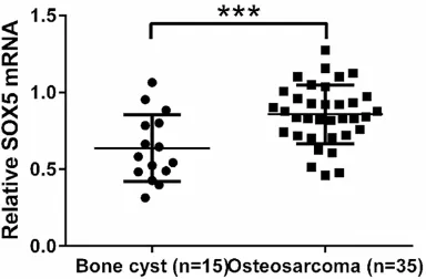
SOX5 mRNA level was elevated in human os-teosarcoma tissues
We first analyzed SOX5 mRNA levels in osteo -sarcoma tissues and bone cyst tissues by real-time PCR. As shown in Figure 1, SOX5
expres-sion was significantly up-regulated in osteosar -coma tissues (n=35), as compared with control bone cyst tissues (n=15, P<0.001).
SOX5 knockdown in osteosarcoma cells
We assessed the mRNA and protein expression of SOX5 in 5 osteosarcoma cell lines by real-time PCR and western blot, respectively. MG63 and U2OS cells showed higher mRNA and pro-tein expression of SOX5 than the other three cell lines, Saos2, SW1353 and HOS cells (Figure 2A).
To investigate the functions of SOX5 in osteo-sarcoma, we knockdown its expression in MG63 and U2OS cells, which expressed high levels of SOX5 by siRNA transfection. Three siR-NAs targeting human SOX5 (1,
siSOX5-2 and siSOX5-3) and a non-specific scramble siRNA (siNC) were synthesized. The efficiency of
siRNA-mediated knockdown of SOX5 in MG63 and U2OS cells was evaluated at both mRNA and protein levels by real-time PCR and west-ern blot, respectively (Figure 2B and 2C). All the three siRNAs targeting SOX5 (1, siSOX5-2 and siSOX5-3) were able decreased the SOX5 mRNA and protein levels in both cell lines. siSOX5-3 was the most effective one among the tested siRNAs with a knockdown ratio of greater than 75% in both cell lines. Therefore, siSOX5-3 was selected for the following experiments.
SOX5 knockdown suppressed the proliferation of osteosarcoma cells
The effect of SOX5 knockdown on osteosarco-ma proliferation was investigated by CCK-8 assay. As shown in Figure 3, SOX5 knockdown in both MG63 and U2OS cells resulted in a
sig-nificant reduction of cell viability at 24 h, 48 h
[image:3.612.93.285.74.200.2]and 72 h compared with cells without knock-down of SOX5 (siNC). These results indicated Figure 1. SOX5 was overexpressed in osteosarcoma
tissues. The mRNA level of SOX5 in osteosarcoma and bone cyst tissues collected from patients ad-mitted to Department of Orthopedics, Zhongnan Hospital of Wuhan University was detected by
real-time PCR. SOX5 mRNA was significantly higher in
that SOX5-siRNA exerted inhibitory effects in the proliferation of osteosarcoma cells.
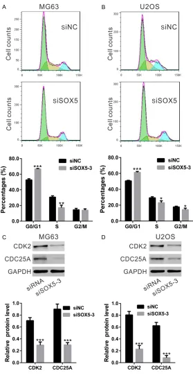
SOX5 knockdown induced G1 phase arrest of osteosarcoma cells
To investigate whether SOX5 influenced cell
cycle distribution was determined by PI staining
[image:4.612.94.522.72.342.2]and flow cytometry analysis (Figure 4A and 4B). SOX5 knockdown in MG63 cells increased the proportion of G0/G1 phase cells from 52.80 ± 1.43% to 66.71 ± 0.62%, while decreased the proportion of S phase cells from 30.72 ± 1.72% to 17.27 ± 3.15%. Similar results were obtained Figure 2. Knockdown of SOX5 in osteosarcoma cells. A. SOX5 expression in 5 osteosarcoma cell lines was analyzed by real-time PCR (upper panel) and Western blot (middle and lower panels). B, C. Knockdown of SOX5 in MG63 and
U2OS cells by real-time PCR (upper panel) and Western blot (middle and lower panels). siNC: non-specific scramble
siRNA transfected cells; siSOX5-1, siSOX5-2 and siSOX5-3: SOX5-siRNA-1, -2 and -3 transfected cells.
Figure 3. Effects of SOX5 knockdown on osteosarcoma cell proliferation in vitro. Proliferation of MG63 (A) and U2OS
(B) cells with knockdown of SOX5 (siSOX5-3) was significantly slower than proliferation of cells without knockdown
[image:4.612.94.522.416.582.2]Moreover, the expression levels of cell cycle
[image:5.612.93.372.70.618.2]regulating proteins were then estimated by differentiation, and proliferation [14]. SOX5, together with SOX6 and SOX13, belongs to Figure 4. Effects of SOX5 knockdown on osteosarcoma cell cycle
distribu-tion. A, B. The percentage of G0/G1 phase population in MG63 and U2OS cells with knockdown of SOX5 (siSOX5-3) was higher than those in cells with-out knockdown of SOX5 (siNC). C, D. Expression of CDK2 and CDC25A was evaluated by Western blot. (*P<0.05, **P<0.01, ***P<0.001 as compared with siNC).
Western blot. SOX5 knock-down in MG63 and U2OS
cells significantly reduced the
expression of CDK2 and CD- C25A (Figure 4C and 4D), indi-cating that SOX5 played an important role in the cell cycle progression of osteosarcoma cells.
SOX5 knockdown suppressed the migration and invasion ability of osteosarcoma cells
To explore the possible func-tion of SOX5 in the metasta-sis, the migration and inva-sion ability of MG63 and U2OS cells were evaluated by Transwell assay (Figure 5).
SOX5 knockdown significantly
reduced the migrated cell number of both osteosarco-ma cells compared with con-trol cells (MG63 cells: siNC, 107 ± 9; siSOX5-3, 52 ± 5; U2OS cells: siNC, 134 ± 9; siSOX5-3, 60 ± 6). The num-ber of invaded knockdown cells was about 37.9% and 32.1% of that of the control cells in MG63 and U2OS cells, respectively.
Moreover, the effect of SOX5 knockdown on the expression levels of important factors to regulate invasion was also explored. The protein levels of MMP-2, MMP-9, Twist1
and Snail1 were significantly
reduced by SOX5 knockdown in both osteosarcoma cells. These results further demon-strated the role of SOX5 in the invasion of osteosarcoma cells.
Discussion
Subgroup D of the SOX family. Previous studies have reported the functions of SOX5 during skeletogenesis [15], neural crest development [16] and gliogenesis [17]. Recently, SOX5 has been linked to various cancers. It may serve as a diagnostic and prognostic marker for glioma patients [6]. SOX5 plays an important role in the regulation of nasopharyngeal carcinoma progression through down-regulating SPARC expression [7]. Furthermore, SOX5 enhances proliferation, migration and invasion of breast cancer [9] and hepatocellular carcinoma cells [11]. In our study, we demonstrated that SOX5 was overexpressed in osteosarcoma (Figure 1) and contributed to cell proliferation (Figure 3), migration and invasion (Figure 5). Our results, consistent with the previous studies, suggest the oncogenic role of SOX5.
Aberrant cell proliferation in most malignancies is mostly due to the inhibition of cell cycle pro-gression. Martinez-Morales et al. has suggest-ed the role of SOX5 in the timing of cell cycle exit of neural progenitors by WNT-catenin sig-naling [18]. Here, cell cycle distribution analysis (Figure 4A and 4B) suggested that SOX5 knock-down in osteosarcoma cells induced G0/G1 cell arrest. We then detected protein expres-sion of CDK2 [19] and CDC25A [20], which were important for cell cycle progression of G1 to S. Consistent with the data of cell prolifera-tion and cell cycle analysis, expression of CDK2
and CDC25A were significantly down-regulated
by SOX5 knockdown (Figure 4C and 4D). Our data that SOX5 knockdown suppressed cell proliferation of osteosarcoma cells via inhibit-ing cell cycle progression.
Previous studies have shown the role of SOX5 in cell migration and invasion via regulating Twist1 expression [9, 11]. In the current study, our data demonstrated that knockdown of
SOX5 significantly reduced the migration and
invasion capacity of osteosarcoma cells (Figure 5A and 5B). Matrix metalloproteinases, includ-ing MMP-2 and MMP-9, are responsible for the degradation of the extracellular matrix, thus involving in tumor invasion and metastasis
[21]. Twist and snail, transcription factors involved in epithelial to mesenchymal
transi-tion (EMT), have significant role in the patho -genesis of osteosarcoma [22]. In this study, we showed that regulation of SOX5 down-regulated the expression of MMP-2, MMP-9 and Twist1 and snail (Figure 5C and 5D). Thus, we proposed that SOX5 induces cell invasion by regulation of MMPs expression and EMT. Taken together, we found that SOX5 was over-expressed in osteosarcoma tissues. Further in vitro experiments demonstrated that SOX5 knockdown suppressed cell proliferation and invasion of osteosarcoma cells. Our study sug-gests the far-reaching clinical implications of SOX5. SOX5 may serve as a potential diagnos-tic biomarker and a therapeudiagnos-tic target for osteosarcoma.
Disclosure of conflict of interest
None.
Address correspondence to: Dr. Wei Jin, Depart- ment of Orthopedics, Zhongnan Hospital of Wuhan University, 169 Donghu Road, Wuchang District, Wuhan 430000, Hubei, People’s Republic of China. Tel: +86 27 87330795; E-mail: weijinwh@sina.com
References
[1] Zhu H, Tang J, Tang M and Cai H. Upregulation of SOX9 in osteosarcoma and its association with tumor progression and patients’ progno-sis. Diagn Pathol 2013; 8: 183.
[2] Bakhshi S and Radhakrishnan V. Prognostic markers in osteosarcoma. Expert Rev Anti- cancer Ther 2010; 10: 271-287.
[3] Jaffe N. Osteosarcoma: review of the past, im-pact on the future. The American experience. Cancer Treat Res 2009; 152: 239-262. [4] Wunderle VM, Critcher R, Ashworth A and
Goodfellow PN. Cloning and characterization of SOX5, a new member of the human SOX gene family. Genomics 1996; 36: 354-358. [5] Dy P, Han Y and Lefebvre V. Generation of mice
harboring a Sox5 conditional null allele. Genesis 2008; 46: 294-299.
[6] Ueda R, Yoshida K, Kawase T, Kawakami Y and Toda M. Preferential expression and frequent IgG responses of a tumor antigen, SOX5, in glioma patients. Int J Cancer 2007; 120: 1704-1711.
[7] Huang DY, Lin YT, Jan PS, Hwang YC, Liang ST, Peng Y, Huang CY, Wu HC and Lin CT. Transcription factor SOX-5 enhances nasopha-ryngeal carcinoma progression by down-regu-lating SPARC gene expression. J Pathol 2008; 214: 445-455.
[8] Ma S, Chan YP, Woolcock B, Hu L, Wong KY, Ling MT, Bainbridge T, Webber D, Chan TH, Guan XY, Lam W, Vielkind J and Chan KW. DNA
fingerprinting tags novel altered chromosomal regions and identifies the involvement of SOX5
in the progression of prostate cancer. Int J Cancer 2009; 124: 2323-2332.
[9] Pei XH, Lv XQ and Li HX. Sox5 induces epithe-lial to mesenchymal transition by transactiva-tion of Twist1. Biochem Biophys Res Commun 2014; 446: 322-327.
[10] Renjie W and Haiqian L. MiR-132, miR-15a and miR-16 synergistically inhibit pituitary tu-mor cell proliferation, invasion and migration by targeting Sox5. Cancer Lett 2015; 356: 568-578.
[11] Wang D, Han S, Wang X, Peng R and Li X. SOX5 promotes epithelial-mesenchymal transition and cell invasion via regulation of Twist1 in he-patocellular carcinoma. Med Oncol 2015; 32: 461.
[12] Basu-Roy U, Seo E, Ramanathapuram L, Rapp TB, Perry JA, Orkin SH, Mansukhani A and Basilico C. Sox2 maintains self renewal of tumor-initiating cells in osteosarcomas. Oncogene 2012; 31: 2270-2282.
[13] Wu Z, Liu J, Wang J and Zhang F. SOX18 knock-down suppresses the proliferation and metas-tasis, and induces the apoptosis of osteosar-coma cells. Mol Med Rep 2016; 13: 497-504.
[14] Lefebvre V, Dumitriu B, Penzo-Mendez A, Han Y and Pallavi B. Control of cell fate and differen-tiation by Sry-related high-mobility-group box (Sox) transcription factors. Int J Biochem Cell Biol 2007; 39: 2195-2214.
[15] Smits P, Li P, Mandel J, Zhang Z, Deng JM, Behringer RR, de Crombrugghe B and Lefebvre V. The transcription factors L-Sox5 and Sox6 are essential for cartilage formation. Dev Cell 2001; 1: 277-290.
[16] Perez-Alcala S, Nieto MA and Barbas JA. LSox5 regulates RhoB expression in the neural tube and promotes generation of the neural crest. Development 2004; 131: 4455-4465. [17] Stolt CC, Schlierf A, Lommes P, Hillgartner S,
Werner T, Kosian T, Sock E, Kessaris N, Richardson WD, Lefebvre V and Wegner M.
SoxD proteins influence multiple stages of oli -godendrocyte development and modulate SoxE protein function. Dev Cell 2006; 11: 697-709.
[18] Martinez-Morales PL, Quiroga AC, Barbas JA and Morales AV. SOX5 controls cell cycle pro-gression in neural progenitors by interfering with the WNT-beta-catenin pathway. EMBO Rep 2010; 11: 466-472.
[19] Reed SI. Control of the G1/S transition. Cancer Surv 1997; 29: 7-23.
[20] Nilsson I and Hoffmann I. Cell cycle regulation by the Cdc25 phosphatase family. Prog Cell Cycle Res 2000; 4: 107-114.
[21] Curran S and Murray GI. Matrix metalloprotein-ases in tumour invasion and metastasis. J Pathol 1999; 189: 300-308.