Radiologic-Pathologic Correlation
Ocular Melanoma
Karen A. Tong, Anne G. Osborn, 1 Nick fvfamalis, Roger P. Harrie, and N. Branson Call
From the Departments of Radiology and Ophthalmology, University of Utah School of
Medicine, Salt Lake City, UT (AO, NM, RPH, NBC); and the Department of Radiology, Lorna
Linda University Medical Center, Lorna Linda, CA (KT)
History
Clinical
A 65-year-old white man had impaired visual acuity in his left eye that persisted for 1 year.
Examination revealed a small 2 X 3-mm
cream-colored elevated left submacular nod-ule. Fluorescein angiography disclosed a
cho-roidal lesion that suggested metastatic tumor.
Laboratory tests were normal. At 6-month fol-low-up there was no evidence of progression. One year later, the patient complained of left
proptosis (Fig 1) and worsening visual acuity.
Ophthalmoscopy showed a large amelanotic mass filling the left posterior segment (Fig 2).
Imaging
A-mode ultrasound demonstrated a steep spike corresponding to the tumor surface (Fig 3A, t) anterior to the scleral spike (Fig 3A, 5).
During the ultrasound examination,
sponta-neous rapidly oscillating internal echoes were also observed, denoting vascularity within the mass. Low reflective echoes posterior to the scleral spike indicated an extraocular compo-nent (Fig 3A, e). On the B-mode study a
collar-button-shaped solid intraocular mass (Fig 3B,
t) with a hypoechoic base and a large relatively
Received April 13, 1993; accepted June 21.
1 Address reprint requests to Anne G. Osborn, MD, Department of
Radiology, University of Utah Medical Center, 50 North Medical Drive, Salt Lake City, UT 84132.
Index terms: Melanoma; Eyes, neoplasms; Eyes, ultrasound; Eyes, magnetic resonance; Radiologic-pathologic correlations
AJNR 14:1359-1366, Nov/Dec 1993 0195-6108/93/1406-1359 © American Society of Neuroradiology
sonolucent extrascleral retrobulbar component
was seen (Fig 3B, e).
Precontrast computed tomographic (CT)
scan disclosed a hyperdense left orbital mass
(Fig 4A, outlined arrows) composed of a small
intraocular component and a larger
retro-orbital mass. Minimal enhancement after
con-trast administration was observed (Figs
4B-4D). The optic nerve (Fig 4D, black
arrow-heads) was surrounded by tumor. Intracranial
CT was normal. Left orbital exenteration was
performed.
Pathology
Gross pathology showed a mildly pigmented
mushroom-shaped choroidal lesion that
dis-placed the retina anteriorly almost to the
pos-terior lens capsule (Figs SA, 5B, and 6). The
tumor extended around the optic nerve without
invading the dural sheath. A slightly more
pigmented large extrascleral component had
extensive central hemorrhage and necrosis.
Microscopic examination revealed
predomi-nantly fusiform cells with spindle-shaped nuclei
and prominent nucleoli. These spindle cells
were interspersed with larger pleomorphic
ep-ithelioid cells (Fig 7).
Diagnosis
Predominantly amelanotic ocular melanoma
(mixed spindle-S and epithelioid type).
Discussion
Incidence and Age
With an incidence of six per million per year, ocular melanomas are the most common
1360 TONG
Fig. 1. Clinical examination shows mild left proptosis. The
abnormal fundus is indicated by the white arrow.
mary intraocular malignancy in adults (1). They are the second most common intraocular neo-plasm overall, after metastasis. Ocular mela-nomas are generally unilateral, although bilat-eral melanomas occur sporadically (2). There is a slight male predominance and an eightfold higher incidence in whites than blacks. The median age at diagnosis is 55 years, with 70% of cases presenting in the fifth to seventh decade. Ocular melanoma is rare in childhood
( 1 ).
Etiology
Ocular melanoma is not considered an
in-herited disease, although recent studies suggest a genetic component. Possible predis-posing factors include preexisting nevi, other melanocytic conditions such as ocular melan-osis and nevus of Ota, impaired immunity,
light-colored irides, occupational exposure (eg, welding), and trauma (1, 3).
Definition and Location
Melanomas are malignant neoplasms of me-lanocytes. Ocular melanomas occur in the most vascular part of the globe, the uvea. The uvea is the middle layer of the wall of the eye
and is composed of the choroid, ciliary body,
and iris. Ocular melanoma most commonly
occurs in the choroid (85%); 9% and 6% arise in the ciliary body and iris, respectively (4).
Melanoma rarely involves the conjunctiva,
eye-lid, or nasolacrimal duct (3).
AJNR: 14, November /December 1993
Gross Pathology
Melanomas are divided into amelanotic or melanotic types by macroscopic visualization of pigmentation. Most melanomas have ho-mogeneous texture, although larger tumors may be hemorrhagic or necrotic.
Choroidal melanomas are usually located posteriorly and are classified by size. Small tumors (less than 10 mm) are typically discoid and confined to the choroid. Medium-sized tumors (11 to 15 mm) usually have a classic collar-button or mushroom appearance. This is secondary to tumor herniation through a ruptured Bruch membrane, the transparent layer between the retinal pigment epithelium and the choroid (Fig 6). Larger tumors (greater than 15 mm) can fill the globe and extend through the sclera.
Ciliary melanomas tend to be small and may invade the anterior angle and iris. Iris melano-mas are also small and can extend into the ciliary body or seed the anterior chamber (5).
Microscopic Pathology
There are two basic melanoma cell types: spindle and epithelioid. Spindle cells are fusi-form in shape and are arranged in tight cohe-sive bundles with a syncitial appearance. Their cytoplasm has a fibrillar character. Spindle-A nuclei are thin with longitudinal folds or stripes, whereas spindle-S nuclei are larger and more oval with prominent nucleoli. Epithelioid cells
[image:2.614.57.296.91.223.2] [image:2.614.343.556.547.669.2]AJNR: 14, November/December 1993
are large, pleomorphic, and occasionally multi-nucleated. These cells have distinct borders abundant glassy cytoplasm, large round nuclei: irregular nuclear envelopes, coarse marginated nuclear chromatin, and larger eosinophilic cen
-tral nucleoli. They tend to have greater mitotic activity. In addition to spindle (A and B) and epithelioid melanomas, there are mixed and necrotic types (5).
Associated Findings
Ocular melanoma is often associated with exudative retinal detachment (6) extending from the tumor margins and over the apex (7). Diffuse vitreous hyperintensity on magnetic resonance (MR) is occasionally seen (8, 9), which may be caused by increased protein from an impaired blood-retinal barrier vitritis or breaching of the Bruch
membran~
byth~
tumor.Diagnosis
Clinical Presentation
Presenting signs and symptoms of ocular melanoma include decreased visual acuity, field defects, blurred vision, floaters, photopsia, and ocular pain. Many patients are asympto-matic, and diagnosis is made incidentally dur -ing routine examination (10). Ocular melanoma is suspected when a visible subretinal mass or exudative retinal detachment is seen by ophthalmoscopy. Fluorescein angiography shows hypervascularity.
Ocular Ultrasound
OCULAR MELANOMA 1361
Fig. 3. A, A-mode ultrasound
shows a steep spike of tumor surface with subsequent decreasing echoes
anterior to the scleral spike. Poste -riorly, low reflective echoes indicate extraocular tumor. v indicates vitre-ous body; T, tumor surface; t, tumor; S, sclera; e, extraocular tumor; and 8,
bone.
8, B-mode ultrasound shows solid
collar-button-shaped intraocular
mass (arrows) with large retrobulbar
extension. The curved black arrow
indicates penetration of tumor
through the Bruch membrane. v I
n-dicates vitreous body; t, tumor; S,
scleral; and e, extraocular component.
A-mode characteristics include a fixed solid mass, low to medium reflectivity, and regular vascularity (seen dynamically as spontaneous rapid spike movements). Small melanomas have uniform echo texture. Larger tumors at-tenuate sound, resulting in progressively de-creasing echo amplitude through the tumor (Fig 3A, t). Extrascleral tumor is depicted by low reflective echoes in the orbit (Fig. 3A, e), posterior to the scleral spike (Fig. 3A, S) (7).
B-mode findings include characteristic mushroom or collar-button shape (Fig 3B) and choroidal excavation (loss of normally highly reflective choroid echoes) (11). Large tumors show echolucency in the tumor base because of sound attenuation and can be heterogeneous secondary to hemorrhage or necrosis (12). Co-existing retinal detachment is common!
seen extending from the tumor margins and
over the tumor apex. Extrascleral e tension is usually small and nodular adjacent to the tumor base but occasionall can be large.
Rarely calcification occurs as a mall focu on the surface under! ing localized re ·nal detachment (7).
[image:3.612.55.398.85.251.2]1362 TONG
Fig. 4. A, Precontrast axial CT
shows hyperdense oblong, left orbital mass (arrows). The lesion has both an
intraocular and retrobulbar
compo-nent.
B-0, Postcontrast axial CTs show
mild homogeneous enhancement of the lesion. Note anterior displacement
of the sclera (double black arrows) by the large extraocular component. The
optic nerve and sheath (black
arrow-heads) can be faintly seen surrounded
by the tumor.
Fig. 5. A, Gross pathologic
speci-men shows a minimally pigmented
mushroom-shaped choroidal mass
(outlined arrows) that displaces the retina anteriorly, almost to the
poste-rior lens capsule. A slightly more pig-mented extrascleral component
(curved white arrows) surrounds the
optic nerve (black arrowheads). B, Low-power magnification
whole-mount specimen shows
cho-roidal melanoma arising from the
pos-terior globe, with large extrascleral
extension. The characteristic coll ar-button or mushroom shape is caused
by rupture and herniation through the
Bruch membrane (see Fig 6).
MR
c
A
Early MR studies reported that melanomas
demonstrated marked T1 shortening
com-pared with other malignant tumors (11 ). Mela-nin has paramagnetic properties thought to be
AJNR: 14, November/December 1993
D
8
secondary to stable free radicals that have dipole-dipole interactions with protons. Sub-sequent proton relaxation enhancement results
[image:4.615.220.560.91.631.2]short-repetition-AJNR: 14, November/December 1993
Short Posterior Ciliary a.
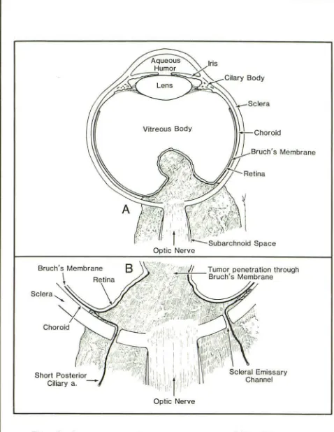
-Fig. 6. Diagrammatic representation of Fig 58.
B, Magnified view shows tumor penetration through the Bruch membrane and extrascleral extension through emissary
channels for neurovascular structures such as the short-pos-terior choroidal arteries.
time/short-echo-time images and lower signal
on long-repetition-time/short-echo-time
im-ages (4). This has been confirmed by most MR
studies (9, 13-22).
More recent studies have reported a wider spectrum of MR signal patterns for melanoma (8, 23). One study demonstrated that T1 values of melanomas varied proportionally with field strength. Conversely higher melanin content has been associated with decreased T 1 and T2 relaxation times (24). However, grossly
amela-notic tumors also may produce T1 shortening,
perhaps because of microscopic amounts of
melanin (22). Other studies have reported
vari-able appearance of amelanotic melanomas on
MR ( 16, 18). Therefore, signal intensity may
vary with field strength and may not be solely related to melanin content.
Melanoma can be complicated by
hemor-rhage or necrosis. Ocular melanoma and
subacute hemorrhage both can have high
sig-OCULAR MELANOMA 1363
Fig. 7. High-power magnification demonstrates primarily
fusiform-shaped cells (curved arrow) characteristic of spindl
e-S melanoma, although several large pleomorphic epithelioid cells (outlined arrow) are also seen, indicating mixed mela
-noma.
nal on T1-weighted images. On T2-weighted
images, earlier hemorrhage is typically
hypoin-tense, whereas later hemorrhage is hyperin-tense, and melanin tends to be mildly hypoin-tense (6).
MR can be helpful in differentiating between
melanoma and subretinal fluid. Subretinal
ex-udates typically appear hyperintense on both
T1- and T2-weighted images because of high
protein content (9, 16, 17, 21, 22). However,
subretinal fluid itself also can be complicated
by hemorrhage.
Several lesions can mimic the MR
appear-ance of uveal melanoma. These include
melan-ocytoma, retinoblastoma, choroidal
hemor-rhage, subretinal hemorrhage, subretinal
fibrosis, senile disciform macular degeneration,
and choroidal metastasis (8, 22). T1 shortening
may be secondary to mucin, other
proteina-ceous fluid, or hemorrhage. Therefore, the T1
and T2 shortening in uveal melanoma should
not be considered pathognomonic (8).
Several studies report difficulty
demonstrat-ing lesions less than 2 to 3 mm on MR (9, 14,
15, 17, 22). Small lesions or subtle extrascleral
extensions not identified on MR have been
detected by ultrasound (19). MR contrast
agents increase the visibility of small tumors
[image:5.612.56.295.86.395.2] [image:5.612.311.555.100.278.2]1364 TONG
Differential Diagnosis
Early studies reported clinical misdiagnosis
rates as high as 40%. Accurate diagnostic
examinations have reduced the frequency of
error. In 1990, the Collaborative Ocular
Mela-noma Study reported a misdiagnosis rate of
TABLE 1: Differential diagnosis of intraocular mass in adult
Ophthalmoscopy Ultrasound
Choroidal Variably pigmented B-scan: collar-button or melanoma circumscribed mushroom-shaped
mass of variable mass with regular size; may have texture
overlying retinal A-scan: low to medium detachment regular reflectivity
often with spont
a-neous vascular pul sa-tions
Orbital echolucency in extrascleral extension
Choroidal Flat or minimally el- B-scan: slightly ele
-nevus evated gray le- vated mass with reg-sions with slightly ular texture indistinct margins A-scan: high internal
reflectivity without spontaneous pul sa-tions
Choroidal Yellow to golden B-scan: dome-shaped
metastasis brown mildly ele- mass with irregular
vated mass with texture
irregular surface A-scan: irregular inter -and borders; may nal reflectivity usu -have overlying ally without spont a-retinal detach- neous pulsations ment; may be
multiple, bilateral
Choroidal Orange-red mildly B-scan: mildly elevated hemangioma elevated mass; dome shaped mass
may have overly- with regular texture ing retinal de- A-scan: high regular in
-tachment ternal reflectivity without vascular pul -sations
Occasional calcification
Subretinal or sub- Round globular dark B-scan: dome-shaped, choroidal hem- brown or green- mildly elevated with
orrhage brown mass variable texture
A-scan: variable inter
-nal reflectivity; may
have spontaneous
pulsations; mobility of surface spike (may
be subtle)
AJNR: 14, November/December 1993
0.48% (27). However, several ocular diseases
continue to mimic uveal melanoma clinically,
most commonly choroidal nevi, benign
melan-ocytoma, choroidal hemangioma, choroidal
hemorrhage, and choroidal metastasis (6, 28)
(Table 1).
MR Contrast
CT
Enhancement Tl-Weighted T2-Weighted
Focal mass hy- Moderate to lsointense or Mild to
perdense to marked hy- hypointense moderate
vitreous perintensity to vitreous
to vitreous
Usually too Usually too Mild to
small to be small to be moderate
detected detected w
ith-out contrast
Focal thickening Variable Variable Mild to
or mass moderate
Ill-defined mass Variable Variable Intense
Focal mass Variable Variable None
[image:6.612.57.557.230.722.2]AJNR: 14, November/December 1993
Prognosis and Treatment
Natural History and Prognosis
Extrascleral local extension occurs via scleral emissary channels for neurovascular structures and is associated with increased risk of recurrence (6) and hematogenous dissemi-nation (5). Metastasis usually occurs hematog-enously and is more common with choroidal
melanomas (1). The liver is the most common
remote site of metastasis (5). Other sites in-clude the lung, spine, skin, subcutaneous tis-sue, nodes, brain and adrenal (29, 30). Metas-tasis usually occurs within five years of pres-entation and occasionally after long intervals. Rarely optic nerve invasion can result in intra-cranial metastasis (31, 32).
Uveal melanoma is a potentially fatal disease
in adults. The 5-, 10-, and 15-year survival
rates are 65%, 52%, and 46%, respectively.
The median survival after hepatic metastasis is 2 to 4 months (1). Epithelioid cell type and tumor diameter greater than 1 0 mm indicate poor prognosis. Other indicators of poor
prog-nosis include elevated mitotic rate, diffusely
infiltrating tumor, scleral/ extrascleral
exten-sion, necrosis, and lymphocytic infiltration (5,
8).
Therapy
Treatment is largely determined by tumor
size. Other important factors include tumor
location, tumor activity, status of the
contra-lateral eye, age, general health, and presence
of metastasis. Although the traditional method
of treatment has been enucleation, currently most small dormant tumors without extra-scleral extensions are managed conservatively and followed by ultrasound observation. Local en-bloc resection has been performed for small
anterior uveal lesions. Laser photocoagulation
is occasionally used for small choroidal
mela-nomas. Radiation therapy (radioactive plaque
therapy or external beam irradiation) is per-formed for many medium-sized and even some
large melanomas. Exenteration for extensive
or recurrent tumor is performed sometimes,
although prognosis is poor. Metastatic mela
-OCULAR MELANOMA 1365
noma has been treated with chemotherapy with minimal effect. Experimental therapies
include hyperthermia and immunotherapy (5,
33). 0
Summary
Uveal melanoma is the second most frequent ocular malignancy after metastasis and the most common primary ocular malignant
neo-plasm in adults. The diagnosis is usually made
from clinical examination and ocular ultra-sound. CT and MR may be helpful for further
evaluation.
References
I. Egan KM, Seddon JM, Glynn RJ, et al. Major review:
epidemio-logic aspects of uveal melanoma. Surv Ophthalmol 1988;32: 239-251
2. Seregard S, Daunius C, Kock E, Popovic V. Two cases of primary
bilateral malignant melanoma of the choroid. Br J Ophtha/mol
1988; 72:244-245
3. Rennie IG. Diagnosis and treatment of ocular melanomas. Br J Hasp Med 1991;46:144-156
4. Char DH, Umsold R, Sobel DF, et al. Computed tomography:
ocular and orbital pathology. In: Newton TH, Bilaniuk L T, eds.
Modern neuroradiology. Vol. 4: Radiology of the eye and orbit.
Kentfield, Calif: Raven, 1990:9.4-9.5
5. McLean IW, Bernier MN, Zimmerman LE, Jakobiec FA. Tumors of the eye and ocular adnexae: atlas of tumor pathology. 3rd series. Washington, DC: Armed Forces Institute of Pathology (in
press)
6. Atlas S. Magnetic resonance imaging of the brain and spine. New
York: Raven, 1991:721-724
7. Byrne SF, Green RL. Ultrasound of the eye and orbit. St. Louis:
Mosby-Year Book, 1992:134-173
8. Bloom PA, Ferris JD, Laidlaw DAH, Goddard PR. Magnetic r
eso-nance imaging: diverse appearances of uveal malignant mel
ano-mas. Arch Ophthalmol 1992; II 0: II 05-1111
9. Mafee MF, Peyman GA, Grisolano JE, et al. Malignant uveal melanoma and simulating lesions: MR imaging evaluation. Ra-diology 1986; 160:773-780
10. Servodidio CA, Abramson DH. Presenting signs and symptoms of choroidal melanoma: what do they mean? Ann Ophthalmol
1992;24: 190-194
II. Hodes BL. Ultrasonographic diagnosis of choroidal malignant
melanoma. Surv Ophtha/mo/1977:22:29-40
12. Munk PL, Velie! AD, Levin M, et al. Sonography of the eye. AJR: Am J Roentgeno/1991 ;157:1079-1086
13. Damadian R, Zaner K, HerD, DiMaio T. Human tumors detected by nuclear magnetic resonance. Proc Nat/ Acad Sci USA
1974;71 :1471-1473
14. Chambers RB, Davidorf FH, McAdoo JF, Chakeres DW. Magnetic
resonance imaging of uveal melanomas. Arch Ophtha/mol
1987;105:917-921
1366 TONG
imaging in the evaluation and differentiation of uveal melanoma. Ophthalmology 1987;94:341-348
16. Mihara F, Gupta KL. Murayama S. et al. MR imaging of malignant uveal melanoma: role of pulse sequence and contrast agent. AJNR: Am J Neuroradio/1991; 12:991-996
17. Peyman GA. Mafee MF. Uveal melanoma and similar lesions: the role of magnetic resonance imaging and computed tomography. Radio/ Clin North Am 1987;24:471-486
18. Peyster RG. Augsburger JJ. Shields JA. et al. Intraocular tumors: evaluation with MR imaging. Radiology 1988; 168:773-779 19. Raymond WR. Char DH, Norm D, Protzko EE. Magnetic resonance
imaging evaluation of uveal tumors. Am J Ophthalmol
1991 ;111 :633-641
20. Sobel DF, Kelly W, Kjos BO. et al. MR imaging of orbital and ocular disease. AJNR: Am J Neuroradiol 1985;6:259-264 21. Sullivan JA, Harms SE. Surface-coil MR imaging of orbital
neo-plasms. AJNR: Am J Neuroradio/1986;7:29-34
22. Wilms G, Marchal G, Van Fraeyenhoven L. et al. Shortcomings and pitfalls of ocular MRI. Neuroradiology 1991 ;33:320-325 23. Marx HF, Colletti PM. Raval JK, et al. Magnetic resonance imaging
features in melanoma. Magn Reson Imaging 1990;3:223-239 24. Gomori JM. Grossman Rl. Shields JA. et al. Choroidal melanomas:
correlation of NMR spectroscopy and MR imaging. Radiology 1986; 158:443-445
AJNR: 14, November /December 1993
25. Adam G, Brab M. Bohndorf K. Gunther RW. Gadolinium-DTPA -enhanced MRI of intraocular tumors. Magn Reson Imaging 1990;8:683-689
26. Bond JB. Haik BG. Mihara F. Gupta KL. Magnetic resonance imaging of choroidal melanoma with and without gadolinium contrast enhancement. Ophthalmology 1991 ;98:459-466 27. Albert DM. Accuracy of diagnosis of choroidal melanomas in the
collaborative ocular melanoma study. Arch Ophthalmol
1990; 108: 1268-1273
28. Shields JA. Augsburger JJ, Brown GC, Stephens RF. The differ -ential diagnosis of posterior uveal melanoma. Ophthalmology 1980;87:518-522
29. Lorigan JG. Wallace S, Mavligit GM. The prevalence and location of metastases from ocular melanoma: Imaging study in 110 patients. AJR: Am J Roentgeno/1991 ;157:1279-1281
30. Shields JA. Shields CL, Shakin EP. Kobetz LE. Metastasis of choroidal melanoma to the contralateral choroid, orbit, and eyelid. Br J Ophtha/mol 1988; 72:456-460
31. Al-Haddab S. Hidayat A, Tabbara KF. Ciliary body melanoma with optic nerve invasion. Br J Ophtha/mo/1990;74:123-124 32. Jones DR, Scobie IN. Sarkies NJC. Intracerebral metastases from
ocular melanoma. Br J Ophtha/mo/1988;72:246-247