Diffusion Tensor Imaging of the Hippocampal
Formation in Temporal Lobe Epilepsy
Bassam A. Assaf, Feroze B. Mohamed, Karine J. Abou-Khaled, J. Michael Williams, May S. Yazeji, John Haselgrove, and Scott H. Faro
BACKGROUND AND PURPOSE:Diffusion tensor imaging (DTI) is a noninvasive technique that can be used to assess the integrity of cerebral tissue. The purpose of this study was to assess DTI measurements in the hippocampal formation (HF) and to investigate the role of DTI in lateralizing the seizure focus in temporal lobe epilepsy (TLE).
METHODS: We evaluated 12 patients with unilateral TLE and 14 healthy subjects. We collected diffusion-weighted images along six different directions with a b value of 1000 s/mm2, as well as an image acquired without diffusion weighting (bⴝ 0 s/mm2). A 1.5-T imager was used to acquire 17 (3-mm) coronal sections covering the temporal lobes. We compared the mean diffusivity (traceD) and fractional anisotropy (FA) from symmetrical voxels by sampling the anterior HF bilaterally. We compared measurements with the EEG, high-resolution MR imaging, and clinical information.
RESULTS:The patient group had significantly increased diffusivity and decreased FA in the HF ipsilateral to the seizure focus, as compared with values in the contralateral HF. When compared with healthy subjects, patients had significantly higher mean diffusivity in the ipsilateral HF; ipsilateral FA values were lower and did not reach statistical significance. Measurements in the contralateral HF did not show differences. Left-right and absolute diffusivity indices lateralized the abnormal HF in eight and five of 12 patients, respectively.
CONCLUSION: Abnormal DTI measurements and the epileptogenic HF are associated in unilateral TLE. This finding may reflect hippocampal sclerosis and may aid in presurgical evaluation.
Diffusion tensor imaging (DTI) is a new imaging technique that can be used to noninvasively assess the molecular and biochemical environment of cerebral tissue (1–2). DTI can aid in characterizing and mea-suring the diffusive transport of water molecules by means of an effective diffusion tensor,D. These sym-metric tensor measurements contain useful informa-tion about the tissue microstructure and architecture. Of the several indices used to characterize diffusion tensor, those most commonly used are the trace of the tensor, which measures mean diffusivity (trace D), and fractional anisotropy (FA) (3– 4). These charac-teristic measurements may represent the changes in cerebral structure that occur in various neurologic conditions (1, 2). DTI has been shown to be useful in
the study of diseases, such as cerebral ischemia (5), acute stroke (6), multiple sclerosis (7) and schizo-phrenia (8).
Noninvasive MR imaging techniques are becoming increasingly important in lateralizing and localizing the seizure focus in a noninvasive manner. Only a few studies have addressed the utility of DTI in epilepsy (9 –11). In general, these studies have demonstrated increased diffusivity and decreased FA in cerebral tissue, which corresponded to the seizure focus in the entire group of the patients evaluated. However, these studies included a mixture of temporal epilepsy and extratemporal epilepsy with or without lesions. In addition, these studies failed to demonstrate a high yield of abnormal DTI measurements in individual-ized patients. A recent study evaluated DTI of white matter in temporal lobe epilepsy (TLE) (12). In that study, significantly lower diffusion anisotropy and higher diffusivity in directions perpendicular to the axons were detected in several white matter struc-tures in the patients, when compared with control subjects. However, none of these measured structures were in the temporal lobes. Another recent study (13) was conducted to evaluate apparent diffusion coeffi-Received March 6, 2003; accepted after revision May 1.
From the Departments of Neurology (B.A.A., K.J.A.-K.), Radi-ology (F.B.M., M.S.Y., S.H.F.), and PsychRadi-ology (J.M.W.), Drexel University College of Medicine, and the Department of Radiology, Children’s Hospital of Philadelphia (J.H.), PA.
Address reprint requests to Bassam Assaf, MD, Department of Neurology, Drexel University College of Medicine, Broad St and Vine St, M.S. 308, Philadelphia, PA 19102.
©American Society of Neuroradiology
cient (ADC) values in patients with unilateral hip-pocampal sclerosis. The investigators found abnormal values on the side with hippocampal sclerosis in all patients, as compared with healthy volunteers. That study, however, was limited to unilateral hippocampal sclerosis findings on MR images, and it involved large regions of interest (ROIs) on axial sections, which are subject to partial-volume effects.
The purpose of this study was to obtain DTI mea-surements of the hippocampal regions from thin coronal sections in patients with clinically proved TLE and in healthy control subjects. In addition, we investigated the diagnostic utility of DTI in detecting hippocampal abnormalities and in lateralizing the temporal-lobe seizure focus in a consecutive series of patients with TLE who have been evaluated for epi-lepsy surgery at our comprehensive epiepi-lepsy center. Furthermore, we compared the DTI measurements and high-resolution brain MR images in these pa-tients.
Methods
We evaluated 12 patients with unilateral nonlesional TLE and 14 healthy subjects by using DTI of the brain. Their age and sex information is provided in Tables 1 and 2. The age range was 27–59 years for the patient group (mean age, 41.42 years; SD, 8.52). The control group had an age range of 24 – 49 years (mean age, 31.29 years; SD 8.19). Informed consent was obtained from all subjects, per the guidelines of the institu-tional review board. The diagnosis of unilateral TLE was based on clinical and EEG findings after a careful review of the patient’s medical history, high-resolution brain MR images, routine EEGs, and continuous audiovisual/EEG monitoring results. This information was interpreted by a board-certified epileptologist and electrophysiologist (B.A.A.) who was blinded to the DTI quantitative measurements. This process resulted in the diagnosis of definite unilateral TLE proved with unilateral ictal EEG recordings during continuous audiovisual/ EEG monitoring in nine patients and in the diagnosis of prob-able unilateral TLE based on exclusively unilateral interictal EEG findings during prolonged continuous audiovisual/EEG monitoring and other concordant clinical information in three patients. This classification was similar to that used by Arfa-nakis et al (12) for TLE diagnosis.
DTI was performed in all patients after they had been seizure-free for at least 24 hours. This approach was intended to ensure measurements from a baseline state as much as possible, because prior studies indicated that seizures from status epilepticus may affect diffusivity measurements (14 –15). DTI was performed by using a 1.5-T MR unit (Vision; Siemens Medical Systems, Erlangen, Germany), with a single-shot echo-planar diffusion-weighted imaging sequence. To fully deter-mine the diffusion tensor, we collected diffusion-weighted im-ages along six different directions with a b value of 1000 s/mm2, as well as an image without diffusion weighting (b⫽0 s/mm2). Seventeen 3-mm coronal sections were acquired to cover the entire temporal lobes. The imaging parameters included the following: TR/TE, 6000/100; FOV, 240 mm; matrix, 98⫻128; and four acquisitions. The total imaging time to collect the DTI images was 4.5 minutes.
After image acquisition, the data were transferred to an independent workstation (Silicon Graphics, Mountain View, CA) for calculation of the DTI indices. The maps of mean diffusivity (rotationally invariant scalar indices of diffusion) and FA were calculated from the diffusion-weighted images by using a method based on that proposed earlier (3, 4, 16). Software written in IDL (Interactive Data Language, Boulder, CO) was used. See the Appendix for details.
We compared the mean diffusivity and FA indices from symmetrical-voxel sampling regions from the anterior hip-TABLE 1: Summary of the patient population evaluated with DTI
Patient/Sex/Age, y TLE HR MRI
TraceD,
10⫺3mm2/s TraceD FA
L R Lateralization L-R L-R Lateralization L R L-R
1/F/40 R R 0.94 1.10 R ⫺0.16 R 0.025 0.015 0.010
2/F/27 R R 0.82 0.93 R ⫺0.11 R 0.017 0.017 0
3/M/31 L L 0.78 0.77 N 0.01 N 0.014 0.021 ⫺0.007
4/F/42 L L 0.89 0.76 N 0.13 L 0.012 0.019 ⫺0.007
5/F/38 L N 0.88 0.86 N 0.02 N 0.012 0.019 ⫺0.007
6/F/59 R N 0.83 0.82 N 0.01 N 0.015 0.014 0.001
7/F/43 L L 0.97 0.83 L 0.14 L 0.010 0.020 ⫺0.010
8/M/49 R R 0.72 0.86 N ⫺0.14 R 0.013 0.017 ⫺0.004
9/F/42 L L 1.20 0.84 L 0.36 L 0.011 0.021 ⫺0.010
10/M/39 L N 0.85 0.74 N 0.11 L 0.012 0.013 ⫺0.001
11/M/50 L L 0.88 0.80 N 0.08 N 0.013 0.013 0
12/F/37 L N 0.96 0.84 L 0.12 L 0.011 0.008 0.003
[image:2.603.53.536.71.240.2]Note.—HR MRI indicates high-resolution MR imaging; N, normal.
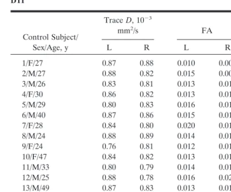
TABLE 2: Summary of the control-subject population evaluated with DTI
Control Subject/ Sex/Age, y
TraceD, 10⫺3 mm2
/s FA
L R L R
1/F/27 0.87 0.88 0.010 0.009
2/M/27 0.88 0.82 0.015 0.009
3/M/26 0.83 0.81 0.013 0.012
4/F/30 0.86 0.82 0.013 0.016
5/M/29 0.80 0.83 0.016 0.019
6/M/40 0.87 0.86 0.015 0.014
7/F/28 0.84 0.80 0.020 0.016
8/M/24 0.88 0.89 0.014 0.016
9/F/24 0.76 0.81 0.012 0.016
10/F/47 0.84 0.82 0.013 0.016
11/M/33 0.80 0.79 0.014 0.017
12/M/25 0.88 0.78 0.016 0.020
13/M/49 0.87 0.83 0.013 0.016
[image:2.603.305.533.286.476.2]pocampal formation (HF) on both sides, in patients and in control subjects. The ROIs were selected on the coronal sec-tion from a 4⫻8-mm rectangular area drawn in the center of the anterior HF. The selections were made by a neurologist (K.J.A.-K.) and a board-certified neuroradiologist (S.H.F.) who were blinded to the side of the seizure focus, the patient’s history. and other clinical information. Figure 1A shows the coronal T2-weighted image through a section showing the tem-poral lobes. Figure 1B shows the placement of the ROIs in the left and right HFs.
Summary data for the patient group and the control sub-jects, along with the relevant medical history and DTI mea-surements are shown in Tables 1 and 2. In our analysis, we compared the group HF mean diffusivity and FA ments ipsilateral with the epileptogenic side to the measure-ments in the contralateral HF. We also compared the group ipsilateral and contralateral DTI measurements in the patient population with the control values derived from a total of 28 HF measurements on both sides in all 14 healthy subjects. In addition, we calculated the left-right diffusivity and FA indices of the patient group and compared them with the mean left-right index of the normal group. The significance of the differ-ences was assessed by using the Studentttest. Only differences with P ⬍ .05 were considered significant. Subsequently, we identified the individual subjects with TLE who had a signifi-cant difference in left-right DTI measurement and also those with DTI measurements differing from the mean for the con-trol subjects. When identifying these patients with abnormal values, we compared the abnormal side in the patients with the mean value of the corresponding side in the control group. We later compared the DTI measurements with the eventual side of TLE, with the high-resolution MR images, and with the other clinical information (Tables 1 and 2).
Results
The statistical measurements of mean diffusivity and FA ipsilateral and contralateral to the epilepto-genic HF in the patient group and the entire mea-surements in the control group are summarized in
Tables 3 and 4. There was no statistically significant difference in diffusivity or FA measurements between the sides in the control measurements, with a mean diffusivity of 0.84 and 0.83 on the left and right sides, respectively, and a mean FA of 0.0146 and 0.0152 on the left and right sides, respectively. The mean diffu-sivity and FA values for all 28 HF measurements in the control group were 0.84⫾0.03 and 0.015⫾0.007, respectively.
The patient group, however, had significantly in-creased mean diffusivity and dein-creased FA values in the HF ipsilateral to the seizure focus, when com-pared with the contralateral HF measurements, and with T(11)⫽ ⫺2.55 and T(11)⫽ 4.14 (P⬍ .05 and P ⬍ .05, respectively). When the patient group was compared with the control subjects, the ipsilateral HF had significantly increased mean diffusivity, with T(24) ⫽ 2.73 and P ⬍ .05. Ipsilateral FA was de-creased, but the difference did attain statistical signif-icance, with T(24) ⫽ ⫺1.82 and P ⫽ .08. The con-tralateral HF measurements did not demonstrate differences when compared with those of the control group.
When left-right diffusivity measurements for indi-vidual patients were compared with those of healthy subjects, eight of 12 patients (patients 1, 2, 4, 7–10, and 12; sensitivity of 66%) had values that were 2 SDs beyond the mean, while three of 12 patients (patients 1, 7, and 9) had abnormal left-right FA measurements that were 2 SDs below or above the mean. When the individual mean diffusivity measurements were com-pared with the mean value of healthy subjects on the same side, five of 12 patients (patients 1, 2, 7, 9, and 12 in Table 1; sensitivity of 42%) had increased dif-fusivity measurements beyond 2 SDs from the mean
FIG 1. Images in a patient with left-sided
TLE.
A, Coronal T2-weighted MR image. Note the abnormal anterior hippocampus with mild deformation on the left side compared with the right side.
[image:3.603.54.383.58.206.2]B, DTI at the same coronal level as inA. Note the marking of the ROI in the left and right HFs. The images in the upper right corner correspond to the diffusion (B0, top), trace D (middle), and FA (bottom) images.
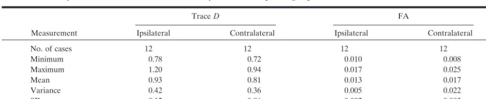
TABLE 3: Summary of statistical measurements of diffusivity and FA in the patient group
Measurement
TraceD FA
Ipsilateral Contralateral Ipsilateral Contralateral
No. of cases 12 12 12 12
Minimum 0.78 0.72 0.010 0.008
Maximum 1.20 0.94 0.017 0.025
Mean 0.93 0.81 0.013 0.017
Variance 0.42 0.36 0.005 0.022
[image:3.603.54.534.234.332.2]for the control subjects. The contralateral HF mea-surements did not demonstrate differences when compared with those of the healthy control group.
We compared the sex and age in both the patient group and the healthy subject group. There was no statistically significant difference in sex, with2(1)⫽
2.48 andP ⬍ .12. There was a 10.13-year mean dif-ference in age between the patient group and the control group, with T(23.1) ⫽ 3.08 and P ⬍ .05. However, age was not correlated with any of the dependent measures (the DTI measurements) there-fore; an analysis of covariance for age was not con-ducted.
The review of clinical information revealed that both high-resolution brain MR imaging results and left-right DTI measurements were lateralizing in eight of 12 pa-tients, whereas they were normal in four cases (Table 1). The results were concordant in six patients and comple-mentary to each other in four other cases. In only two of 12 patients were both measurements normal. Neither imaging technique demonstrated discordant findings with the side of the epilepsy or with the other technique. Three patients (patients 1, 6, and 7) underwent an-teromesial temporal lobectomy and became seizure-free. In these patients, pathologic evaluation revealed temporal-lobe sclerosis with gliosis and a diminished neuronal count. Other clinical information is summa-rized in Table 1.
Discussion
The hippocampus is still an essential structure in the epileptogenicity of TLE, and hippocampal sclero-sis is the major pathology underlying nonlesional TLE (17–18). While visual inspection of high-resolution brain MR images and, often, volumetric HF measure-ments is the standard in evaluating brain MR abnor-malities in patients with TLE, hippocampal atrophy on brain MR images is not always indicative of epi-leptogenicity (19 –20). In this study, we investigated the role of DTI in lateralizing the seizure focus and the potential pathophysiology underlying DTI findings.
The patient group analyzed in this study demon-strated a significant difference in the mean diffusivity and FA measurements in the epileptogenic HF, as compared with values on the contralateral side. The finding of significantly increased mean diffusivity and the trend of decreased FA that did not reach statis-tical significance indicate that diffusivity measure-ments are more sensitive than FA in detecting the epileptogenic side, when values were compared with
those of the healthy group. These findings concur with the results that Rugg-Gunn et al (10) reported in TLE patients. The observation may best be explained by the concept that expansion in the extracellular space, which causes increased diffusivity, is probably associated with a relatively preserved structural orga-nization of the fiber bundle.
This study also demonstrates higher sensitivity with the left-right diffusivity index in lateralizing the epi-leptogenic temporal lobe than with other indices or findings in previous studies. In eight of our 12 pa-tients (sensitivity of 66%), the left-right diffusivity difference was abnormal, when compared with the difference in the control group. Three of 12 patients had an abnormal left-right FA difference. The sensi-tivity of the left-right diffusivity index was also higher than that of the HF diffusivity index (sensitivity of 42%) in lateralizing the seizure focus. These repre-sent significant findings in our study, as compared with the results of previous studies. Rugg-Gunn et el (10) found that, despite the sensitivity of the diffusiv-ity index in what they called “acquired epilepsy with lesions,” diffusivity did not permit the identification of a clinically concordant abnormality in most their cases with what they called “cryptogenic epilepsy”; these included 15 patients with TLE. In their series, only one of 15 patients with TLE demonstrated indi-vidualized changes when investigators performed a voxel-by-voxel comparison by using the statistical parametric mapping model. While the results from the group comparison of diffusivity and anisotropy were statistically significant in our study as well as in theirs, we believe that the higher sensitivity of indi-vidualized left-right and direct HF diffusivity mea-surement in our study (66% and 42%, respectively) reflects the higher accuracy of our manual method. The selection of ROI in the anterior body of the HF from relatively thin (3-mm) sections is likely to pro-duce accurate and consistent measurement from the HF while avoiding partial-volume effects. In addition, measurements in the cases of unilateral TLE in our study with relatively healthier contralateral HF could have produced left-right diffusivity difference more robust than that observed in a series with mixed unilateral and bilateral TLE. The increased sensitivity of the left-right measurement difference may be re-lated to the inherent control of this index with regard to other variables that may affect the DTI measure-ments (such as patient age, age of onset, duration of epilepsy, among others).
[image:4.603.53.533.70.158.2]Yoo et al (13) reported ADC measurements in 18
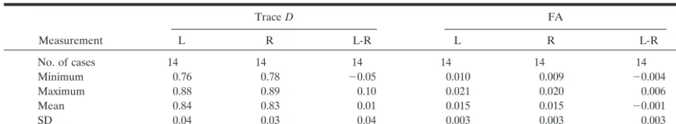
TABLE 4: Summary of statistical measurements of diffusivity and FA in the healthy control group
Measurement
TraceD FA
L R L-R L R L-R
No. of cases 14 14 14 14 14 14
Minimum 0.76 0.78 ⫺0.05 0.010 0.009 ⫺0.004
Maximum 0.88 0.89 0.10 0.021 0.020 0.006
Mean 0.84 0.83 0.01 0.015 0.015 ⫺0.001
patients with TLE and unilateral MR imaging–posi-tive hippocampal sclerosis. The researchers found sig-nificant abnormalities in all sclerotic hippocampal measurements, when compared with those in healthy subjects. Although DTI is more sensitive than ADC measurement, the higher sensitivity in their study is related to two factors: One pertains to their restricted selection of patients with TLE to those with unilateral hippocampal sclerosis, as shown on brain MR images, and the other factor relates to the selection of the ROI from axial images. This method is likely to pro-duce a partial-volume effect that propro-duces higher ADC values in the sclerotic hippocampi due to asso-ciated hippocampal atrophy and CSF sampling (13). Our analysis was based on manual selection of the ROI as a region in the center of the anterior HF in all patient and healthy subjects. We avoided outlining the hippocampal margins to ensure complete sampling from the HF and to avoid CSF sampling. This method was consistent for multiple measurements from each subject and for the two investigators (K.J.A.-K., S.H.F.). In addition, the section thickness limited to 3 mm en-sured sampling from only the HF. The measurements were unlikely to be affected by sampling outside the HF as a result of the convoluted shape of the HF in the healthy population and of the added deformation in-curred in the case of mesial temporal sclerosis in TLE. The lack of significant differences in the left and right HF DTI measurements in the healthy population in our study further supports the consistency and validity of this manual method. The consistency and reproducibil-ity of DTI measurements obtained by using the manual selection of ROI is also reported in other DTI studies of TLE in which ROIs were manually selected to measure DTI indices in the white matter (12).
This and other studies (9, 10, 13) indicate a poten-tial role for DTI in localizing the seizure focus in TLE. High-resolution brain MR imaging is still the standard neuroimaging technique for lateralizing the seizure focus in TLE, but negative MR imaging re-sults can be encountered in cases of unilateral TLE. In four of 12 patients, high-resolution brain MR find-ings were negative. Two of these patients had abnor-mal left-right HF diffusivity measurements. On the other hand, two of four patients with normal left-right HF diffusivity measurements had abnormal high-res-olution brain MR images. This observation supports a complementary role of these two imaging modalities, although a study in larger patient sample may be needed to satisfactorily address this issue. This poten-tial complementary role of DTI may be explained by the fact that each imaging technique is used to mea-sure a relatively different aspect of the seizure focus. We believe that the improved technical design and understanding of the pathophysiology of DTI mea-surements may further improve the role of this imag-ing technique in evaluatimag-ing patients with TLE. The improved sensitivity in our analysis technique proba-bly accounts for the discrepancy between the signifi-cant differences with group analysis and the lack of difference between measurements in individualized patients and mean control subjects in other studies
(10). In addition, further understanding of other fac-tors influencing the DTI indices, such as the temporal occurrence and frequency of seizures and interictal spikes, is likely to better define the significance of these DTI measurements and will probably increase their sensitivity. Seizure activity in status epilepticus has been shown to decrease the diffusivity of water molecules (14, 15, 21). Furthermore, improved un-derstanding would better clarify the role of DTI in the presurgical workup of patients with intractable TLE, in comparison to the role of high-resolution MR tech-niques, which have high sensitivity to changes in the mesial structures in mesial TLE (22).
We evaluated only patients with unilateral TLE to assess the sensitivity and utility of this refined DTI technique in a well-defined patient population. While this technique is demonstrably sensitive in identifying the epileptogenic hippocampus in this group of pa-tients, further and larger studies in patients with TLE—including patients with bilateral independent temporal lobe seizures—are needed. Such studies may have to be controlled for seizure and spike oc-currence, as well as for other variables (such as sub-jects’ age, age at seizure onset, and duration of epi-lepsy) to improve our understanding of the role of this technique in assessing the various TLE entities.
Conclusion
The results of this study demonstrate the ability of DTI to depict changes to the HF architecture and to lateralize the seizure focus in the entire group of patients and in individualized patients with TLE. Fur-ther studies are needed to delineate the role of this technique in the evaluation of patients with various categories of TLE.
Appendix
When using a method based on the work of Basser and Pierpaoli (16) to obtain an overall evaluation of the diffusivity in a region, one must avoid the anisotropic diffusion effects and limit the results to an invariant. One such invariant, the trace of the diffusion tensor was used to calculate the mean diffusivity in this study. The trace is given by Equation 1, as follows:
1) Trace具D典⫽Dxx⫹Dyy⫹Dzz
⫽3具D典,
where Dxx, Dyy, and Dzz are the three diagonal
ele-ments of the diffusion tensor and their average,具D典, is the mean diffusivity. Regarding FA, this invariant index measures the diffusion anisotropy of fiber struc-ture, as shown in Equation 2:
2)
FA⫽
冑
3关共1⫺具典兲2⫹共
2⫺具典兲2⫹共3⫺具典兲2兴
冑
2共12⫹22⫹32兲,
where具典 ⫽ (1⫹ 2⫹3)/3 and1,2, and3are
References
1. Le Bihan D, Mangin JF, Poupon C, Clark C, Pappata S, Molko N, and Chabriat H.Diffusion tensor imaging: concepts and applica-tions.J Magn Reson Imag2001;13:534 –546
2. Alsop DC, Connelly A, Duncan JS, Hufnagel A, Pierpaoli C, and Rugg-Gunn FJ.Diffusion and perfusion MRI in epilepsy.Epilepsia 2002;43(Suppl 1):69 –77
3. Pierpaoli C, Jezzard P, Basser PJ, Barnett A, Di Chiro G.Diffusion tensor MR imaging of the human brain.Radiology1996;201:637– 648
4. Basser PJ, Pierpaoli C.Microstructural and physiological features of tissues elucidated by quantitative-diffusion-tensor MRI.J Magn Reson B1996;111:209 –219
5. Lythgoe MF, Busza AL, Calamante F, et al.Effects of diffusion anisotropy on lesion delineation in a rat model of cerebral isch-emia.Magn Reson Med1997;38:662– 668
6. Van Gelderen P, de Vleeschouwer MHM, DesPres D, Pekar J, Van Zijl PCM, Moonen CTW.Water diffusion and acute stroke.Magn Reson Med1994;31:154 –163
7. Werring DJ, Clark CA, Barker GJ, Thompson AJ, Miller DH. Diffusion tensor imaging of lesions and normal-appearing white matter in multiple sclerosis.Neurology1999;52:1626 –1632 8. Lim KO, Hedehus M, Moseley M, de Crespigny A, Sullivan EV,
Pfefferbaum A. Compromised white matter tract integrity in schizophrenia inferred from diffusion tensor imaging.Arch Gen Psychiatry1999;56:367–374
9. Wieshmann UC, Clark CA, Symms MR, Barker GJ, Birnie KD, Shorvon SD.Water diffusion in the human hippocampus in epi-lepsy.Magn Reson Imaging1999;17:29 –36
10. Rugg-Gunn FJ, Eriksson SH, Symms MR, Barker GJ, and Duncan JS.Diffusion tensor imaging of cryptogenic and acquired partial epilepsies.Brain2001;124:627– 636
11. Eriksson SH, Rugg-Gunn FJ, Symms MR, Barker GJ, and Duncan JS.Diffusion tensor imaging in patients with epilepsy and malfor-mation of cortical development.Brain2001;124:617– 626
12. Arfanakis K, Hermann BP, Rogers BP, Carew JD, Seidenberg M, and Meyerand ME.Diffusion tensor MRI in temporal lobe epi-lepsy.Magn Reson Imag2002;20:511–519
13. Yoo SY, Chang KH, Song IC, et al.Apparent diffusion coefficient value of the hippocampus in patients with hippocampal sclerosis and in healthy volunteers.AJNR Am J Neuroradiol2002;23:809 – 812
14. Righini A, Pierpaoli C, Alger JR, Di Chiro G.Brain parenchyma apparent diffusion coefficient alterations associated with experi-mental complex partial status epilepticus.Magn Reson Imag1994; 12:865– 871
15. Wieshmann UC, Symms MR, Shorvon SD.Diffusion changes in status epilepticus.Lancet1997;350:493– 494
16. Basser PJ, Pierpaoli C.A simplified method to measure the diffu-sion tensor from seven MR images.Magn Reson Med1998;39:928 – 934
17. Babb TL.Pathology of the temporal hippocampal sclerosis. In: Luders HO and Comair YG.Epilepsy Surgery.2nd ed. Philadelphia: Lippincott, Williams & Wilkins; 2001;901–906
18. Mathern GW. Babb TL, Leite JP, Pretorius JK, Yeoman KM, Kuhlman PA.The pathogenic and progressive features of chronic human hippocampal epilepsy.Epilepsy Res1996;26:151–161 19. Gilliam F, Bowling S, Billir E, et al.Association of combined MRI,
interictal EEG and ictal EEG results with outcome and pathology after temporal lobectomy.Epilepsia1997;38:1315–1320
20. King D, Spencer SS, McCarthy G, Spencer DD.Surface and depth EEG findings in patients with hippocampal atrophy.Neurology 1997;48:1363–1367
21. Lynch LA, Lythgoe DJ, Haga EK, et al.Temporal evolution of CNS damage in a rat model in chronic epilepsy.In:Proceedings of the Fourth Scientific Meeting of the International Society for Magnetic Resonance in Medicine.Berkeley: International Society for Mag-netic Resonance in Medicine; 1996:521