One of the most striking examples of central nervous system (CNS) plasticity is the recovery from unilateral labyrinthectomy (UL) observed in all classes of vertebrates. An acute UL syndrome in terrestrial animals involves disturbances of body posture (bending of the trunk and neck, turning of the head, asymmetry in the limb muscular tone) as well as abnormal eye position and, in some species, spontaneous ocular nystagmus. Both postural and oculomotor reflexes are
also damaged (for reviews, see Smith and Curthoys, 1989; Dieringer, 1995). In aquatic animals (fish, lamprey, tadpole), UL results in even more severe motor disorders, i.e. in a complete loss of the postural stability with continuous rotation during swimming (de Burlet and Versteegh, 1930; Burt and Flohr, 1991a; von Holst, 1935; Ott and Platt, 1988; Löwenstein, 1932; Schoen, 1950; Rayer et al. 1983). With time, all symptoms gradually become less pronounced and JEB0678
Removal of a vestibular organ (unilateral labyrinthectomy, UL) in the lamprey (Lampetra fluviatilis) results in a loss of equilibrium, so that the animal rolls (rotates around its longitudinal axis) almost continuously when swimming. This paper describes (i) UL-evoked disturbances of the pattern of locomotory movements responsible for rolling, (ii) recovery of equilibrium control after UL (vestibular compensation), and (iii) the role of vision in the recovery of equilibrium control.
It was found that rolling is caused by an asymmetry in the undulatory locomotory movements, with larger deviations of the head towards the side with an intact labyrinth. The rolling appeared to be synchronized with the undulatory locomotory rhythm: during one complete roll turn (360 °), two cycles of locomotion were performed. A characteristic feature of the UL-induced motor deficit in the lamprey is the alternation of episodes of impaired swimming (with a distortion of the body shape and of the locomotor pattern and with a loss of equilibrium) with episodes of normal swimming (without any marked distortion of the locomotor pattern or loss of equilibrium). In the course of recovery after UL, the duration and frequency of the appearance of episodes of normal swimming increased, whereas episodes of impaired swimming became less frequent and shorter.
The recovery of equilibrium control and the role of vision in recovery were investigated in lampreys with different combinations of lesions to the vestibular and
visual sensory organs. In group 1 (UL only) animals, the time required for 80 % recovery was, on average, 33 days. In group 2 (UL and removal of both eyes) and in group 3 (UL and removal of the contralateral eye) animals, vestibular compensation was considerably retarded, and normal functioning of the roll control system was not regained even 3 months after UL. In contrast, in group 4 (UL and removal of the ipsilateral eye) animals, no impairment of the equilibrium control was observed, and the animals swam without rolling immediately after surgery. These findings indicate (i) that the visual system is important for the process of vestibular compensation, and (ii) that the deficiency in equilibrium control caused by UL can be abolished by means of unilateral (contralateral to UL) visual input.
The hypothesis is advanced that the main UL-evoked motor deficit in the lamprey (loss of equilibrium) is primarily caused not by a persistent static distortion of the body shape, but by a loss of function of the roll control system responsible for stabilization of the dorsal-side-up orientation during swimming. A conceptual model of the roll control system of the lamprey, formulated in our previous studies, is used here to present arguments in favour of this hypothesis.
Key words: lamprey, Lampetra fluviatilis, locomotion, spatial orientation, equilibrium control, vestibular compensation, visuo-vestibular interaction, reticulospinal neurones.
Summary
Introduction
VESTIBULAR COMPENSATION IN LAMPREYS: IMPAIRMENT AND RECOVERY OF
EQUILIBRIUM CONTROL DURING LOCOMOTION
T. G. DELIAGINA*
Nobel Institute for Neurophysiology, Department of Neuroscience, Karolinska Institute, S-17177 Stockholm, Sweden
and A.N. Belozersky Institute of Physico-Chemical Biology, Moscow State University, Moscow 119 899, Russia
Accepted 14 March 1997
some of them disappear. This process is usually referred to as ‘vestibular compensation’. The characteristic time for recovery varies considerably between species and for different symptoms, ranging from hours to months (Smith and Curthoys, 1989; Dieringer, 1995). Vestibular compensation presents a convenient model for studying functional recovery in the CNS. The present paper describes one aspect of vestibular compensation, that is recovery of equilibrium control, in the lamprey (a lower vertebrate, cyclostome). For this animal, vestibular compensation has not been described in any detail. While the general organization of the CNS in the lamprey is similar to that of higher vertebrates (Kappers et al. 1936), the lamprey has a number of advantages as an experimental model for studying the problems of spatial orientation and postural control. First, spatial orientation of both lamprey and fish can be characterized by a small number of variables: the roll angle in the transverse plane and the pitch angle in the sagittal plane determine the orientation of the animal in the gravity field. This advantage was first exploited by von Holst (1935), who characterized the postural control system in fish, the role of visual and vestibular inputs for postural control, as well as postural deficits after UL, by using only one main quantitative index, the roll tilt angle. Second, the lamprey presents a number of opportunities for analytical studies of the neuronal networks controlling body orientation and equilibrium, mainly because an in vitro preparation (brainstem isolated with the labyrinths and eyes) has been developed that exhibits ‘fictive’ spatial orientation behaviour (Orlovsky et al. 1992). By utilizing this preparation, some of the neuronal mechanisms responsible for postural control, body orientation and equilibrium have been characterized at the network and cellular levels in our previous electrophysiological studies (Deliagina
et al. 1992a,b, 1993a,b, 1995; Orlovsky et al. 1992). On the
basis of these data, a conceptual model of the roll control system in the lamprey has been formulated (Deliagina et al. 1993a; Grillner et al. 1995). The model can explain stabilization of the body orientation in the transverse plane in swimming lampreys, as well as the modulatory effects of asymmetrical visual input on the vestibular-driven postural mechanisms. As shown in the present paper, a number of phenomena related to the effects of UL in the lamprey can also be explained in the framework of this model.
Normally, the lamprey swims with its dorsal side up (Fig. 1Ai,iii; see Williams et al. 1989; Ullén et al. 1995a, for a description of swimming), and any deflection from this orientation (tilt around the longitudinal axis α, Fig. 1Aii) evokes a correcting motor response aimed at restoring the dorsal-side-up orientation (Ullén et al. 1995a). Vestibular input is very important for the system controlling body orientation and equilibrium. Blinded animals, as well as intact animals in darkness, orient themselves perfectly with the help of the vestibular apparatus. However, after bilateral labyrinthectomy, the lampreys cannot maintain any fixed orientation in space (de Burlet and Versteegh, 1930; Ullén et al. 1995b). Visual input exerts only a modulatory effect on the body orientation. Illumination of one of the eyes evokes roll
tilt towards the source of light (Ullén et al. 1995b). This phenomenon was first described more extensively for bony fish and was termed ‘the dorsal light response’ (von Holst, 1935; Platt, 1983).
The present study has the following goals: (1) to describe in detail the UL-induced disturbances of the body orientation in the swimming lamprey; (2) to study the recovery of function of the system controlling body orientation and equilibrium (vestibular compensation); (3) to evaluate the role of vision in vestibular compensation, by comparing the rate of recovery from UL in animals with intact vision and in animals with different combinations of lesions to the vestibular and visual sensory organs; and (4) to explain the UL-evoked motor deficit and its compensation on the basis of information about the neuronal organization of the system controlling body orientation. A study of the activity of the impaired system may also promote better understanding of its function under normal conditions.
Preliminary results of parts of this study have been published previously (Deliagina, 1994a,b, 1995).
Materials and methods
Experiments were performed on adult (20–25 cm) river lampreys (Lampetra fluviatilis L.), which were kept in an aerated freshwater aquarium (110 cm×35 cm×40 cm depth) at 7 °C with a 12 h:12 h light:dark cycle (illumination between 08:00 and 20:00 h). Five groups of animals were investigated (see Fig. 1B): the control group (intact animals, N=8) and four groups (1–4) with different combinations of lesions to the visual and vestibular sensory organs. In animals of group 1 (N=11), one of the labyrinths was removed. In animals of group 2 (N=5), one labyrinth and both eyes were removed. In two groups of animals, one labyrinth and one eye were removed, either on opposite sides (group 3, N=4) or on the same side (group 4, N=15). Operations on the animals were carried out under MS-222 (Sandoz; 100 mg l−1) anaesthesia. To
remove the labyrinth, a hole was made on the dorso-lateral aspect of the vestibular capsule. Through this hole, the labyrinth was completely removed by means of a pair of fine forceps under visual control. After removal, the intact medial wall of the vestibular capsule and a stump of the eighth nerve could be seen. In approximately 20 % of animals, post mortem investigation of the vestibular capsule was performed. In all cases, complete removal of the vestibular organ without damage to the medial wall of the capsule was confirmed.
rather diffuse illumination within the aquarium. After adaptation of the animal to the new environment (for approximately 1–3 min), swimming was recorded with a video camera (25 frames s−1) mounted above the aquarium. When the animal spontaneously terminated swimming and attached to the bottom with its sucker mouth, it was gently detached and thus stimulated to resume swimming. Each test lasted for 3–5 min, which usually included swimming episodes with a total duration of 2.5–3 min. During the initial 10 days after surgery, the animals were tested relatively frequently (every day or every second day). Later, testing was performed less frequently. In some cases, swimming was video-recorded not in the shallow aquarium but in the deep aquarium (100 cm×35 cm×40 cm depth) in which the animals were normally maintained. Video recordings were analyzed frame by frame. For each of the tests, the speed of forward progression and the frequency of rolling (if rolling was present) were calculated. For some animals (the control group and group 4, not exhibiting rolling), the locomotor frequency (i.e. the frequency of lateral body undulations) was calculated. All the measurements were performed for the rectilinear parts of the trajectories, that is during the time between collisions with the side walls. To evaluate the degree of impairment of the roll control system, a quality of swimming (Q) was calculated for each test, as:
Q = Tnorm/(Tnorm+Troll) ,
where Trollis the duration of swimming with rolling and Tnorm is the duration of swimming without rolling. Brief episodes of circling behaviour (see below) were also counted as normal swimming since, during these episodes, rolling was absent. Also, episodes of swimming with some tilt around the longitudinal axis (provided rolling was absent) were considered as normal swimming. In continuously rolling
animals, Q=0; in the absence of rolling, Q=1. It was found (see Results) that the value of Q characterizes well both the impairment of the roll control system evoked by UL and the recovery of this system during the process of compensation of the vestibular deficit.
With an average speed of swimming of approximately 30 cm s−1(see Results, Fig. 1C) and the size of the aquarium being 80 cm×80 cm, the lamprey will collide with an aquarium wall approximately every 3 s, which gives approximately 50 collisions during each 3 min test. Collision with a wall always resulted in an immediate change in the direction of swimming. Therefore, our experimental paradigm allowed the system responsible for stabilization of the dorsal-side-up orientation to be tested both under close-to-static conditions (rectilinear swimming between the walls) and under dynamic conditions (sharp turns).
Results
Intact animals
In intact lampreys (N=8), locomotor episodes usually alternated with periods of quiescence, when the animals were attached to the bottom or walls of the aquarium with their sucker mouths. In quiescent animals, locomotion could be evoked by detaching them from the substratum. Intact lampreys could usually swim at different depths, both close to the walls and in the central area of the aquarium. The speed of swimming usually ranged from 10 to 50 cm s−1, with a mean value of 32 cm s−1(Fig. 1C). In this group of animals, the mean frequency of lateral body undulations (‘locomotor frequency’) was 1.41±0.5 Hz (mean ± 1 S.D.). The intact animals always swam with the dorsal side up; they exhibited neither rolling nor circling behaviour (which was characteristic of the operated
B
V V E
V E E
V E
V V
E E
C
D
Ai Aii
Aiii
40
0 10 20 30 50
Velocity (cm
s
−
1) Gravity Gravity
0 0.5 1.0
Frequency of rotation
(turns
s
−
1)
C 1 2 3 4
C 1 2 3 4
Control Group 1 Group 2 Group 3 Group 4 Fig. 1. (A) Orientation of the lamprey
during swimming. Normally, the lamprey is oriented with its dorsal side up as shown schematically in frontal view (i) and side view (iii). A deflection from this orientation, i.e. tilt around the longitudinal
axis α(ii) is counteracted by the postural
control system. (B) Different groups of experimental animals. Lesions to sensory organs are shown schematically, with intact organs indicated (V, vestibular organ; E, eye). (C) Speed of locomotion
(mean + S.D.) in intact animals (C) and in
groups 1–4 (all animals of each group were tested, N=4–11 in different groups). (D) Frequency of rotation during rolling
(mean + S.D.) averaged over different
[image:3.609.218.570.486.739.2]animals of groups 1–3, see below). During swimming, the tail was always deflected ventrally (Fig. 1Aiii). When they hit a wall, the lampreys either swam along the wall or turned away from it. However, a collision with a wall did not result in loss of equilibrium, i.e. the lampreys remained oriented with the dorsal side up.
Group 1 animals
The locomotor performance of the lampreys in group 1 (UL only) was, in some aspects, similar to that of intact animals. Locomotor episodes usually alternated with periods of quiescence. In quiescent animals, locomotion could be evoked by detaching them from the substratum. When swimming, group 1 lampreys exhibited, on average, a speed of forward progression close to that in intact animals (Fig. 1C). During swimming, the tail was deflected ventrally, as in the intact animals (Fig. 1Aiii). The principal difference between the group 1 animals and the intact ones was that group 1 lampreys very often lost equilibrium and exhibited swimming with continuous rolling, i.e. rotation around the longitudinal axis. This meant that direct measurement of the frequency of lateral body undulations (locomotor frequency) in this group of animals was difficult. So, for some animals, an indirect method of evaluating locomotor frequency was used (see below). While rolling, the lesioned side moved downwards when the
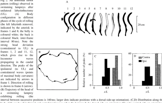
animal passed the normal (dorsal-side-up) orientation. Fig. 2A shows the motor pattern responsible for rolling in a lamprey with the left labyrinth removed (note that the belly is white and the back is black; the lesioned side is marked with an asterisk in frames 1 and 8). The following positions are seen in the sequence in Fig. 2A: 1, 2, dorsal side up; 3–5, the right side moves up and the ventral side (white area) gradually becomes increasingly exposed to the video camera starting from the head, which demonstrates body twisting; 7, 8, the whole body (except for the tail) is positioned with its ventral side up; 9, 10, the left side gradually becomes increasingly exposed to the video camera; 11, 12, the dorsal side is uppermost. In UL animals exhibiting rolling, the lateral undulations of the anterior part of the body were not symmetrical: flexions towards the side of the intact labyrinth had a much larger amplitude than flexions towards the lesioned side; these large flexions are clearly seen in frames 1, 2 and 11, 12 in Fig. 2A. No notable flexion in the opposite direction is seen in any frame. Because of this asymmetry, the anterior part of the body was, on average, flexed towards the side of the intact labyrinth. In contrast, the posterior part of the body was always deflected ventrally (frames 6 and 10 of Fig. 2A, see also Fig. 1Aiii for the intact animal). These two flexions, i.e. the lateral bending of the anterior part of the body and the ventral bending of the posterior part, gave the lamprey’s body a spiral, screw-like
C B
D A
40
0 20
60 60
0 20 40
Relative number of peaks (%)
20 cm
0 0.5 1.0 0 0.5 1.0
Contra Ipsi
1 2 3 4 5 6 7 8 9 10 11 12
x/L x/L
[image:4.609.40.557.386.716.2]*
*
Fig. 2. An abnormal motor pattern (rolling) observed in swimming lampreys after unilateral labyrinthectomy (UL). (A) Body configuration in different phases of the cycle of rolling (the left labyrinth removed, indicated by the asterisk in frames 1 and 8; the belly is coloured white; the back is coloured black; inter-frame interval 80 ms). Note the strong head deviation (contralateral to UL) in frames 1, 2 and 11, 12, which gives rise to the locomotor waves propagating in the caudal direction. The peaks of the ipsilateral (to UL) and contralateral waves (points of maximal body curvature) are indicated by arrows in frame 1. Direction of rolling is shown in frame 6 (arrow). (B) Trajectory of the head of a swimming lamprey exhibiting rolling (the
shape, which presumably explains the continuous rolling when the lamprey moved forwards. As a result, the swimming trajectory was not linear but helical (in Fig. 2B, a view of the spiral trajectory is shown from above). The frequency of rotation usually ranged from 0.5 to 1 turns s−1, with mean value of 0.73 turns s−1 (Fig. 1D). Another motor disorder in UL animals was body twisting towards the lesioned side, which is clearly seen in Fig. 2A (frames 3–6). This twisting may lead to the appearance of a rotatory component in the lateral undulatory locomotor waves propagating along the body, which could also help to elicit rolling.
[image:5.609.355.514.354.633.2]Since the periodic repetition of body shape coincided with the rhythm of rolling, it was evident that the locomotor rhythm and the rhythm of rolling were synchronized. The periodic changes of body shape due to the propagation of lateral undulatory locomotor waves were thus in phase with the rotation of the animal around its longitudinal axis. At a given roll angle, the animal always had the same body shape. In frames 1 and 12 (Fig. 2A), the animal was in the same phase of the roll turn (dorsal side up) in two consecutive cycles of rolling, and the body configuration is almost the same in the two frames. To analyze this phenomenon in greater detail, the positions of the peaks of the locomotor waves (the points of maximal body curvature) were determined along the body axis for the dorsal-side-up orientation and for the ventral-side-up orientation of the animal. This is illustrated in frame 1 of Fig. 2A (the dorsal-side-up orientation), where the ipsilateral (to UL) peak is marked by a black arrow and the contralateral one by a white arrow. Such an analysis was carried out for a few (3–6) consecutive cycles in six different animals, and the results are presented in Fig. 2C,D. This shows a distribution of the peak positions along the body axis for the dorsal-side-up orientation (Fig. 2C) and for the ventral-side-up orientation (Fig. 2D). One can see that the ipsilateral peak is always located in the midbody area, while the contralateral peak is always located closer to the tail; this applies to both the dorsal-side-up and the ventral-dorsal-side-up orientations. This finding indicates that the two rhythms are synchronized and that the locomotor frequency relates to the frequency of rolling as 2:1. In the course of vestibular compensation, the duration and number of swimming episodes with rolling, when the roll control system was not able to maintain equilibrium (‘impaired swimming’), gradually decreased, while the duration and number of swimming episodes without rolling, when the equilibrium was maintained (‘normal swimming’), increased (see below). However, in all animals, the frequency of rotation during rolling hardly changed, as illustrated for one animal in Fig. 3A. Both at the non-compensated stage (Q=0.08) and 2 weeks later, when the animal had achieved a good level of compensation (Q=0.81), the average frequency of rolling was practically the same. Thus, the parameter Q, which reflects only the relative duration of impaired swimming but not the frequency of rolling (see Materials and methods), is a good indicator of the degree of impairment of the roll control system evoked by UL.
During episodes of normal swimming, the locomotor pattern
and the body configuration rarely differed from those in intact animals: body twisting was absent, the lateral locomotor undulations were symmetrical in relation to the body axis and the lamprey swam along a rectilinear trajectory (for a description of normal swimming, see Williams et al. 1989; Ullén et al. 1995a). In six animals (54 %), however, a tilt around the longitudinal axis towards the operated side (15–30 °, sometimes 45 °) could be observed (the tilt angle was estimated by the size of the dorsal fin visible from above, see Ullén et al. 1995b). In one animal, the locomotor undulations were asymmetrical, with a larger lateral deviation of the head towards the intact labyrinth. This animal swam along a circular trajectory.
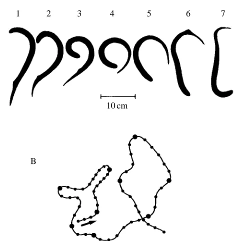
In addition to rolling, animals of group 1 exhibited a different type of motor behaviour, circling, which was not observed in intact animals. This behaviour pattern is illustrated in Fig. 4 for an animal with the right labyrinth removed. Fig. 4A shows a single turn of the lamprey, the turn being directed towards the side of the intact labyrinth. The turn started with a lateral bending of the anterior part of the body; the bending subsequently propagated towards the tail. Such turns appeared in sequence, one after the other, which resulted
B A
0.5
0 1.0
Frequency of rotation
(turns
s
−
1)
0.5
0 1.0
Quality of swimming,
Q
Q=0.08 Q=0.81
Day 1 Day 16
Circling
Normal
Fig. 3. (A) Frequency of rotation during rolling (mean + S.D.) for one
in continuous circling of the animal. The trajectory of two complete circles is shown in Fig. 4B. This trajectory was accomplished as a sequence of nine single turns (the beginning of each turn is indicated by the larger dots in Fig. 4B). The circling movements always occurred in the horizontal plane and, during circling, the lamprey was oriented with its dorsal side up. Episodes of circling appeared spontaneously and usually lasted 2–10 s. Since rolling was absent in these episodes, they were counted as normal swimming when the quality of swimming (Q) was calculated (see Materials and methods). At the early stage of recovery from UL, it was usually only during these episodes that the dorsal-side-up orientation was maintained and rolling was absent. In other words, only when episodes of circling occurred was the quality of swimming greater than zero in most animals (Fig. 3B, day 1). At later stages of recovery, however, the proportion of circling in the non-rolling swimming episodes was reduced (Fig. 3B, day 16). Finally, in well-compensated animals, circling disappeared completely.
A recovery of equilibrium control after UL, reflected in a gradual increase in Q, was observed in all 11 lampreys of group 1. However, individual animals differed strongly in the degree of impairment of the roll control system evoked by UL (i.e. in the initial value of Q), in the rate of recovery (i.e. in the slope of the curve relating Q and time) and in the duration of the recovery period. All these characteristics of recovery are presented in Fig. 5. Fig. 5A shows Q plotted against time for individual animals. Most of the animals (8 out of 11) had rather small initial values of Q (0.08–0.22), although three animals
(marked by asterisks in Fig. 5A) had higher initial values of Q (0.5, 0.75 and 0.87). In contrast to the other animals, these three lampreys initially swam slowly almost all the time and were very close to the bottom, touching it with their sucker mouths. Usually, Q gradually increased with time but, in some animals, a temporary decrease of Q (‘decompensation’) was also observed. By the seventieth day, 10 out of 11 animals had achieved an almost complete compensation, and only one animal took approximately 3 months to reach Q=0.8 (marked with a cross in Fig. 5A). This lamprey was also much less active than the other animals and was excluded from further statistical analysis. We used the value of Q=0.8 as a criterion to evaluate the duration of recovery, which ranged in individual animals from 0 to 100 days (Fig. 5A). The mean value of the duration of recovery was 33±34 days (mean ±1 S.D., N=10). Fig. 5B shows the quality of swimming (mean ±1 S.E.M.) as a function of time post-UL averaged over 10 animals of group 1.
During recovery, some changes in the locomotor behaviour of lampreys were observed. At the earlier stages of recovery, episodes of normal swimming (without rolling) occurred more often when animals moved closer to the bottom of the aquarium (sometimes touching the bottom), and the probability of loss of equilibrium increased in the upper layers of water. Also, during the episodes of normal swimming, some animals
B A
1 2 3
10 cm
[image:6.609.323.556.70.310.2]4 5 6 7
Fig. 4. An abnormal motor pattern (circling) observed in swimming lampreys after UL. (A) A single turn (interval between successive frames, 160 ms). (B) Trajectory of the head during two complete loops of circling accomplished as nine sequential turns (the beginning of each turn is indicated by a larger dot (interframe intervals, 80 ms).
B A
1.0
0 0.5 1.0
0 0.5
Quality of swimming,
Q
5 10 50 100
0 0
V E E
Group 1 N=11
5 10 50 100
Time (days)
*
*
[image:6.609.48.284.437.678.2]*
×Fig. 5. Recovery of equilibrium control after UL in group 1 lampreys. (A) Quality of swimming (Q) as a function of time post-UL in individual animals. Asterisks indicate the cases with lesser impairment of equilibrium control (these animals often swam touching the bottom). During the period of observation, all 11 animals reached the criterion of good compensation (Q=0.8, broken line).
(B) Quality of swimming (mean ±S.E.M.) averaged over the whole
exhibited tilt around the longitudinal axis towards the lesioned side. The tilt angle was smaller (30–45 °) when the lamprey swam closer to the bottom and larger (60–90 °) when it swam in the upper layers of water. This observation suggests that the UL lampreys have a capability, albeit rather limited, to orient themselves in relation to the bottom of the aquarium (probably by using the visual and/or lateral line systems). In the course of recovery, however, the role of additional sensory inputs in postural control diminished. This was confirmed by testing lampreys in a deep aquarium (see Materials and methods). A fourfold increase in depth (compared with that of the shallow aquarium) considerably reduced the possibility for lampreys to orient themselves in relation to the bottom. As shown in Fig. 6, under these conditions, the quality of swimming decreased dramatically (from Q=0.91 in the shallow aquarium to Q=0.41 in the deep aquarium) in the group of animals that reached the criterion of good swimming (Q=0.8) 10–15 days before the test. Later (30–55 days after reaching Q=0.8), the same animals were able to swim equally well both in the shallow and in the deep aquaria (Fig. 6).
In the quiescent state, when the lampreys were attached to the bottom by their sucker mouths, no distortion of body shape was observed. The body axis was rectilinear, body twisting was absent and the dorsal fins exhibited no lateral deviation. However, some tilt around the longitudinal axis towards the lesioned side (a turn around the snout due to the activity of surrounding muscles) and displacement of the eyes, with the ipsilateral eye deviating downwards and the contralateral one upwards, could be seen. These symptoms were observed not only in the animals exhibiting impairment of equilibrium during locomotion, but also in the animals that had recovered their equilibrium control.
Group 2 and 3 animals
Since animals in group 2 (one labyrinth and both eyes removed) and group 3 (one labyrinth and the contralateral eye
removed) had the same level of locomotor activity and similar motor deficits, these groups will be considered together.
The level of locomotor activity in these animals was similar to that of intact animals. Locomotor episodes alternated with periods of quiescence. However, when attached to the bottom, the animals exhibited a slight roll tilt towards the side of UL (a turn around the snout due to the activity of surrounding muscles) and (in group 3) a downward deviation of the remaining eye. The speed of locomotion was similar to that of intact animals (Fig. 1C). Animals of groups 2 and 3 exhibited locomotor deficits similar to those of group 1 animals, that is they rolled continuously when swimming. Short episodes of circling were also observed. The motor patterns of rolling and circling were the same as in those for the animals of group 1 (Figs 2, 4), and the frequency of rotation during rolling was similar to that seen in group 1 (Fig. 1D).
The initial quality of swimming (i.e. the value of Q observed in the first days after surgery) in groups 2 and 3 (Fig. 7A) was low (approximately 0.1) and similar to that found in group 1 (if group 1 animals swimming near the bottom were discounted). In contrast to group 1, recovery from UL in groups 2 and 3 was poor (Fig. 7A). In approximately half of the animals, no recovery was seen during the whole period of observations (2–3 months). In the remaining animals, recovery started much later than in group 1 (compare Fig. 7A and Fig. 5A), and the final value of Q (i.e. that reached by the end of the period of observations) was rather low; only two animals in group 2 and one animal in group 3 reached Q=0.5 by day 55 (Fig. 7A). The very low rate of recovery is also illustrated
1.0
0.5
0
Quality of swimming,
Q
10–15 30–55
Time (days)
Fig. 6. Quality of swimming of group 1 animals in the shallow (open
columns) and deep (filled columns) aquaria (mean + S.E.M., N=10) at
different stages of recovery (10–15 days and 30–55 days after reaching a criterion of good swimming, Q=0.8).
B A
V Group 2
N=5
Group 3
N=4 V
E
0.5 1.0
0
1.0
0 0.5
Quality of swimming,
Q
0 5 10 50 100
0 5 10 50 100
Time (days)
Fig. 7. (A) Quality of swimming in individual animals of group 2 (dashed lines) and group 3 (solid lines) as a function of time post-UL. (B) Quality of swimming averaged over groups 2 and 3 (mean ±
[image:7.609.331.565.443.680.2] [image:7.609.95.241.524.689.2]in Fig. 7B, which shows the mean values of Q over time for groups 2 and 3 taken together.
Group 4 animals
Animals in group 4 (one labyrinth and the ipsilateral eye removed) were kept in two different lighting conditions: either under a 12 h:12 h light:dark cycle (N=6) or in continuous darkness (N=9). No essential difference was found between these two subgroups of animals, and they will be considered together. The level of locomotor activity in the animals in group 4 was similar to that of intact animals and animals in groups 1–3 (Fig. 1C); the locomotor frequency (1.61±0.39) was similar to that of intact animals. In contrast to groups 1–3, however, animals of group 4 swam practically without rolling (Fig. 1D). Fig. 8A shows that the value of Q in most animals ranged between 0.9 and 1.0 from the very beginning of the period of observation. Even the animals (N=3) tested on the day of surgery, just after recovery from anaesthesia (day 0), had a high value of Q. When averaged over all the animals of group 4, the value of Q was very close to 1.0 throughout the period of observations (Fig. 8B). Animals from group 4 swam equally well both in close contact with the bottom and in free water, as well as in a deep aquarium. Hitting the wall did not usually evoke rolling.
Although the main symptom of vestibular deficiency (rolling) was absent in group 4 animals, some distortion of spatial orientation was observed during the 1–3 weeks following UL, namely a tilt around the longitudinal axis, which was dependent on the light conditions. In the majority of tests (67 %), the animals which were kept under 12 h:12 h light:dark conditions and taken for testing during the daytime (i.e. were adapted to light) swam with a tilt around the longitudinal axis
towards the UL side. In 33 % of the tests no tilt was seen (Fig. 9A). The animals that were adapted to darkness and tested in light exhibited no tilt in 44 % of tests, a tilt to the operated side in 33 % of tests and a tilt to the contralateral side (i.e. towards the intact eye), which was never observed in the animals adapted to light, in 23 % of tests (Fig. 9B).
In the quiescent state, when attached to the bottom, all animals of group 4 exhibited some tilt around the longitudinal axis towards the UL side (due to a turn around the sucker mouth) and upward deviation of the remaining eye during the whole period of observations.
Discussion
Motor disorders evoked by UL
The most dramatic motor disorder evoked by UL in the lamprey is a complete loss of equilibrium during locomotion so that, when swimming, the animal rotates continuously around its longitudinal axis towards the lesioned side. This type of UL-induced motor deficit, continuous rolling, was first described by de Burlet and Versteegh (1930). In the present study, it was shown that rolling was correlated with a specific
B A 0 0.5 1.0
Quality of swimming,
Q
0 5 10 50 100
5
V E
10 50 100
0
Time (days)
Group 4
[image:8.609.353.499.73.396.2]N=15
Fig. 8. Quality of swimming in group 4 animals as a function of time post-UL for individual animals (A) and for the whole group (mean ±
S.E.M., N=15) (B).
B A
50
0
100
50
0 100
Percentage of animals
Light (N=6, n=12)
Darkness (N=9, n=18)
Ipsi No tilt
[image:8.609.57.291.464.699.2]Contra
distortion of the body shape. Periodic locomotor undulations of the anterior part of the body were not symmetrical, and there was a prevailing deviation towards the intact labyrinth (Fig. 2A). The anterior part of the body was therefore, on average, flexed towards the side of the intact labyrinth. This flexion of the anterior part, combined with the ventral deviation of the posterior part, gave the body a spiral-like shape. When moving forwards, the locomotor waves propagating towards the tail will cause a lamprey with such a body configuration swim along a spiral trajectory and to rotate continuously around its longitudinal axis. An additional cause of rolling may be body twisting (Fig. 2A), which can result in the appearance of the rotatory component in the lateral undulatory locomotor waves propagating along the body.
UL-induced rolling during locomotion has also been observed in teleost fish. However, in contrast to the lamprey, which may exhibit rolling for several weeks after UL, rolling in teleost fish lasts only for a short period, from 10–30 min to a few hours, depending on lighting conditions (Burt and Flohr, 1991a; Ott and Platt, 1988; Löwenstein, 1932; Schoen, 1950). This is probably due to a much stronger contribution of the visual system in spatial orientation in teleost fish than in the
lamprey. Deprived of two labyrinths, some fish can maintain a fixed orientation in space by relying exclusively on visual input (Graf and Meyer, 1983; von Holst, 1935). In contrast, a bilaterally labyrinthectomized lamprey cannot maintain any fixed orientation in space when swimming (de Burlet and Versteegh, 1930; Ullén et al. 1995a).
The dramatic consequences of UL in the lamprey, that is a complete loss of equilibrium which may last for weeks, strongly contrasts with the UL-evoked deficits in terrestrial vertebrates. The latter exhibit numerous postural disorders but do not lose the ability to maintain an upright body posture, except during a short initial period after UL when rolling is observed (Magnus, 1924; Smith and Curthoys, 1989; Schaefer and Meyer, 1973, 1974; Dieringer, 1995). This is largely due to the presence of mechanical support in terrestrial animals when they are sitting, standing and walking. The support is absent in swimming animals. In addition, the somatosensory system of terrestrial animals, which signals the position of the body and its segments in relation to each other and to the supporting surface, presents an important input for the postural control mechanisms that supplements visual and vestibular inputs (Magnus, 1924; Azzena, 1969; Jensen, 1979; Schaefer
B
C D E F
A
L 180 90 0 90 180 R
L 180 90 0 90 180 R
L 180 90 0 90 180 R
L 180 90 0 90 180 R
L 180 90 0 90 180 R
RS(L)
RS(R)
RS(L)
Group 1 RS(R) Group 2 Group 3 Group 4
RS(R)
RS(R)
RS(L)
RS(L) RS(L)
Command to spinal cord
Correcting motor response E(L)
V(L)
+ + + +
V(R)
E(R)
RS(R)
Roll tilt angle (degrees) Equilibrium point
Equilibrium point Control
V E
V V
E E
V E E
V E
V RS(L)
RS(R)
Roll tilt angle (degrees)
.... ..
[image:9.609.79.516.71.343.2]and Meyer, 1973, 1974; Park et al. 1995; see Horak and Macpherson, 1995, for a review).
In addition to rolling, UL also evoked circling in the lamprey, although this was observed much less frequently. In the episodes of circling, large waves of unilateral flexion (towards the intact labyrinth) propagated repeatedly along the body in a caudal direction (Fig. 4A). This pattern does not seem to be a normal locomotor pattern distorted by the asymmetry in muscular tone induced by UL, but rather a special natural pattern used by intact animals for turns (Wallén
et al. 1994). In intact lampreys, turning is usually a brief
transitional state which occurs between episodes of forward progression, but turns in the UL lamprey are repeated again and again. This circling behaviour is also observed in mammals for a short period after UL, although, in contrast to the lamprey, their circling is directed towards the operated side (Magnus, 1924; Schaefer and Meyer, 1973, 1974).
Two more motor disorders were observed when the lamprey was in a quiescent state – a tilt around the longitudinal axis towards UL (due to a turn around the sucker mouth) and a deviation of the eyes. The latter symptom has also been described for bony fish (Graf and Meyer, 1983). No distortion of the body shape (lateral bending and twisting) was observed in quiescent lampreys, in contrast to terrestrial vertebrates for which a variety of UL-induced ‘static symptoms’, such as the lateral deviation of the head, body twisting, etc., have been described (see Smith and Curthoys, 1989, for a review).
Compensation of UL-induced motor disorders
Animals in group 1 (subjected to UL alone) exhibited a gradual recovery of equilibrium control and finally reached a high quality of swimming (Fig. 5). This was due to a reduction in the duration and frequency of appearance of the episodes of impaired swimming compared with normal swimming. A similar phenomenon has been described for the goldfish (Burt and Flohr, 1991a,b). On average, the recovery took 33 days. This period is characteristic for post-UL recovery of postural control in some other species of lower vertebrate, e.g. in the frog and tadpole (Dieringer, 1995; Rayer et al. 1983). Interestingly, the postural deficits exhibited by group 1 animals in a quiescent state, i.e. a tilt around the longitudinal axis towards UL (a turn around the snout) and deviation of the eyes, were evident not only in animals with an impaired equilibrium but also in those showing a complete recovery of equilibrium control. A large difference in the rate of recovery of different symptoms after UL, and the absence of complete recovery for some of them, is characteristic of other species as well (see Magnus, 1924; Smith and Curthoys, 1989; Dieringer, 1995, for reviews) and is probably an indication that plastic changes underlying compensation of different symptoms occur in different parts of the CNS (see Dieringer, 1995, for discussion of this problem).
The process of the gradual recovery of equilibrium control after UL, which is one of the most important aspects of vestibular compensation in the lamprey, is strongly retarded in animals with impaired visual input. By the sixtieth day, the animals with intact vision (group 1) reached an average value
for Q of 0.89 (Fig. 5B); in contrast, blinded animals (group 2) and animals with the ipsilateral (to UL) eye removed (group 3) had reached a value for Q of only 0.21 (Fig. 7B). Vision is also very important for vestibular compensation in teleost fish (Burt and Flohr, 1991a,b; von Holst, 1935; Löwenstein, 1932; Ott and Platt, 1988). In mammals, visual input has little or no effect on the compensation of some of the vestibular deficits, such as the spontaneous nystagmus (Courjon et al. 1977; Fetter et al. 1988; Smith et al. 1986) and yaw head deviation (Smith et al. 1986), whereas others (for example, the head roll tilt) depend on visual input (Smith et al. 1986; Putkonen et al. 1977; Park et al. 1995). As shown in the present study, the ipsilateral and contralateral (to UL) eyes play different roles in vestibular compensation. The ipsilateral eye does not promote compensation since animals of group 3 exhibited very slow recovery (Fig. 7A). In contrast, removal of the ipsilateral (to UL) eye resulted in a dramatic effect: animals of group 4 exhibited no impairment of the equilibrium control and, therefore, no process of vestibular compensation in the usual meaning of this term (Fig. 8). A contribution of input from the ipsilateral (to UL) eye to vestibular compensation has been reported in other species as well. In bony fish, it was shown that rolling was absent if UL was combined with removal of the ipsilateral eye (von Holst, 1935). In frogs, normalization of the head posture was significantly slowed if the optic nerve on the intact side, but not on the UL side, was sectioned (Kolb, 1955). We have also found that, in rats, transection of the ipsilateral (to UL) optic nerve significantly reduced the head roll tilt (T. G. Deliagina and L. B. Popova, unpublished data). Possible explanations for these effects are discussed in the next section. The present study has shown that the impairment of equilibrium control by UL and, in particular, the rate of recovery differed strongly between individual animals in group 1 (Fig. 5A). In contrast, the variability was much smaller in groups 2 and 3 (Fig. 7A) and was practically absent in group 4 (Fig. 8A). The larger variability in group 1 was not caused by different degrees of surgical impairment of the vestibular system (surgical procedures were standard for all the groups, see Materials and methods), but was probably caused by the presence of two eyes and by the differing abilities of individual animals to develop an asymmetry in visual inputs, which seems to be necessary to compensate for UL-induced postural deficits (Burt and Flohr, 1991a,b). The early recovery of equilibrium control in three animals (marked by asterisks in Fig. 5A) was probably caused by a new ‘strategy’ they used, that of touching the bottom with their sucker mouth when swimming.
Origin of motor disorders
(that is periods with normal functioning of the equilibrium control mechanisms) and impaired swimming (when these mechanisms do not function or are unable to maintain equilibrium). Thus, to understand the origin of motor disorders evoked by UL, it is necessary to consider the nervous mechanisms responsible for equilibrium control in the lamprey. These mechanisms were studied in our previous electrophysiological experiments (Deliagina et al. 1992a,b, 1993a,b; Orlovsky et al. 1992). The principal elements of the roll control system are represented in Fig. 10A in the form of a conceptual model (Grillner et al. 1995). The model is based on the following experimental findings. (1) In the lamprey, the main descending pathway transmitting vestibular commands from the brain to the spinal cord is formed by reticulospinal (RS) neurones (Brodin et al. 1988). Left and right sub-populations of RS neurones, RS(L) and RS(R) (Fig. 10A), are driven by excitatory input from the contralateral labyrinth, inputs V(R) and V(L) (Deliagina et al. 1992b), which are activated by contralateral roll tilt, with a peak of activity occurring at approximately 90 ° (Deliagina et al. 1992a) (Fig. 10B). (2) Illumination of one eye results in an activation of the ipsilateral RS neurones, inputs E(R) and E(L) in Fig. 10A (Deliagina et al. 1993a). (3) RS neurones mediate vestibular reflexes counteracting the initial roll tilt (Deliagina et al. 1993b). In response to vestibular input, they send a command to the spinal cord (Fig. 10A) which evokes a correcting motor response aimed at restoring the normal (dorsal-side-up) orientation of the animal. RS(L) and RS(R) evoke roll tilt in opposite directions (indicated by arrows in Fig. 10A,B). The system has an equilibrium point at 0 °, i.e. at the dorsal-side-up orientation of the animal (Fig. 10B). At this orientation, the activities of the two sub-populations of RS neurones are equal, and no correcting motor response will occur. Any deflection from this orientation will increase the activity in one sub-population and decrease the activity in the other, so that the most active sub-population will evoke a correcting motor response.
Although the model was initially designed to explain only two phenomena, i.e. the stabilization of the dorsal-side-up orientation and the ‘dorsal light response’, a number of the experimental findings of the present study can also be explained by this model. These are as follows.
UL evokes rolling. The rolling evoked by UL in groups 1–3
can be explained in the following way. In group 1, removal of, for example, the right vestibular organ [V(R) in Fig. 10A] deprives RS(L) of vestibular input, so that its activity, determined only by tonic visual input evoked by a diffuse illumination of the left eye, E(L), is reduced. In contrast, the activity of RS(R), determined by vestibular input V(L) (depending on the roll tilt) and by tonic visual input E(R), remains high. The two activity curves, RS(L) and RS(R), do not intersect, and the system has no equilibrium point (Fig. 10C). At any orientation in space, the dominating sub-population RS(R) will evoke rolling towards the lesioned side, as indicated by the arrows.
In group 2 (Fig. 10D), visual inputs to RS(L) and RS(R) are absent, which will result in an equal downward translation of both activity curves compared with Fig. 10C. In this case,
RS(R) is driven by vestibular input, whereas RS(L) is deprived of any input. The curves still do not intersect, the system has no equilibrium point, and the animal will rotate.
The ipsilateral eye does not counteract rolling. In group 3
animals, without a labyrinth and the contralateral eye, the situation is even worse than in group 2 (Fig. 10E). The RS(R) neurones receive excitatory inputs from both the vestibular organ and the eye, whereas the RS(L) neurones are completely deafferented, which makes the difference in activities of RS(L) and RS(R) neurones even larger. The system has no equilibrium point, and the animal will rotate.
The contralateral eye counteracts rolling. The model
indicates an important way of eliminating UL-induced rolling. The tonic activity in the deafferented RS(L) neurones has to be increased to a level at which the two curves, RS(L) and RS(R), intersect. In group 4 animals (UL combined with removal of the ipsilateral eye), this is achieved by input from the contralateral (to UL) eye, whereas the ipsilateral visual input is eliminated to produce a downward translation of the RS(R) versus roll angle curve (Fig. 10F). In these animals, one sub-population of RS neurones, RS(R), receives excitation from the labyrinth, and the activity of these neurones depends on the spatial orientation of the animal (i.e. on the roll tilt). The antagonistic sub-population, RS(L), receives tonic input from the eye (subjected to diffuse illumination), and so the activity of these neurones does not depend on the orientation of the animal in space. If input from the eye is strong enough, the RS(L) and RS(R) curves will intersect, the equilibrium point in the roll control system will be reached, and the animal will not rotate. Our experiments have shown that visual input is powerful enough to achieve this, and animals of group 4 maintain equilibrium perfectly well. Even collisions with the wall and sharp turns did not result in a loss of equilibrium. However, some swimming animals exhibited a tilt around the longitudinal axis towards the lesioned side (Fig. 9A), suggesting that visual input is not strong enough to have the equilibrium point exactly at 0 ° (i.e. at the dorsal-side-up position), and the intersection point of RS(L) and RS(R) is shifted towards the lesioned side, as illustrated in Fig. 10F. The present study has also shown that the ipsilateral (towards UL) roll tilt can be abolished and even reversed with stronger visual input, when the animals were initially adapted to darkness and subsequently tested in light (Fig. 9B), an observation which can be easily explained in the framework of the model.
Other ways to counteract rolling. Removal of the ipsilateral
eye is not the only way to re-create an equilibrium point in the roll control system. According to the model, any means of promoting activation of RS neurones that are deprived of vestibular input may be efficient. Some of these methods, namely (i) tonic electrical stimulation of the optic nerve contralateral to UL, (ii) illumination of the eye contralateral to UL, and (iii) tonic electrical stimulation of the vestibular nerve ipsilateral to UL, were tested experimentally and were found to restore the equilibrium control (Deliagina, 1994a,b, 1995).
Alternation of normal and impaired swimming. The model
impaired swimming. The two forms correspond to the two possible modes of operation of the roll control system: first, that the RS(L) and RS(R) curves intersect and the system has an equilibrium point and, second, that they do not intersect and the system has no equilibrium point (Fig. 10).
It appears likely that the plastic changes underlying vestibular compensation in the lamprey involve a restoration of tonic activity in the deafferented RS neurones. This corresponds to a widely accepted point of view that a fundamental step in the process of recovery after UL in all species is the restoration of tonic activity in those brainstem motor centres that have lost an excitatory input from the labyrinth (see, for example, Smith and Curthoys, 1989). One cannot exclude, however, the possibility that compensatory processes occur not only in the brainstem but also in the spinal cord (see, for example, Jensen, 1979; Dieringer, 1995). In the initial stages of recovery after UL, when activity of deafferented RS neurones is still rather low, even a slight reduction of this activity can result in the disappearance of the equilibrium point in the roll control system. Later, when activity is restored in these neurones, the probability of a loss of equilibrium decreases. This may explain the gradual increase in the value of Q during the course of compensation and the persistence of the same motor deficit in the episodes of impaired swimming at different stages of vestibular compensation.
Synchronization of rolling and locomotor rhythm. The model
can also explain distortions of the locomotor pattern occurring during impaired swimming. In UL animals, the activity of RS neurones with an intact vestibular input [RS(R), Fig. 10C–E] strongly depends on the roll tilt, with its peak at 90 ° of the contralateral roll tilt. Evidently, in animals exhibiting continuous rolling, this activity will not be constant but will change rhythmically, with a period equal to one body turn. The rhythmically active RS neurones send a command to the spinal cord where it interacts with the segmental network generating the locomotory rhythm (Grillner et al. 1995). This will result, first, in mutual synchronization of the locomotory rhythm and the rhythm of rotatory body movements. Measurements performed on the non-rolling animals (the control group and group 4) have shown that the locomotor frequency averaged approximately 1.5 Hz while the frequency of rolling (in groups 1, 2 and 3) was approximately 0.7 Hz (Fig. 1D). This may explain the 2:1 relationship between the two frequencies during rolling, when the rhythm of the spinal locomotor network and the rhythm of rolling are synchronized. Second, the periodic asymmetrical descending signals may evoke distortion of the symmetry in the locomotor undulations of the body. Both these effects were observed in the present study (Fig. 2).
From Fig. 10C–E, one can see that the central asymmetry, that is a difference between the RS(R) and RS(L) activity, depends on the configuration of lesions to the visual system. The asymmetry is higher in group 3 (Fig. 10E) than in groups 1 and 2 (Fig. 10C,D). For the following reasons, there appear to be no behavioural correlates for the difference in the central asymmetry in groups 1–3. (i) The frequency of rolling was similar in these groups (Fig. 1D), which can be explained by the
fact that the frequency of rolling was synchronized by the locomotor frequency, which was presumably similar in all the groups. (ii) During the initial period after UL, animals in all the groups were similar in that they exhibited almost continuous rolling, as shown in Fig. 5A (the curves not marked by asterisks) and Fig. 7A. This finding means that both the larger central asymmetry (group 3) and the smaller asymmetry (groups 1 and 2) were sufficient to cause the equilibrium point in the roll control system to disappear, which makes the behaviour of these animals similar. (iii) The rate of recovery after UL appeared to be much faster in group 1 (Fig. 5) than in group 2 with the same degree of asymmetry. This can be explained by a crucial role of input from the contralateral eye for vestibular compensation.
Thus, the model of the roll control system (Fig. 10A,B) gives a satisfactory explanation of the six major phenomena (listed above) observed in the UL lamprey. All of them relate to the initial period after UL. Later, the compensatory process will modify the roll control system. Predictions from the model are: (1) that these modifications will lead to recovery of tonic activity in the RS neurones deprived of excitatory vestibular input, and (2) that input from the contralateral (to UL) eye will play an important role in the recovery of this activity. These predictions will be tested in future experiments.
As shown in the present study, animals of group 1, in the early stages of recovery after UL, exhibited better postural stability when swimming closer to the bottom and especially when touching the bottom. This observation suggests that they may use tactile receptors, as well as the lateral line system (Hassan, 1989), to obtain information about spatial orientation when the main (vestibular) sensory input is damaged. With the recovery of the main, vestibular-driven postural control system, the role of the additional inputs diminishes (Fig. 5). A vicarious function of different sensory inputs at the early stage of vestibular compensation has also been shown for other species (for a review, see Smith and Curthoys, 1989).
The author is grateful to Drs G. Orlovsky, S. Grillner, Y. Arshavsky and F. Ullén for valuable comments on the manuscript. This work was supported by an International Research Scholars grant from the Howard Hughes Medical Institute, by the Royal Swedish Academy of Sciences (Research Grant for Swedish–Russian Scientific Cooperation), by the Swedish Medical Research Council (Grant no. 11554) and by the Gösta Fraenckels Fund.
References
AZZENA, G. B. (1969). Role of the spinal cord in compensating the effects of hemilabyrinthectomy. Archs Ital. Biol. 107, 43–53. BRODIN, L., GRILLNER, S., DUBUC, R., OHTA, Y., KASICKI, S. AND
HÖKFELT, T. (1988). Reticulospinal neurons in lamprey: transmitters, synaptic interactions and their role during locomotion.
Archs Ital. Biol. 126, 317–345.
BURT, A. ANDFLOHR, H. (1991a). Role of the visual input in recovery
of function following unilateral vestibular lesion in the goldfish. I. Short-term behavioural changes. Behav. Brain Res. 42, 201–211.
of function following unilateral vestibular lesion in the goldfish. II. Long-term behavioural changes. Behav. Brain Res. 42, 213–225. COURJON, J. H., JEANNEROD, M., OSSUZIO, I. ANDSCHMID, R. (1977).
The role of vision in compensation of vestibulo-ocular reflex after hamilabyrinthectomy in the cat. Exp. Brain Res. 28, 235–248. DEBURLET, H. M. ANDVERSTEEGH, C. (1930). Uber Ban und Funktion
des Petromyzonlabyrinthes. Acta oto-laringol.( Suppl.) 13, 5–58. DELIAGINA, T. G. (1994a). An asymmetrical visual input can abolish
postural disturbances evoked by unilateral labyrinthectomy in the lamprey. Abstr. 24th Meeting Soc. Neurosci. 575.20, p. 1408. DELIAGINA, T. G. (1994b). Postural disturbances evoked by unilateral
labyrinthectomy in the lamprey can be abolished by an asymmetrical visual input. Acta physiol. scand. 151, 15A. DELIAGINA, T. G. (1995). Vestibular compensation in the lamprey.
NeuroReport 6, 2599–2603.
DELIAGINA, T. G., GRILLNER, S., ORLOVSKY, G. N. ANDULLÉN, F. (1993a). Visual input affects the response to roll in reticulospinal neurons of the lamprey. Exp. Brain Res. 95, 421–428.
DELIAGINA, T. G., ORLOVSKY, G. N. AND GRILLNER, S. (1993b). Vestibulospinal reflexes in lamprey. Abstr. ENA Meeting 1079, p.278. DELIAGINA, T. G., ORLOVSKY, G. N., GRILLNER, S. ANDWALLÉN, P. (1992a). Vestibular control of swimming in lamprey. II. Characteristics of spatial sensitivity of reticulospinal neurons. Exp.
Brain Res. 90, 489–498.
DELIAGINA, T. G., ORLOVSKY, G. N., GRILLNER, S. ANDWALLÉN, P. (1992b). Vestibular control of swimming in lamprey. III. Activity of vestibular afferents. Convergence of vestibular inputs on reticulospinal neurons. Exp. Brain Res. 90, 499–507.
DELIAGINA, T. G., ULLÉN, F., GONZALES, M. J., EHRSSON, H., ORLOVSKY, G. N., GRILLNER, S. (1995). Initiation of locomotion in larval and adult lamprey by stimulation of lateral line
photoreceptors. J. exp. Biol. 198, 2581–2591.
DIERINGER, N. (1995). ‘Vestibular compensation’: Neural plasticity and its relations to functional recovery after labyrinthine lesions in frogs and other vertebrates. Prog. Neurobiol. 46, 97–129. FETTER, M., ZEE, D. S. ANDPROCTOR, L. R. (1988). Effect of lack of
vision and of occipital lobectomy upon recovery from unilateral labyrinthectomy in rhesus monkey. J. Neurophysiol. 59, 399–407.
GRAF, W. ANDMEYER, D. L. (1983). Central mechanisms counteract
visually induced tonus asymmetries: a study of ocular responses to unilateral illumination in goldfish. J. comp. Physiol. 150, 473–481. GRILLNER, S., DELIAGINA, T., EKEBERG, Ö., ELMANIRA, A., HILL, R., LANSNER, A., ORLOVSKY, G. AND WALLÉN, P. (1995). Neural networks controlling locomotion and body orientation in lamprey.
Trends Neurosci. 18, 270–279.
HASSAN, E. S. (1989). Hydrodynamic imaging of the surroundings by the lateral line of the blind cave fish Anoptichthys jordani. In The
Mechanosensory Lateral Line. Neurobiology and Evolution (ed. S.
Coombs, P. Görner and H. Munz), pp. 217–227. New York: Springer-Verlag.
HORAK, F. B. ANDMACPHERSON, J. M. (1995). Postural orientation
and equilibrium. In Handbook of Physiology, section 12,
Integration of Motor, Circulatory, Respiratory and Metabolic Control during Exercise (ed. J. Shepard and L. Rowell), pp. 1–39.
New York: Oxford University Press.
JENSEN, D. W. (1979). Reflex control of acute postural asymmetry and compensatory symmetry after unilateral vestibular lesion.
Neurosci. 4, 1059–1073.
KAPPERS, A. C. U., HUBER, G. C. AND CROSBY, E. (1936). The
Comparative Anatomy of the Nervous System of Vertebrates, Including Man. New York: Macmillan.
KOLB, E. (1955). Untersuchungen uber zentrale Kompensation und
Kompensationsbewegungen einseitig entstateter Frösche. Z. vergl.
Physiol. 37, 136–160.
LÖWENSTEIN, O. (1932). Experimentelle Untersuchungen über den Gleichgewichtssinn der Elritze (Phoxinus laevis L.). Z. vergl.
Physiol. 17, 806–854.
MAGNUS, R. (1924). Körperstellung. Berlin: Springer.
ORLOVSKY, G. N., DELIAGINA,T. G. ANDWALLÉN, P. (1992). Vestibular control of swimming in lamprey. I. Responses of reticulospinal neurons to roll and pitch. Exp. Brain Res. 90, 479–488.
OTT, J. F. ANDPLATT, C. (1988). Early abrupt recovery from ataxia
during vestibular compensation in goldfish. J. exp. Biol. 138, 345–357.
PARK, B. R., SUN, J. S., KIM, M. S., JEONG, Y. Y., CHUN, S. W. AND
LEE, Y. H. (1995). Effect of sensory deprivation or electrical
stimulation on acute vestibular symptoms following unilateral labyrinthectomy in rabbit. Acta oto-laringol. (Suppl.) 519, 162–167.
PLATT, C. (1983). The peripheral vestibular system of fishes. In Fish
Neurobiology: Brainstem and Sense Organs, vol. 1 (ed. R. G.
Northcutt and R. E. Davis), pp. 89–123. Ann Arbor: University of Michigan Press.
PUTKONEN, P. T., COURJON, J. H. AND JEANNEROD, M. (1977). Compensation of postural effects of hemilabyrinthectomy in the cat. A sensory substitution process? Exp. Brain Res. 28, 249–257.
RAYER, B., CAGOL, E. AND HORN, E. (1983). Compensation of
vestibular-induced deficits in relation to the development of the southern clawed toad, Xenopus laevis Daudin. A behavioural study.
J. comp. Physiol. A 151, 487–498.
SCHAEFER, K.-P. AND MEYER, D. L. (1973). Compensatory mechanisms following labyrinthectomy lesions in the guinea pig. A simple model of learning. In Memory and Transfer of Information (ed. H. P. Zipel), pp. 203–232. New York: Plenum Press.
SCHAEFER, K.-P. AND MEYER, D. L. (1974). Compensation of vestibular lesions. In Handbook of Sensory Physiology, vol. VI/2 (ed. H. H. Kornhuber), pp. 463–490. Berlin: Springer Verlag. SCHOEN, L. (1950). Quantitative Untersuchungen über die zentrale
Kompensation nach einseitiger Utriculusausschaltung bei Fischen.
Z. vergl. Physiol. 32, 121–150.
SMITH, P. F. ANDCURTHOYS, I. S. (1989). Mechanisms of recovery
following unilateral labyrinthectomy: a review. Brain Res. Rev. 14, 155–180.
SMITH, P. F., DARLINGTON, C. L. ANDCURTHOYS, I. S. (1986). The
effect of visual deprivation on vestibular compensation in the guinea pig. Brain Res. 364, 195–198.
ULLÉN, F., DELIAGINA, T. G., ORLOVSKY, G. N. AND GRILLNER, S.
(1995a). Spatial orientation in lamprey. I. Control of pitch and roll.
J. exp. Biol. 198, 665–673.
ULLÉN, F., DELIAGINA, T. G., ORLOVSKY, G. N. AND GRILLNER, S.
(1995b). Spatial orientation in lamprey. II. Visual influence on orientation during locomotion and in the attached state. J. exp. Biol. 198, 675–681.
VONHOLST, E. (1935). Über den Lichtrückenreflex bei Fishen. Pubbl.
Staz. Zool. Napoli. 15, 143–158.
WALLÉN, P., ULLÉN, F., DELIAGINA, T. G., ORLOVSKY, G. N. AND GRILLNER, S. (1994). Modulations of the normal locomotor pattern during visually evoked yaw turns in the lamprey. Abstr. 24th
Meeting Soc. Neurosci. 575.19, p. 1408.