Robert C. Brasch' Danute E. Nitecki' Michael Brant-Zawadzki' Dieter R. Enzmann2 George E. Wesbey' Thomas N. Tozer3 L. Dallas Tuck3 Christopher E. Cann' John R. Fike4 Phillip Sheldon'
This article appears in the September / Octo-ber 1983 issue of AJNR and the NovemOcto-ber 1983 issue of AJR.
Received October 29, 1982; accepted alter revision February 14, 1983.
Presented at the Symposium Neuroradiologi-cum, Washington, DC, October 1982.
This work was supported in parts by grants from the Dean of Medicine, University of Califor-nia, San Francisco; Radiology Research and Ed-ucalion Foundation, University of California, San Francisco; and National Institutes of Health grants ROl AM 31937, National Institute of Arthritis, Di-abetes, Digestive and Kidney Diseases, and
CA-30445, National Cancer Institute.
, Department of Radiology, University of Cali-fornia School of Medicine, San Francisco, CA
94143. Address reprint requests to R. C. Brasch.
2 Department of Radiology, Stanford University School of Medicine, Stanford, CA 94305.
3 Department of Pharmaceutical Chemistry, University of California School of Medicine, San Francisco, CA 94143.
, Department of Radiation Therapy, University of California School of Medicine, San Francisco, CA 94143.
AJNR 4:1035-1039, September/October 1983 0195-6108/83/0405-1035 $00.00
© American Roenlgen Ray Society
Brain Nuclear Magnetic
Resonance
Imaging
Enhanced
by
a
Paramagnetic Nitroxide
Contrast Agent:
Preliminary Report
1035
Contrast-enhancing agents for demonstrating abnormalities of the blood-brain barrier may extend the diagnostic utility of proton nuclear magnetic resonance (NMR) imaging.
"TES," a nitroxide stable free radical derivative, was tested as a central nervous system contrast enhancer in dogs with experimentally induced unilateral cerebritis or radiation cerebral damage. After intravenous injection of TES, the normal brain showed no change in NMR appearance, but areas of disease demonstrated a dramatic increase (up to 45%) in spin-echo intensity and a decrease in T, relaxation times. The areas of disease defined by TES enhancement were either not evident on the nonenhanced NMR images or were better defined after contrast administration. In-depth tests of toxicity, stability, and metabolism of this promising NMR contrast agent are now in progress.
Contrast-enhancing pharmaceutical agents may extend the diagnostic capa-bilities of nuclear magnetic resonance (NMR) imaging. Paramagnetic substances tested as NMR contrast agents include the ions of manganese and iron and nitroxide stable free radicals (NSFRs) [1, 2]; all have been shown to decrease proton relaxation times, namely T, and T 2 [3]. Thus, paramagnetic substances enhance contrast differences between those tissues containing the contrast agent and magnetically similar tissues without it.
In separate reports we have described the relative advantages and disadvan-tages of various methods to manipulate NMR contrast [2,4] and the potential to directly evaluate renal function in experimental animals using NSFRs as uro-graphic NMR contrast agents [2, 5].
NSFRs are a group of synthetic, strongly paramagnetic organic compounds that for two decades have been used as "spin labels" for in vitro biologic studies [6]. A water-soluble piperidinyl NSFR derivative, "TES," is rapidly excreted into the urine after intravenous administration, an excretion pattern useful for NMR urographic studies [2, 5]. TES demonstrates additional properties suggesting promise as a clinically useful NMR contrast agent; these include chemical stability of solutions over a broad range of pH and temperature, limited in vivo metabolism, and broad chemical versatility [2, 6]. The ability to chemically attach TES to a variety of biomolecules, drugs, and particles may permit the synthesis of tissue-specific NMR contrast agents. Preliminary toxicity studies of NSFRs, the subject of future reports, are also favorable for the continued development of NSFRs as pharmaceuticals.
1036 BRASCH ET AL. AJNR:4, Sep.IOct. 1983
Materials and Methods
Experimental Animals
Alpha-streptococcus brain abscesses were produced in two
mon-grel dogs using a procedure described previously [7). Briefly, a bacteria agarose mixture (1 0"_1 09 alpha streptococci) was injected through a burr hole to a depth of 3-4 mm into the left parietal
cortex. The infected dogs, weighing 15 and 25 kg, were imaged on postoperative day 6 (late cerebritis stage) or postoperative day 13 (early capsule stage) by computed tomographic (CT) (Varian) and
NMR techniques. CT brain scans were obtained before and 30 min
after intravenous administration of methylglucamine iothalamate 60% (1 ml/kg).
In a third mongrel dog, 16 kg, cerebral radiation damage was
induced using a removable 125
1 source (3M Co.) placed through a
burr hole into the right parietal white matter adjacent to the lateral
ventricle for a period of 3 days. The source strength was 31.9 mCi
(1,180.3 MBq). The radiation dose was 3,000 rad (30 Gy) 1 cm
from the source. CT imaging was performed at monthly intervals
using a General Electric 7800 scanner with and without iothalamate contrast enhancement. Four months after brain irradiation the
ani-mal was imaged using NMR.
All three dogs were sacrificed following the NMR imaging pro-cedure and brain tissue samples were obtained for in vitro nitroxide
measurement by electron spin resonance spectrometry and for histologic evaluation. The histologic specimens were fixed in 10% formalin for a period of at least 5 weeks before routine hematoxylin and eosin staining.
CT and NMR images were qualitatively compared for diagnostic content and particularly for contrast enhancement. In vitro me as-urements of nitroxide tissue concentrations and histologic data were used to verify the imaging data.
NMR Imaging
Our NMR imaging unit, previously described in detail [8], uses a 3.5 kG superconducting magnet. High-resolution mode yielded pixels of 1.6
x 1.4
mm (XY) on a 128 x 128 matrix. Five contiguous 7 mm transverse sections through the brain were sampled in a 13 min period. Four separate spin-echo intensity images were obtained for each section, each based on a different instrument setting forpulse interval "a" (0.5 or 1 .0 sec) or echo sampling "b" (28 or 56 msec). The spin-echo sequence utilizes a 90° radiofrequency pulse followed by two 180° pulses. From these intensity data, estimated
T, and T2 relaxation values, given in milliseconds, were calculated.
A computer program permitted averaging of intensity (I), T 1, and T 2 relaxation values for selected regions of interest. Such reg
ion-of-interest measurements were made for multiple areas of the brain before and after administration of NMR contrast agent.
Preparation of NSFR Contrast Agent
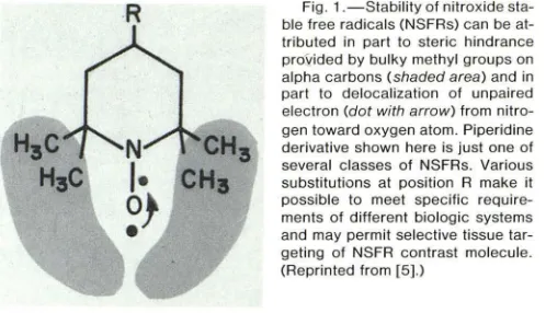
The general structure of a piperidinyl NSFR compound "TES" is shown in figure 1 . An unpaired electron within this NSFR compound
confers paramagnetic properties. This electron is protected from
chemical interaction by steric hindrance provided by four methyl groups on the alpha carbons and by delocalization of the unpaired electron toward the center of the molecule. TES, synthesized in our
laboratory, was stored as dry powder or as a concentrated aqueous solution, buffered to pH 7.2, before intravenous administration. The dose of TES, chosen arbitrarily, was 0.9 glkg body weight. Nitroxide Concentration in Tissue Samples
Samples of brain tissue (30-250 mg) were homogenized in 2N sodium hydroxide and kept for 24 hr at room temperature to extract
R
Fig. 1 .-Stability of nitroxidesta-ble free radicals (NSFRs) can be at-tributed in part to steric hindrance
prOvided by bulky methyl groups on alpha carbons (shaded area) and in
part to delocalization of unpaired electron (dot with arrow) from
nitro-gen toward oxygen atom. Piperidine
derivative shown here is just one of several classes of NSFRs. Various
substitutions at position R make it possible to meet specific
require-ments of different biologic systems and may permit selective tissue tar-geting of NSFR contrast molecule. (Reprinted from [5].)
TES. After filtration, the extracts were measured for nitroxide co
n-centration by electron spin resonance spectrometry. Freshly
pre-pared nitroxide solutions of known concentrations were used as standards.
Our electron spin resonance spectrometer, previously described
[9] and modified with a Varian 100 kcycle field modulation and control unit and a multipurpose cavity, was used to measure nitr
ox-ide concentrations within extracts of autopsied nervous tissue. The aqueous extracts were placed in a Varian flat cell with a volume of 0.04 ml within the microwave cavity.
Results
Experimentally induced cerebritis was demonstrated on both CT and NMR images. The CT manifestations of
cere-britis in our study confirmed those previously reported [7]
and included peripheral contrast enhancement with gradual
diffusion of contrast material into the central, low-density
part of the lesion. The peripheral area of contrast
enhance-ment on the CT images correlated best with the extensive surrounding cerebritis. The central, low-density area seen
on the CT corresponded to the pathologically demonstrated necrotic center. The CT scan in both the late cerebritis and
the early capsule stages retained this pattern of peripheral
enhancement. The extent of surrounding inflammation
tended to regress by the early capsule stage (day 13), and
the area of contrast enhancement became more
circum-scribed corresponding to the development capsule (figs. 2A
and 2B).
The brain infections, well seen on the nonenhanced NMR
images, were depicted as high-intensity rings with low-in-tensity central necrotic areas. The non enhanced NMR
ap-pearance of these brain abscesses has recently been fully
deqcribed [10].
An obvious contrast-enhancing effect was noted on the NMR images after intravenous administration of TES. In the late cerebritis stage (day 6) the size of the abnormality on TES-enhanced images was considerably larger than on the nonenhanced images (fig. 3). TES produced a marked in-crease of intensity within the central part of the lesion
attributed to diffusion of contrast molecules into the necrotic
core. The apparent increase in overall area of the lesion
after contrast enhancement is probably due to diffusion of
contrast material into the surrounding regions of brain
[image:2.612.317.566.84.227.2]AJNR:4, Sep./Oct. 1983 CONTRAST-ENHANCED NMR OF BRAIN 1037
A
B
c
D
E
F
Fig. 2.- 00g with abscess in early capsule stage. Coronal CT scans before (A) and after (B) intravenous administration of iothalamate (1 ml/kg). (This animal moved slightly between pre-and postcontrast scans.) Cir
cum-scribed pattern of contrast enhancement corresponded to developing
cap-sule. NMR spin-echo images (a = 28 msec; b = 1.0 sec) through same
anatomic section before (C) and 10 (0), 20 (E), and 60 (F) min after
intravenous administration of TES. C, Low-intensity central focus is
sur-rounded by rim of higher intensity, which pathologically corresponds to
developing capsule. Contrast enhancement of lesion is maximum in E and is partly faded by F. O-F showed marked increase in intensity at periphery of
lesion and within central necrotic core. Incidental finding is contrast enha
nce-ment in soft tissues overlying site of prior craniotomy, possibly reflecting local
vascular damage.
There also appeared to be a "blush" on the walls of the
lateral ventricles after contrast enhancement. Similar o bser-vations have been noted with contrast-enhanced CT images
[11]. The explanation for this phenomenon is unknown but
may be related to the subependymal veins [12].
In the early capsule stage (day 13) there was also a
marked increase in intensity within the abscess (10, 20, and
60 min) after TES administration (figs. 2C-2F). In the center
of the abscess the intensity (mean ± SO) increased 45%
(2.2
±
0.08 to 3.2±
0.14) by 20 min. The periphery of theabscess was also enhanced in a discrete circular pattern,
[image:3.614.51.298.83.485.2]A
B
Fig. 3.- 00g with late cerebritis. NMR spin-echo images (a = 28 msec;
b = 1.0 sec) before (A) and 20 min after (B) TES administration show
contrast-enhancing effect in cerebral cortex at abscess site. Contrast agent
produces increase in intensity in central, necrotic core of cerebritis lesion as well as diffuse peripheral contrast enhancement around inflammatory lesion. Overall size of lesion appears larger after contrast administration. Also noted is increase in intensity or "blush" in ventricular walls.
3500
3000
INTENSITY
2500
2000
f
NORMAL .&.- '" T1 2000 DRAIN 6- --6. INl(NSITY
f
AREA OF - - . . T1 (EREBRITIS 0 - - - 0 INTENSITY~,//
"'", , , I I
NSFA ADMINISTRATION
I ! I ! I I
10 20 30 40 50 60 TIME Im;n)
1500 T I VALUE
(msec)
1000
500
Fig. 4.- Same dog as in fig. 2, with early capsule inflammatory lesion. Area-of-interest measurements from normal right cerebral white matter and left-sided area of cerebritis show significant increase in intensity and s imul-taneous decline in T I relaxation times in area of cerebritis. Contrast-enhanc
-ing effect is maximum at 20 min.
not diffusely as in the late cerebritis lesion. The degree of
contrast enhancement diminished by 60 min when the a
ni-mal was sacrificed. Correspondingly, T, values decreased
in the center and rim of the abscess after TES administration
(fig. 4). T2 values remained relatively constant on the pre
-and postcontrast calculations (70 and 62 msec, r
espec-tively). T 2 values of the normal brain varied from 66 to 59
msec.
The electron spin resonance measurements of tissue ni-troxide concentration showed a large difference in con cen-tration 1 hr postinjection between the abscess and the contralateral unaffected brain. The necrotic center of the
abscess had a concentration of 0.45 mM/g and the rim of
[image:3.614.315.557.83.232.2] [image:3.614.320.555.321.507.2]dis-1038 BRASCH ET AL. AJNR:4, Sep./Oct. 1983
Fig. S.-Dog with radiation-induced damage in right, periventricutar white
matter. NMR spin-echo images (a = 28 msec; b = 1.0 sec). A, Precontrast image shows ventricutar asymmetry but no focat intensity abnormality in
damaged region. B, 30 min after contrast. Focus of increased intensity at site of pathologically confirmed noninflammatory necrosis.
tance of 1 cm from the rim there was a detectable
accu-mulation of nitroxide, 0.1 mM/g. Four tissue samples taken
from the contralateral (right) cerebral cortex had no
detect-able nitroxide activity by electron spin resonance, indicating
less than 0.03 mM/g.
For the animal with experimentally induced radiation
dam-age NMR demonstrated ventricular asymmetry on the
non-enhanced images but no focal abnormality could be
de-tected (intensity, T, or T2 images). NMR images obtained
after TES administration showed an obvious area of
in-creased intensity in the right cerebrum adjacent to the
lateral ventricle, the site of 1251 source implantation 4 months
before (fig. 5). The lesion intensity (mean value
±
SO) wasslightly higher on the 30 min image than on the 10 min
image. The lesion intensity increased from 1.6
±
0.11(precontrast) to 1.9
±
0.19 (30 min postcontrast) in theregion of radiation damage. The corresponding contralateral
cerebral tissue maintained a uniform intensity (1.7 ± 0.11
precontrast and postcontrast). The observed change in
in-tensity within the damaged brain was again associated with
a decrease in T 1 relaxation values from 1,045 precontrast to 624 30 min after TES administration. T2 values did not
change significantly (54 to 56 msec).
Pathologically 1251 irradiation produced white-matter atro
-phy, noninflammatory coagulation necrosis, and edema. Fibrinoid necrosis of vessel walls with adjacent spongiosis
and gliosis were observed [13].
Electron spin resonance analysis of pathologic and
nor-mal brain specimens showed no detectable nitroxide within
the normal left cerebrum (less than 0.008 mM/g). In the
region of radiation necrosis, a nitroxide concentration of
0.02 mM/g was observed.
Discussion
The mechanism of TES contrast enhancement is attrib
-utable to its strong paramagnetic properties. Paramagnetic
substances when placed in a magnetic field exert a relaxing
effect on hydrogen nuclei in the microchemical environment.
This effect, termed "proton relaxation enhancement,"
pro-duces a decrease in T 1 and T 2 relaxation values of
neigh-boring hydrogen nuclei [3]. Our data indicate that the
de-crease in T, values is relatively greater than the change in T2 values at the concentrations of TES employed. The
proton relaxation enhancement effects are temporary and
disappear when the paramagnetic contrast agent is cleared
from the tissue.
Numerous reports demonstrate the utility of NMR imaging
for the identification and diagnosis of central nervous system
disease [14-16]. For certain conditions including
demyeli-nating disease and brainstem infarction, NMR appears to be
more useful than CT. To date, one of the possible
disadvan-tages of NMR has been the lack of contrast agents to
identify subtle breaks in the BBB [14].
NSFRs potentially fulfill this role and, if developed into
clinically employed pharmaceuticals, may extend the
diag-nostic sensitivity and specificity of the NMR technique.
Although our experiments include only two types of cerebral
pathology (bacterial cerebritis and radiation necrosis), the
results vividly demonstrate a contrast-enhancing effect of
TES in abnormal tissues when compared with normal,
non-enhanced tissues. The pathophysiology of NSFR
accumu-lation in abnormal tissues is postulated to be the same as
that of the commonly used radiographic agents; in
particu-lar, contrast molecules leak into the brain substance only in
regions of BBB breakdown. Contrast enhancement of BBB
defects by CT imaging is widely used and clinically valuable;
the clinical role of parallel contrast agents for NMR imaging
remains to be determined.
The time course of TES accumulation and clearance from
brain lesions appears to be well suited to clinical use. After
intravenous injection, the contrast effects (decreased T,
values and increased intensity) were maximum in the brain
lesions from 20 to 30 min after administration and tended to
diminish by 1 hr. The renal clearance of TES in the healthy
cat is 10 ml/min, a value approximating glomerular filtration.
Half-life of TES in the cat is about 38 min [2, 5]. Thus, the
entire bolus of vascular contrast agents should be eliminated
from the body within a few hours. More detailed studies of
NSFR distribution, clearance, and metabolism are in
pro-gress.
Our studies are inadequate to indicate an "ideal" or a
"Ieast-possible-but-useful" dose for TES in demonstrating
cerebral lesions. From previous in vitro studies we have
shown a 40% intensity increase of aqueous solutions of 1.0
mM TES [2]. By narrowing the NMR gray scale, as is the
routine practice in CT scanning, it may be possible to readily
identify intensity alterations of 5% or less. Thus, the levels
of contrast enhancement achieved in our animal, as high as
45% intensity increase above background, exceed the
nec-essary or even the desired contrast enhancement. The TES
dose of 0.9 g/kg of body weight was chosen arbitrarily and can surely be reduced in future studies without loss of contrast effect. The appropriate dose of the NMR contrast agent may also decrease in future studies as the resolving
capabilities of NMR improve, a likely prediction based on
AJNR:4, Sep./Oct. 1983 CONTRAST-ENHANCED NMR OF BRAIN 1039 REFERENCES
1. Goldman MR, Brady T J, Pykett IL, et al. Application of NMR imaging to the heart. Radiology 1982; 142: 246-248
2. Brasch RC, Nitecki DE, London 0, et al. Evaluation of nitroxide stable free radicals for contrast enhancement in NMR imaging
(abstr). J Nuclear Magnetic Resonance Med. (in press) 3. Slichter CPo Principles of magnetic resonance. New York:
Harper & Row, 1963: 1-50, 137-183
4. Brasch RC. Methods of contrast enhancement for NMR im
ag-ing and potential applications. Radiology 1983;147: 781 -788 5. Brasch RC, Tozer TN, London DA, et al. Nuclear magnetic
resonance study of a paramagnetic nitroxide contrast agent for
enhancement of renal structures in experimental animals. Ra
-diology 1983;147:773-779
6. Stone T J, Buckman T, Nardio PL, McConnell HM. Spin-labeled
biomolecules. Proc Natl Acad Sci USA 1965;54: 1 01 0-1 01 7 7. Enzmann DR, Britt RH, Yeager AS. Experimental brain abscess
evaluation; computed tomographic and neuropathologic co r-relation. Radiology 1979; 133: 11 3-1 22
8. Crooks L, Arakawa M, Hoenninger J, et al. Nuclear magnetic
resonance whole-body imager operating at 3.5 KGauss. Ra
-diology 1982; 143: 1 69-1 74
9. Tuck LD, Schieser OW. Electron spin resonance of some
nitrogen-containing aromatic free radicals. J Phys Chem
1962;66: 937 -939
10. Brant-Zawadzki M, Enzmann DR, Placone RC Jr, et al. NMR
imaging of experimental brain abscess: comparison with CT.
AJNR 1983;4:250-253
11. Naidich TP, Padlowski RM, Leeds NE, Naidich JB, Chisholm
AJ, Rifkin MD. The normal contrast-enhanced computed axial tomogram of the brain. J Comput Assist Tomogr 1977; 1 : 16-29
12. Sage MR. Blood-brain barrier: phenomenon of increasing im
-portance to the imaging clinician. AJNR 1982;3: 1 27 -138, AJR 1982;138:887-898
13. Fike JR, Cann CE, Berstein M, et al. Radiation damage to the
canine brain after 125
1 implantation (abstr). Radiat Res
1982;91 : 370
14. Bydder GM, Steiner RF, Young IR, et al. Clinical NMR imaging of the brain: 140 cases. AJNR 1982;3: 459-480, AJR 1982;139:215-236
15. Young IR, Hall AS, Pallis CA, Legg NJ, Bydder BM, Steiner RE. Nuclear magnetic resonance imaging of the brain in mul -tiple sclerosis. Lancet 1982;2: 1 063-1 066
16. Brant-Zawadzki M, Davis PL, Crooks LE, et al. NMR demon
-stration of cerebral abnormalities: comparison with CT. AJNR